Article

Surface Modification of Monolithic Zirconia Using Sandblasting and Laser Methods

by
Ximena Estrada Sotelo
1,
Humberto Alejandro Monreal Romero
1,2,*,
Laura Isabel Duarte Chávez
1,
Luis Gerardo Maldonado Muñoz
1,
Manuel Antonio Lujan Aguilar
1,
Guillermo Acosta Barriga
1,
Claudia López Meléndez
2,3,
Héctor Alfredo López Aguilar
2,3,
José Guadalupe Chacón-Nava
4 and
Caleb Carreño-Gallardo
4
1
Department of Biomaterials Science and Nanotechnology, Autonomous University of Chihuahua (UACH), Chihuahua C.P. 31000, Mexico
2
Department of Engineering and Materials, La Salle University, Avenue Lomas de Majalca 1120, Chihuahua C.P. 31625, Mexico
3
Faculty of Agrotechnological Sciences, Universidad Autónoma de Chihuahua, Chihuahua C.P. 31160, Mexico
4
Centro de Investigación en Materiales Avanzados (CIMAV), Av. Miguel de Cervantes No. 120, Complejo Industrial Chihuahua, Chihuahua C.P. 31136, Mexico
*
Author to whom correspondence should be addressed.
Crystals 2026, 16(8), 483; https://doi.org/10.3390/cryst16080483
Submission received: 25 June 2026 / Revised: 21 July 2026 / Accepted: 22 July 2026 / Published: 24 July 2026

Abstract

In this study, zirconia specimens were subjected to Al2O3 airborne-particle abrasion and Er laser irradiation to investigate their effects on surface characteristics. The treated and untreated zirconia surfaces were characterized using scanning electron microscopy (SEM), atomic force microscopy (AFM), qualitative elemental analysis by laser-induced breakdown spectroscopy (LIBS), energy-dispersive X-ray spectroscopy (EDS), Raman spectroscopy, XRD analysis, and power spectral density (PSD) analysis. The Er laser-treated group showed the highest mean surface roughness (Sa: 7.763 ± 2.449 µm), compared with the Al2O3-treated group (3.640 ± 2.164 μm); however, the differences in Sa among the experimental groups were not statistically significant (Kruskal–Wallis, p = 0.095). Likewise, no statistically significant differences were observed for Sz (one-way ANOVA, p = 0.567). SEM, AFM, and PSD analyses provided complementary information on the morphological and spatial characteristics of the surfaces produced by the different treatments. Under the experimental conditions evaluated, Er laser irradiation produced distinct surface topographic features and a descriptive trend toward higher mean Sa values, but statistical superiority over Al2O3 airborne-particle abrasion or the untreated control was not demonstrated. Further studies are required to determine whether these surface modifications translate into functional or clinical benefits.

1. Introduction

The morphological evaluation of zirconia using laser and sandblasting methods has increased interest in studying several characteristics, including physical, optical, and mechanical properties [1,2]. Research into novel surface characterization approaches for zirconia has focused on evaluating surface roughness and morphological changes using techniques such as laser treatment, grinding/abrasion, sandblasting, and alumina sandblasting [3,4,5,6]. The use of metal oxides such as zirconia represents a favorable alternative for these activities due to their mechanical resistance and durability. It has been applied in various areas, such as dentistry for the fabrication of prostheses, drug delivery, nano-catalysts, etc. Zirconia’s physicochemical properties and biological characteristics can enhance chemical reactions on the zirconia surface and molecular interactions in aqueous solution demonstrating its versatility in these fields. Recent reviews have emphasized that the continuous evolution of zirconia microstructure, processing technologies, and adhesive strategies has significantly improved its clinical performance while maintaining excellent mechanical properties and long-term reliability. These developments have also highlighted the importance of selecting appropriate surface conditioning protocols according to the zirconia composition and intended clinical application [7,8]. In various systems, including semiconductor materials, neuroscience, energy storage, dental implants, fuel cells, etc., this fact reduces fluid resistance by immobilizing ionic density during electrostatic interactions [9]. Among other things, zirconia has good tissue compatibility and low thermal conductivity, making it suitable for use in prostheses, coatings, thermal insulators, and more. Similarly, the significance of employing two distinct methods, namely the sandblasting and laser methods, is underscored by the various strategies for surface modification [10,11,12]. Therefore, it has been proposed to complement sandblasting with chemical agents such as 10-MDP (10-methacryloyloxydecyl dihydrogen phosphate), a functional monomer that chemically interacts with zirconia oxide. In addition, the quantitative characterization of zirconia surface morphology requires the use of standardized areal roughness parameters, such as Sa, Sz, Sq, Ssk, and Sku, which provide complementary information regarding surface topography and functional behavior. On the other hand, when analyzing the impact of alternative contemporary treatments on the surface of 3Y-TZP, laser irradiation has been shown to produce significant morphological alterations, generating complex topographies that markedly contrast with the morphological effectiveness and continuity achieved through conventional sandblasting [13,14,15,16]. Other techniques used to study the surface roughness of zirconia include glazing, finishing, and polishing, which exhibit distinct characteristics [17,18]. Laser treatment has been proposed as an innovative alternative for modifying the surface of zirconia oxide without compromising its mechanical properties. Recent experimental evidence demonstrated that Er:YAG laser irradiation can modify zirconia surface morphology without causing clinically relevant deterioration of its mechanical properties when appropriate irradiation parameters are used [15,19,20]. Unlike sandblasting, which acts mechanically and can induce microcracks, laser treatment enables more precise, controlled modification of surface microtopography. Various types of lasers have been studied in dentistry for the treatment of zirconia, including CO2, Er:YAG, Nd:YAG, and, more recently, femtosecond lasers. Each interacts differently with the ceramic surface, depending on its wavelength, power, pulse duration, and application mode. The femtosecond laser, for its part, enables extremely precise ablation with minimal thermal damage, producing a complex, highly rough topography without affecting the material’s internal structure [21,22,23]. Recent evidence strongly supports the effectiveness of this technology for topographic alteration. In vitro studies have demonstrated that Er:YAG laser application significantly affects the surface properties of translucent zirconias, revealing a decrease in the contact angle (improving wettability) and a notable increase in average surface roughness and aging resistance. The increased formation of roughness through sandblasting and laser processes plays an important role in the development of various zirconia-based compounds for use in dentistry, biomaterials for tissue reconstruction, and the ceramic industry, among others [15,24,25]. Furthermore, a particularly interesting area in the development of high-roughness surfaces achieved through sandblasting and laser techniques is the design of implants using materials such as zirconia, titanium, and Ni-Cr [26,27,28]. The purpose of this research is the comparison of the sandblasting and laser techniques in the modification of the zirconia surface by increasing the roughness.

2. Materials and Methods

2.1. Preparation of Zirconia Samples Using CAD/CAM

A total of 33 zirconia specimens were randomly allocated into three experimental groups (n = 11 per group): untreated control, Al2O3 airborne-particle abrasion, and Er laser treatment. Of the 11 specimens in each group, six were used for AFM-based surface roughness analysis, with six different surface areas evaluated per specimen. The specimen was considered the experimental unit for statistical analysis. One additional representative specimen from each group was used for complementary characterization by SEM, EDS, LIBS, Raman spectroscopy, XRD, and PSD analysis. Data obtained from these complementary techniques were interpreted descriptively and were not used as independent replicates for inferential statistical analysis. The remaining four specimens in each group were retained as reserve specimens to account for potential specimen loss, damage, or defects during preparation and experimental procedures. As no replacement was required, these reserve specimens were not subjected to further analysis.
For sample preparation, 3D models of the 15 × 4 × 1.5 mm zirconia samples were designed in Meshmixer (Autodesk Inc., San Francisco, CA, USA) and exported as STL files. The digital files (STL) were imported into the Chitubox software (Autodesk Meshmixer version 3.5 and CHITUBOX Dental version v1.2.0, Shenzhen, China), which was used to configure machining parameters and prepare milling paths for an Ivoclar Mill Dry machine. The subtractive manufacturing (milling) of the specimens was then performed using 3 mol% yttria-stabilized tetragonal zirconia polycrystal discs from Amann Girrbach (Mäder, Austria). Finally, to consolidate the material structure and achieve its final mechanical properties, the milled samples were sintered in an Ivoclar HS2 furnace at 1450 °C, then sandblasted and laser processed.

2.2. Preparation of the Sample of Zirconia Through the Sandblasting Method

The zirconia samples measuring 15 × 4 × 1.5 mm were treated with aluminum oxide powder (AquaCare, Velopex International, London, UK) containing 53 µm particles at a pressure of 2.5 bar, a distance of 10 mm, for 10 s, and an angle of incidence of 90° in the AquaCare sandblasting equipment.

2.3. Preparation of the Sample of Zirconia Through Laser Method

The 15 × 4 × 1.5 mm zirconia samples were treated with a Lite Touch Er:YAG laser (Light Instruments Ltd., Yokneam, Israel) using a wavelength of 2.94 µm in short pulse (SP) 50–100 µs with an energy of 150 mJ per pulse, a frequency of 15 Hz, and an average power of 2.25 W. Each sample’s entire treated surface is uniformly swept by the 0.8-mm sapphire conical laser tip, which is kept at a focal distance of 1–2 mm from the surface. This ensures a uniform distribution of energy across the surface. Laser application was performed using a manual technique, consisting of a single irradiation pass per sample. Additionally, the integrated air/water cooling system of the Lite Touch laser was used during irradiation. This allows modification of the surface roughness and topography of the zirconia without generating microcracks or structural damage, thereby ensuring sample integrity during the procedure.

2.4. Characterization of Zirconia Specimens by Roughness and LIBS Analysis

The characterization of the compounds was carried out using a VHX-X1 Series digital microscope (KEYENCE Corporation, Osaka, Japan) equipped with an EA-300 Series laser-based elemental analyzer for laser-induced breakdown spectroscopy (LIBS) (KEYENCE Corporation, Osaka, Japan), which was used to perform semi-quantitative elemental analysis. Laser-induced breakdown spectroscopy (LIBS) was employed to qualitatively compare the emission spectra of zirconia surfaces after the different surface treatments. The technique was used to identify the characteristic spectral features of the constituent elements and to evaluate possible qualitative changes in the surface composition. The preparation of the samples for LIBS was performed as follows: a 5 × 5 dot matrix was used for each one of the six zones, with a total of 150 points of analysis per group, with a dot spacing of 180 µm; the laser was then focused and pulsed, creating plasma around each dot. In this way, the bonded electrons are separated from the atoms that make up the material; as the plasma cools, the atoms recombine with the electrons and, in the process, emit light in the UV, visible and infrared regimes. All roughness parameters (Sa, Sz, Sq, Ssk, Sku) and elemental concentrations (wt%) are reported as arithmetic means and standard deviations. Prior to statistical processing, the LIBS data recorded in decimal format in the source files (points 16 to 25) were converted to a percentage format to ensure data integrity. Statistical analyses were performed using Python 3.11 (Python Software Foundation, Wilmington, DE, USA) with the SciPy statistical package version 1.11.4. Data normality was evaluated using the Shapiro–Wilk test, and homogeneity of variances was assessed using Levene’s test. Variables meeting the assumptions of normality and homogeneity of variances were analyzed using one-way analysis of variance (ANOVA). For variables that did not satisfy the normality assumption, the non-parametric Kruskal–Wallis test was applied. Statistical significance was established at p < 0.05.

2.5. Characterization of Zirconia Specimens by AFM

Afterward, the fragments were extracted and analyzed using an easyScan 2 atomic force microscope (Nanosurf AG, Liestal, Switzerland), equipped with PPP-NCHR-20 silicon tips (NANOSENSORS®, NanoWorld AG, Neuchâtel, Switzerland), operating in non-contact mode. The resonance frequency was set to 204–497 kHz, and the constant force was maintained at 10–130 N/m. The AFM topographic images were acquired over scan areas of approximately 65 × 65 µm and 25 × 25 µm. The estimated lateral resolutions were approximately 127 nm/pixel and 49 nm/pixel, respectively.

3. Results and Discussion

3.1. Characterization of Zirconia in the Presence of Sandblasting and the Laser Method by SEM

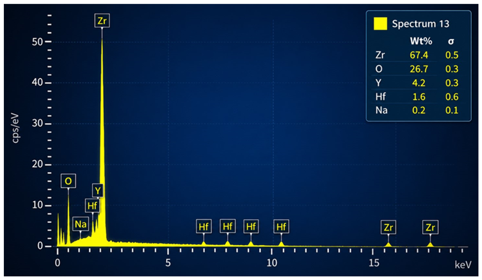
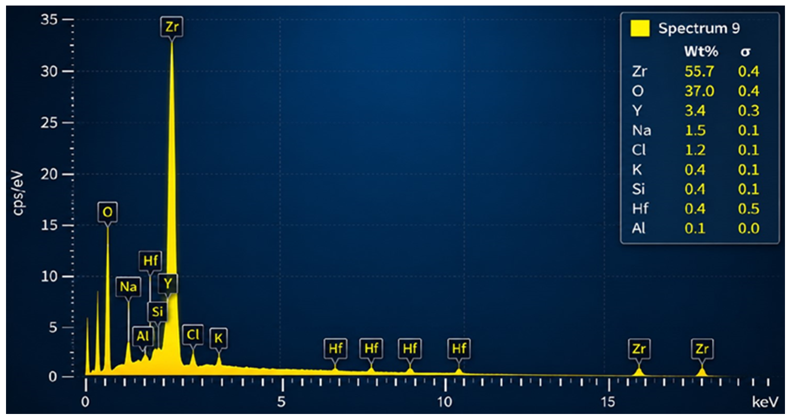
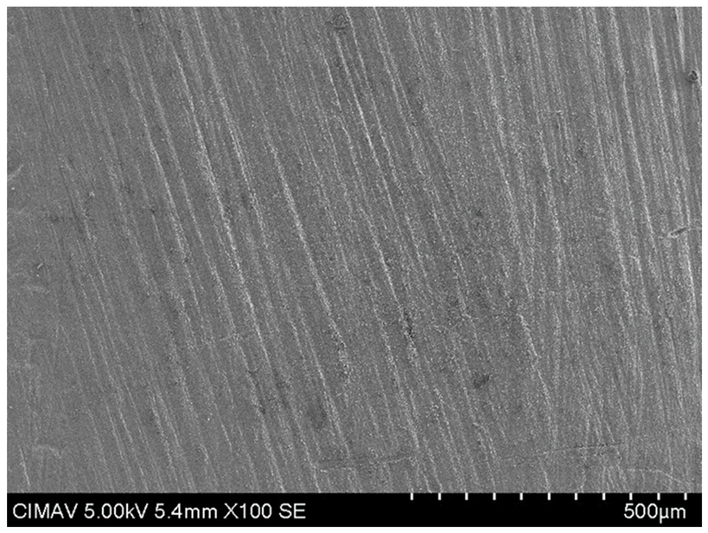
Figure 1 shows the SEM image of the zirconia sample without sandblasting or laser treatment; the surface exhibits a continuous, uniform morphology. EDS was employed to qualitatively evaluate the elemental composition and detect possible changes in surface chemistry after the different surface treatments. The elemental composition of the untreated zirconia specimen, determined by EDS, is shown in Figure 2. Due to the overlap between the Zr L, Y L, and Hf M emission lines, quantitative values obtained by EDS should be interpreted with caution. Therefore, the results are discussed as qualitative evidence of elemental composition rather than absolute concentrations. All spectra confirmed the presence of zirconium, oxygen, yttrium, and hafnium, with no evidence of major compositional changes after the different surface treatments.
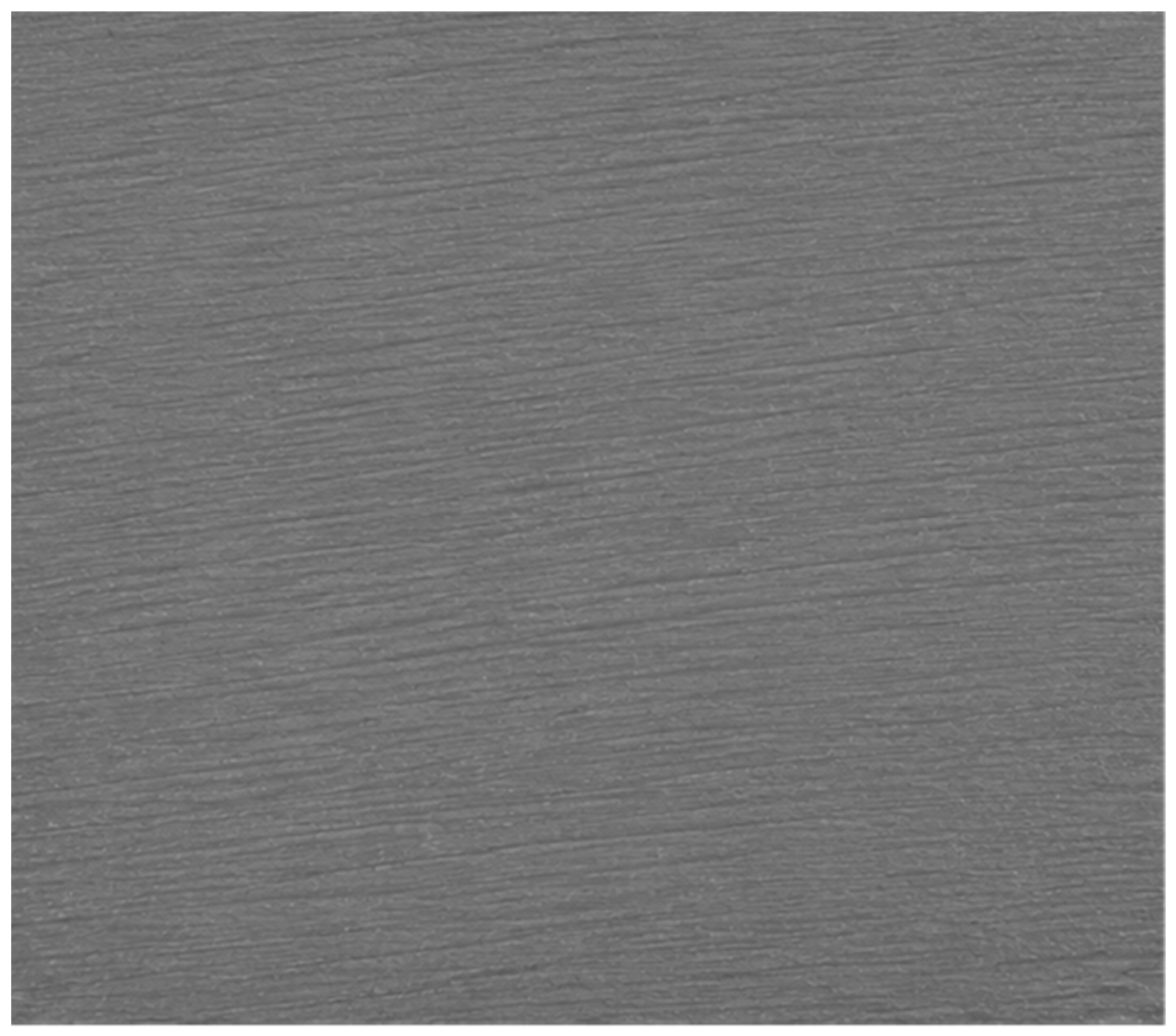
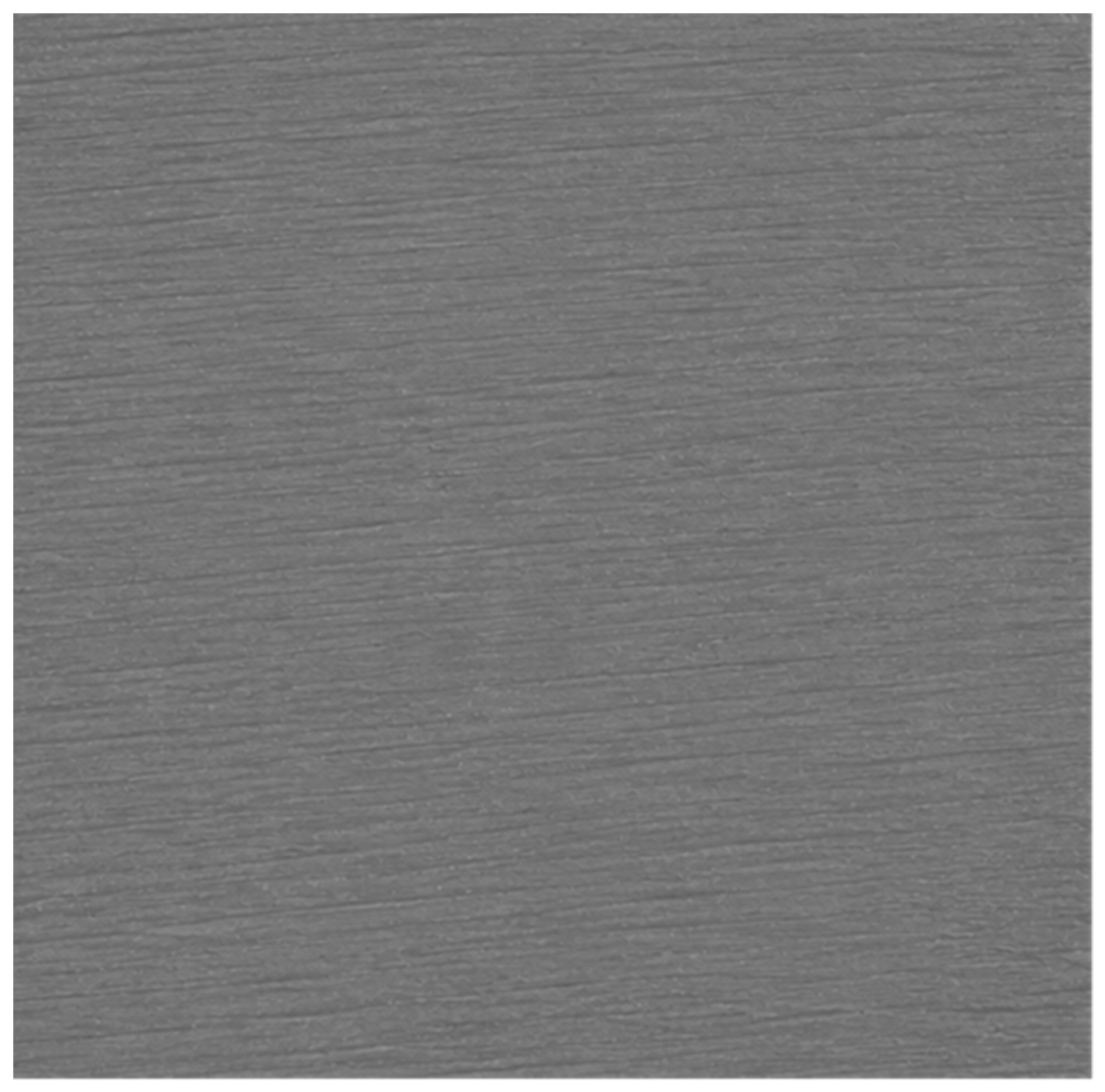
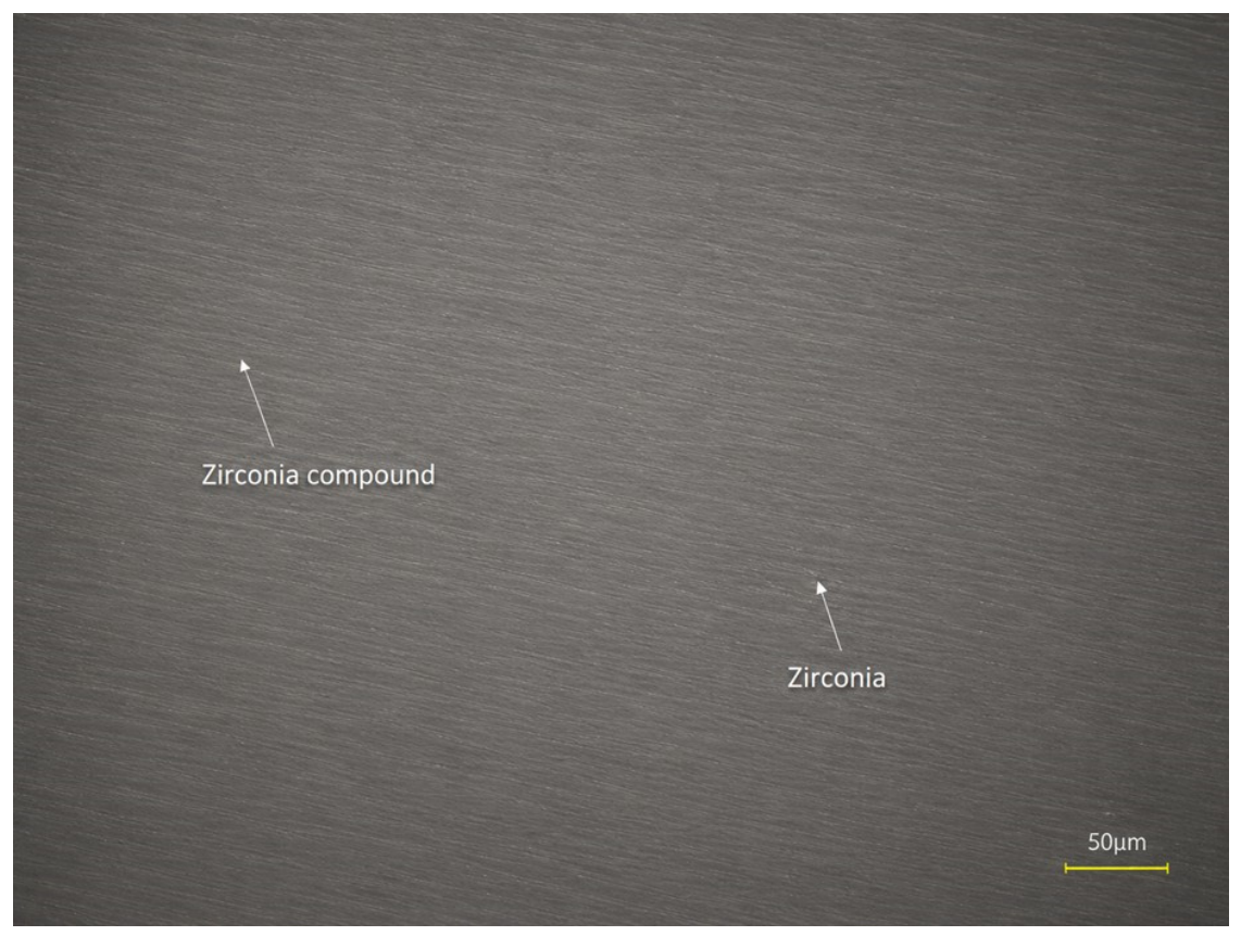
Figure 3 shows the roughness analysis of the untreated zirconia specimen. The surface exhibits a relatively homogeneous morphology with fine, uniformly distributed linear features.
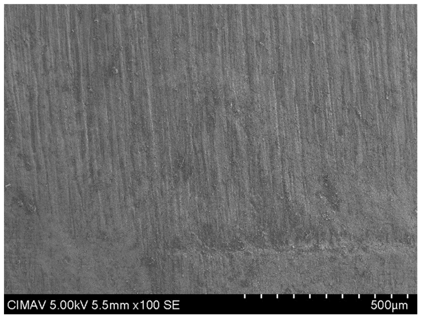
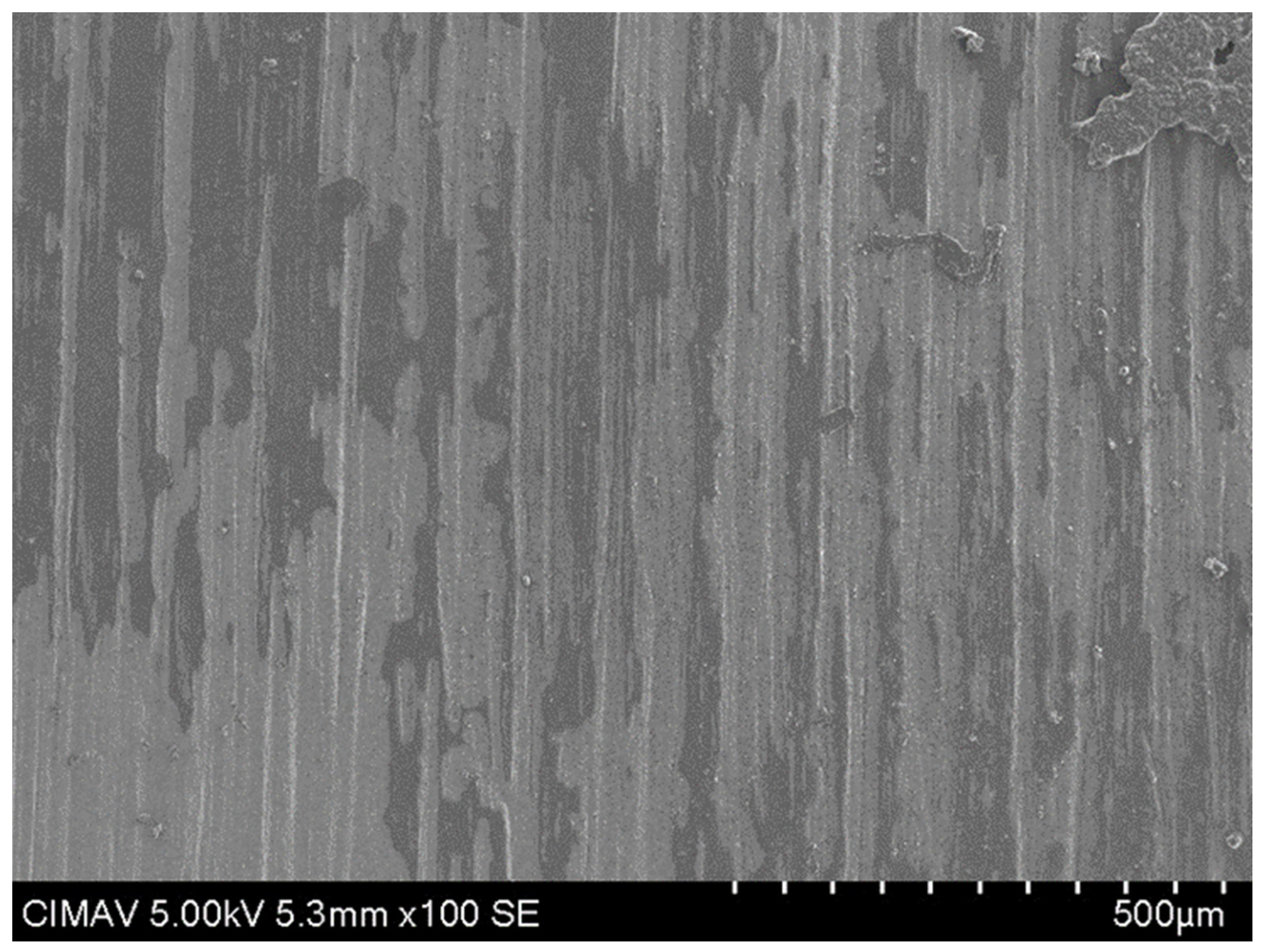
In Figure 4 the sample surface after sandblasting is examined under the SEM.
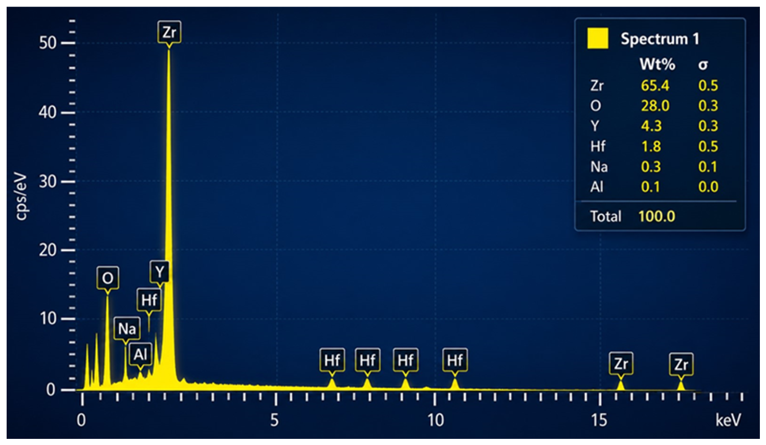
Figure 5 presents the energy-dispersive X-ray (EDS) spectrum of the zirconia specimen after the sandblasting treatment.
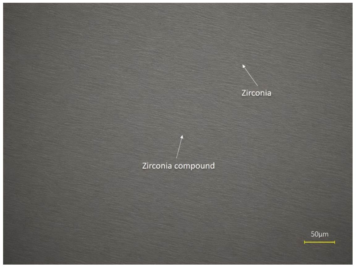
Figure 6 shows a more pronounced directional texture with visible grooves extending across the scanned area. The surface exhibits greater heterogeneity than the first specimen, indicating that the treatment process produced localized modifications and increased microstructural complexity. The grooves appear deeper and more continuous, suggesting enhanced material removal or restructuring. These topographical features can influence the LIBS laser–material interaction by promoting localized variations in laser absorption. Surface asperities and valleys may act as preferential sites for plasma initiation, potentially increasing the efficiency of material ablation while maintaining relatively uniform elemental detection.
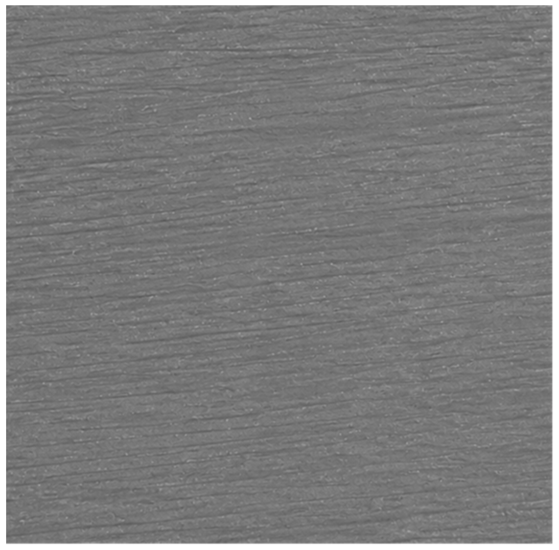
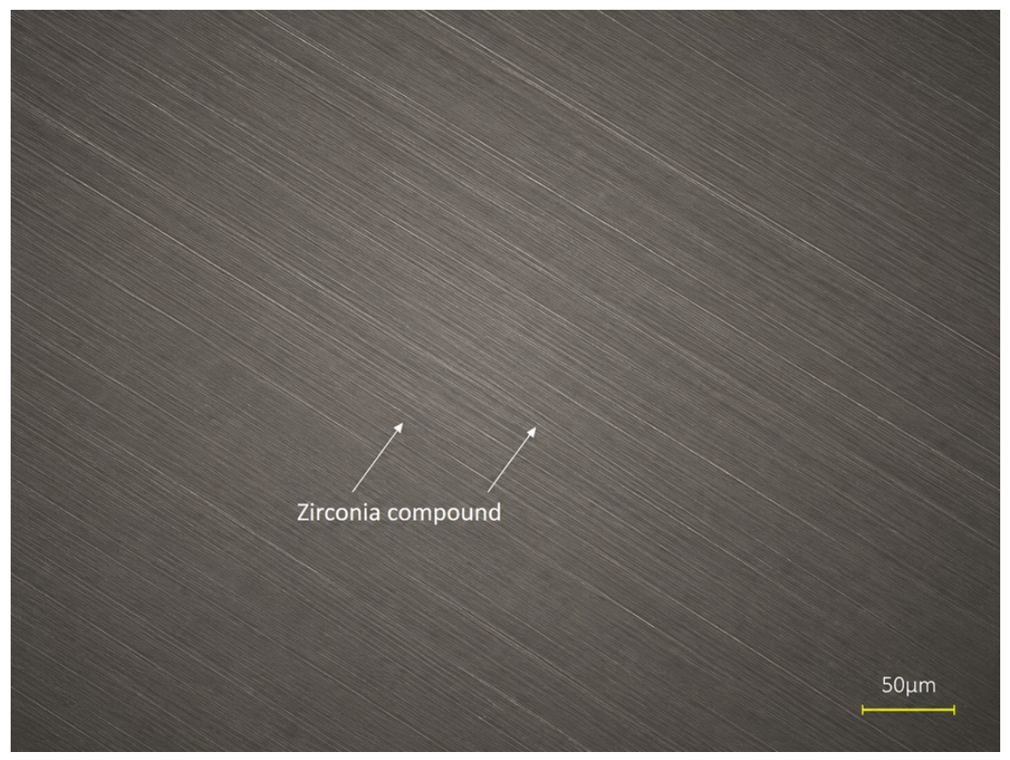
The SEM image in Figure 7 shows the specimen prepared using the laser method. Figure 8 illustrates the energy-dispersive X-ray analysis using the laser method. The EDS spectra confirmed the presence of the main elements expected for yttria-stabilized zirconia (Zr, O, Y, and Hf) in all experimental groups, without evidence of additional elements associated with chemical contamination after surface treatment. Because of the known overlap between the Zr L, Y L, and Hf M emission lines in conventional EDS, the elemental percentages should be regarded as qualitative rather than absolute quantitative values. Consequently, the EDS analysis was used only to verify the elemental composition of the specimens and not to establish quantitative compositional differences among the groups. Likewise, Figure 9 presents the roughness analysis by LIBS. Sandblasting and direct laser writing have been used to study the stability of zirconia 3Y-TZP [15,16,29,30]. The LIBS spectra exhibited the characteristic emission features expected for yttria-stabilized zirconia in all groups. The spectra were used for qualitative comparison of the different surface treatments rather than for quantitative elemental analysis. The image displays various morphological features, and the use of a laser likely increases roughness. In this sense, energy-dispersive X-ray spectroscopy (EDS) was employed to determine the elemental composition of the zirconia surfaces after the different surface treatments. The analysis was performed using an EDS detector coupled to a scanning electron microscope (SEM), allowing simultaneous observation of surface morphology and chemical characterization. Representative spectra were collected from selected regions of each specimen under identical acquisition conditions to ensure comparability among groups. The comparison of elemental distributions among groups provided information regarding possible chemical changes induced by laser irradiation, sandblasting, or other surface modification techniques while maintaining the fundamental zirconia composition. Overall, the EDS methodology enabled the verification of the chemical stability of the zirconia substrate and the detection of minor compositional variations associated with the different surface treatments, contributing to the understanding of the relationship between surface chemistry, morphology, and subsequent material performance. In another context, the three LIBS-derived surface maps reveal distinct topographical characteristics associated with different surface conditions of zirconia. Although all specimens exhibit directional surface features, important differences can be observed in the morphology, texture uniformity, and distribution of surface irregularities. The surface appears smooth with shallow grooves and limited topographical contrast across the analyzed region. The profile traces indicate gradual height variations without abrupt changes, suggesting a relatively uniform energy interaction with the surface. The predominance of subtle directional marks is consistent with a polished or minimally modified surface where material removal has been limited and the original microstructure remains largely preserved. Figure 9 presents the most developed surface texture among the analyzed specimens. Numerous parallel grooves and micro-retentive features are evident throughout the scanned area, producing a markedly roughened morphology. The surface displays greater contrast between elevated and depressed regions, indicating substantial modification of the original topography. The presence of well-defined microgrooves suggests a more intense interaction between the treatment process and the zirconia substrate. Such features increase the effective surface area and create multiple sites for laser energy concentration. In LIBS analysis, this morphology may enhance plasma generation through increased local absorption and multiple scattering effects, resulting in stronger emission signals and improved sensitivity for elemental characterization. From a surface engineering standpoint, the increased density of micro-retentive features observed in Figure 9 indicates a greater capacity for mechanical interlocking and enhanced surface activity. The more developed texture observed in Figure 9 is therefore expected to promote more efficient laser–material interaction while preserving the characteristic directional morphology of the zirconia surface. Furthermore, EDS and LIBS were used primarily for qualitative elemental characterization. Although elemental weight percentages may be generated by the analytical software, these values should be interpreted with caution because spectral overlap, matrix effects, differences in sampling depth, and other technique-specific limitations may affect quantitative accuracy. In particular, spectral interferences involving Zr, Y, and Hf limit the interpretation of small numerical differences among the experimental groups. Therefore, the present study does not interpret variations in elemental weight percentages or spectral intensities as evidence of treatment-induced compositional changes. Instead, the EDS and LIBS results are used to identify the principal elemental signals present in the analyzed specimens. Minor elemental signals may originate from trace constituents, manufacturing or processing procedures, treatment residues, handling, or environmental contamination. Accordingly, these findings should be considered descriptive and qualitative rather than evidence of quantitative differences in zirconia composition.

3.2. Semi-Quantitative Elemental Analysis (LIBS)

Table 1 summarizes the average elemental composition of the control, Al2O3-treated, and laser-treated zirconia groups, expressed as the mean and standard deviation.
Table 2 consolidates the roughness parameters for the three experimental groups. The mean and standard deviation values are derived from the 6 zones analyzed per group and are expressed in micrometers (µm) to three decimal places. The table summarizes five areal surface texture parameters (Sa, Sz, Sq, Ssk, and Sku) for the control, Al2O3-treated, and laser-treated zirconia groups. These parameters provide complementary information regarding surface roughness magnitude, height distribution, and topographical complexity. The laser group exhibited the highest Sa value (7.763 µm), corresponding to 60.6% higher roughness than the control group and 113.3% higher roughness than the Al2O3 group [15,16,31]. The Al2O3 group showed the lowest Sa value, suggesting that sandblasting produced a less pronounced average roughness than laser irradiation. The sample’s coefficient of variation (CV) was as follows: control 90.3%, Al2O3 59.5%, and laser 31.5%. The laser group exhibits substantially lower relative variability, indicating a more reproducible surface modification process. The relative increases between the samples were +14.7% for laser vs. control and +35.8% for laser vs. Al2O3. These findings indicate that laser irradiation creates deeper valleys and higher peaks than alumina abrasion. The variability of the sample was: control 66.8%, Al2O3 38.6% and laser 24.8%. Again, the laser group demonstrates the highest consistency. Based on the root mean square roughness (Sq) analysis, the laser-treated surface demonstrates a 47.7% increase in Sq compared to the control and a 99.6% increase in Sq compared to Al2O3. The nearly twofold increase compared with Al2O3 suggests that laser treatment generates a more pronounced three-dimensional topography [18,23,30]. Furthermore, all groups exhibit Ssk < 0, which indicates surfaces dominated by valleys rather than peaks. This is an important observation because the negative skewness is generally associated with enhanced fluid retention. Valley-dominated surfaces may improve resin penetration and micromechanical interlocking [24,26,32,33,34]. The values are relatively close to zero, suggesting that none of the surfaces exhibit strong asymmetry. The statistical trends reveal a clear distinction among treatments. Of this manner, Er:YAG laser treatment showed the highest mean Sa values and a descriptive trend toward increased surface roughness; however, these differences were not statistically significant [16,20,31]. The present findings differ from those of previous studies reporting statistically significant increases in zirconia surface roughness following laser irradiation [16,20,31,34]. Such discrepancies may be explained by methodological differences, including laser wavelength, energy per pulse, pulse duration, repetition rate, irradiation distance, scanning protocol, cooling conditions, zirconia composition, initial surface characteristics, and the technique used for surface roughness assessment [15,23,35]. These factors influence laser–material interactions and may result in different degrees of surface modification. In the present study, Er laser treatment produced the highest mean Sa values, indicating a descriptive trend toward increased surface roughness; however, the differences among groups did not reach statistical significance. The relatively large within-group variability may have contributed to this statistical outcome by increasing the overlap among the distributions of the experimental groups and reducing the ability to detect between-group differences. Notably, the laser-treated group showed lower relative variability than the control and Al2O3-treated groups, suggesting a more consistent surface response under the experimental conditions evaluated. Nevertheless, this greater consistency should not be interpreted as evidence of superior treatment efficacy in the absence of statistically significant between-group differences [16]. Additionally, Table 3 presents several statistical tests. The Shapiro–Wilk test showed that the Sa values for the Al2O3 group significantly deviated from a normal distribution (W = 0.777, p = 0.036), whereas the Control (W = 0.816, p = 0.082) and Laser (W = 0.889, p = 0.312) groups did not. In contrast, all Sz datasets satisfied the normality assumption (Control: W = 0.855, p = 0.174; Al2O3: W = 0.885, p = 0.291; Laser: W = 0.862, p = 0.196). Levene’s test confirmed homogeneity of variances for both Sa (F = 0.470, p = 0.634) and Sz (F = 1.375, p = 0.283). Accordingly, the non-parametric Kruskal–Wallis test was applied to Sa and revealed no significant differences among groups (H = 4.713, p = 0.095). One-way ANOVA was used for Sz and likewise showed no statistically significant differences among groups (F(2,15) = 0.589, p = 0.567).
Figure 10, Figure 11 and Figure 12 display micrographs of the quantum analysis performed by LIBS on the specimens. These findings indicate that laser irradiation increases the amount of energy absorbed by the zirconia surface, promoting electronic excitation and, under sufficiently high energy densities, partial ionization of the surface layer. The absorbed laser energy is rapidly converted into heat, generating highly localized temperature increases and steep thermal gradients within the irradiated region. Because zirconia exhibits relatively low thermal conductivity, heat dissipation is limited, leading to the accumulation of thermal stresses during the rapid heating and cooling cycles. When these thermally induced stresses exceed the mechanical strength of the superficial layer, localized surface fractures and microcrack formation may occur. At the same time, the combined thermal and photomechanical effects of the laser promote microabrasion through the removal of superficial material. In areas where the surface temperature approaches or exceeds the local melting temperature, partial melting is followed by rapid resolidification, producing irregular topographical features such as microprotrusions, shallow depressions, and resolidified granular structures. The combined effects of laser energy absorption, electronic excitation, localized heating, thermal stress, microabrasion, surface fracture, and rapid resolidification are responsible for the observed surface restructuring and the significant increase in surface roughness [15,20,23]. An additional factor that should be considered when interpreting the present findings is the manual application of the Er laser using a single irradiation pass [35,36]. Although the main irradiation parameters, focal distance, and cooling conditions were standardized, manual scanning may introduce local variations in scanning speed, pulse overlap, energy distribution, and effective fluence across the treated surface. These factors may contribute to spatial heterogeneity in laser-induced surface modification and could partly explain the variability observed in the roughness measurements. Such within-group variability may have increased the overlap among the experimental groups and reduced the ability to detect statistically significant differences. Therefore, the higher mean Sa observed in the laser-treated group should be interpreted as a descriptive trend rather than evidence of treatment superiority. Future studies using automated or computer-controlled scanning systems are recommended to improve irradiation reproducibility and to more precisely determine the effects of Er laser treatment on zirconia surface characteristics [36,37]. It is important to distinguish the laser–material interaction responsible for the experimental surface modification from that involved in LIBS analysis [23,37]. Er irradiation was used as a surface-treatment procedure to modify zirconia topography through the interaction of 2.94-µm laser energy with the material surface under controlled irradiation and cooling conditions. In contrast, LIBS was employed exclusively as an analytical technique, in which a separate pulsed laser produces localized ablation and plasma formation, and the resulting optical emission is analyzed for qualitative elemental identification. Thus, the plasma-generating process inherent to LIBS was not part of the surface-treatment mechanism investigated in this study. The minor elemental signals detected in the LIBS spectra may have several possible origins, including trace constituents or impurities in the commercial zirconia material, residues introduced during CAD/CAM manufacturing and sintering, surface-treatment procedures, specimen handling, or environmental contamination. In particular, aluminum detected after Al2O3 airborne-particle abrasion may be associated with residual abrasive particles retained on the treated surface. However, because LIBS was used for qualitative elemental characterization and minor spectral signals may be influenced by matrix effects and spectral overlap, the origin and relative abundance of these elements cannot be definitively established from the present data alone.

3.3. Characterization of the Surface of Zirconia by Power Spectral Density (PSD) Analysis

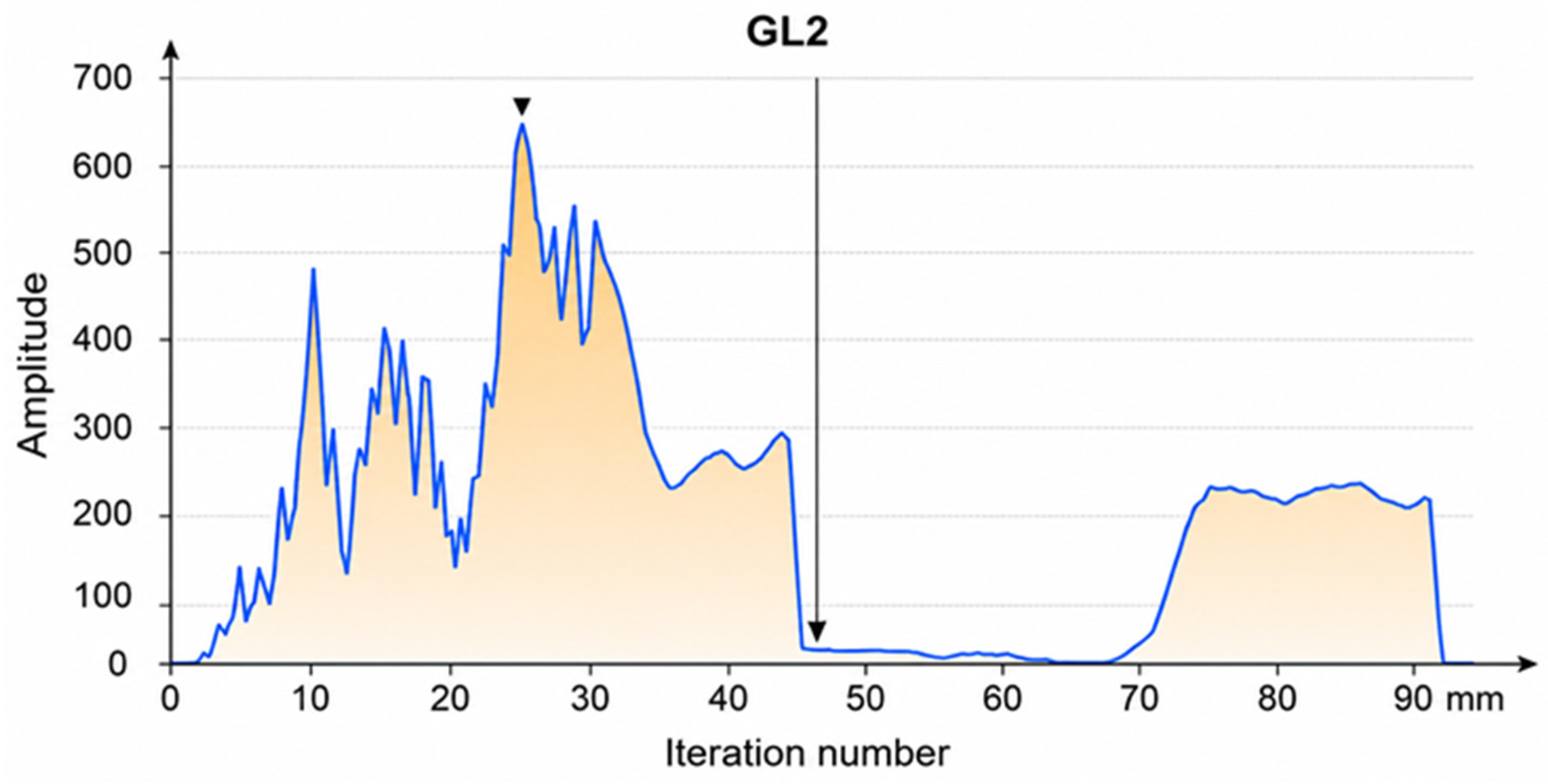
The power spectral density (PSD) acts as a tool for normalizing the spectrum, as it distributes the signal’s energy according to spatial frequency. This facilitates the comparison between surfaces and allows the identification of dominant frequency ranges associated with manufacturing processes or surface treatments. Figure 13 shows the analysis of the PSD of the zirconia surface. This analysis describes how the energy of the surface texture is distributed across spatial scales. A dominant peak is observed around 26.15 mm, with an amplitude of 6.366 GL (Grain Level 2), indicating the presence of a predominant characteristic wavelength on the surface. Subsequently, the curve descends to approximately 40 mm, followed by decreasing secondary peaks, suggesting a hierarchical distribution of roughness and a progressive reduction in the energetic contribution of longer wavelengths. Overall, these results enable quantitative interpretation of the surface topography relating the spectral distribution of roughness to possible implications for the material’s functional performance, such as interactions with light or tribological behavior. The amplitude profile complements the frequency spectrum analysis by quantifying the contribution of different spatial frequencies to the overall surface texture. The high-amplitude peaks observed between iterations 10 and 35 indicate dominant surface wavelengths that contribute significantly to roughness formation. The maximum amplitude, reaching approximately 650 arbitrary units, corresponds to the most prominent topographical features detected on the surface. In contrast, the region between iterations 45 and 70 exhibits very low amplitudes, indicating a reduced contribution of intermediate spatial frequencies and suggesting a relatively smooth transition zone. The pronounced central intensity observed in the frequency spectrum corresponds to the dominant background component, indicating that the surface texture is mainly controlled by low spatial frequencies associated with large-scale topographical features. The dominant peak at 25.15 in the GL2 spectrum corresponds to the principal spatial frequency of the surface texture. Its high amplitude indicates that the treated surface is primarily characterized by topographical features of this characteristic wavelength, demonstrating the presence of a well-developed roughness pattern that contributes significantly to the overall surface morphology and potential bonding performance.
PSD analysis provided complementary information to the conventional areal roughness parameters used in this study. Whereas Sa and Sz describe the amplitude of surface irregularities, PSD characterizes how topographic variations are distributed across different spatial frequencies or wavelengths. Consequently, surfaces with comparable overall roughness values may still differ in the characteristic size, spacing, and spatial organization of their topographic features. In the present study, PSD was therefore used to complement the comparison among the control, Al2O3-treated, and Er laser-treated groups by identifying differences in the spatial organization of their surface features. However, because the PSD analysis was primarily descriptive and was not subjected to inferential statistical testing, these findings should be interpreted as complementary evidence of distinct surface patterns rather than as proof of statistically significant differences or treatment superiority.

3.4. Characterization of the Surface of Zirconia by AFM

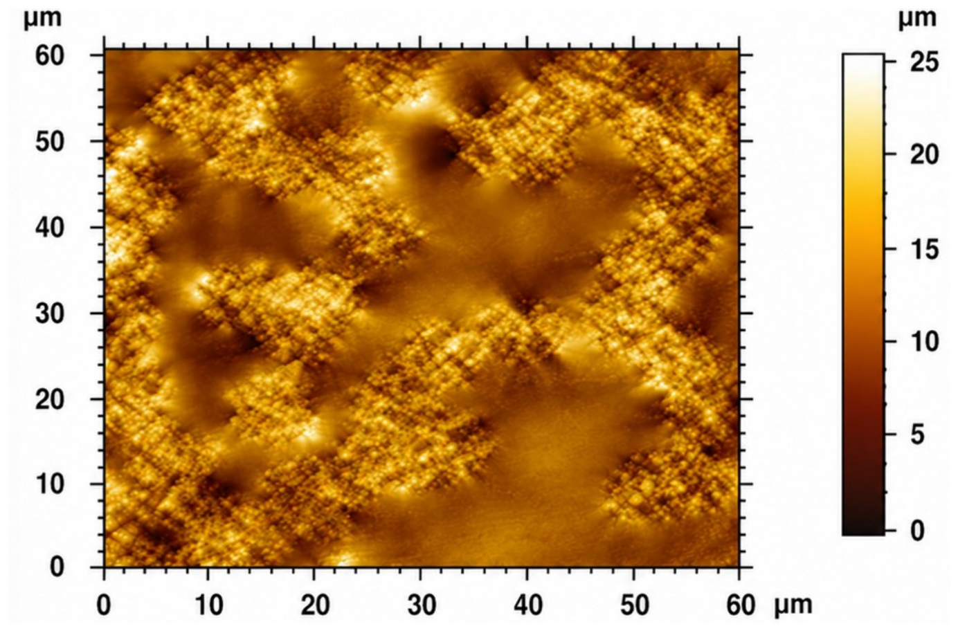
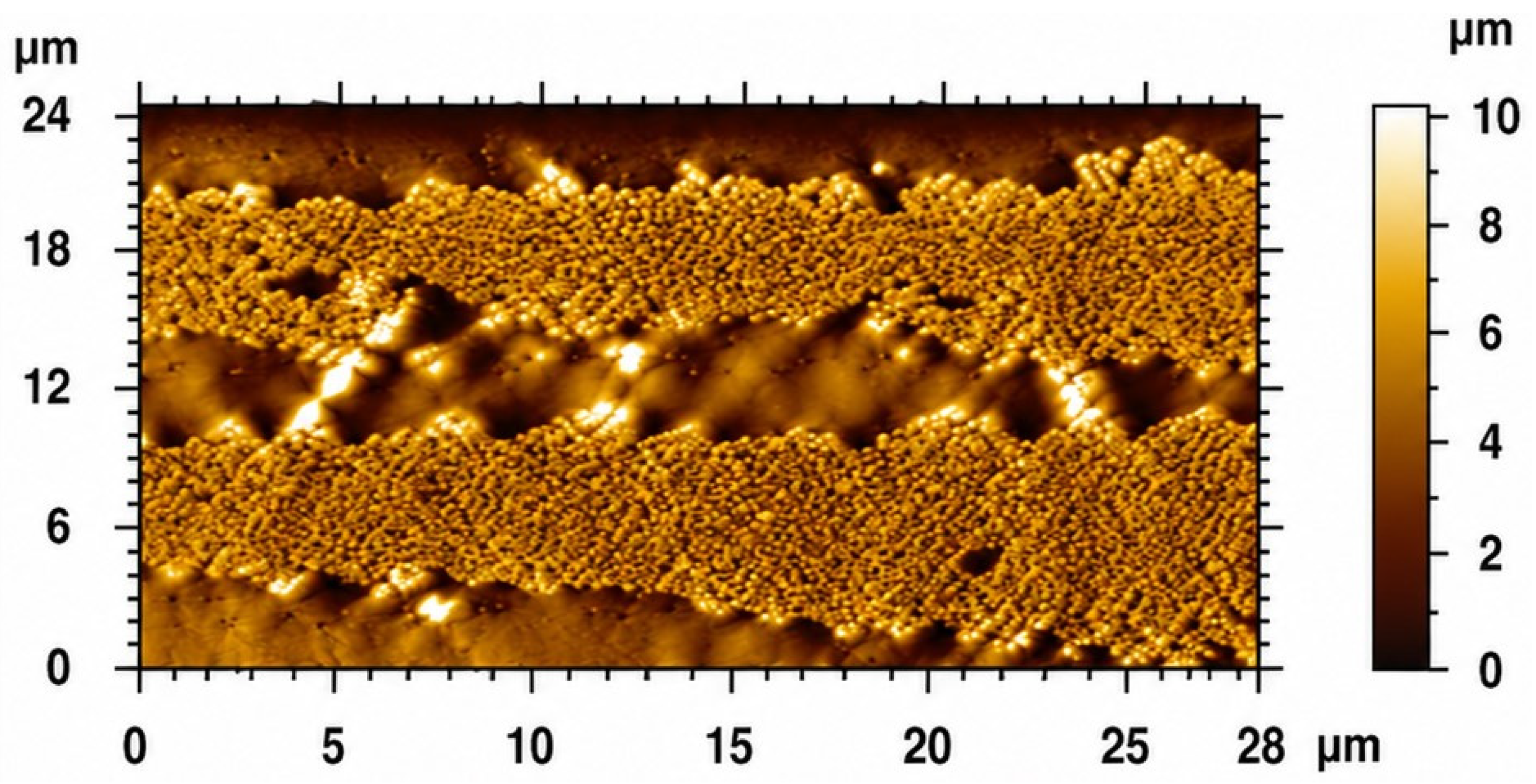
Figure 14 illustrates an AFM topography-mode image of a surface with a heterogeneous morphology and, to a lesser extent, the following characteristics: granular/aggregated structure, consisting of domains or “islands” of micrometric size that are distributed non-uniformly. These regions have a finer internal texture, with substructures suggesting particle aggregation. Irregular height distribution: the color map indicates height variations ranging approximately from 0 to 25 nm. The lighter areas correspond to peaks or elevated regions, while the darker ones indicate valleys or depressions. Local repetitive pattern: in several areas, a kind of reticulated or “lattice” pattern on a smaller scale can be observed. The presence of multiple peaks and valleys distributed across the entire surface suggests a roughness without completely smooth regions. Figure 15 shows the morphology of the zirconia, which has a clearly defined surface marked by horizontal bands or stripes throughout the analyzed area. The granular texture suggests localized surface modification and an increase in effective surface area, which may enhance mechanical interlocking and surface reactivity, as shown in Figure 15. Figure 15 displays a markedly anisotropic morphology characterized by parallel ridges and grooves extending across the scanned area [15,27]. The surface features exhibit a preferential orientation, forming a periodic pattern with height variations of up to approximately 10 µm. These aligned structures indicate directional surface modification, likely associated with machining, polishing, or laser-scanning processes. A comparison of the AFM images demonstrates distinct surface morphologies. The upper image presents a randomly roughened surface with a homogeneous distribution of nanoscale peaks and valleys, whereas the lower image exhibits a directional topography composed of organized grooves and ridges. The anisotropic pattern observed in the lower image suggests a stronger influence of the surface treatment process, producing well-defined microstructural features and a greater degree of topographical organization than the irregular morphology observed in the upper image [15,20]. Surface alterations in zirconia were characterized using STM and SEM in a separate study [38].
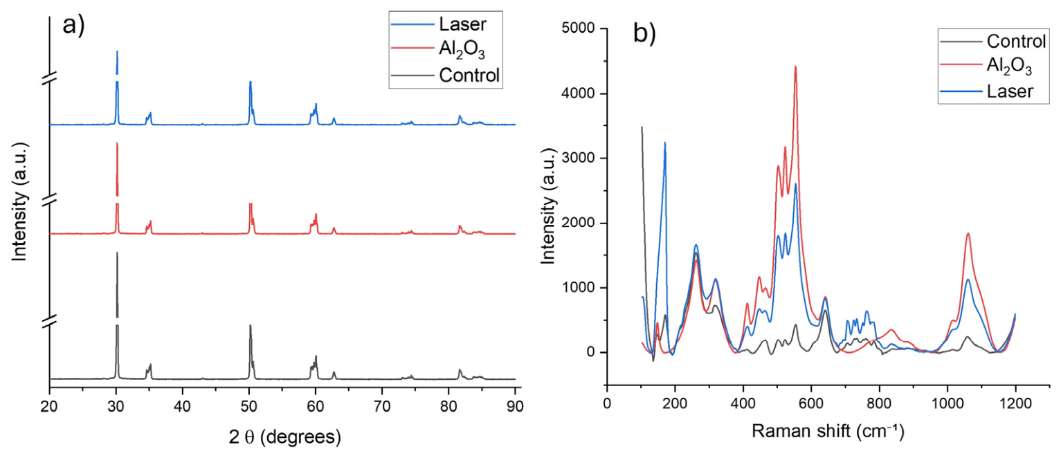
The XRD patterns of the control, Al2O3-sandblasted, and laser-treated zirconia samples show similar diffraction profiles, with the main reflections located at approximately 30°, 35°, 50°, 60°, 63°, 74°, and 82° 2θ. These reflections are consistent with yttria-stabilized zirconia, mainly associated with the tetragonal/cubic crystalline structure. No intense additional peaks attributable to monoclinic zirconia were clearly observed, suggesting that the surface treatments did not induce a significant bulk tetragonal-to-monoclinic transformation. However, the Raman spectra revealed noticeable variations in band intensity and definition after Al2O3 sandblasting and laser treatment, particularly in the 400–650 cm−1 region. These changes may be attributed to surface roughness, residual stresses, defect generation, and local lattice rearrangements induced by the surface modification processes. Therefore, Raman spectroscopy indicates surface-level structural modifications (Figure 16b), whereas XRD confirms the preservation of the main crystalline structure of the zirconia substrate (Figure 16a).
The present study evaluated the effects of airborne-particle abrasion and Er laser irradiation on the surface characteristics of CAD/CAM zirconia using complementary morphological, topographical, chemical, and structural characterization techniques. The combined results demonstrate that although both surface treatments modified the zirconia surface, the extent and nature of these modifications differed according to the characterization method employed. SEM observations revealed that airborne-particle abrasion generated a more irregular surface characterized by sharp-edged depressions and grooves produced by the mechanical impact of Al2O3 particles. In contrast, Er laser irradiation produced localized melting, shallow craters, and surface resolidification features, indicating that the interaction mechanism was predominantly thermal rather than mechanical. These observations are consistent with previous studies reporting that Er laser energy induces localized heating capable of modifying zirconia microtopography without causing extensive structural damage when appropriate irradiation parameters are used [15,20,31,33]. AFM analysis further confirmed that both treatments altered the surface morphology at the micrometer scale [27,28]. Current evidence indicates that successful bonding to zirconia requires an adequate combination of surface conditioning, functional phosphate monomers, and appropriate cementation protocols rather than relying exclusively on surface roughness [20,28,33,34,39]. Although the laser-treated specimens exhibited higher mean Sa values than the untreated and airborne-particle-abraded groups, the statistical analysis demonstrated that these differences were not significant (p > 0.05) [16,20,31]. Likewise, no significant differences were observed for Sz. These findings suggest that, under the irradiation conditions employed (150 mJ, 15 Hz, 2.25 W), Er laser treatment produced measurable topographical modifications; however, these changes were not sufficiently large relative to the variability among specimens to produce statistically significant differences. This highlights the importance of combining qualitative surface characterization with quantitative roughness analysis when evaluating zirconia surface treatments. Power spectral density (PSD) analysis provided additional insight into the spatial distribution of surface features. The dominant spectral peak observed in the laser-treated specimens indicates that laser irradiation generated repetitive surface structures with characteristic wavelengths. Rather than simply increasing roughness, the laser modified the spatial organization of the surface texture, suggesting that PSD analysis can reveal morphological changes that may not be fully reflected by conventional roughness parameters such as Sa and Sz. A limitation of this study is that both EDS and LIBS were employed as complementary qualitative characterization techniques. Conventional EDS is affected by the overlap of the Zr L, Y L, and Hf M emission lines, while LIBS presents partial overlap of the characteristic emission lines of Zr, Y, and Hf within the ultraviolet spectral region. These spectral interferences reduce the accuracy of quantitative elemental analysis. Therefore, both techniques were interpreted qualitatively to confirm the expected elemental composition of yttria-stabilized zirconia and to verify the absence of significant surface contamination after the different treatments. Importantly, the principal conclusions of this investigation are independent of the quantitative elemental values obtained by EDS or LIBS. The effects of the different surface treatments were established primarily from the surface topography observed by SEM, the quantitative roughness parameters obtained by AFM (Sa, Sq, Sz, Ssk, and Sku), the power spectral density (PSD) analysis, and the corresponding statistical comparisons. Consequently, the EDS and LIBS analyses serve only as complementary qualitative evidence supporting the surface characterization and do not constitute the basis for the conclusions regarding the effects of the treatments. The present findings are generally consistent with previous investigations reporting that Er laser irradiation can increase zirconia surface irregularity. However, discrepancies among published studies regarding the magnitude of roughness changes may be explained by differences in laser parameters, pulse duration, energy density, scanning speed, cooling conditions, and roughness evaluation methods. Such methodological variability makes direct comparisons difficult and highlights the need for standardized experimental protocols [15,16,20,31]. Recent comprehensive reviews have further emphasized that no single surface treatment is universally superior for all zirconia systems and that treatment selection should be based on zirconia composition, adhesive system, and clinical application [8,32,33,36]. An important strength of this study is the integration of multiple complementary characterization techniques, allowing the correlation of morphological, topographical, chemical, and structural findings. Nevertheless, some limitations should be acknowledged.
Recent evidence indicates that the surface response of zirconia to laser irradiation is highly dependent on the laser source and irradiation protocol, including wavelength, pulse energy, pulse duration, repetition rate, focal distance, cooling conditions, and scanning strategy [37,38,39,40]. Consequently, laser irradiation does not necessarily produce a statistically significant increase in surface roughness under all experimental conditions. Indeed, visible surface alterations after Er irradiation have been reported without significant changes in average surface roughness [41,42], supporting the distinction between morphological surface modification and statistically significant changes in conventional roughness parameters. In the present study, the higher mean Sa observed in the laser-treated group should therefore be interpreted as a descriptive trend. Furthermore, the manual single-pass irradiation protocol may have introduced local variations in scanning speed, pulse overlap, energy distribution, and effective fluence. Recent controlled laser-texturing approaches emphasize the importance of scanning parameters and irradiation-path control in determining surface morphology and treatment reproducibility [41,42,43]. The AFM analysis was performed on a limited number of specimens and represents localized surface measurements. Furthermore, the present investigation focused exclusively on surface characterization and did not evaluate the effect of these surface modifications on bond strength or long-term clinical performance [28,32,39,43]. Future studies should correlate these surface changes with adhesive behavior, fatigue resistance, and aging stability to determine their clinical relevance. Future investigations should also evaluate the long-term stability of these surface modifications after thermocycling and artificial aging, since bonding durability remains one of the principal determinants of clinical success for zirconia restorations [28,33,39]. Overall, the results indicate that Er laser irradiation represents a conservative surface treatment capable of modifying zirconia surface topography. Although significant differences in conventional roughness parameters were not detected, the combined evidence obtained from SEM, AFM, PSD, Raman spectroscopy, EDS, and XRD demonstrates that laser irradiation produces distinct surface modifications may be relevant for future investigations on adhesive procedures. Although surface topography and roughness are relevant characteristics when investigating zirconia surface treatments, overall, the results indicate that Er laser irradiation represents a conservative surface treatment capable of modifying zirconia surface topography. Although significant differences in conventional roughness parameters were not detected, the combined evidence obtained from SEM, AFM, PSD, Raman spectroscopy, EDS, and XRD demonstrates that laser irradiation produces distinct surface modifications that may be advantageous for subsequent adhesive procedures. The present study did not directly evaluate bonding performance, mechanical interlocking, tribological behavior, or clinical outcomes. Therefore, the observed surface modifications should not be interpreted as evidence of improved adhesion, micromechanical retention, wear resistance, or clinical performance. Rather, these findings provide a basis for future studies designed to determine whether the surface characteristics produced by Er laser irradiation or Al2O3 sandblasting translate into functional benefits. Further investigations should incorporate direct bond-strength testing, artificial aging, thermocycling, fatigue testing, and tribological analyses to establish the potential clinical relevance of these surface modifications.

4. Conclusions

Within the limitations of this study, Er laser treatment produced distinct surface topographic characteristics and the highest mean Sa values among the evaluated groups. However, the differences in Sa and Sz did not reach statistical significance, and therefore the present findings do not demonstrate that Er laser treatment is superior to Al2O3 airborne-particle abrasion or the untreated control in increasing zirconia surface roughness. The observed differences should be interpreted as descriptive trends in surface modification. Further studies with larger sample sizes and direct evaluation of functional outcomes are needed to determine the potential relevance of these surface characteristics.

Author Contributions

Methodology, X.E.S., H.A.M.R., C.C.-G. and L.G.M.M.; Software, L.G.M.M.; Formal analysis, H.A.M.R., H.A.L.A. and J.G.C.-N.; Investigation, L.I.D.C., X.E.S. and C.L.M.; Data curation, L.G.M.M., M.A.L.A. and G.A.B. All authors have read and agreed to the published version of the manuscript.

Funding

This research received no external funding.

Informed Consent Statement

Not applicable.

Data Availability Statement

Data are contained within this article. For more information, one can contact the corresponding author.

Acknowledgments

The authors thank the Advanced Materials Research Center (CIMAV), Mexico, for its technical support.

Conflicts of Interest

The authors declare no conflicts of interest.

References

  1. Schabbach, L.M.; Dos Santos, B.C.; De Bortoli, L.S.; Fabris, D.; Fredel, M.C.; Henriques, B. Translucent multilayered zirconia: Sandblasting effect on optical and mechanical properties. Dent. Mater. 2023, 39, 807–819. [Google Scholar] [CrossRef] [PubMed]
  2. Tzanakakis, E.; Kontonasaki, E.; Voyiatzis, G.; Andrikopoulos, K.; Tzoutzas, I. Surface characterization of monolithic zirconia submitted to different surface treatments applying optical interferometry and Raman spectrometry. Dent. Mater. J. 2019, 39, 111–117. [Google Scholar] [PubMed]
  3. Ruyter, E.I.; Vajeeston, N.; Knarvang, T.; Kvam, K. A novel etching technique for surface treatment of zirconia ceramics to improve adhesion of resin-based luting cements. Acta Biomater. Odontol. Scand. 2017, 3, 36–46. [Google Scholar] [CrossRef] [PubMed]
  4. Egilmez, F.; Ergun, G.; Cekic-Nagas, I.; Vallittu, P.K.; Lassila, L.V. Factors affecting the mechanical behavior of Y-TZP. J. Mech. Behav. Biomed. Mater. 2014, 37, 78–87. [Google Scholar] [CrossRef] [PubMed]
  5. Flury, S.; Peutzfeldt, A.; Lussi, A. Influence of surface roughness on mechanical properties of two computer-aided design/computer-aided manufacturing (CAD/CAM) ceramic materials. Oper. Dent. 2012, 37, 617–624. [Google Scholar] [CrossRef] [PubMed]
  6. Abdou, A.; Hussein, N.; Kusumasari, C.; Abo-Alazm, E.A.; Rizk, A. Alumina and glass-bead blasting effect on bond strength of zirconia using 10-methacryloyloxydecyl dihydrogen phosphate (MDP) containing self-adhesive resin cement and primers. Sci. Rep. 2023, 13, 19127. [Google Scholar] [CrossRef] [PubMed]
  7. Sulaiman, T.A.; Suliman, A.A.; Abdulmajeed, A.A.; Zhang, Y. Zirconia restoration types, properties, tooth preparation design, and bonding: A narrative review. J. Esthet. Restor. Dent. 2024, 36, 78–84. [Google Scholar] [CrossRef] [PubMed]
  8. Cesar, P.F.; Miranda, R.B.P.; Santos, K.F.; Scherrer, S.S.; Zhang, Y. Recent advances in dental zirconia: 15 years of material and processing evolution. Dent. Mater. 2024, 40, 824–836. [Google Scholar] [CrossRef] [PubMed]
  9. Chitoria, A.K.; Mir, A.; Shah, M.A. A review of ZrO2 nanoparticles applications and recent advancements. Ceram. Int. 2023, 49, 32343–32358. [Google Scholar] [CrossRef]
  10. Finger, C.; Stiesch, M.; Eisenburger, M.; Breidenstein, B.; Busemann, S.; Greuling, A. Effect of sandblasting on the surface roughness and residual stress of 3Y-TZP (zirconia). SN Appl. Sci. 2020, 2, 1700. [Google Scholar] [CrossRef]
  11. Martins, F.V.; Mattos, C.T.; Cordeiro, W.J.B.; Fonseca, E.M. Evaluation of zirconia surface roughness after aluminum oxide airborne-particle abrasion and the erbium-YAG, neodymium-doped YAG, or CO2 lasers: A systematic review and meta-analysis. J. Prosthet. Dent. 2019, 121, 895–903. [Google Scholar] [CrossRef] [PubMed]
  12. Shalabi, A.; Kanout, C.; Hamadah, O. Evaluation of surface roughness of translucent zirconia after applying Nd:YAG and CO2 lasers: An in vitro study. Cureus 2024, 16, e64009. [Google Scholar] [CrossRef] [PubMed]
  13. Lung, C.Y.K.; Botelho, M.G.; Heinonen, M.; Matinlinna, J.P. Resin zirconia bonding promotion with some novel coupling agents. Dent. Mater. 2012, 28, 863–872. [Google Scholar] [CrossRef] [PubMed]
  14. Ruzova, T.A.; Haddadi, B. Surface roughness and its measurement methods: Analytical review. Results Surf. Interfaces 2025, 19, 100441. [Google Scholar] [CrossRef]
  15. Sahoo, N.; Carvalho, O.; Özcan, M.; Eyüboğlu, T.F.; Souza, J.C.M.; Silva, F.; Henriques, B. Short-pulse direct laser writing of 3Y-TZP zirconia: A superior surface modification strategy for enhanced resin bonding and adhesion durability. Ceram. Int. 2025, 51, 47830–47843. [Google Scholar] [CrossRef]
  16. Zhu, H.; Tao, H.; Wei, M.; Liu, P.; Yuan, L.; Zhang, Y. Effects of different frequencies of Er:YAG laser on the bonding properties of zirconia ceramic. Lasers Med. Sci. 2023, 38, 4. [Google Scholar] [CrossRef]
  17. Ezzat, M.E.; Mohsen, C.A.; Asaad, R.S. Effect of different glazing methods on the surface roughness and hardness of zirconia ceramics: An in vitro study. BMC Oral Health 2025, 25, 1508. [Google Scholar] [CrossRef] [PubMed]
  18. Silva, S.E.G.; Silva, B.C.D.; Crispim, A.H.T.; Dal Piva, A.M.O.; Kleverlaan, C.J.; Assunção e Souza, R.O. Effect of different finishing/polishing techniques and glaze application on the flexural strength of ultratranslucent zirconia after hydro-thermal aging. J. Mech. Behav. Biomed. Mater. 2025, 164, 106924. [Google Scholar] [CrossRef] [PubMed]
  19. Zhang, X.; Dong, H.; Wu, X.; Li, Q.; Zhao, J.; Guo, C. Effects of Er:YAG laser debonding on changes in the properties of dental zirconia. PLoS ONE 2024, 19, e0313739. [Google Scholar] [CrossRef] [PubMed]
  20. Hager, A.; Nagy, A.; Hanaa, F.; Dalia, S. Effect of Er:YAG laser surface treatment on surface properties and shear-bond strength of resin-cement to three translucent zirconia: An in-vitro study. Discov. Mater. 2024, 4, 35. [Google Scholar] [CrossRef]
  21. Kirmali, O.; Akin, H.; Kapdan, A. Evaluation of the surface roughness of zirconia ceramics after different surface treatments. Acta Odontol. Scand. 2014, 72, 432–439. [Google Scholar] [CrossRef] [PubMed]
  22. Daskalova, A.; Angelova, L.; Carvalho, A.; Trifonov, A.; Nathala, C.; Monteiro, F.; Buchvarov, I. Effect of surface modification by femtosecond laser on zirconia-based ceramics for screening of cell–surface interaction. Appl. Surf. Sci. 2020, 513, 145914. [Google Scholar] [CrossRef]
  23. Han, J.; Zhang, F.; Van Meerbeek, B.; Vleugels, J.; Braem, A.; Castagne, S. Laser surface texturing of zirconia-based ceramics for dental applications: A review. Mater. Sci. Eng. C 2021, 123, 112034. [Google Scholar] [CrossRef]
  24. Kontonasaki, E.; Giasimakopoulos, P.; Rigos, A.E. Strength and aging resistance of monolithic zirconia: An update to current knowledge. Jpn. Dent. Sci. Rev. 2020, 56, 1–23. [Google Scholar] [CrossRef] [PubMed]
  25. Tosiriwatanapong, T.; Singhatanadgit, W. Zirconia-based biomaterials for hard tissue reconstruction. Bone Tissue Regen. Insights 2018, 9, 1179061X18767886. [Google Scholar] [CrossRef]
  26. Yang, L.; Chen, B.; Meng, H.; Zhang, H.; He, F.; Xie, H.; Chen, C. Bond durability when applying phosphate ester monomer-containing primers vs. self-adhesive resin cements to zirconia: Evaluation after different aging conditions. J. Prosthodont. Res. 2020, 64, 193–201. [Google Scholar] [CrossRef] [PubMed]
  27. Saade, J.; Skienhe, H.; Ounsi, H.F.; Matinlinna, J.P.; Salameh, Z. Evaluation of the effect of different surface treatments, aging and enzymatic degradation on zirconia-resin micro-shear bond strength. Clin. Cosmet. Investig. Dent. 2020, 12, 1–8. [Google Scholar] [CrossRef] [PubMed]
  28. Al-Amari, A.S.; Saleh, M.S.; Albadah, A.A.; Almousa, A.A.; Mahjoub, W.K.; Al-Otaibi, R.M.; Alanazi, E.M.; Alshammari, A.K.; Malki, A.T.; Alghelaiqah, K.F.; et al. A comprehensive review of techniques for enhancing zirconia bond strength: Current approaches and emerging innovations. Cureus 2024, 16, e70893. [Google Scholar] [CrossRef] [PubMed]
  29. Fratucelli, E.D.O.; Candido, L.M.; Pinelli, L.A.P. Surface properties and flexural strength of a monolithic zirconia submitted to grinding and regenerative heat treatment. Int. J. Appl. Ceram. Technol. 2021, 18, 525–531. [Google Scholar]
  30. Liu, X.; Aarts, J.M.; Ma, S.; Choi, J.J.E. The influence of polishing on the mechanical properties of zirconia: A systematic review. Oral 2023, 3, 101–122. [Google Scholar] [CrossRef]
  31. Celso, B.F.; Izabela, C.M.; Vivian, C.; Adriana, C.F.; Érica, A.G. Is the Er:YAG laser affect the surface characteristics and bond strength of Y-TZP? Braz. Dent. J. 2021, 32, 83–92. [Google Scholar] [CrossRef]
  32. Subasi, M. Evaluation of the topographical surface changes and roughness of zirconia after different surface treatments. Lasers Med. Sci. 2012, 27, 735–742. [Google Scholar] [PubMed]
  33. Li, X.; Liang, S.; Inokoshi, M.; Zhao, S.; Hong, G.; Yao, C.; Huang, C. Different surface treatments and adhesive monomers for zirconia–resin bonds: A systematic review and network meta-analysis. Jpn. Dent. Sci. Rev. 2024, 60, 175–189. [Google Scholar] [CrossRef] [PubMed]
  34. Iulian-Costin, L.; Tatarciuc, M.S.; Bobu, L.I. Bonding strategies for zirconia fixed restorations: A scoping review of surface treatments, cementation protocols, and long-term durability. Biomimetics 2025, 10, 632. [Google Scholar] [CrossRef]
  35. Ebadian, B.; Moazami, A.; Jowkar, M. Effect of Er:YAG laser application and sandblasting on shear bond strength of veneering ceramic to zirconia core. Front. Dent. 2024, 21, 25. [Google Scholar] [CrossRef] [PubMed]
  36. Cunha, W.; Carvalho, O.; Henriques, B.; Silva, F.S.; Özcan, M.; Souza, J.C.M. Surface modification of zirconia dental implants by laser texturing. Lasers Med. Sci. 2022, 37, 77–93. [Google Scholar] [CrossRef] [PubMed]
  37. Hartung, F.; Moss, C.; Seitz, H.; Schnell, G. Effect of laser scanning parameters on surface morphology and topography of glass solder-coated zirconia substrate. J. Funct. Biomater. 2025, 16, 324. [Google Scholar] [CrossRef] [PubMed]
  38. Young, Y. Improving zirconia–resin cement bonding through laser surface texturing: A comparative study. Prosthesis 2025, 7, 19. [Google Scholar] [CrossRef]
  39. Rigos, A.E.; Sarafidou, K.; Kontonasaki, E. Zirconia bond strength durability following artificial aging: A systematic review and meta-analysis of in vitro studies. Jpn. Dent. Sci. Rev. 2023, 59, 138–159. [Google Scholar] [CrossRef] [PubMed]
  40. García, N.; Jiménez, E.; Roa, J.; Mas-Moruno, C. A review of surface topographical modification strategies of 3Y-TZP: Effect in the physicochemical properties, microstructure, mechanical reliability, and biological response. J. Eur. Ceram. Soc. 2023, 43, 2977–3004. [Google Scholar] [CrossRef]
  41. Zang, N.; Tao, H.; Liang, Z.; Liu, H.; He, J.; Wang, B. Comparison of the shear rebond strength of zirconia brackets treated with different Er:YAG laser energies. Dent. Mater. J. 2023, 42, 405–411. [Google Scholar] [CrossRef] [PubMed]
  42. Assery, N.; Alomeir, N.; Zeng, Y.; Xiao, J.; Tsigarida, A. The effect of Er:YAG laser treatment on biofilm formation on titanium and zirconia disc surfaces. J. Periodontol. 2023, 94, 344–353. [Google Scholar] [PubMed]
  43. Alotaibi, A.O.; Alzahrani, T.A. The impact of different bonding protocols and thermal cycling on the final bond strength of zirconia restorations: Literature review. Am. J. Clin. Med. Res. 2025, 13, 46–50. [Google Scholar] [CrossRef]
Figure 1. SEM image of zirconia in the absence of sandblasting and laser treatment.
Crystals 16 00483 g001
Figure 2. Energy-dispersive X-ray analysis of zirconia.
Crystals 16 00483 g002
Figure 3. Roughness analysis of zirconia in the absence of sandblasting and the laser method.
Crystals 16 00483 g003
Figure 4. SEM image of zirconia after sandblasting.
Crystals 16 00483 g004
Figure 5. Energy-dispersive X-ray analysis of zirconia in the presence of the sandblasting method.
Crystals 16 00483 g005
Figure 6. Roughness analysis of the zirconia using the sandblasting method.
Crystals 16 00483 g006
Figure 7. SEM image of zirconia using the laser method.
Crystals 16 00483 g007
Figure 8. Energy-dispersive X-ray analysis of zirconia using the laser method.
Crystals 16 00483 g008
Figure 9. Roughness analysis of the zirconia using the laser method.
Crystals 16 00483 g009
Figure 10. Element analysis of zirconia in the absence of sandblasting and the laser method.
Crystals 16 00483 g010
Figure 11. Element analysis of zirconia in the presence of the sandblasting method.
Crystals 16 00483 g011
Figure 12. Element analysis of zirconia by laser method.
Crystals 16 00483 g012
Figure 13. Power spectral density analysis of the laser-treated zirconia surface. The symbols included in the figure indicate the characteristic peak position and the reference marker used in the PSD analysis.
Crystals 16 00483 g013
Figure 14. AFM image of zirconia after sandblasting.
Crystals 16 00483 g014
Figure 15. AFM image of zirconia under the laser method.
Crystals 16 00483 g015
Figure 16. (a) X-ray diffraction (XRD) patterns and (b) Raman spectra of the control, Al2O3-treated, and laser-treated zirconia specimens.
Crystals 16 00483 g016
Table 1. Average Elemental Composition.
ElementControl Group
(Mean ± SD)
Al2O3 Group
(Mean ± SD)
Laser Group
(Mean ± SD)
Zr81.76 ± 3.1269.85 ± 8.4577.42 ± 6.15
O11.65 ± 1.4815.42 ± 3.2212.38 ± 1.95
C1.85 ± 0.756.12 ± 4.352.84 ± 1.12
Si0.84 ± 0.921.15 ± 1.251.76 ± 0.88
Na0.05 ± 0.120.85 ± 1.100.45 ± 0.65
S4.22 ± 3.150.08 ± 0.051.25 ± 2.30
K0.00 ± 0.000.15 ± 0.450.01 ± 0.02
Al0.00 ± 0.000.25 ± 0.850.00 ± 0.00
Ca0.00 ± 0.000.02 ± 0.010.01 ± 0.01
ClTrace/N.D.Trace/N.D.Trace/N.D.
Table 2. Roughness parameters for the samples.
ParameterControl Group ( x ¯ ± σ)Al2O3 Group ( x ¯ ± σ)Laser Group ( x ¯ ± σ)
Sa (µm)4.834 ± 4.3633.640 ± 2.1647.763 ± 2.449
Sz (µm)39.429 ± 26.33133.298 ± 12.83745.225 ± 11.235
Sq (µm)6.261 ± 5.5614.633 ± 2.6809.247 ± 2.859
Ssk−0.248 ± 0.686−0.136 ± 0.301−0.152 ± 0.536
Sku3.504 ± 0.8653.363 ± 0.6272.425 ± 0.314
Table 3. Statistical analysis of Sa and Sz parameters.
1.1. Normality assessment (Shapiro–Wilk test).
ParameterControlAl2O3LaserInterpretation
SaW = 0.816,
p = 0.0822
W = 0.777,
p = 0.0361
W = 0.889,
p = 0.3120
The Al2O3 group did not satisfy the normality assumption (p < 0.05).
SzW = 0.855,
p = 0.1739
W = 0.885,
p = 0.2912
W = 0.862,
p = 0.1957
All groups satisfied the normality assumption.
2.2. Homogeneity of variances (Levene’s test).
ParameterF statisticp-valueInterpretation
Sa0.4700.6342Homogeneity of variances was confirmed.
Sz1.3750.2830Homogeneity of variances was confirmed.
3.3. Comparison among experimental groups.
ParameterStatistical testTest statisticp-valueInterpretation
SaKruskal–WallisH = 4.7130.0947No statistically significant differences among groups (α = 0.05).
SzOne-way ANOVAF(2,15) = 0.5890.5671No statistically significant differences among groups (α = 0.05).
Disclaimer/Publisher’s Note: The statements, opinions and data contained in all publications are solely those of the individual author(s) and contributor(s) and not of MDPI and/or the editor(s). MDPI and/or the editor(s) disclaim responsibility for any injury to people or property resulting from any ideas, methods, instructions or products referred to in the content.