Article

Characterization of Novel Composite Scintillators Based on the Epitaxial Structures of TbAG:Ce/GAGG:Ce and TbAG:Ce,Mg/GAGG:Ce Garnets in Mixed Radiation Fields

by
Agnieszka Syntfeld-Każuch
1,*,
Tomasz Szczęśniak
1,
Abdellah Bachiri
1,
Kamil Brylew
1,
Vitalii I. Gorbenko
2,
Tetiana Zorenko
2,
Yurii Syrotych
2,
Oleg Sidletskiy
3,4,5 and
Yuriy Zorenko
2,6
1
National Centre for Nuclear Research, 05-400 Otwock, Poland
2
Department of Physics, Kazimierz Wielki University in Bydgoszcz, 85-090 Bydgoszcz, Poland
3
Institute for Scintillation Materials, NAS of Ukraine, 61072 Kharkiv, Ukraine
4
Centre of Excellence ENSEMBLE3 Sp. z o.o., 01-919 Warsaw, Poland
5
O.I. Akhiezer Department for Nuclear Physics and High Energy Physics, V.N. Karazin Kharkiv National University, 61022 Kharkiv, Ukraine
6
Medical Physics Department, Prof. Franciszek Łukaszczyk Oncology Center, 85-796 Bydgoszcz, Poland
*
Author to whom correspondence should be addressed.
Crystals 2026, 16(4), 230; https://doi.org/10.3390/cryst16040230
Submission received: 31 January 2026 / Revised: 24 March 2026 / Accepted: 25 March 2026 / Published: 30 March 2026
(This article belongs to the Section Inorganic Crystalline Materials)

Abstract

In this work, we present a study of newly developed two-layered composite scintillators based on epitaxial structures of garnet compounds for the simultaneous registration of different components of mixed radiation fluxes, and we evaluate their α/β/γ discrimination performance. The composite scintillators under study were doubly layered structures composed of TbAG:Ce or TbAG:Ce,Mg single-crystalline film grown onto Czochralski-grown GAGG:Ce single-crystal substrates using the liquid-phase epitaxy (LPE) method. The spectrometry measurements were performed with four different radioactive sources: 137Cs (emitting 661.6-keV γ rays), 241Am (5.5-MeV α particles and 59.5-keV γ rays), 90Sr (β particles with energies up to 2 MeV), and 14C (β particles with energies up to 156 keV). The pulse-height spectra (PHS) were recorded with a shaping time of 10 μs in an amplifier due to the presence of long scintillation components in the tested samples. Scintillation time profiles were measured under excitation of 661.6-keV γ rays, 5.5-MeV α particles, and β particles from 90Sr/90Y and 14C. Both types of TbAG:Ce film/GAGG:Ce substrate and TbAG:Ce,Mg film/GAGG:Ce substrate composites show good ability for the simultaneous registration of the mentioned components in the mixed radiation field with very reasonable Figure-of-Merit values: FoM(τ) greater than 0.2 and FoM(PSD) greater than 1.0.

1. Introduction

Scintillation detectors are widely used in nuclear physics, radiation protection, and medical applications due to their high detection efficiency, fast response, and suitability for real-time measurements. In many practical scenarios, detectors operate in mixed radiation fields, where α, β, and γ radiation are simultaneously present. Reliable discrimination among these radiation components is essential for accurate particle identification and dose evaluation. However, achieving efficient α/β/γ discrimination within a single detector remains a considerable challenge.
One promising approach to this problem is the development of multilayer composite scintillators based on epitaxial heterostructures [1]. In such detectors, a thin scintillating layer is grown onto a bulk scintillating substrate, enabling selective interaction with particles of different penetration depths. Short-range particles, such as α particles and low-energy β particles, predominantly deposit their energy in the surface layer, whereas γ rays interact mainly within the bulk crystal. This architecture allows simultaneous detection of multiple radiation components while preserving good optical coupling between layers. Among available fabrication techniques, liquid-phase epitaxy (LPE) is particularly well- suited for producing high-quality single-crystalline films (SCFs) with controlled thickness and composition on lattice-matched substrates.
Ce3+-doped garnet scintillators are especially attractive for composite detector designs due to their high density, high effective atomic number, and favourable scintillation properties. In particular, Gd3Al2.5Ga2.5O12:Ce (GAGG:Ce) single crystals (SCs) exhibit high light yield and good radiation hardness, making them suitable as bulk substrates [2]. Terbium aluminum garnet (Tb3Al5O12, TbAG) activated with Ce3+ ions offers distinct scintillation decay characteristics compared to GAGG:Ce [3,4], which can be further modified by co-doping with divalent ions such as Mg2+. These differences in scintillation kinetics between the film and substrate materials underlie radiation discrimination in composite scintillators.
Differences in scintillation pulse shapes induced by various types of ionizing radiation can be exploited through pulse-shape discrimination (PSD) techniques. PSD methods are well-established in organic scintillators for neutron–γ discrimination [5,6,7,8,9] and have been successfully applied in this work to inorganic scintillators exhibiting complex decay kinetics. In composite scintillators, the coexistence of scintillation components with different decay times enhances the potential for particle separation based on temporal characteristics rather than solely on energy deposition.
Recent studies have demonstrated that multilayer scintillators based on epitaxial garnet compounds enable effective simultaneous registration of different components of mixed radiation fields [1]. In particular, SCF/SC composite scintillators based on Ce-doped aluminum and mixed aluminum–gallium garnets have shown distinct responses to α- and γ-radiation in both light yield and scintillation decay profiles [10]. Additionally, co-doping with divalent ions was reported to modify charge-compensation mechanisms and accelerate scintillation decay [11,12,13], thereby potentially improving PSD performance. Despite these advances, systematic investigations of TbAG-based SCF/GAGG:Ce composite scintillators under α, β, and γ excitation remain limited.
Although multilayer garnet-based scintillators have been investigated previously for mixed radiation detection, most reported studies have focused on composite structures based on Gd–Al–Ga garnet systems or other Ce-doped garnet combinations. In particular, earlier works [1,10] demonstrated that SCF/SC structures can provide different responses to α and γ radiation due to differences in scintillation kinetics and particle stopping power in the film and substrate layers. However, the potential of TbAG:Ce-based SCFs grown onto GAGG:Ce SC substrates has not yet been systematically explored for simultaneous α/β/γ discrimination.
In this context, the present study introduces a new class of composite scintillators based on TbAG:Ce and TbAG:Ce,Mg SCFs grown by the LPE method on GAGG:Ce SCs. In addition to investigating their scintillation response under α, β, and γ excitation, special attention was paid to the effect of Mg2+ co-doping on scintillation decay kinetics and pulse-shape discrimination performance. Compared with previously reported garnet composites, the studied structures provide a distinct combination of layered architecture and tunable scintillation kinetics, enabling effective discrimination of different radiation components in mixed fields. The discrimination capability is evaluated through both decay time analysis and digital PSD measurements, demonstrating promising FoM values for α/β/γ separation.
In this work, we present a detailed study of two-layer composite scintillators composed of TbAG:Ce and TbAG:Ce,Mg SCFs grown by the liquid-phase epitaxy (LPE) method onto Czochralski-grown GAGG:Ce SC substrates, with particular emphasis on their capability for simultaneous detection and discrimination of α, β, and γ radiation in mixed radiation fields. Advanced spectrometric measurements, scintillation decay kinetics analysis, and digital pulse shape discrimination techniques are employed to evaluate their discrimination performance when excited by α particles, β particles of different energies, and γ rays. The influence of Mg2+ co-doping on scintillation efficiency, decay characteristics, and the discrimination Figure-of-Merit (FoM) is also investigated to assess the potential of these composite scintillators for radiation detection and dosimetry. The results demonstrate that both TbAG:Ce SCF/GAGG:Ce SC and TbAG:Ce,Mg SCF/GAGG:Ce SC composite scintillators exhibit effective α/β/γ discrimination, confirming their suitability for use in radiation detectors and dosimeters operating in mixed radiation fields.

2. Composite Scintillators Preparation and Their Architecture

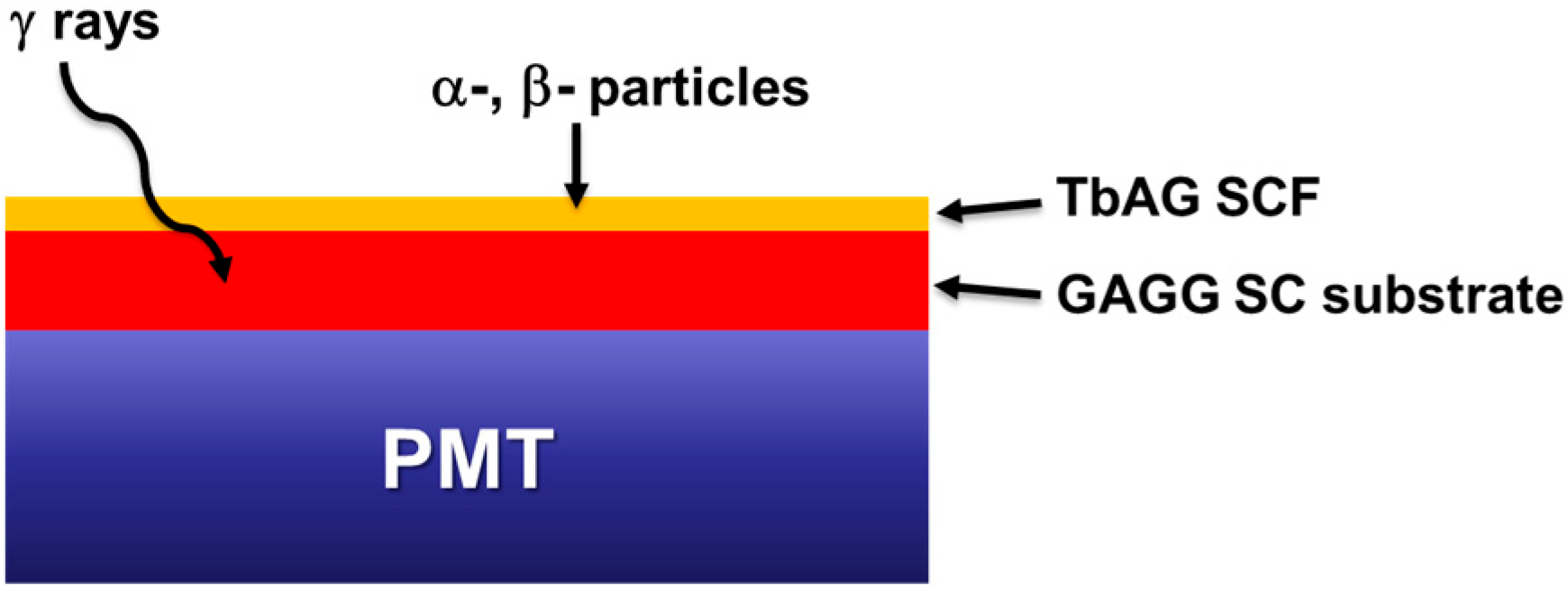
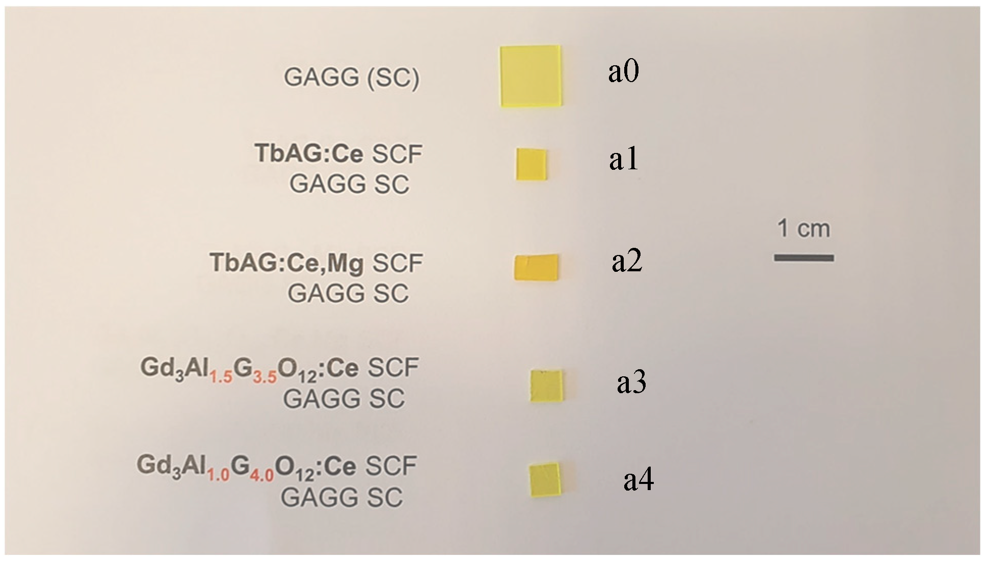
The composite scintillators under study were double-layered structures comprising SCF grown by the LPE method on GAGG:Ce bulk SC substrates (see the schematic layout in Figure 1) at the Epitaxy Laboratory of Chair for Optoelectronic Materials in the Institute of Physics of Kazimierz Wielki University (UKW) in Bydgoszcz, Poland. The crystal for the substrates was grown by the Czochralski method at approximately 1850 °C by the Institute for Materials Research (IMR), Tohoku University, Sendai, Japan. In this work, five samples (see Figure 2) were investigated:
  • GAGG:Ce (Gd3Al2.5Ga2.5O12:Ce bulk SC substrate), labelled as a0 sample;
  • TbAG:Ce/GAGG:Ce (Tb3Al5O12:Ce SCF grown onto GAGG:Ce SC substrate), a1 sample;
  • TbAG:Ce,Mg/GAGG:Ce (Tb3Al5O12:Ce,Mg SCF grown onto GAGG:Ce SC substrate), a2 sample;
  • Gd3Al1.5Ga3.5O12:Ce/GAGG:Ce (Gd3Al1.5Ga3.5O12:Ce SCF grown onto GAGG:Ce SC substrate), a3 sample;
  • Gd3AlGa4O12:Ce/GAGG:Ce (Gd3Al1.0Ga4.0O12:Ce SCF grown onto GAGG:Ce SC substrate), a4 sample.
Samples a1 and a2 were grown from a PbO–B2O3 flux, whereas samples a3 and a4 were prepared using the LPE method from a BaO–BaF2–BaCO3 flux. The use of different fluxes results in different segregation coefficients of Ga3+ cations (0.60–0.65 for the PbO-based flux and above 1.0 for the BaO-based flux). Therefore, for the SCF samples a3 and a4, grown from BaO-based flux, the Ga real content in the SCF samples was significantly higher (3.5–4) than the optimal range of 2.25–3. At such high Ga concentrations, the Ce3+ ions are buried by Ga-related electronic states forming the bottom of the conduction band of the respective garnet compounds [2,14]. This results in significantly lower LY for samples a3 and a4 compared with samples a1 and a2 (Table 1).
Furthermore, two double-layer composite scintillators, Gd3Al1.5Ga3.5O12:Ce SCF/GAGG:Ce SC (a3) and Gd3Al1Ga4O12:Ce SCF/GAGG:Ce SC (a4), also exhibit very low light yield and show no measurable response to α particles from 241Am due to their very high Ga content. For this reason, samples a3 and a4 were excluded from subsequent studies, as they cannot discriminate among mixed (α/β/γ) radiation. Table 1 presents a standard parameter set for the studied samples, including luminescence characteristics (light yield, decay time constants, emission peak wavelength), along with other physical parameters such as density, effective atomic number (Zeff), and growth temperature. The atomic percentage of Ce doping and Mg co-doping is also shown.
The Ce content in the GAGG:Ce SC scintillator, as determined by XRD microanalysis, was 0.0182 at.%. During liquid-phase epitaxy (LPE) growth in all SCFs scintillators investigated in this work, the nominal Ce3+ concentration in the melt was 10 mol%. However, the segregation coefficient of large Ce ions during LPE growth in garnet hosts is due to the very low (0.005–0.006) value, and the actual Ce3+ concentration incorporated into the SCFs was significantly lower. The resulting Ce3+ contents in the scintillating films were approximately 0.1 at.% for TbAG:Ce, 0.111 at.% for TbAG:Ce,Mg, 0.215 at.% for Gd3Al1.5Ga3.5O12:Ce, and 0.084 at.% for Gd3Al1Ga4O12:Ce. The Mg content in TbAG:Ce,Mg was 0.175 at.%.
The light yield (LY) of the investigated SCFs and SC substrate was evaluated relative to a reference YAG:Ce single-crystalline film, which exhibited a light yield of 2650 photons·MeV−1 under excitation by α particles from a 239Pu source (see Table 1). The decay time constants were estimated during the growth process: first for a bulk substrate, then for the grown composite scintillators, under excitation with α particles from 239Pu.

3. Methods

Each of five samples, the substrate GAGG:Ce SC and four double-layered composite scintillators, was sequentially coupled to a photomultiplier tube (PMT) window using silicon grease, which enhanced light collection at the PMT’s photocathode. The scintillator connected to the PMT (see the schematic layout in Figure 1) was placed in a lightproof box to protect the photocathode against ambient light. Pulse-height spectra (PHS) and decay kinetics under excitation of γ-rays of 137Cs (661 keV), α-particles (~5.5 MeV) and 59-keV γ-rays from 241Am, and β-particles from 90Sr/90Y and 14C were recorded and analyzed. The scintillator’s ability to discriminate mixed radiation was tested with 241Am combined with the second radioactive source emitting γ-rays: 60Co, 137Cs, and 51Cr for GAGG:Ce SC, TbAG:Ce SCF/GAGG SC, and TbAG:Ce,Mg SCF/GAGG SC, respectively. The radioactive sources used in this work are collected in Table 2.

3.1. α-, β- and γ Spectrometry

In spectrometry studies of a scintillator response to α-, β-, and γ-radiation, the output signal from a Hamamatsu R6231-100 PMT (Hamamatsu Photonics K.K., Hamamatsu City, Shizuoka, Japan) anode was fed to a charge-sensitive preamplifier and then to an ORTEC 672 high-performance energy spectroscopy amplifier (EG&G/ORTEC, Oak Ridge, TN, USA) and processed at a 10 μs shaping time constant. The Hamamatsu R6231-100 PMT features a high-quantum-efficiency photocathode for detecting Ce3+ emission at ~550 nm. The pulse-height (energy) spectra (PHS) were acquired using a TUKAN8k multichannel analyzer, MCA-TUKAN8k (National Centre for Nuclear Research, Otwock, Poland) [15].
The alpha source used (241Am) was electrodeposited onto a 0.127 mm thick platinum foil with a diameter of 1.27 cm. The active source diameter was nominally 3 mm, and the equivalent source thickness was less than 8 keV. The source holder was stainless steel with an outside diameter of 1.68 cm. The geometry of the source allowed the registration of α particles at a distance as low as 0.7 mm from the active source. Due to the low activity of the source, the distance between the active source and the scintillator was kept as close as possible. However, the greater distance and broadening of the energy spectrum do not limit the applicability of the PSD approach used in this work for discrimination among different radiation types.

3.2. Light Pulse Shapes

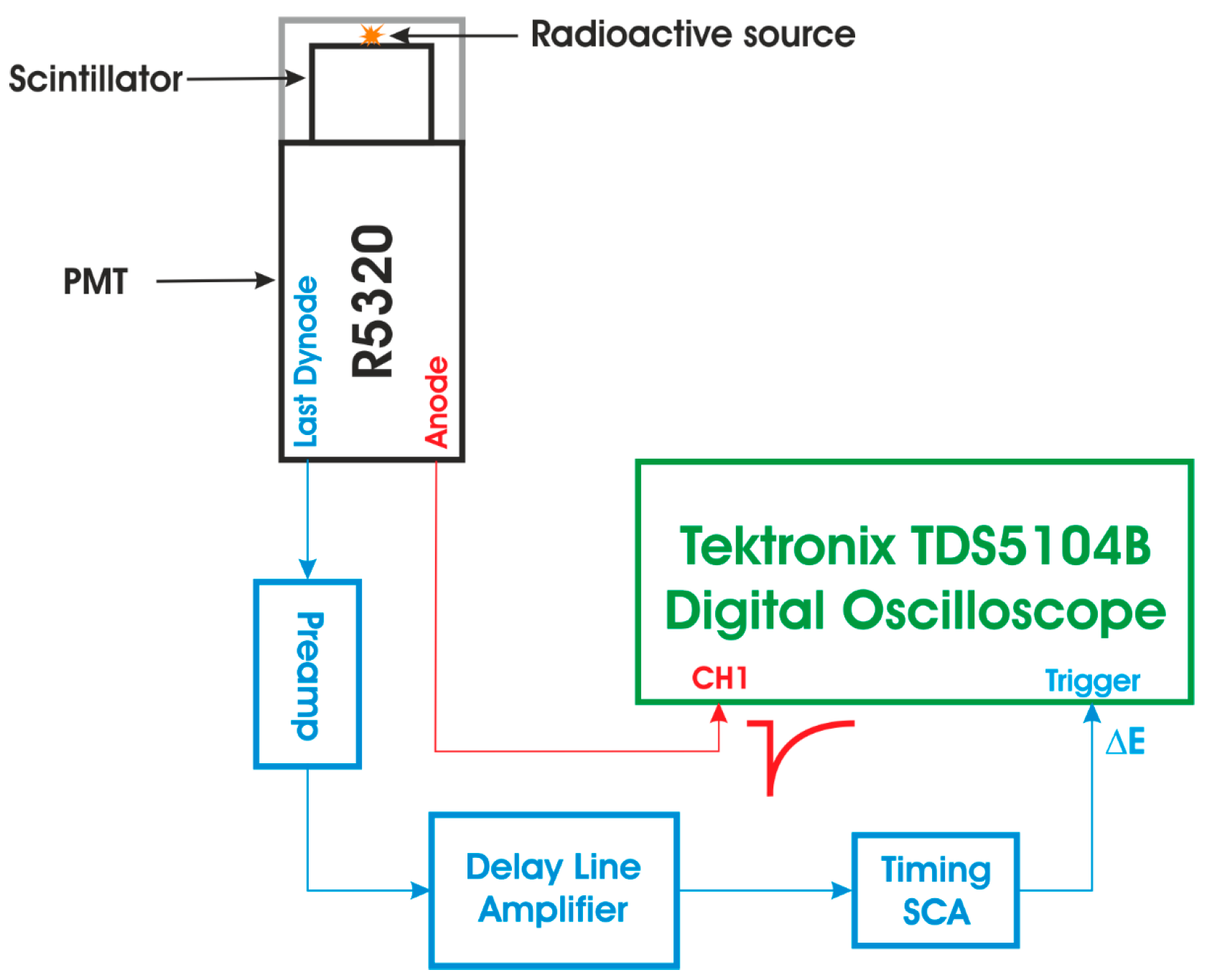
Time-profile measurements for the studied samples were performed for each type of ionizing radiation in separate runs. The method for measuring a single energy-gated light pulse using a digital oscilloscope was adapted from [16] to capture scintillation decays following interaction of the sample with α, β, or γ radiation. A fast Hamamatsu R5320 photomultiplier tube (PMT) (Hamamatsu Photonics K.K., Hamamatsu City, Shizuoka, Japan) [17], with a transit time of 10 ns, a low time jitter of 140 ps, and a rise time of 0.7 ns, was used as a photodetector for time-profile measurements. Anode pulses from the R5320 PMT were sent directly to the Tektronix TDS5104B Digital Oscilloscope, 1 GHz, 5 GS/s (Tektronix, Inc., Beaverton, OR, USA). At the same time, the last dynode signal was used to set an energy gate (energy window, ΔE) and to trigger data acquisition (measurements of the average light pulse shapes at a given energy deposition) [16]. The dynode signal was pre-amplified with a Canberra 2005 charge-sensitive preamplifier (Canberra Industries, Meriden, CT, USA) and sent to an ORTEC 460 Delay Line Amplifier (DLA) manufactured by EG&G/ORTEC, Oak Ridge, TN, USA. Then, the delayed signal from the DLA was fed into a TUKAN8k multichannel analyzer (MCA-TUKAN8k), which recorded pulse-height spectra from the dynode. The other DLA output signal (bipolar) was fed into an ORTEC 551 Timing Single-Channel Analyzer (TSCA) provided by EG&G/ORTEC, Oak Ridge, TN, USA, whose output signal triggered data acquisition in the Tektronix TDS5104B Digital Oscilloscope. The other output signal from TSCA was routed to an ORTEC 416A Gate and Delay Generator (EG&G/ORTEC, Oak Ridge, TN, USA) to generate the gate for data acquisition in the TUKAN8k multichannel analyzer (MCA-TUKAN8k). The energy window, ΔE, was adjusted with the TUKAN8k in coincidence with the gate signal from the Gate and Delay Generator. Figure 3 shows the schematic layout of the experimental setup used to measure the scintillation decay time at a given energy window (ΔE).
Scintillation time profiles were measured for the energy deposition corresponding to the ΔE = 662 keV full-energy peak (137Cs), the energy deposition of α-particle broad peak (~5.5 MeV 241Am), and the whole β-particle energy range (90Sr/90Y and 14C).

3.3. Pulse Shape Discrimination (PSD)

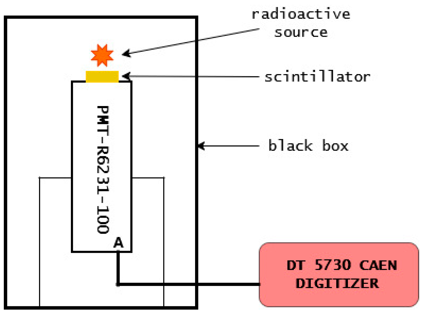
Digital pulse shape discrimination (PSD) to separate mixed radiation detected by scintillators was performed using an 8-channel, 14-bit, 500 MS/s CAEN DT5730 digitizer (2 Vpp dynamic range) running Digital Pulse Processing (DPP) firmware designed for multiparametric data acquisition. The CAEN DT5730 digitizer was provided by CAEN S.p.A., Viareggio, Italy. It enables simultaneous recording of energy and PSD information. The simple experimental setup for the PSD measurements is shown in Figure 4. The tested samples were coupled to the Hamamatsu R6233-100 PMT (Hamamatsu Photonics K.K., Hamamatsu City, Shizuoka, Japan) with silicon grease and placed in a black box in a vertical orientation (see Figure 4). The PSD performance of composite materials was tested with 65Zn + 241Am (GAGG:Ce SC), 65Zn + 241Am (TbAG:Ce SCF/GAGG:Ce SC) and 51Cr + 241Am (TbAG:Ce,Mg SCF/GAGG:Ce SC).
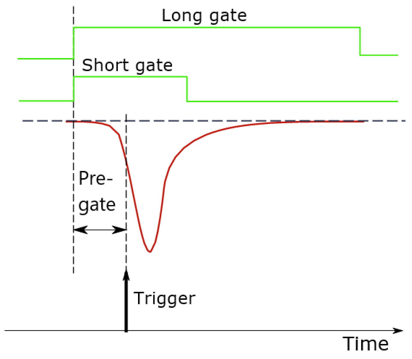
The CAEN DT5730 digitizer firmware provides a standard PSD option [18]. It is based on the charge comparison method (CCM), in which two integrals of the input signal are compared, and the PSD parameter is calculated as (Qlong gate − Qshort gate)/Qlong gate; see Figure 5. By definition, the Qlong gate integral should cover the entirety of the signal, thus representing the incident particle energy deposition. In case of several longer-lived components of the light pulse, the Qlong gate is chosen to give the optimal particle discrimination. The second integral, the Qshort gate, covers only a small portion of the signal. The pre-gate corresponds to the starting position of the long and short gates, before the trigger time. If α particle and γ-rays exhibit different scintillation light pulse shapes where intensities of light pulse components vary with the type of impinging particle, they will separate from each other in the 2D heatmap of PSD values as a function of particle energy.
The discrimination capability of the studied a0, a1 and a2 composite scintillators was quantified by Figure-of-Merit values, FoM(PSD), calculated as follows:
F o M P S D = C α C γ F W H M α + F W H M γ
where Cα and Cγ stand for centroids of the peaks corresponding to α particles and γ-rays detection as projected onto the PSD parameter axis. FWHM stands for the full width at half maximum of the alpha and gamma peaks determined by fitting a Gaussian distribution to each PSD peak.

4. Results

4.1. α-, β- and γ Spectrometry

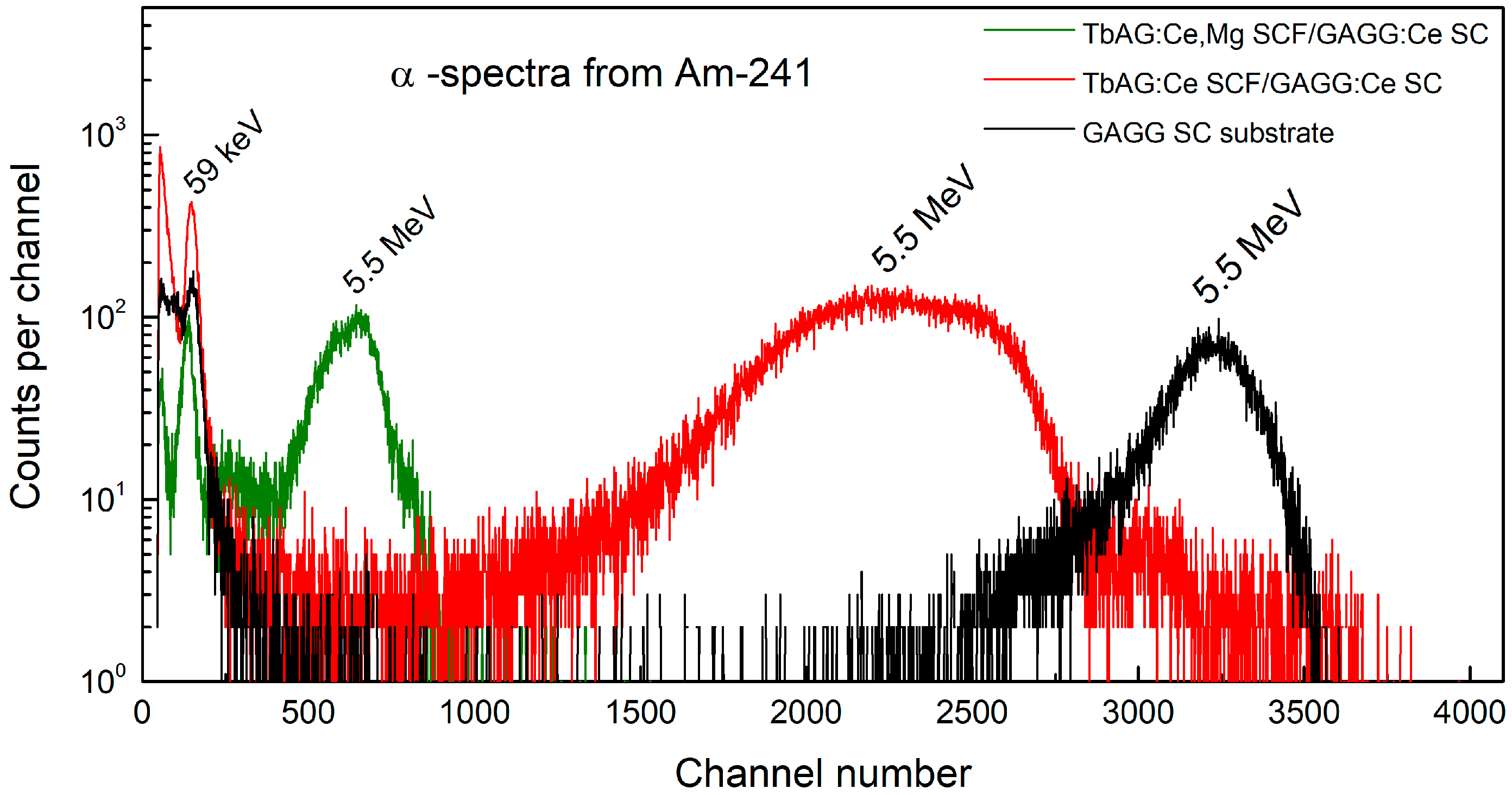
Figure 6 shows the α spectra measured for TbAG:Ce SCF/GAGG:Ce SC and TbAG:Ce,Mg SCF/GAGG:Ce SC composite scintillators in comparison with GAGG:Ce SC substrate under excitation by 5.5-MeV α particles from 241Am. The spectra were recorded at the same amplifier’s gain to compare the relative positions of the broad α peaks of the studied scintillators. Due to the uncollimated 241Am source used in the measurements and the ~0.7 mm distance between the source and the scintillator, all studied samples exhibited broader α peaks with distorted energy spectra.
For the TbAG:Ce SCF/GAGG:Ce SC (a1) scintillator, two partially overlapping peaks were registered at 10 μs shaping time. Additional measurement at short (0.5 μs) shaping time values showed that both α peaks were well resolved, suggesting a folded response of the TbAG:Ce SCF/GAGG:Ce SC composite to α-particles. In the case of TbAG:Ce,Mg SCF/GAGG:Ce SC (a2), the quenching of light output was observed for all types of radiation due to Mg2+ co-doping of the SCF scintillator.
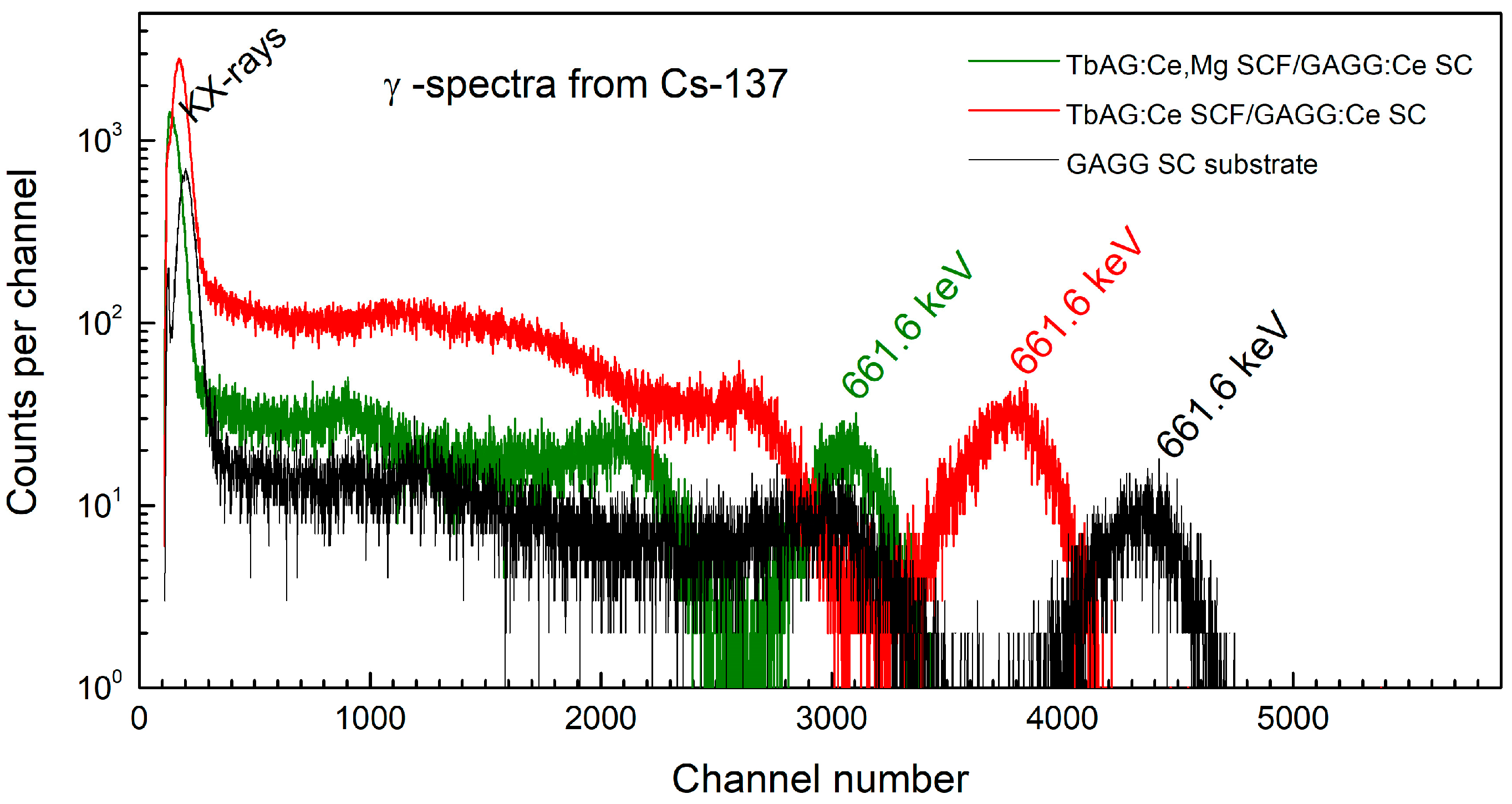
Figure 7 depicts the γ-ray spectra of composite scintillators recorded for 137Cs. The most substantial decrease in the light output for TbAG:Ce,Mg SCF component is due to the presence of Mg2+ co-doping. The two other composite scintillators, Gd3Al1.5Ga3.5O12:Ce SCF/GAGG:Ce SC (a3) and Gd3Al1.0Ga4.0O12:Ce SCF/GAGG:Ce SC (a4), did not show any measurable response to α-particles from 241Am (see Section 2). This behaviour is attributed to a pronounced reduction in radiative emission following absorption of highly ionizing α radiation, resulting from the complete quenching of Ce3+ luminescence due to the overlap of the Ce3+ excited levels with the bottom of the conduction band of the GAGG host [14].
Figure 8 shows measured responses of the TbAG:Ce,Mg SCF/GAGG:Ce SC composite scintillator to α particles from 241Am, β particles from 90Sr/90Y and 14C, and γ-rays from 137Cs. The spectra were recorded at the same shaping time and amplifier gain.

4.2. Light Pulse Shapes

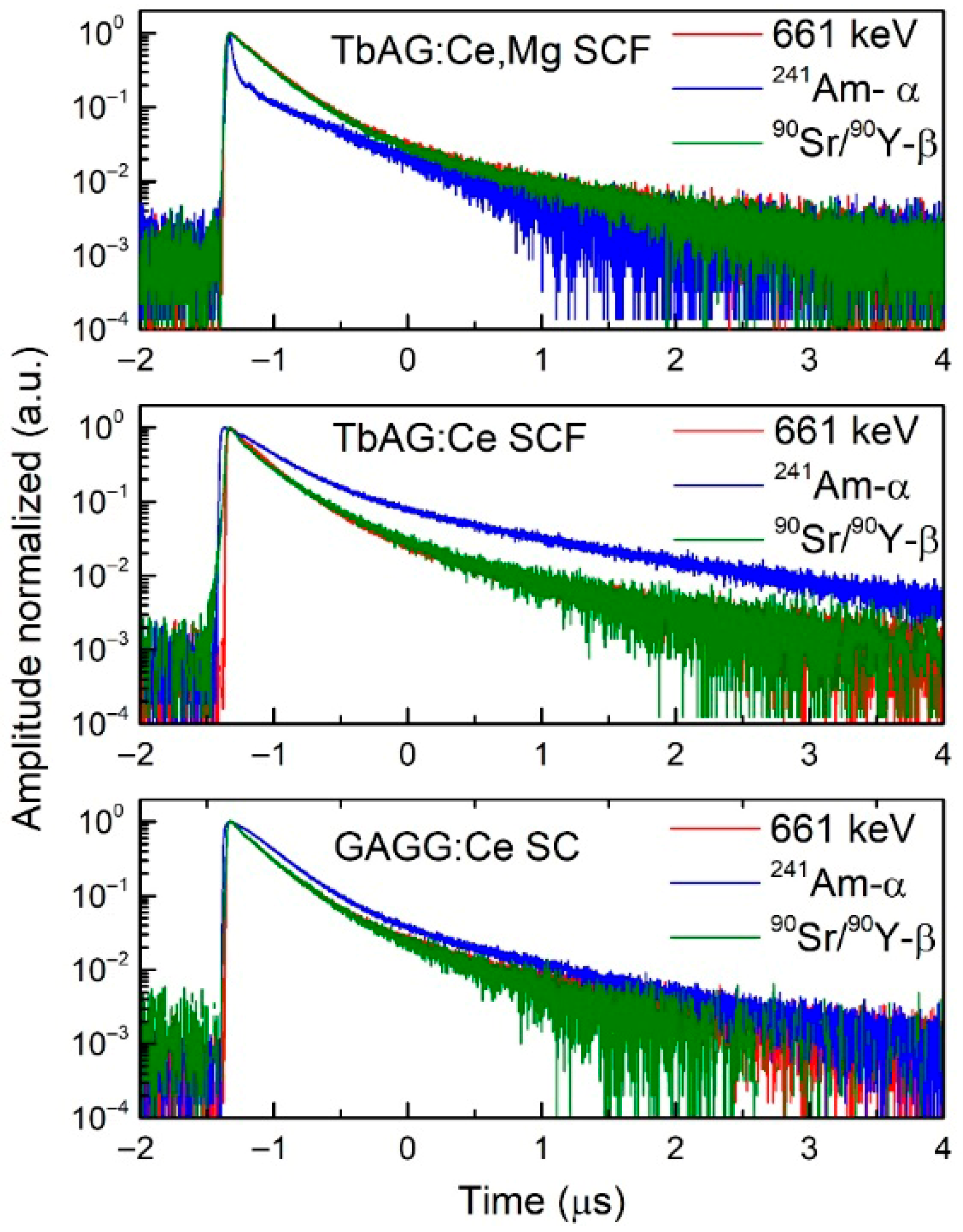
The decay time for each scintillator was measured using a Tektronix TDS5104B digital oscilloscope, which recorded 1000 averaged waveforms from a fast Hamamatsu R5320 PMT. The plots of time profiles for three tested scintillators, TbAG:Ce SCF/GAGG:Ce SC (a1), TbAG:Ce,Mg SCF/GAGG:Ce SC (a2), and GAGG:Ce SC substrate (a0), are presented in Figure 9. TbAG:Ce SCF/GAGG:Ce SC (a1) and TbAG:Ce,Mg SCF/GAGG:Ce SC (a2) are characterized by a distinct time profile for alphas when compared to γ-rays and β particles. Acceleration of the scintillation decay kinetics was achieved via co-doping TbAG:Ce with divalent Mg2+ ions.
To describe quantitatively the light pulse shape of a scintillator that de-excites via two or more (up to k) decay modes, the following analytical fitting function was used:
y(t) = ∑(i=1 to k)Ai exp(−(t − t0)/τi) + y0,
where τi is the decay time constant, Ai is the amplitude of the ith-component, and t0 and y0 are offsets in the fitting function. The intensity Ii of component i at each deposited energy E was calculated as a product of the amplitude Ai and decay time constant τi resulting from a fit to a registered averaged scintillation pulse:
Ii = Ai τi,
Due to slow (submicrosecond) decay modes in the studied samples, a timescale was set up to 10 μs in the oscilloscope.
The decay times, τi, and intensities Ii of scintillation light components of the three samples capable of discriminating α- and γ-radiation are depicted in Table 3. The scintillation decay times measured in this work for TbAG:Ce SCF/GAGG:Ce SC at the 662-keV energy window (137Cs) and ~5 MeV energy window (241Am-alpha) are comparable to those presented in [10], where they were measured for scintillation pulses corresponding to the whole energy spectrum. Both types of TbAG:Ce SCF/GAGG:Ce SC and TbAG:Ce,Mg SCF/GAGG:Ce SC composites show good ability for the simultaneous registration of the mentioned components in mixed radiation fields. This ability, reflected in differences in decay time constants, can be qualitatively described using a Figure-of-Merit (FoM(τ)) value that is defined for each pair of ionizing radiations as follows:
FoM(τ) (α/γ) = |(τα − τγ)|/(τα + τγ)
FoM(τ) (α/β) = |(τα − τβ)|/(τα + τβ)
FoM(τ) (β/γ) = |(τβ − τγ)|/(τβ + τγ)
Table 3 presents the FoM(τ) values for the three tested samples: GAGG:Ce substrate, TbAG:Ce SCF/GAGG: Ce SC, and TbAG:Ce,Mg SCF/GAGG:Ce SC excited with α particles (the broad peak around 5.5 MeV), β particles (continuous energy spectrum), and γ-rays (the full-energy peak at 662 keV). The studied samples were characterized by two scintillation components (τ1, I1; τ2, I2); thus, the FoM(τ) was calculated separately for each component. FoM(τ) values greater than 0.2 indicate a good ability of the tested sample to discriminate different types of exciting radiation.

4.3. Pulse Shape Discrimination (PSD)

The observed difference in scintillation light pulse shapes (see Figure 9), expressed by the FoM(tau) values around 0.2 or greater, as shown in Table 3, suggests that TbAG:Ce/GAGG:Ce (a1) and TbAG:Ce,Mg/GAGG:Ce (a2) composite scintillators are capable of discriminating α particles against β particles and γ rays. Thus, both double-layer samples and the GAGG:Ce SC (a0) substrate (used for comparison) were subjected to digital PSD analysis based on a charge comparison algorithm using the CAEN5730 high-performance waveform digitizer (see Section 3.3). The discriminator settings used in the measurements were as follows: leading-edge mode, 12 mV threshold, 50 ns pre-gate and 144 ns pre-trigger. The entire waveform record lasted 4000 ns. The optimal values of the short gate and long gate were as follows: short gate = 150 ns, long gate = 1000 ns (GAGG:Ce substrate, a0), short gate = 270 ns, long gate = 1400 ns (a1), and short gate = 80 ns, long gate = 800 ns (a2).
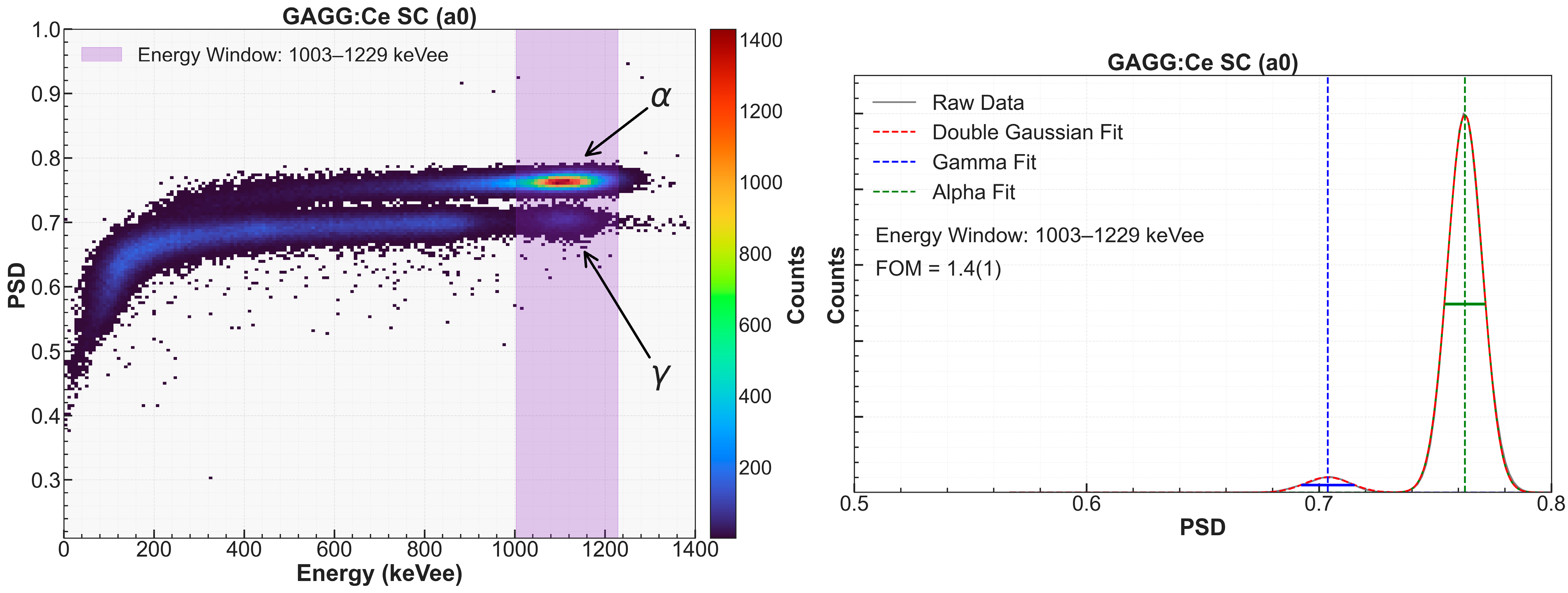
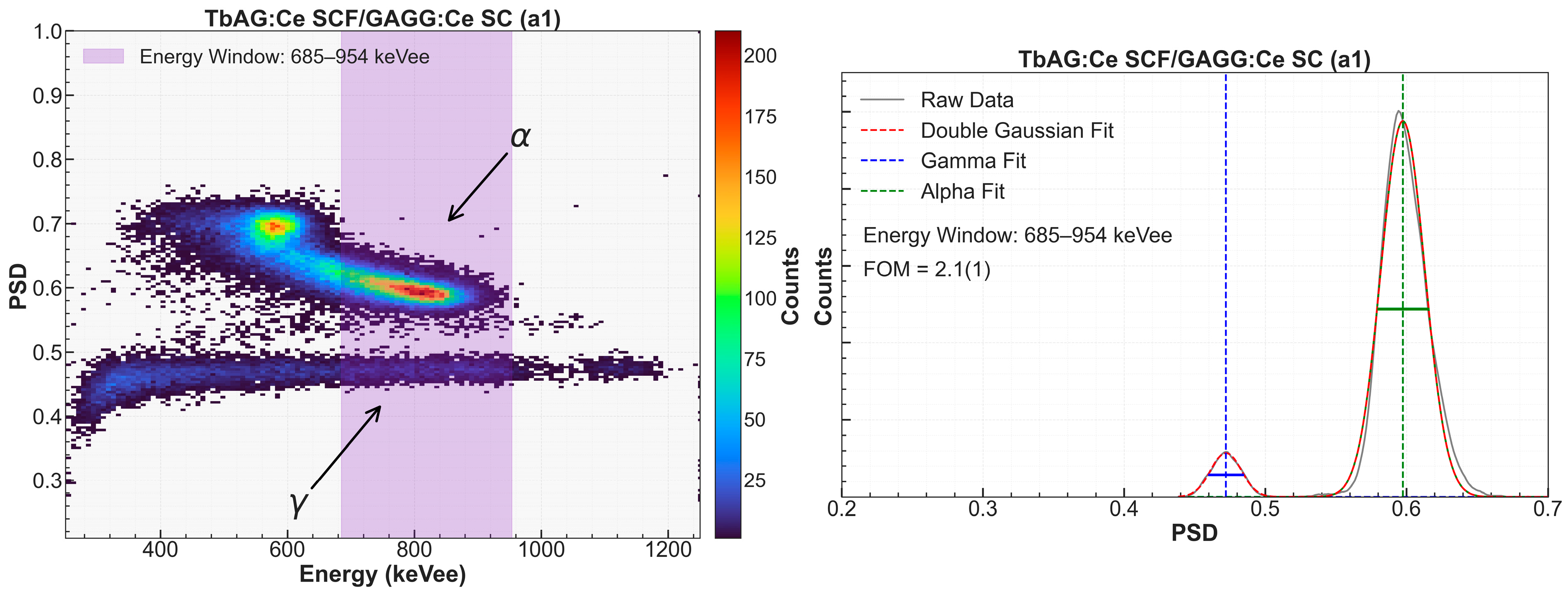
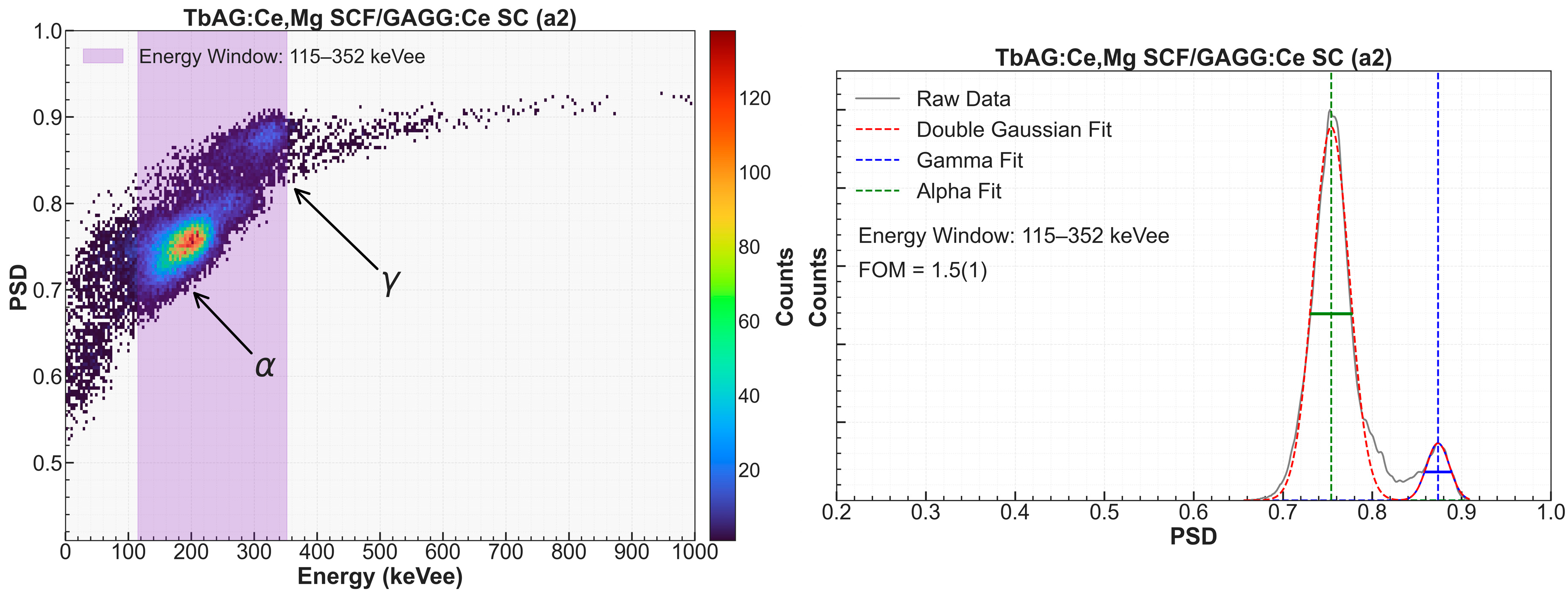
Negligible differences in scintillation time profiles of γ-rays and β particles absorbed in the GAGG:Ce substrate (see Table 3) did not allow discriminating between these two radiation types via the charge comparison method. More pronounced difference in scintillation decay times is observed for α particles and γ-rays (or β particles). Figure 10, Figure 11 and Figure 12 show 2D heatmaps (left panels) of the PSD parameter versus deposited energy expressed in keVee (kiloelectronvolt electron-equivalent) unit for GAGG:Ce single crystal (substrate, a0), TbAG:Ce SCF/GAGG:Ce SC (a1) and TbAG:Ce,Mg SCF/GAGG:Ce SC (a2), respectively. Note that the scale labelled in keVee is valid just for electrons interacting within the scintillator; the primary α particle’s energy is not properly translated into the keVee scale due to the light quenching that is observed when detecting highly ionizing particles.
The right panels in Figure 10, Figure 11 and Figure 12 represent the PSD distribution at a given energy window. Alpha and gamma peaks were fitted with a double Gaussian function, and relevant FoM(PSD) values were determined. The higher the energy deposited in the scintillator, the better FoM(PSD) value is measured [7,8,9]. Hence, a scintillator with a high FoM(PSD) value measured at lower energies is the most required. The superior α–γ discrimination is observed for a1 where α particles from 241Am were simultaneously measured with γ-rays from 65Zn (1115.6-keV full-energy peak). Yet, the energy spectrum exhibited a broad structure, indicating a complex interaction between alphas and the TbAG:Ce SCF/GAGG:Ce SC (a1) composite. The FoM(PSD) value for the 685–954 keVee energy slice was found to be as good as 2.1(1). Good α-γ separation was also achieved for a2, with FoM(PSD) equal to 1.5(1), where the energy window was set to the low-energy range of 115–352 keVee. In case of GAGG:Ce substrate (a0), high light yield for α particles allowed us to determine FoM(PSD) at a high-energy slice between 1003 and 1229 keVee, which achieved the value of 1.4(1).

5. Discussion

The results presented in this work demonstrate that composite scintillators based on TbAG:Ce and TbAG:Ce,Mg SCFs grown on GAGG:Ce SC substrates exhibit distinct scintillation responses to α, β, and γ radiation, enabling effective particle discrimination in mixed radiation fields. The observed behaviour results from a combination of the layered detector architecture, differences in stopping power for charged particles, and intrinsic differences in scintillation decay kinetics between the film and substrate materials.
In the composite structures studied, short-range α particles and low-energy β particles predominantly deposit their energy in the thin TbAG-based SCF layer. In contrast, higher-energy γ rays interact mainly within the bulk GAGG:Ce SC substrate. This spatial separation of energy deposition is reflected in the PHS, where folded or partially overlapping peaks are observed for α excitation in TbAG:Ce SCF/GAGG:Ce SC composites, confirming the contribution of both scintillating layers. Such behaviour is consistent with earlier reports on multilayer garnet-based scintillators [1] and confirms the suitability of epitaxial SCF/SC structures for radiation detection in mixed fields.
It is also important to emphasize that many previously reported scintillators primarily demonstrate α–γ or neutron–γ discrimination. In contrast, the TbAG-based composite scintillators investigated in this work enable simultaneous detection and discrimination of α, β, and γ radiation components within a single detector architecture, which is particularly advantageous for applications involving complex radiation environments.
Analysis of scintillation decay kinetics further reveals pronounced differences among α-, β-, and γ-induced light pulses, especially for the TbAG:Ce and TbAG:Ce,Mg-based composite scintillators. Both materials exhibit at least two dominant decay components whose relative intensities and time constants depend on the type of incident radiation. The resulting FoM(τ) values, which exceed 0.2 for several radiation pairs, indicate a significant intrinsic capability for pulse-shape-based particle discrimination.
Co-doping of TbAG:Ce SCFs with Mg2+ ions leads to a substantial modification of scintillation kinetics. The acceleration of decay components observed in TbAG:Ce,Mg SCFs is consistent with earlier studies reporting that divalent co-dopants influence charge-compensation mechanisms and trap-related processes in Ce-activated scintillators [11,12,13]. In garnet lattices, the introduction of Mg2+ ions may promote charge compensation through the partial conversion of Ce3+ to Ce4+ states. This process modifies the recombination dynamics of charge carriers and reduces the probability of carrier trapping in shallow defect states. As a result, delayed recombination channels associated with trapping and detrapping processes are suppressed, leading to faster scintillation decay kinetics.
The modification of trap populations and carrier transport pathways also affects the relative contributions of fast and slow scintillation components under different types of ionizing radiation. Because highly ionizing α particles generate a higher density of electron–hole pairs than γ radiation, these changes enhance the differences in recombination dynamics for the two excitation conditions. Consequently, Mg2+ co-doping increases the contrast between α- and γ-induced scintillation pulse shapes, thereby improving pulse-shape discrimination capability.
At the same time, Mg co-doping reduces the overall light yield. However, the increased difference between scintillation pulse shapes generated by α and γ excitation improves α/γ discrimination in digital PSD measurements. This trade-off between light yield and discrimination performance represents an important factor in optimizing scintillator materials for specific radiation detection applications.
The present study also demonstrates that composite structures based on Gd–Al–Ga garnet SCFs with high Ga content (Gd3Al1.5Ga3.5O12:Ce and Gd3Al1Ga4O12:Ce) are not suitable for α detection due to strong quenching of Ce3+ luminescence. This behaviour can be explained by band-structure effects, in which Ga-related electronic states overlap with the excited levels of Ce3+ ions, thereby increasing non-radiative recombination. These observations highlight the importance of careful band-gap engineering and composition control in the design of multilayer scintillators intended for mixed-field radiation detection.
Digital PSD measurements using a high-speed waveform digitizer confirm and extend the conclusions drawn from the analogue decay time analysis. The two-dimensional PSD heatmaps clearly demonstrate separation of α and γ events for both TbAG:Ce SCF/GAGG:Ce SC and, in particular, TbAG:Ce,Mg SCF/GAGG:Ce SC composites. The superior α–γ separation observed for the Mg-co-doped composite indicates that tailoring scintillation kinetics through controlled co-doping represents an effective strategy for enhancing PSD performance in inorganic composite scintillators.
Compared with previously reported SCF/SC garnet composite scintillators [1,10], the structures investigated in this work introduce two important modifications. First, the use of TbAG-based epitaxial films on GAGG:Ce substrates provides a larger difference in scintillation kinetics between the film and substrate components. Second, Mg2+ co-doping of the TbAG:Ce film allows controlled tuning of decay times, which enhances differences between α- and γ-induced scintillation pulses. As a result, the TbAG:Ce,Mg SCF/GAGG:Ce SC composite demonstrates improved pulse-shape discrimination behaviour, as confirmed by both FoM(τ) analysis and digital PSD measurements. These features highlight the potential of TbAG-based epitaxial composites as a versatile platform for detectors operating in mixed radiation environments.
To place the discrimination performance obtained in this work in a broader context, it is useful to compare the FoM values with those reported for other inorganic scintillators capable of particle discrimination. In previously reported composite garnet scintillators based on SCF/SC structures, FoM(τ) values for α–γ discrimination typically range from approximately 0.2 to 0.5 when evaluated using decay time differences or digital PSD techniques [1,10]. Similar performance levels have been reported for several Ce-doped inorganic scintillators, in which differences in scintillation kinetics under high-ionization-density excitation enable particle identification.
In the present study, the TbAG:Ce SCF/GAGG:Ce SC and TbAG:Ce,Mg SCF/GAGG:Ce SC composite scintillators exhibit FoM(τ) values exceeding 0.2 for several radiation pairs and digital PSD FoM values greater than 1.0. These results indicate a discrimination capability comparable to or better than that observed in previously reported garnet-based composite scintillators. In particular, the TbAG:Ce,Mg SCF/GAGG:Ce SC structure demonstrates improved α–γ separation due to the modification of scintillation kinetics induced by Mg2+ co-doping (Table 4). This comparison confirms that tailoring scintillation decay characteristics in layered garnet structures represents an effective strategy for enhancing particle discrimination performance in inorganic scintillators.
Overall, the combination of high density and effective atomic number in garnet materials, together with engineered scintillation kinetics and layered detector geometry, provides a versatile platform for developing composite scintillators capable of simultaneous α/β/γ detection and discrimination. The present results are consistent with previous studies on garnet-based SCF/SC composites and further extend their applicability to TbAG-based systems.
Future research may focus on further optimizing the multilayer scintillator architecture to improve both light yield and particle discrimination efficiency. In particular, systematic studies of SCF and substrate thickness, their content, and Ce3+ concentration could help optimize the balance between scintillation efficiency and pulse-shape discrimination performance. In addition, exploring other co-doping ions (e.g., Ca2+, Sr2+, or aliovalent dopants such as Pb2+) may offer further opportunities to control charge-compensation processes, trap populations, and recombination dynamics in garnet-based scintillators. Finally, evaluation of the developed composite scintillators under realistic radiation fields, including mixed neutron–gamma or alpha–gamma environments, is important in assessing their practical applicability in radiation detection systems.

6. Conclusions

In this work, novel double-layer composite scintillators based on TbAG:Ce and TbAG:Ce,Mg single-crystalline films, grown by liquid-phase epitaxy on GAGG:Ce single-crystal substrates, were systematically investigated under α, β, and γ radiation.
The main conclusions can be summarized as follows:
The TbAG-based SCF/GAGG:Ce composite scintillators exhibit apparent differences in scintillation response and decay kinetics for α, β, and γ excitation, enabling effective discrimination of mixed radiation fields.
Scintillation decay time analysis shows multiple decay components whose relative contributions depend on the type of incident radiation. The resulting Figure-of-Merit values (FoM(τ) ≥ 0.2 for several cases) confirm good intrinsic discrimination capability.
Mg2+ co-doping of TbAG:Ce significantly accelerates scintillation decay and enhances differences in pulse shapes between α and γ excitation. Although the light yield is reduced, the TbAG:Ce,Mg SCF/GAGG:Ce SC composite demonstrates superior α–γ discrimination in digital PSD measurements.
Digital pulse shape discrimination using a fast waveform digitizer confirms the capability of both TbAG:Ce SCF/GAGG:Ce SC and TbAG:Ce,Mg SCF/GAGG:Ce SC composites to separate α and γ events (FoM(PSD) > 1.0), with the best performance observed for the Mg co-doped composite at low energy depositions.
The results obtained demonstrate that TbAG-based composite scintillators are promising candidates for applications requiring simultaneous detection and discrimination of α, β, and γ radiation, including radiation monitoring, dosimetry, and mixed-field measurements. Future work will focus on developing double- and multilayer composite architectures and further optimizing composition and co-doping strategies to achieve an optimal balance among light yield, timing characteristics, and discrimination performance.

Author Contributions

Conceptualization, A.S.-K. and Y.Z.; methodology, investigation, analyses A.S.-K., T.S. and A.B.; data curation, A.S.-K.; writing—original draft preparation, A.S.-K.; writing—review and editing, A.S.-K.,Y.Z., T.Z., O.S. and Y.S.; growth of films, V.I.G.; data visualization, A.S.-K., K.B. and A.B. All authors have read and agreed to the published version of the manuscript.

Funding

The work was performed in the frame of the Polish National Science Centre (NCN) Opus 24 LAP No 2022/47/I/ST8/02600 and Opus 28 No 2024/55/B/ST7/02680 projects.

Data Availability Statement

The raw data supporting the conclusions of this article will be made available by the authors on request.

Conflicts of Interest

Author Oleg Sidletskiy was employed by the company Centre of Excellence ENSEMBLE3 Sp. z o.o. The remaining authors declare that the research was conducted in the absence of any commercial or financial relationships that could be construed as a potential conflict of interest.

References

  1. Witkiewicz-Lukaszek, S.; Gorbenko, V.; Zorenko, T.; Syrotych, Y.; Mares, J.A.; Nikl, M.; Sidletskiy, O.; Bilski, P.; Yoshikawa, A.; Zorenko, Y. Composite Detectors Based on Single-Crystalline Films and Single Crystals of Garnet Compounds. Materials 2022, 15, 1249. [Google Scholar] [CrossRef] [Scilit] [PubMed]
  2. Kamada, K.; Yanagida, T.; Pejchal, J.; Nikl, M.; Endo, T.; Tsutsumi, K.; Fujimoto, Y.; Fukabori, A.; Yoshikawa, A. Crystal Growth and Scintillation Properties of Ce Doped Gd3Al2Ga3O12 Single Crystals. IEEE Trans. Nucl. Sci. 2012, 59, 2112–2115. [Google Scholar] [CrossRef] [Scilit]
  3. Zorenko, Y.; Douissard, P.-A.; Martin, T.; Riva, F.; Gorbenko, V.; Zorenko, T.; Paprocki, K.; Iskalieva, A.; Witkiewicz, S.; Fedorov, A.; et al. Scintillating screens based on the LPE grown Tb3Al5O12:Ce single crystalline films. Opt. Mater. 2017, 65, 73–81. [Google Scholar] [CrossRef] [Scilit]
  4. Markovskyi, A.; Gorbenko, V.; Zorenko, T.; Yokosawa, T.; Will, J.; Spiecker, E.; Batentschuk, M.; Elia, J.; Fedorov, A.; Zorenko, Y. LPE growth of Tb3Al5O12:Ce single crystalline film converters for WLED application. CrystEngComm 2021, 23, 3212–3219. [Google Scholar] [CrossRef] [Scilit]
  5. Sosa, C.S.; Flaska, M.; Pozzi, S.A. Comparison of analog and digital pulse-shape-discrimination systems. Nucl. Instrum. Methods A 2016, 826, 72–79. [Google Scholar] [CrossRef] [Scilit]
  6. Zaitseva, N.; Rupert, B.; Pawełczak, I.; Glenn, A.; Martinez, H.P.; Carman, L.; Faust, M.; Cherepy, N.; Payne, S. Plastic scintillators with efficient neutron/gamma pulse shape discrimination. Nucl. Instrum. Methods A 2012, 668, 88–93. [Google Scholar] [CrossRef] [Scilit]
  7. Grodzicka-Kobylka, M.; Szczesniak, T.; Moszyński, M.; Brylew, K.; Swiderski, L.; Valiente-Dobon, J.J.; Schotanus, P.; Grodzicki, K.; Trzaskowska, H. Fast neutron and gamma ray pulse shape discrimination in EJ-276 and EJ-276G plastic scintillators. J. Instrum. 2020, 15, P03030. [Google Scholar] [CrossRef] [Scilit]
  8. Syntfeld-Każuch, A.; Swiderski, L.; Szczesniak, T.; Grodzicka-Kobylka, M.; Brylew, K.; Mianowska, Z.; Maiatska, O.; Pausch, G.; Stein, J.; Ritter, H. Characterization of a novel polyurethane-based plastic scintillator for neutron and gamma detection in mixed radiation fields. Nucl. Instrum. Methods A 2024, 1068, 169764. [Google Scholar] [CrossRef] [Scilit]
  9. Maiatska, O.; Dunnebacke, T.; Kreuels, M.; Pausch, G.; Scherwinski, F.; Stein, J. Polyurethane-based scintillators for neutron and gamma radiation detection in medical and industrial applications. Radiat. Meas. 2026, 193, 107647. [Google Scholar] [CrossRef] [Scilit]
  10. Witkiewicz-Lukaszek, S.; Gorbenko, V.; Zorenko, T.; Paprocki, K.; Sidletskiy, O.; Fedorov, A.; Kucerkova, R.; Mares, J.A.; Nikl, M.; Zorenko, Y. Epitaxial growth of composite scintillators based on Tb3Al5O12:Ce single crystalline films and Gd3Al2.5Ga2.5O12:Ce crystal substrates. CrystEngComm 2018, 20, 3994–4002. [Google Scholar] [CrossRef] [Scilit]
  11. Koschan, M.A.; Melcher, C.L.; Szupryczynski, P.; Carey, A.A. Lutetium Oxyorthosilicate Scintillator Having Improved Scintillation and Optical Properties and Method of Making the Same. U.S. Patent No. 8278624, 2 October 2012. [Google Scholar]
  12. Yang, K.; Melcher, C.L.; Rack, P.D.; Eriksson, L. Effects of Calcium Codoping on Charge Traps in LSO:Ce Crystals. IEEE Trans. Nucl. Sci. 2009, 56, 2960–2965. [Google Scholar] [CrossRef] [Scilit]
  13. Lucchini, M.T.; Babin, V.; Bohacek, P.; Gundacker, S.; Kamada, K.; Nikl, M.; Petrosyan, A.; Yoshikawa, A.; Auffray, E. Effect of Mg2+ ions co-doping on timing performance and radiation tolerance of Cerium doped Gd3Al2Ga3O12 crystals. Nucl. Instrum. Methods A 2016, 816, 176–183. [Google Scholar] [CrossRef] [Scilit]
  14. Fasoli, M.; Vedda, A.; Nikl, M.; Jiang, C.; Uberuaga, B.; Andersson, D.A.; McClellan, K.J.; Stanek, C.R. Band-Gap Engineering for Removing Shallow Traps in Rare-Earth Lu3Al5O12 Garnet Scintillators Using Ga3+ Doping. Phys. Rev. B 2011, 84, 081102. [Google Scholar] [CrossRef] [Scilit]
  15. Guzik, Z.; Borsuk, S.; Traczyk, K.; Plominski, M. TUKAN-an 8K pulse height analyzer and multi-channel scaler with a PCI or a USB interface. IEEE Trans. Nucl. Sci. 2006, 53, 231–235. [Google Scholar] [CrossRef] [Scilit]
  16. Swiderski, L.; Moszynski, M.; Syntfeld-Kazuch, A.; Szawlowski, M.; Szczesniak, T. Measuring the scintillation decay time for different energy depositions in NaI:Tl, LSO:Ce and CeBr3 scintillators. Nucl. Instrum. Methods A 2014, 749, 68–73. [Google Scholar] [CrossRef] [Scilit]
  17. Moszyński, M.; Kapusta, M.; Nassalski, A.; Szczesniak, T.; Wolski, D.; Eriksson, L. New Prospects for Time-of-Flight PET with LSO Scintillators. IEEE Trans. Nucl. Sci. 2006, 53, 2484–2488. [Google Scholar] [CrossRef] [Scilit]
  18. CAEN DT5730 Digitizer. Available online: https://www.caen.it/products/dt5730/ (accessed on 14 February 2014).
Figure 1. The principal scheme of a composite scintillator optically connected to the photomultiplier tube (PMT), which serves as a photodetector.
Crystals 16 00230 g001
Figure 2. The GAGG:Ce SC substrate and composite SCF–SC scintillators measured in this work. The different stoichiometric numbers for the a3 and a4 samples, compared with the GAGG:Ce SC substrate are marked in red (see details in the text).
Crystals 16 00230 g002
Figure 3. Schematic of the experimental setup used to measure the scintillation decay time at a given energy window (ΔE) (not to scale).
Crystals 16 00230 g003
Figure 4. Schematic of the experimental setup for the PSD measurements (not to scale).
Crystals 16 00230 g004
Figure 5. Illustration of the PSD approach implemented in the CAEN digitizer used in this work. In the PSD measurements in this work, the red curve represents the anode pulse from the PMT, and the dashed line is the baseline.
Crystals 16 00230 g005
Figure 6. The pulse-height spectra (PHS) (in log scale) of TbAG:Ce SCF/GAGG:Ce SC (a1) and TbAG:Ce,Mg SCF/GAGG:Ce SC (a2) composite scintillators in comparison with GAGG:Ce SC substrate (a0) under excitation by 5.5-MeV α particles and 59-keV γ rays from 241Am. The shaping time of 10 μs was set in the amplifier.
Crystals 16 00230 g006
Figure 7. The pulse-height spectra (PHS) (in log scale) as measured for GAGG substrate (a0), TbAG:Ce SCF/GAGG SC (a1), and TbAG:Ce,Mg SCF/GAGG SC (a2) with 137Cs at 10 μs shaping time constant.
Crystals 16 00230 g007
Figure 8. The pulse-height spectra (PHS) (in log scale) of the TbAG:Ce,Mg SCF/GAGG:Ce SC (a2) composite scintillator registered for 137Cs (γ-source), 241Am (α-source), 90Sr/90Y (β-source) and 14C (β-source) at the 10 μs shaping time set in the spectroscopy amplifier.
Crystals 16 00230 g008
Figure 9. The light pulse shapes of TbAG:Ce SCF/GAGG:Ce SC, TbAG:Ce,Mg SCF/GAGG:Ce SC and GAGG:Ce SC substrate under excitation by 661-keV γ rays, 5.5-MeV α particles (241Am), and β particles (90Sr/90Y).
Crystals 16 00230 g009
Figure 10. A 2D heatmap of PSD capability of GAGG:Ce SC (a0) (left panel), and distribution of PSD values (right panel) for α and γ radiation. α particles are from 241Am, and γ-rays are emitted by 65Zn (1115.6-keV full-energy peak). The pre-gate was set to 50 ns, the long gate = 1000 ns, and the short gate = 150 ns. The red line (double Gaussian fit) overlaps the grey line (raw data). The solid blue line and solid green line correspond to FWHMγ and FWHMα, respectively.
Crystals 16 00230 g010
Figure 11. A 2D heatmap of PSD capability of TbAG:Ce SCF/GAGG:Ce SC (a1) (left panel), and distribution of PSD values (right panel) for γ– and α radiation. α particles are from 241Am, and γ-rays are emitted by 65Zn (1115.6-keV full-energy peak). The pre-gate was set to 50 ns, the long gate = 1400 ns, and the short gate = 270 ns. The solid blue line and solid green line correspond to FWHMγ and FWHMα, respectively.
Crystals 16 00230 g011
Figure 12. A 2D heatmap of PSD capability of TbAG:Ce,Mg SCF/GAGG:Ce SC (a2) (left panel), and distribution of PSD values (right panel) for γ- and α radiation. α particles are from 241Am, and γ-rays are emitted by 51Cr (320.1-keV full-energy peak). The pre-gate was set to 50 ns, the long gate = 800 ns, and the short gate = 80 ns. The solid blue line and solid green line correspond to FWHMγ and FWHMα, respectively.
Crystals 16 00230 g012
Table 1. Parameters of the studied GAGG:Ce single-crystal substrate (a0) and composite scintillators (a1–a4) under excitation by α particles from a 239Pu radioactive source (for all samples) and a 137Cs radioactive source (only for GAGG:Ce SC substrate).
Scintillator
(Label)
Thickness,
μm
Density ρ, g/cm3Zeffρ×Zeff4,
106 g/cm3
Temperature of Growth, °CCe/Mg Content,
at.%
λem,maxLY,
Photons/MeV
239Pu/137Cs
Decay Time Constant, ns
Gd3Al2.5Ga2.5O12:Ce SC
(GAGG, a0)
10006.635.265118500.0182/
n.m.
5556040/
41,900
343
Tb3Al5O12:Ce SCF
(TbAG:Ce, a1)
106.07348.19950.098/
n.m.
5685830/
n.m.
319
Tb3Al5O12:Ce,Mg SCF
(TbAG:Ce, a2)
126.07348.19800.111/
0.175
5691325/
n.m.
40
Gd3A1.5Ga3.5O12:Ce
(GAGG:Ce, a3) 24-3
206.80 54.45910850.215/
n.m
547.5167/
n.m.
312
Gd3AlGa4O12:Ce
(GAGG:Ce, a4) 24-6
416.95566810900.084/
n.m.
544132/
n.m.
358
n.m. stands for ‘not measured’ ” in the table footer.
Table 2. Radioactive sources used in spectrometry, light pulse shape, and PSD measurements.
Radioactive SourceParticleParticle Energy
137Csγ661.6 keV (γ), 32.1 keV (KX-rays)
241Amα, γ~5.5 MeV (α), 59.5 keV (γ)
51Crγ320.1 keV
65Znγ1115.6 keV
14Cββ end point: 156 keV
90Sr/90Yββ end points: 546 keV and 2.28 MeV
Table 3. The decay times, τ, intensities, I, of scintillation light components, and FoM(τ) values for the samples irradiated by ~5.5 MeV α particles, 661.6-keV γ rays, β particles from 90Sr/90Y and 14C.
SCFSubstrateSample LabelRadiationτ1, μs I1 %τ2, μs I2 %FoM(τ) (α/γ),
τ1/τ2
FoM(τ) (α/β),
τ1/τ2
FoM(τ) (β/γ),
τ1/τ2
GAGG:Cea0γ 137Cs
(661.6 keV)
0.2471.30.9728.70.09/0.08
β 90Sr/90Y0.2367.00.7033.0 0.11/0.180.02/0.10
β 14C0.2671.11.0928.9 0.06/0.020.04/0.06
α 241Am0.2976.61.1423.40.09/0.08
TbAG:CeGAGG:Cea1γ 137Cs
(661.6 keV)
0.2571.91.0028.10.18/0.22
β 90Sr/90Y0.2155.90.6844.1 0.25/0.390.08/0.19
β 14C0.3064.21.2735.8 0.09/0.110.09/0.12
α 241Am0.3559.81.5740.20.18/0.22
TbAG:Ce,MgGAGG:Cea2γ 137Cs
(661.6 keV)
0.2672.40.9927.90.79/0.27
β 90Sr/90Y0.2572.40.9827.6 0.79/0.270.006/0.005
β 14C0.2870.01.1430.0 0.81/0.340.04/0.07
α 241Am0.0318.20.5781.80.79/0.27
Table 4. Discrimination type and comparative values of FoM for different types of composite scintillators in previous publications and this work.
ScintillatorDiscrimination TypeFoMReference
GAGG:Ceα–γ~0.2–0.3Witkiewicz-Lukaszek et al., 2022 [1]
Different garnet
SCF/SC composites
α–γ0.2–0.5Witkiewicz-Lukaszek et al., 2022 [1]
TbAG:Ce SCF
/GAGG:Ce SC
α–β–γ>0.2This work
TbAG:Ce,Mg SCF
/GAGG:Ce SC
α–γ (PSD)>1.0This work
Disclaimer/Publisher’s Note: The statements, opinions and data contained in all publications are solely those of the individual author(s) and contributor(s) and not of MDPI and/or the editor(s). MDPI and/or the editor(s) disclaim responsibility for any injury to people or property resulting from any ideas, methods, instructions or products referred to in the content.