Article

Rapid Synthesis of Highly Crystalline ZnO Nanostructures: Comparative Evaluation of Two Alternative Routes

by
Emely V. Ruiz-Duarte
,
Juan P. Molina-Jiménez
*,
Duber A. Avila
,
Cesar O. Torres
and
Sindi D. Horta-Piñeres
Grupo de Óptica e Informática, Universidad Popular del Cesar, Valledupar 200001, Cesar, Colombia
*
Author to whom correspondence should be addressed.
Crystals 2025, 15(7), 640; https://doi.org/10.3390/cryst15070640
Submission received: 29 May 2025 / Revised: 6 July 2025 / Accepted: 7 July 2025 / Published: 11 July 2025
(This article belongs to the Special Issue Synthesis and Characterization of Oxide Nanoparticles)

Abstract

Zinc oxide (ZnO) is a wide bandgap semiconductor of great scientific and technological interest due to its high exciton binding energy and outstanding structural and optical properties, making it an ideal material for applications in optoelectronics, sensors, and photocatalysis. This study presents the rapid synthesis of highly crystalline ZnO nanostructures using two alternative routes: (1) direct thermal decomposition of zinc acetate and (2) a physical-green route assisted by Mangifera indica extract. Both routes were subjected to identical calcination thermal conditions (400 °C for 2 h), allowing for an objective comparison of their effects on structural, vibrational, morphological, and optical characteristics. X-ray diffraction analyses confirmed the formation of a pure hexagonal wurtzite phase in both samples, highlighting a higher crystallinity index (91.6%) and a larger crystallite size (35 nm) in the sample synthesized using the physical-green route. Raman and FTIR spectra supported these findings, revealing greater structural order. Electron microscopy showed significant morphological differences, and UV-Vis analysis showed a red shift in the absorption peak, associated with a decrease in the optical bandgap (from 3.34 eV to 2.97 eV). These results demonstrate that the physical-green route promotes significant improvements in the structural and functional properties of ZnO, without requiring changes in processing temperature or the use of additional chemicals.

1. Introduction

Zinc oxide (ZnO) is a Group II-VI semiconductor with a direct bandgap (~3.37 eV) and high exciton binding energy (~60 meV), whose structural, optical, and electronic properties make it a versatile material for applications in sensors, optoelectronic devices, piezoelectric systems, and photocatalysis [1,2,3]. Obtaining ZnO nanostructures (NS-ZnO) with high crystallinity, controlled morphology, and phase purity is essential to maximize their performance in these applications [4,5].
Traditionally, NS-ZnO are synthesized by physical-chemical methods such as pulsed laser deposition/ablation [6,7], sol-gel method [8], chemical co-precipitation [9,10], thermal decomposition [11,12], solvothermal [13] and hydrothermal routes [14,15]. Although effective, the latter require long reaction times, high pressures, and sustained temperatures, which limits their scalability and environmental sustainability. Against this backdrop, green methodologies have gained considerable interest due to their ability to replace synthetic chemical agents through the use of plant extracts rich in bioactive compounds such as polyphenols, flavonoids, terpenoids, and tannins, which can act as directing agents, stabilizers, or complexing agents during the formation of nanomaterials [16,17,18,19].
Among the plant species used, Mangifera indica stands out for its abundance of highly reactive secondary metabolites, including mangiferin, phenolic acids, and antioxidant compounds, which have been shown to significantly influence the nucleation and growth of metal oxides [20,21,22]. However, most of the green routes reported in the literature rely on prolonged hydrothermal or solvothermal processes, which still involve controlled conditions and high energy consumption [23].
In this context, the present study proposes an alternative rapid physical-green synthesis route, which consists of incorporating Mangifera indica extract into a zinc acetate precursor solution, followed by direct calcination at 400 °C, without the need for hydrothermal treatment, chemical coprecipitation, autoclaving, or additional pressure or temperature stages. This strategy is rigorously compared with a conventional thermal route under the same calcination conditions, allowing the specific effect of the plant extract on the structural, morphological, vibrational, and optical properties of the obtained NS-ZnO to be isolated and evaluated.
To our knowledge, this is the first systematic comparative evaluation between a physical-green route assisted by natural extracts without hydrothermal conditions or the implementation of other chemical substances, and a purely thermal route, both under identical calcination conditions.
The results presented demonstrate that a physical-green route can generate ZnO nanostructures with superior crystallinity and improved optical properties compared to their conventional counterparts. This provides robust evidence of the ability of phytochemical compounds to improve crystallinity and modify the functional response of ZnO, opening up new possibilities for its sustainable synthesis on an industrial scale.

2. Materials and Methods

Two alternative routes were implemented for the synthesis of zinc oxide nanostructures: a conventional physical route by direct calcination of zinc acetate salt (Route 1) and a physical-green route assisted by Mangifera indica extract (Route 2).
For both routes, Zinc Acetate Dihydrate (CH3COO)2Zn* 2H2O ≥ 99.9% (Merck, Boston, MA, USA) was used as zinc precursor and high purity deionized (DI) water. In the following, the synthesis processes of crystalline NS-ZnO via these two routes are detailed:

2.1. Rapid NS-ZnO Synthesis (Route 1)

Figure 1 presents the schematic diagram of the physical synthesis for this conventional route. Six grams of zinc acetate dihydrate were subjected to a calcination process in a muffle furnace. The temperature was maintained at 400 °C for 2 h. The calcined powder was then mechanically ground in an agate mortar, rapidly obtaining high crystallinity and purity ZnO nanostructures.

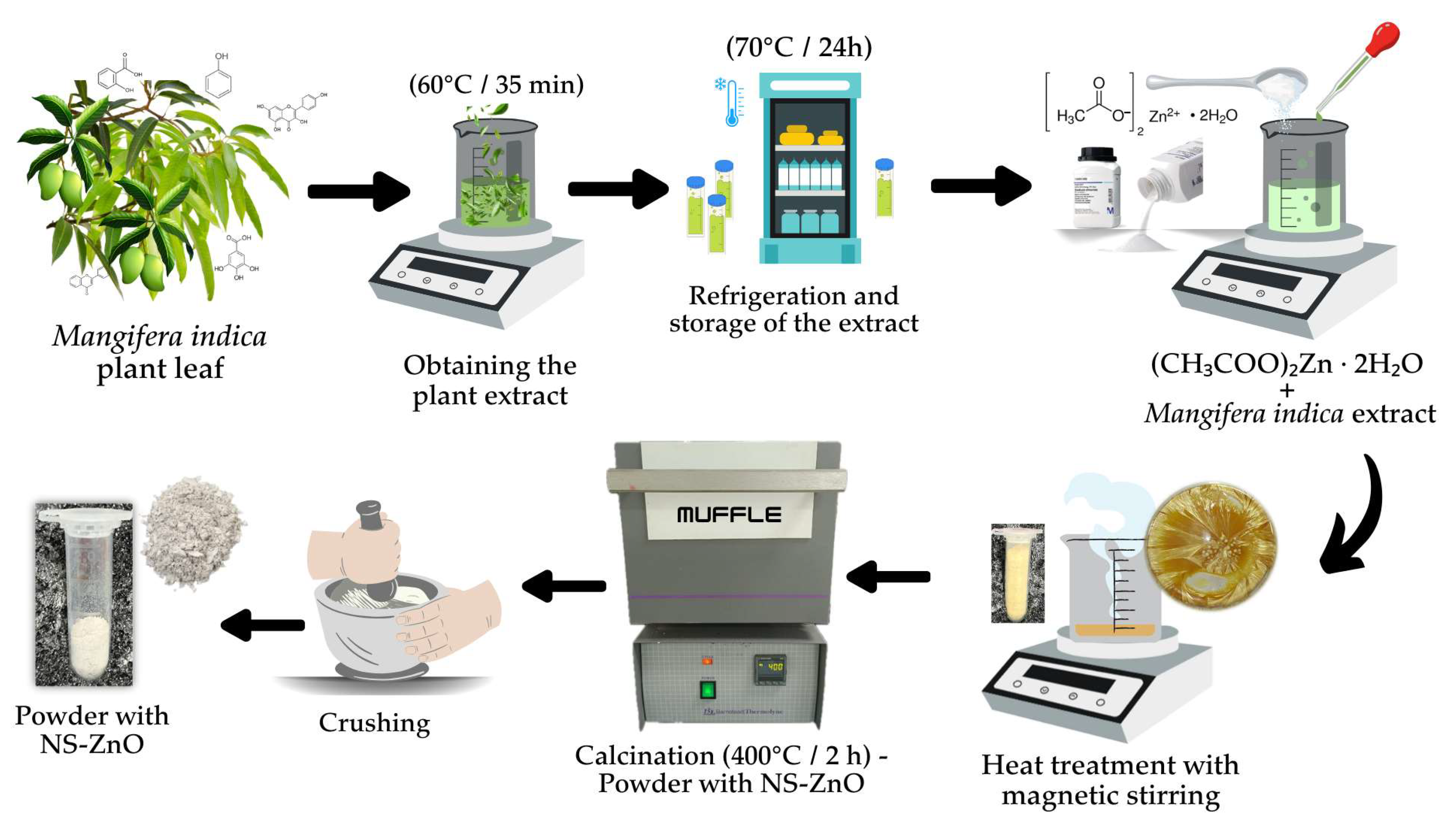
2.2. NS-ZnO Synthesis (Route 2)

The second route implemented to obtain NS-ZnO was carried out in a physical-green synthesis process, the schematic representation of which is illustrated in Figure 2.

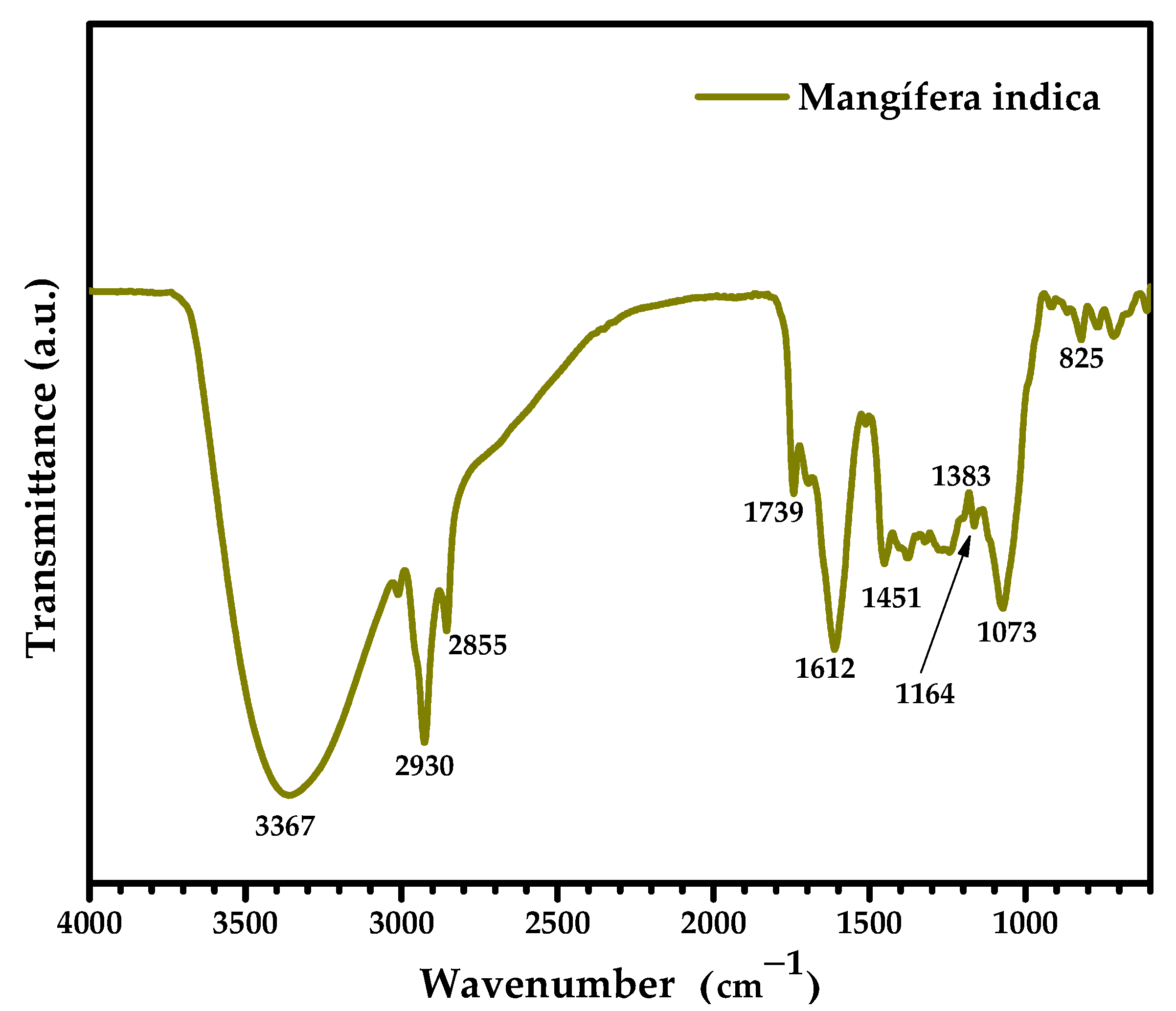
2.2.1. Preparation of Plant Extract

Initially, for the preparation of the plant extract, fresh and young leaves were collected from the Mangifera indica plant located in the department of Cesar, Colombia. The leaves were rinsed three times with deionized water to eliminate impurities and dried at room temperature. Once dry, the stems were removed and the leaves were cut into small pieces. Sixteen grams of chopped leaves were added in a beaker containing 100 mL of deionized water. The mixture was sonicated for 15 min to facilitate the extraction of compounds. Subsequently, the mixture was subjected to heat treatment for 35 min at a temperature of 60 °C to promote the release of phytocomponents. The solution was filtered with Boeco grade 3hw filter paper and centrifuged at 15,000 RPM for 15 min. This resulted in a plant extract free of impurities that was stored and refrigerated as shown in Figure 2.

2.2.2. Physical-Green Synthesis of NS-ZnO

After the preparation of the extract, a solution was prepared by dissolving 4.4 g of zinc acetate dihydrate (0.1 M) in a beaker containing 200 mL of deionized water. The mixture was kept under magnetic stirring for 15 min. After this time, 30 mL of Mangifera indica plant extract was added to the mixture and the mixture was subjected to heat treatment with magnetic stirring for 24 h at a temperature of 70 °C until a crystallized powder was obtained. This powder was scraped from the beaker and mechanically crushed in a mortar. Then, it was taken to a calcination stage with the same conditions of route 1; 400 °C for 2 h. This resulted in a white NS-ZnO powder of high crystallinity and purity.
To ensure the reproducibility of the methodology employed, it is emphasized that the synthesis parameters described in this study are the result of an optimization process based on multiple experimental trials. During this stage, several batches of samples were prepared under identical conditions in different weeks. The observed properties, particularly optical and structural, showed minimal variation between batches, which is evidence of the reliability and consistency of the technique. The sample selected for characterizations and comparative evaluation with its conventional counterpart corresponds to a representative set of all the tests performed.

2.3. Characterization of NS-ZnO

The structural, vibrational, morphological, compositional and optical characteristics of ZnO nanostructures synthesized by two alternative routes were investigated to establish a detailed and comprehensive comparative evaluation.
The structural, vibrational, morphological, compositional, and optical characteristics of the ZnO nanostructures synthesized via two alternative routes were investigated to establish a detailed and comprehensive comparative evaluation.
Confirmation of the hexagonal wurzite phase of the ZnO NS, phase purity, and crystallite size were investigated using a Malvern-PANalytical Model Empyrean 2012 X-ray (Malvern Panalytical, Malvern, United Kingdom) diffractometer (XRD) with a 3D Pixel detector and Cu source (λ = 1.541 Å) at 45 kV and 40 µm; goniophotometer: Omega/2 theta; and platform configuration: Spinner rotating at 4 rpm. Diffractograms were obtained with a step size of 0.05° and a step time of 50 s.
The crystallite size (D) of the ZnO-NS was estimated based on the full width at half maximum (FWHM) of the main reflections by applying Debye Scherrer’s formula [24,25]:
D = 0.9 λ β cos θ
where λ is the incident X-ray wavelength, β is the full width at half maximum (FWHM), and θ is the Bragg angle.
The lattice parameters for the hexagonal unit cell, known as ‘a’ and ‘c’, can be calculated from the XRD spectra of the samples using the following equation [26,27]:
1 d h k l 2 = 4 3 h 2 + h k + k 2 a 2 + l 2 c 2
where h, k, and l are known as the Miller indices. dhkl is the interplanar spacing of the crystal.
The interplanar spacing dhkl is given by Bragg’s law:
d h k l = n λ 2 sin θ
where n is the diffraction order (usually n = 1), λ is the X-ray wavelength, and dhkl is the spacing between the planes of the given Miller indices h, k, and l.
Choosing the (100) and (002) planes for Equations (2) and (3), the lattice parameters ‘a’ and ‘c’ are calculated from the following equations:
a = λ 3 sin θ 100
c = λ sin θ 002
where θ100 and θ002 correspond to the angles of the diffraction peaks corresponding to the (100) and (002) planes, respectively.
The volume of the unit cell with hexagonal geometry can be calculated from the lattice parameters by using the following equation [28]:
V = 3 2 a 2 c
The relative crystallinity index (RCI) can also be determined from the XRD spectra by the following equation [29]:
I c % = A c A c + A a × 100
where Ac is the total area of the crystalline peaks and Aa corresponds to the total area of the amorphous peaks.
The vibrational characteristics of NS-ZnO were analyzed using a Thermo Scientific DXR2 Raman spectrometer (ThermoFisher Scientific, Waltham, MA, USA) from the Raman Instruments line. A 532 nm laser with 5.0 mW power was used and the spectrograph aperture was adjusted to 50 µm.
Infrared spectroscopy (FTIR) analysis was performed using a Pelkin–Elmer Spectrum two FTIR spectrometer (PerkinElmer, Inc., Waltham, MA, USA) with a Smart Orbit diamond attenuated total reflection (UATR) accessory. A transmission analysis temperature of 22 °C with a spectral resolution of 4 cm−1 and operating range of 4000–450 cm−1 was used.
The morphological and compositional characteristics of NS-ZnO were analyzed through SEM micrographs using a Thermo Scientific (ThermoFisher Scientific, Waltham, MA, USA) model Scios 2 LoVac Dual Beam Emission Electron Microscope-(FIB-FESEM). With an EDS microanalysis system (Ul-traDry 129 eV 30 mm2 model No: SDBX-30PM-B) and EBSD (Quasor II CMOS sensor). STEM 3+ detector. Resolution of 0.7 nm at 30 kV STEM mode-1.4 nm at 1 kV FESEM mode with maximum magnification 300,000×.
The optical spectral response was analyzed at room temperature using an ASEQ Instruments UV-Vis spectrometer (ASEQ Instruments, Vancouver, Canada) with a spectral resolution of 0.2 nm and an operating range of 200–1000 nm. The optical bandgap was determined from the diffuse absorbance measurements through the following correlation [30,31]:
( α h υ ) 1 / n = A ( h υ E g )
where α is the absorption coefficient, h is Planck’s constant, υ is the frequency of the incident photon, A is a proportionality constant, Eg is the characteristic bandgap of the material and the parameter n indicates the nature of the transition; where n = 2 for direct transitions allowed. The optical bandgap has been determined according to Tauc’s model, using Equation (8). The value of Eg is obtained from a linear extrapolation of de ( α h υ ) 2 vs. photon energy ( h υ ) at the absorption limit; that is when α ≈ 0.

3. Results and Discussion

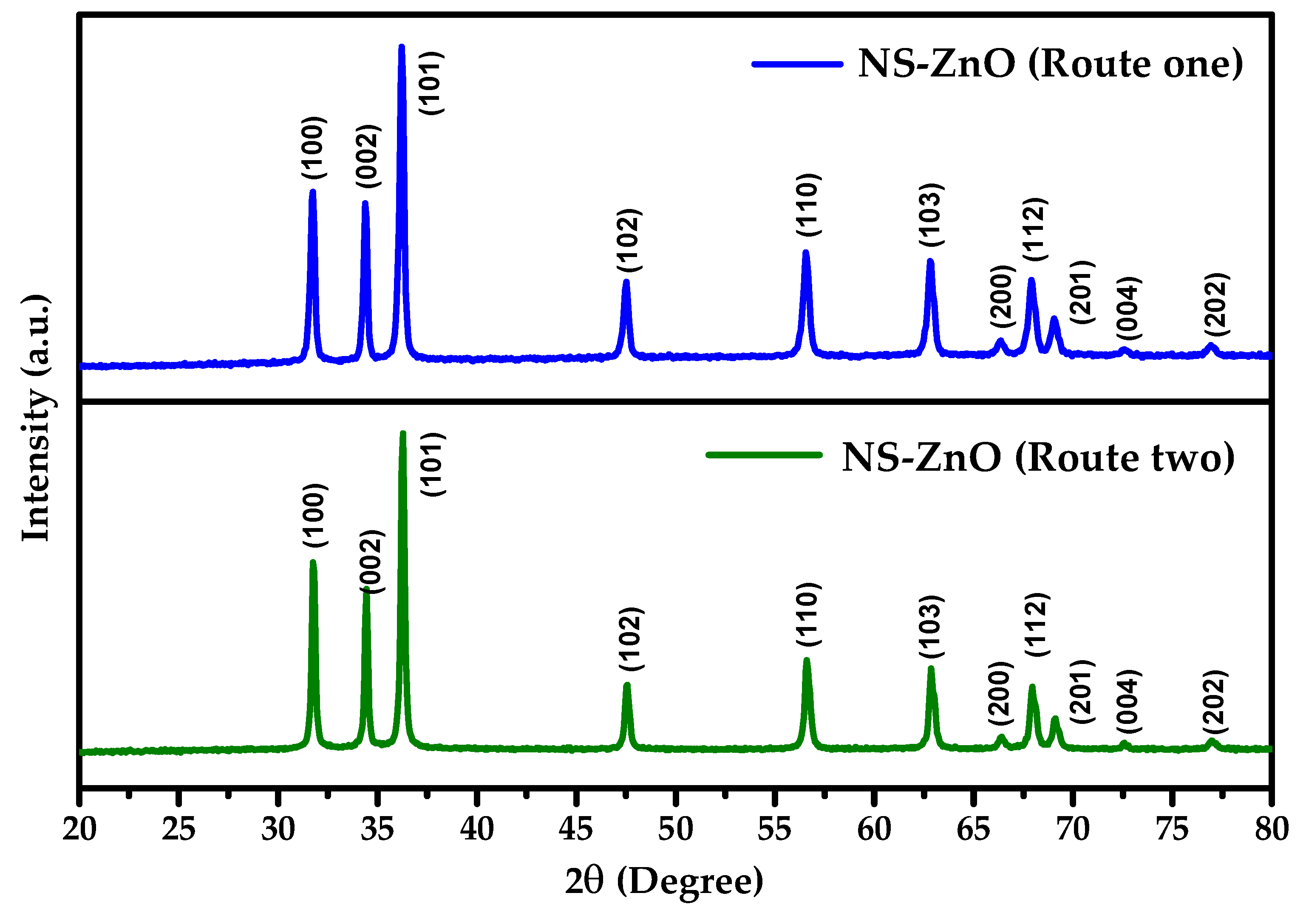
3.1. Structural Analysis

The crystal structure and phase purity of the synthesized ZnO nanostructures were examined by X-ray diffraction. Figure 3 shows the XRD patterns of the NS-ZnO obtained by routes 1 and 2, respectively, for a diffraction angle 2θ ranging from 20° to 80°. The diffraction patterns reveal well-defined peaks that indicate the purity and polycrystallinity of the NS-ZnO obtained by these two routes. It is clearly observed that all diffraction peaks are coincident, excluding the variation in peak intensity, which indicates a change in the morphology of the samples. The peaks indexed to the lattice planes (100), (002), (101), (102), (110), (103), (112), and (004) agree well with the standard ICDD card (#79-0205) of the pure hexagonal (wurtzite) structure of ZnO with space group P63mc (186). Furthermore, no characteristic peaks related to other impurities were observed in the spectra, indicating that the ZnO nanostructures synthesized by the two routes are of high purity.
Table 1 shows the crystallographic parameters of the ZnO nanostructures obtained through the two synthesis routes. According to this table, clear differences in the crystallinity of the ZnO nanostructures synthesized through the conventional physical route (route 1) and physical-green (route 2) are evident. The relative crystallinity index, calculated from the integrated area of the diffraction peaks (for a representative sample per route), was 86.9 % for Route 1 and 91.6 % for Route 2. The higher value observed in the sample synthesized by the green route suggests a higher crystalline ordering. Since, both samples were subjected to a calcination process at the same temperature, it can be considered that the improvement in crystallinity comes from the influence of the bioactive compounds present in the Mangifera indica leaf extract. Previous studies have revealed the formation of ZnO nanostructures with identical diffraction patterns using Mangifera indica plant extract [32,33]. However, the differences in crystallinity indices and crystallographic parameters compared to the conventional procurement method (route 1) have not been explored.
Several studies have shown that polyphenols, flavonoids and other functional molecules present in plant extracts can act as directing and stabilizing agents during the nucleation and growth stages, promoting the formation of larger and more ordered crystalline domains [34,35]. In this sense, the presence of such phytochemicals could have modulated the growth kinetics, favoring a more regular structure in the sample obtained by physical-green synthesis.
The crystallite size, estimated using the Debye–Scherrer equation for the preferred plane associated with the most intense peak (101), was 30 nm for Route 1 and 35 nm for Route 2. This increase in crystallite size is consistent with a more uniform, sustained growth process and a lower dislocation density. This behavior is characteristic of systems stabilized by organic agents and is consistent with the higher crystallinity index observed.
The calculated lattice parameters showed slight expansions compared to the standard values for hexagonal ZnO. Consequently, the calculated cell volumes were 47.76 Å3 and 47.56 Å3 for routes 1 and 2, respectively, revealing a slight increase when compared to the standard value of 47.21 Å3.
Deviations in cell volume, although minimal, are typical in nanocrystalline materials and can be attributed to micro-deformations or internal stresses resulting from incomplete relaxation of the crystal lattice during growth. These stresses are amplified by the high surface-to-volume ratio characteristic of nanocrystals [26,36]. Furthermore, the presence of point defects, such as oxygen vacancies or zinc atoms in interstitial sites, possibly induced during calcination or by interactions with the organic compounds in the plant extract, can locally modify the lattice parameters [37].
Overall, the X-ray diffraction results indicate that both routes allow ZnO nanostructures with high crystallinity and phase purity to be obtained. However, the physical-green route offers clear advantages in terms of the degree of structural order, attributable to the modulating effect of the plant extract on the crystal growth process, without the need to vary the calcination thermal conditions.

3.2. Raman Spectroscopy Analysis
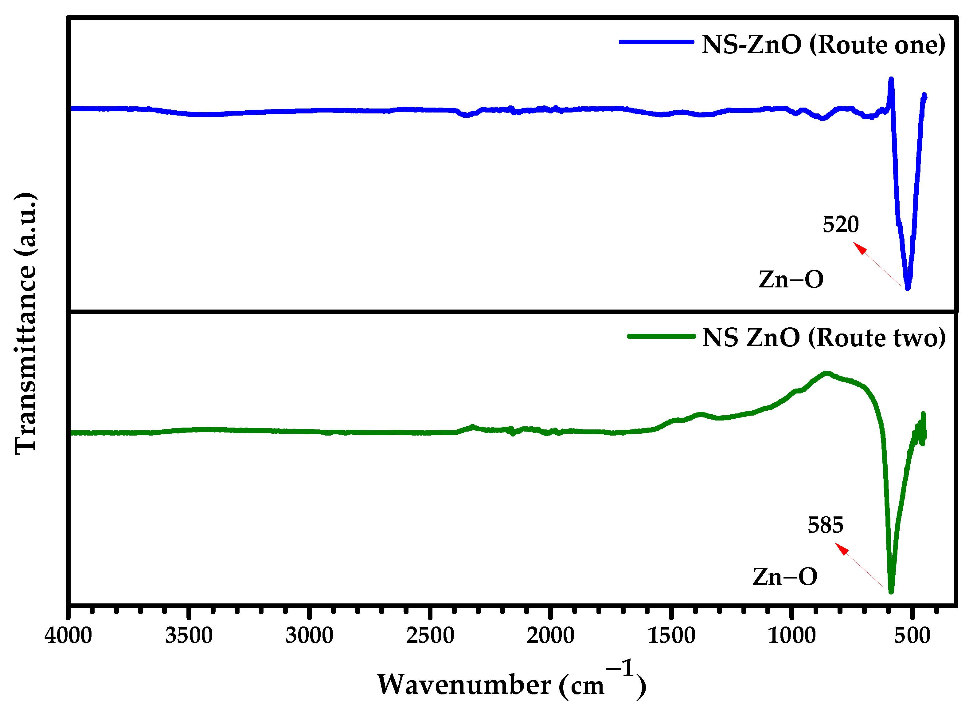
Figure 4 shows the Raman spectra obtained for the ZnO nanostructures synthesized by routes 1 and 2. These spectra reveal the existence of two bands located around 98 and 434 cm−1, which are coincident for both samples, excluding the variation in the intensity of the peaks. As already confirmed in this study, ZnO crystallizes in the wurtzite (hexagonal) structure. According to the space group (P63mc) of hexagonal ZnO, hexagonal, the active optical modes are decomposed as Γ = A 1 + E 1 + 2 E 2 + 2 B 1 , of which, A1 and E1 are polar and divided into transverse optical (A1 TO y E1 TO) and longitudinal optical (A1 LO and E1 LO).
The E2 mode consists of two phonon modes of low and high frequency (low E2 and high E2), which are associated with the vibration of the sublattice of the Zn and O atoms, respectively [38,39]. In our study, the band located at 98 cm−1 is assigned to the E2 (low) mode, associated with Zn sublattice vibrations, while the band at 434 cm−1 corresponds to the E2 (high) mode, characteristic of oxygen vibrations and sensitive to the crystalline quality, grain size and the presence of internal stresses in the nanostructures [40]. This mode is often the most intense in the spectra of highly crystalline ZnO and its presence with high intensity and without appreciable displacements concerning its typical position (~437 cm−1) is evidence of an ordered structure and of the predominant wurtzite phase [39,40].
A difference in the relative intensity of the E2 (gigh) mode was observed between the two samples, suggesting variations in the degree of crystallinity and coherent domain size. In particular, a higher intensity in this mode suggests a higher structural regularity in the sample synthesized by route 2, which is in agreement with the results obtained by X-ray diffraction. Additionally, the absence of defect-activated modes, such as A1 (LO) or E1 (LO) modes, which as opposed to E2 (low and gigh) modes usually appear around 574 and 583 cm−1 in the presence of oxygen vacancies or internal lattice stresses [39,41,42], supports the conclusion that both synthesis routes allow obtaining ZnO nanostructures with excellent structural quality.

3.3. FTIR Analysis

The analysis by Fourier transform infrared spectroscopy (FTIR) allowed confirming the formation of ZnO in the nanostructures obtained by both synthesis routes. According to Figure 5, for the conventional route 1, a vibrational band associated with the Zn–O stretching mode was identified, located at ~520 cm−1, which is in agreement with other published works [43,44]. This band shifted to ~585 cm−1 in the sample obtained by the plant extract-assisted route (route 2). This shift towards higher wavenumbers may be attributed to effects such as lattice tensions, decreased particle size or interactions between the ZnO surface and the organic residues of the Mangifera indica extract.
The observed shift of the Zn–O vibrational band in the FTIR spectrum of the sample synthesized via Route 2 may initially suggest the presence of lattice strain or structural perturbations [45,46]. However, this interpretation must be considered with caution, as the corresponding Raman spectra (Figure 4) do not show any measurable shift in the E2 (high) mode, which is known to be highly sensitive to internal stresses within the ZnO wurtzite lattice. The lack of a Raman shift indicates that significant compressive or tensile strain is unlikely. Therefore, the FTIR shift is more plausibly attributed to changes in the local chemical environment surrounding the Zn–O bond, potentially caused by interactions with residual phytochemicals or organic functional groups from the Mangifera indica extract. These interactions may alter bond polarity or coordination symmetry without introducing detectable crystallographic strain.
Figure 6 shows the FTIR spectrum of the plant extract of Mangifera indica leaves, in which the function groups involved in the early stages of nucleation and growth of ZnO nanostructures can be clearly observed. A predominant peak was detected at 3367 cm−1 corresponding to the stretching mode of the OH group [47], the bands at 2855 and 2930 cm−1 to the stretching mode of the C-H bond, the weak peak at 1739 cm−1 and the strong peak at 1612 cm−1 is associated with the tension mode of the C=O bond [48]. The peak at 1451 cm−1 corresponds to the stretching vibrations of the aromatic ring of mangiferin and the scissor-like deformation of the O-H bond appears at 1383 cm−1. The vibrations at 1612 and 825 cm−1 would correspond to the tensile and bending vibrations of the C=C double bond respectively, while the band around 1073 cm−1 is associated with the vibrations of the C-O bond.
The presence of functional groups may establish some hydrogen bonds or coordination interactions with ZnO, locally modifying its vibrational environment and contributing to the observed shift.
This result is also consistent with the previous analysis of the Raman spectra, in which a greater intensity of the E2 (high) mode (~434 cm−1) was observed in the sample synthesized by route 2. This mode is particularly sensitive to the structural order and to the interstitial presence of oxygen atoms in the lattice of the wurtzite-type ZnO.
The combination of the FTIR analysis with the XRD and Raman structural data suggests that the green route promoted greater structural regularity, possibly facilitated by the modulating effect of the plant extract during the early stages of ZnO nucleation and growth.

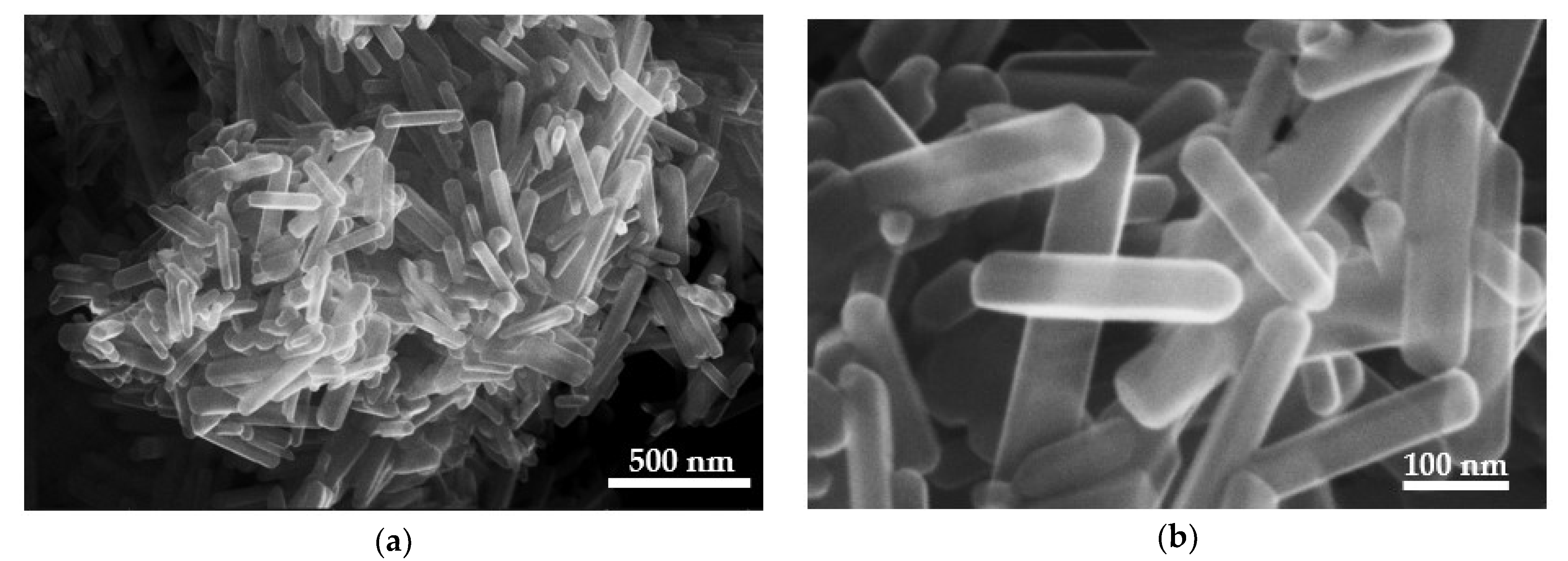
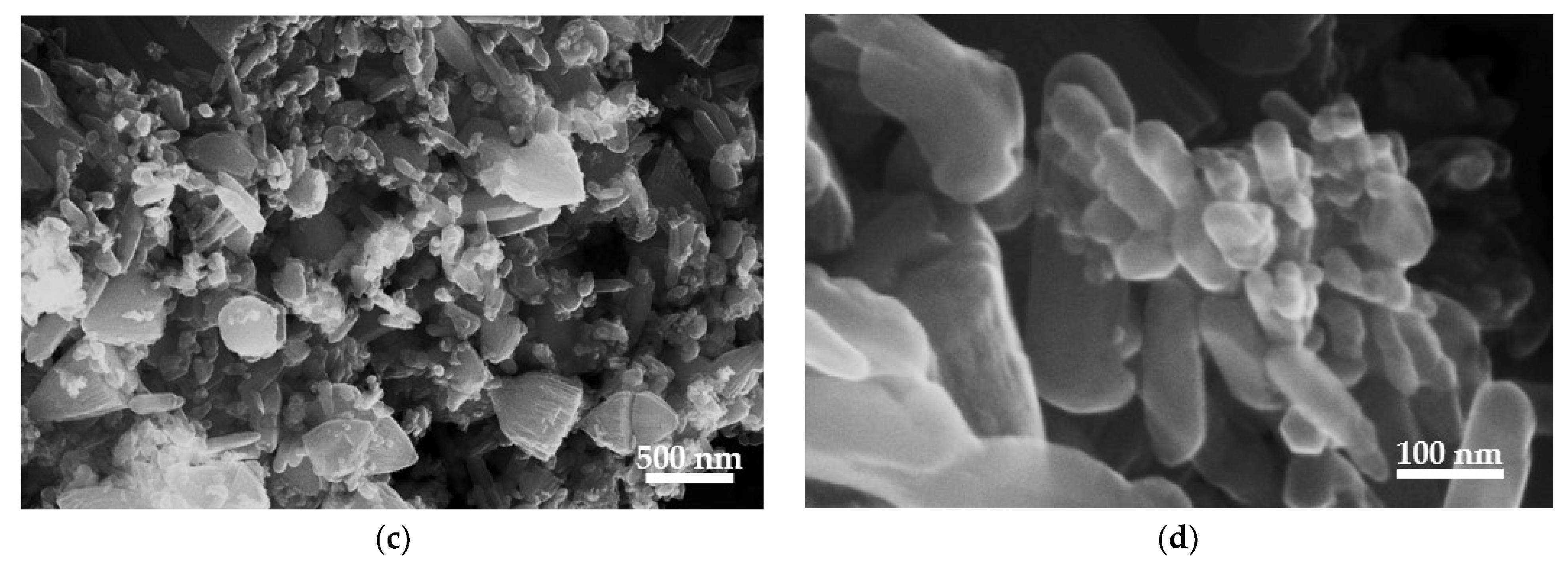
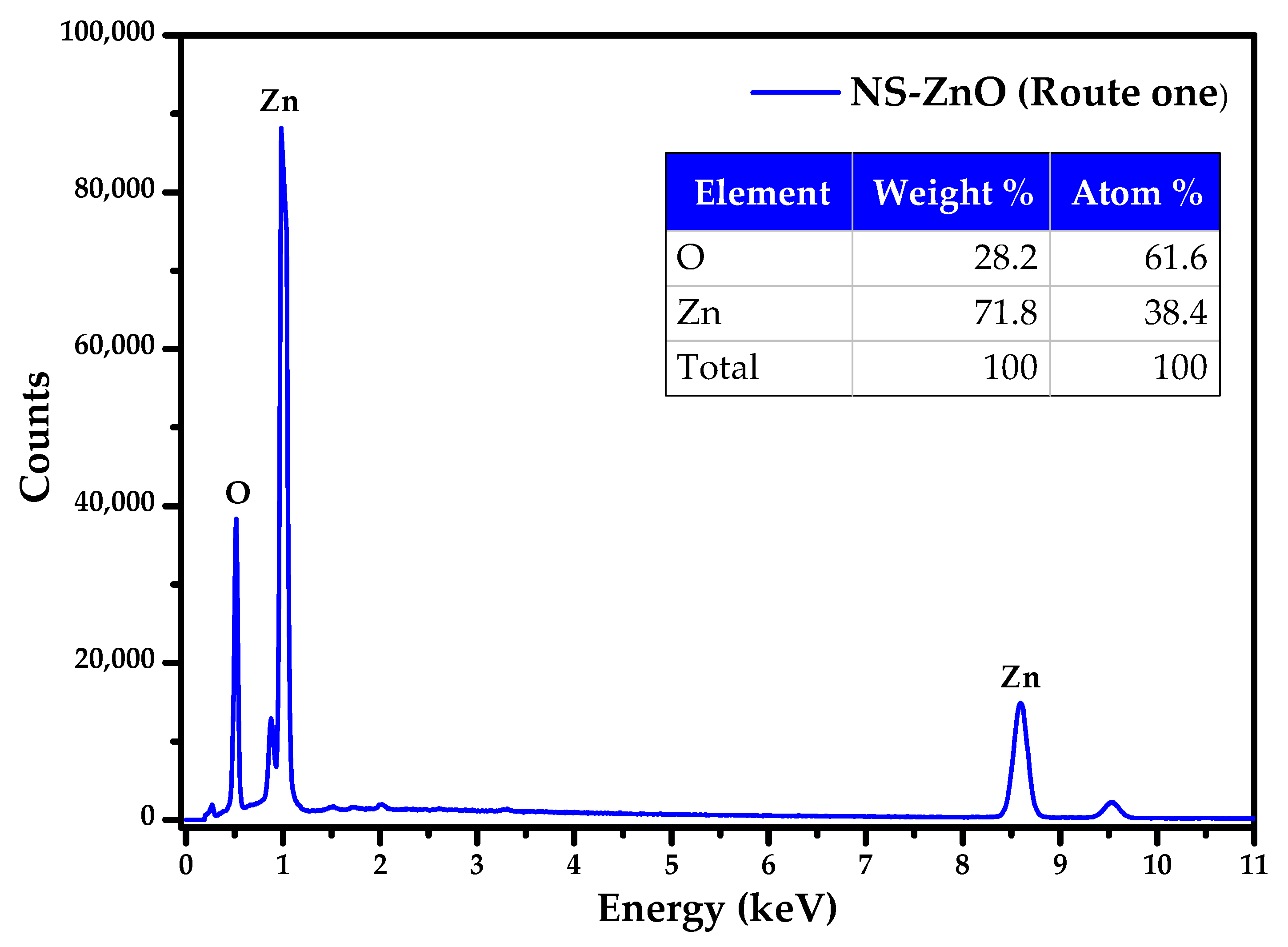
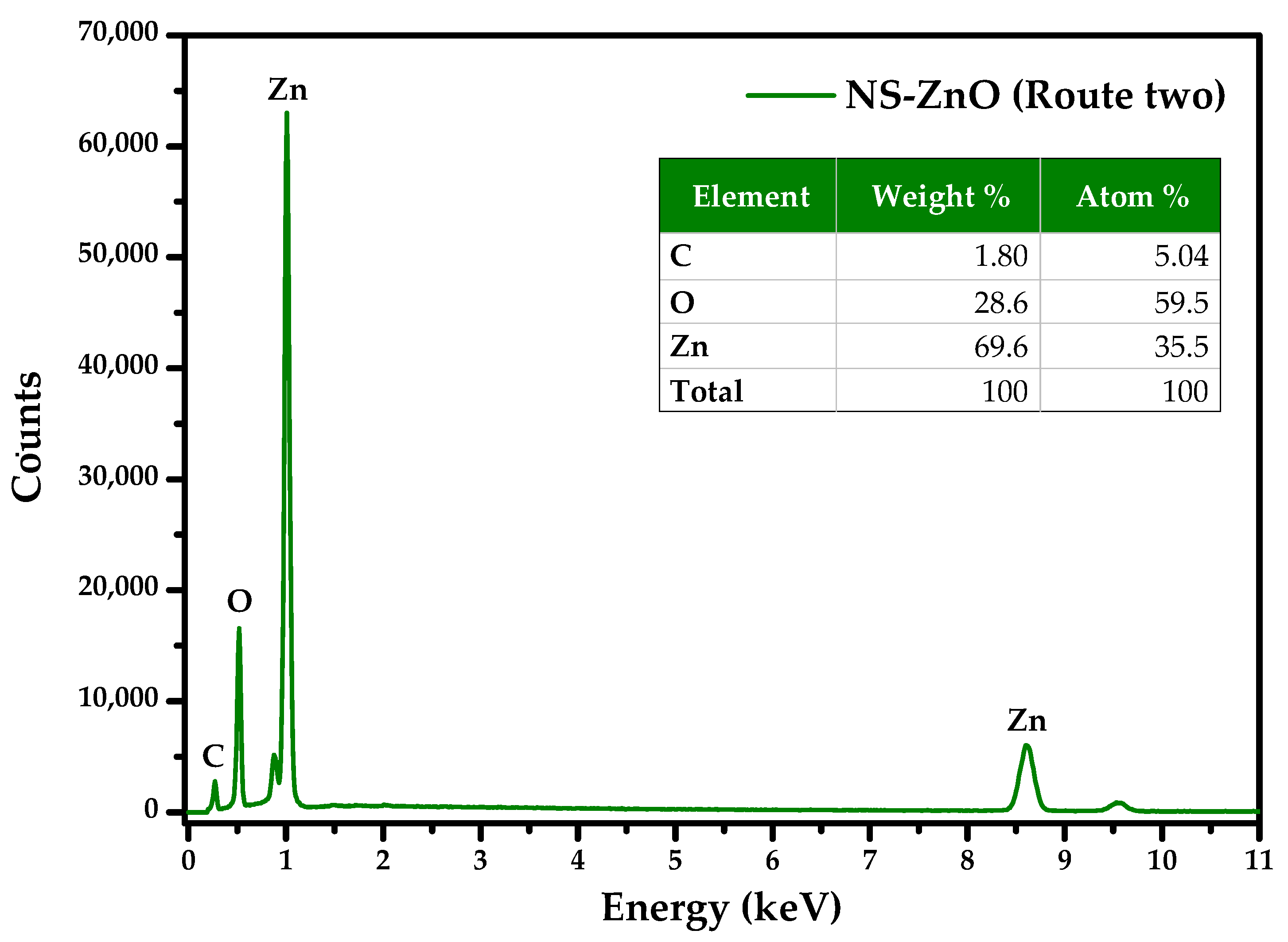
3.4. FE-SEM and EDS Analysis

The surface morphology, size distribution, and elemental composition of the ZnO nanostructures obtained via two alternative routes (1 and 2) were analyzed using field emission scanning electron microscopy (FESEM) and energy-dispersive spectroscopy (EDS). According to the set of SEM micrographs presented in Figure 7, there is a notable correlation between the morphological characteristics of the nanostructures and the synthesis route used. Figure 7a,b shows the successful formation of nanostructures with an elongated rod-like morphology, characteristic of zinc oxide synthesized by thermal decomposition of zinc acetate (route 1). In the image with lower magnification (80,000×) (Figure 7a), a dense and homogeneous assembly of nanorods is observed, indicating relatively uniform growth in morphological terms, although with a certain tendency to agglomeration. This agglomeration can be attributed to Van der Waals forces between the nanostructures, which is common in materials obtained via solid-phase thermal routes [49,50].
In the image with the higher magnification (300,000×) (Figure 7b), the morphology of the nanorods can be seen in greater detail. These have rounded ends and a relatively smooth surface, suggesting controlled growth in a preferential direction, possibly along the c-axis of the hexagonal ZnO system. Measurements made from SEM images using ImageJ version 1.54p software, calibrated with the included scale indicate that the nanorods possess an average length of 252 ± 4 nm and an average width of 51 ± 5 nm, resulting in an approximate aspect ratio of 4.9. This high aspect ratio is indicative of anisotropic growth, characteristic of ZnO when synthesized in the absence of directing agents or surface modifiers, and under suitable thermal conditions [51].
This anisotropic geometry is particularly advantageous for applications requiring efficient charge transport, such as piezoelectric generators, optoelectronic devices and photodetectors, where directional carrier mobility and light-guiding effects are critical. In addition, the regular surface area and alignment of the nanorods indicate fewer surface defects and more coherent crystalline domains, which improves functional performance by minimizing charge recombination. While the present study did not include functional testing, the observed morphological characteristics are consistent with ZnO architectures previously successfully employed in bioanalytical, multifunctional environmental and optical sensing applications [3,52,53].
On the other hand, Figure 7c,d shows the SEM micrographs corresponding to the nanostructures obtained by route 2, which involved mixing the inorganic precursor (zinc acetate) with Mangifera indica extract in a stage prior to calcination. A significant change in morphology is observed compared to the conventional route.
The image at lower magnification (50,000×) (Figure 7c) shows greater morphological heterogeneity, with nanostructures averaging 63.2 ± 4.6 nm in size and irregular morphologies. Although some structures still retain an elongated morphology, many have more equiaxial shapes, are agglomerated, and have less defined contours, indicating that the plant extract has significantly influenced the nucleation and growth process of the crystals, as previously indicated in the structural analysis.
The higher magnification image (300,000×) (Figure 7d) clearly shows this change. The nanostructures outlined in yellow show a more dispersed size distribution and less uniform shapes compared to the defined nanorods obtained by route 1. This morphological transition can be explained by the selective adsorption of phytochemical compounds, such as flavonoids, phenolic acids and tannins, on specific crystallographic facets of the growing ZnO cores. These molecules can inhibit anisotropic growth by preferentially binding to the (001) or (100) planes, thereby suppressing elongation along the c-axis of the wurtzite ZnO lattice. As a result, isotropic or less well-defined particles tend to form, consistent with the irregular and equiaxial shapes observed in SEM. This behavior has been previously reported in green syntheses where bioactive compounds interfere with the intrinsic growth kinetics of ZnO nanostructures, reducing their aspect ratio and promoting morphological heterogeneity [31,32]. The observed morphological suppression thus reflects a classic structure-directing effect exerted by the phytochemicals present in the extract.
In summary, the plant extract may induce different growth kinetics, decreasing the anisotropy of crystalline growth due to preferential adsorption on certain faces of ZnO, resulting in a higher variability in particle size, as observed in the micrographs. Despite this, the presence of rod-shaped structures suggests that subsequent calcination partially preserved the characteristic one-dimensional growth mechanism of ZnO.
Overall, these results show that while the physical-green route reduces the control over morphology, it significantly improves the structural properties of ZnO, such as crystallinity, crystallite size and structural order. This translates into a significant advantage when looking for applications where crystalline quality and structural purity are decisive, such as in sensors, piezoelectric devices or photocatalysts [5].
Regarding chemical composition, EDS spectra (Figure 8 and Figure 9) demonstrate that both routes yielded materials with compositions approaching the 1:1 stoichiometric ratio of ZnO. Route 1 exhibited a Zn:O ratio of 0.62 At.%, while Route 2 showed 0.59 At.%. These minor deviations, consistent with prior reports, indicate adsorbed oxygen species (O2, OH) typical of oxidative synthesis [54]. Furthermore, Route 2 contained 5.04 At.% carbon, arising from carbonized organic traces (flavonoids/phenolic acids) of the plant extract that survived calcination at 400 °C. While this residue may alter electronic properties like conductivity, it enhances photocatalytic applications by facilitating electron-hole pair separation through the formation of surface electron-trapping states [55,56,57,58].

3.5. UV-Vis Absorption Analysis

Analysis of the UV-Vis spectra (Figure 10) shows significant differences in the optical properties of the ZnO nanostructures obtained by both methods, where Method 1 (direct calcination) shows a characteristic absorption peak at 388 nm corresponding to wurtzite-type nanocrystalline ZnO [14], while Method 2 (assisted with Mangifera indica extract) shows a red shift to 422 nm. This shift is indicative of a decrease in bandwidth, which was corroborated from the Tauc diagrams (Figure 11). Linear approximations in the absorption limit allowed estimating an Eg of 3.34 eV for route 1 (Figure 11a) and 2.97 eV for route 2 (Figure 11b). This decrease in the bandgap can be attributed to the incorporation of structural defects induced by bioactive compounds present in the plant extract, which could introduce intermediate levels within the bandgap or induce local stresses in the crystal lattice of the ZnO [59]. Additionally, the size polydispersity and irregular geometry of the nanostructures observed through SEM may increase optical scattering, broadening the absorption spectrum [60,61].
From a functional point of view, improvements in crystallinity and optical properties are very relevant for practical applications. ZnO nanostructures with higher crystalline order and lower bandgap exhibit higher charge carrier mobility and better separation of photogenerated electron-hole pairs, critical parameters in photocatalytic and optoelectronic systems [62,63,64]. For example, a reduced bandgap allows higher absorption in the visible region, which increases the response of the material to solar radiation, while high crystallinity reduces recombination losses. Although this work did not include direct performance testing (e.g., photocatalysis or sensing), the observed structural and optical improvements suggest promising potential for such applications, as demonstrated in related studies of ZnO nanostructures with similar characteristics.

4. Conclusions

A rigorous comparative evaluation of two fast routes for the synthesis of ZnO nanostructures was performed using identical thermal conditions. Both routes allowed obtaining ZnO with pure wurtzite-type hexagonal phase and high crystallinity.
The physical-green route, assisted by Mangifera indica extract, generated nanostructures with higher structural quality, evidenced by a crystallinity index of 91.6% and a larger crystallite size (35 nm). Raman and FTIR analyses corroborated a greater structural regularity in the green samples, while the morphology observed by SEM revealed a more heterogeneous and polydisperse distribution in size with respect to the nanorods defined by the conventional physical route. Optical analysis showed a decrease in bandgap (from 3.34 eV to 2.97 eV), associated with the presence of structural defects induced by residual organic compounds, which broadened the sensitivity of the material towards the visible spectrum.
The results indicate that the incorporation of bioactive compounds through a physical-green route offers an efficient way to improve the structural and functional properties of ZnO, without requiring additional energetic conditions. This strategy represents a promising alternative for the sustainable production of nanomaterials with advanced applications in sensors, photocatalysis and optoelectronic devices, which are the most important future prospects of this study.

Author Contributions

Conceptualization, E.V.R.-D., J.P.M.-J., C.O.T. and S.D.H.-P.; methodology, S.D.H.-P. and C.O.T.; validation, J.P.M.-J., D.A.A., C.O.T. and S.D.H.-P.; formal analysis, J.P.M.-J., D.A.A. and S.D.H.-P.; investigation, E.V.R.-D.; resources, D.A.A. and C.O.T.; data curation, E.V.R.-D.; writing—original draft preparation, E.V.R.-D., J.P.M.-J. and S.D.H.-P.; writing—review and editing, E.V.R.-D., J.P.M.-J., S.D.H.-P., C.O.T. and D.A.A.; visualization, E.V.R.-D., J.P.M.-J., C.O.T. and S.D.H.-P.; supervision, D.A.A., S.D.H.-P. and C.O.T.; funding acquisition, D.A.A. and C.O.T. All authors have read and agreed to the published version of the manuscript.

Funding

This research was financed by Science, Technology and Innovation of the Sistema General de Regalias (SGR)—Colombia through the project “Formacion de Capital Humano maestria y Doctorado para el departamento del Cesar” Universidad Popular del Cesar–Nacional with BPIN code 2021000100130, and the project “Formacion de Capital Humano de Alto Nivel” Universidad Popular del Cesar–Nacional with BPIN code 2019000100010.

Institutional Review Board Statement

Not applicable.

Informed Consent Statement

Not applicable.

Data Availability Statement

Data are contained within the article.

Acknowledgments

The authors would like to acknowledge the Universidad Popular del Cesar for providing the facilities of the optics and informatics laboratory for the partial development of this research.

Conflicts of Interest

The authors declare no conflicts of interest.

References

  1. Borysiewicz, M.A. ZnO as a Functional Material, a Review. Crystals 2019, 9, 505. [Google Scholar] [CrossRef]
  2. Sha, R.; Basak, A.; Maity, P.C.; Badhulika, S. ZnO Nano-Structured Based Devices for Chemical and Optical Sensing Applications. Sens. Actuators Rep. 2022, 4, 100098. [Google Scholar] [CrossRef]
  3. Sorbiun, M.; Shayegan Mehr, E.; Ramazani, A.; Taghavi Fardood, S. Green Synthesis of Zinc Oxide and Copper Oxide Nanoparticles Using Aqueous Extract of Oak Fruit Hull (Jaft) and Comparing Their Photocatalytic Degradation of Basic Violet 3. Int. J. Environ. Res. 2018, 12, 29–37. [Google Scholar] [CrossRef]
  4. Ding, M.; Guo, Z.; Zhou, L.; Fang, X.; Zhang, L.; Zeng, L.; Xie, L.; Zhao, H. One-Dimensional Zinc Oxide Nanomaterials for Application in High-Performance Advanced Optoelectronic Devices. Crystals 2018, 8, 223. [Google Scholar] [CrossRef]
  5. Leal-Perez, J.E.; Almaral-Sanchez, J.L.; Hurtado-Macias, A.; Cortez-Valadez, M.; Borquez-Mendívil, A.; García-Grajeda, B.A.; Mendivil-Escalante, J.M.; Flores-Valenzuela, J. Structural and chemical analysis of Zn ion exchange in thermally modified zelite A4. Rev. Mex. Ing. Quím. 2024, 23, Mat24264. [Google Scholar] [CrossRef]
  6. Karnati, P.; Haque, A.; Taufique, M.F.N.; Ghosh, K. A Systematic Study on the Structural and Optical Properties of Vertically Aligned Zinc Oxide Nanorods Grown by High Pressure Assisted Pulsed Laser Deposition Technique. Nanomaterials 2018, 8, 62. [Google Scholar] [CrossRef]
  7. Chen, W.; Yao, C.; Gan, J.; Jiang, K.; Hu, Z.; Lin, J.; Xu, N.; Sun, J.; Wu, J. ZnO Colloids and ZnO Nanoparticles Synthesized by Pulsed Laser Ablation of Zinc Powders in Water. Mater. Sci. Semicond. Process. 2020, 109, 104918. [Google Scholar] [CrossRef]
  8. Rathore, M.S.; Verma, H.; Akhani, S.B.; Pathak, J.; Joshi, U.; Joshi, A.; Prakash, C.; Kaur, K.; Oza, A. Photoluminescence and Antibacterial Performance of Sol–Gel Synthesized ZnO Nanoparticles. Mater. Adv. 2024, 5, 3472–3481. [Google Scholar] [CrossRef]
  9. Sumaya, M.U.; Maria, K.H.; Toma, F.T.Z.; Zubair, M.A.; Chowdhury, M.T. Effect of Stabilizer Content in Different Solvents on the Synthesis of ZnO Nanoparticles Using the Chemical Precipitation Method. Heliyon 2023, 9, e20871. [Google Scholar] [CrossRef]
  10. Vittaya, L.; Chalad, C.; Sirimahachai, U. Green Synthesis and Biological Activities of Zinc Oxide Nanoparticles Using Ampelocissus martini Rhizome Extract. ChemistrySelect 2024, 9, e202304671. [Google Scholar] [CrossRef]
  11. Das, D.; Nath, B.C.; Phukon, P.; Kalita, A.; Dolui, S.K. Synthesis of ZnO Nanoparticles and Evaluation of Antioxidant and Cytotoxic Activity. Colloids Surf. B Biointerfaces 2013, 111, 556–560. [Google Scholar] [CrossRef] [PubMed]
  12. Jayswal, S.; Moirangthem, R.S. Thermal decomposition route to synthesize ZnO nanoparticles for photocatalytic application. AIP Conf. Proc. 2018, 2009, 020023. [Google Scholar] [CrossRef]
  13. Motelica, L.; Oprea, O.-C.; Vasile, B.-S.; Ficai, A.; Ficai, D.; Andronescu, E.; Holban, A.M. Antibacterial Activity of Solvothermal Obtained ZnO Nanoparticles with Different Morphology and Photocatalytic Activity against a Dye Mixture: Methylene Blue, Rhodamine B and Methyl Orange. Int. J. Mol. Sci. 2023, 24, 5677. [Google Scholar] [CrossRef]
  14. Josun, J.; Sharma, P.; Kumar Garg, V. Optical and Structural Behavior of Hydrothermally Synthesized ZnO Nanoparticles at Various Temperatures with NaOH Molar Ratios. Results Opt. 2024, 14, 100601. [Google Scholar] [CrossRef]
  15. Abdullah, J.A.A.; Guerrero, A.; Romero, A. Efficient and Sustainable Synthesis of Zinc Salt-Dependent Polycrystal Zinc Oxide Nanoparticles: Comprehensive Assessment of Physicochemical and Functional Properties. Appl. Sci. 2024, 14, 1815. [Google Scholar] [CrossRef]
  16. Villagrán, Z.; Anaya-Esparza, L.M.; Velázquez-Carriles, C.A.; Silva-Jara, J.M.; Ruvalcaba-Gómez, J.M.; Aurora-Vigo, E.F.; Rodríguez-Lafitte, E.; Rodríguez-Barajas, N.; Balderas-León, I.; Martínez-Esquivias, F. Plant-Based Extracts as Reducing, Capping, and Stabilizing Agents for the Green Synthesis of Inorganic Nanoparticles. Resources 2024, 13, 70. [Google Scholar] [CrossRef]
  17. El-Seedi, H.R.; El-Shabasy, R.M.; Khalifa, S.A.M.; Saeed, A.; Shah, A.; Shah, R.; Iftikhar, F.J.; Abdel-Daim, M.M.; Omri, A.; Hajrahand, N.H.; et al. Metal Nanoparticles Fabricated by Green Chemistry Using Natural Extracts: Biosynthesis, Mechanisms, and Applications. RSC Adv. 2019, 9, 24539–24559. [Google Scholar] [CrossRef]
  18. Rani, N.; Singh, P.; Kumar, S.; Kumar, P.; Bhankar, V.; Kumar, K. Plant-Mediated Synthesis of Nanoparticles and Their Applications: A Review. Mater. Res. Bull. 2023, 163, 112233. [Google Scholar] [CrossRef]
  19. Kharissova, O.V.; Dias, H.V.R.; Kharisov, B.I.; Pérez, B.O.; Pérez, V.M.J. The Greener Synthesis of Nanoparticles. Trends Biotechnol. 2013, 31, 240–248. [Google Scholar] [CrossRef]
  20. Huong, L.M.; Tai, L.T.; Dat, N.M.; Hai, N.D.; An, H.; Nam, N.T.H.; Thi Thu Hanh, T.; Hieu, N.H. Non-Metallic X (X = C, N, S, and P) Co-Doped Copper Oxide Derived from Mangifera indica Leaf Extract: Synthesis, Characterization, Density Functional Theory Simulation of Structure, and Photoactivities. Colloids Surf. A Physicochem. Eng. Asp. 2024, 687, 133393. [Google Scholar] [CrossRef]
  21. Khan, Z.U.H.; Gul, N.S.; Mehmood, F.; Sabahat, S.; Muhammad, N.; Rahim, A.; Iqbal, J.; Khasim, S.; Salam, M.A.; Khan, T.M.; et al. Green Synthesis of Lead Oxide Nanoparticles for Photo-Electrocatalytic and Antimicrobial Applications. Front. Chem. 2023, 11, 1175114. [Google Scholar] [CrossRef] [PubMed]
  22. Saputra, I.S.; Nurfani, E.; Fahmi, A.G.; Saputro, A.H.; Apriandanu, D.O.B.; Annas, D.; Yulizar, Y. Effect of Secondary Metabolites from Several Leaf Extracts on the Green Synthesized-ZnO Nanoparticles. Vacuum 2024, 227, 113434. [Google Scholar] [CrossRef]
  23. da Biron, D.S.; dos Santos, V.; Bergmann, C.P. Synthesis and characterization of zinc oxide obtained by combining zinc nitrate with sodium hydroxide in polyol medium. Mater. Res. 2020, 23, e20200080. [Google Scholar] [CrossRef]
  24. Mahajan, M.; Kumar, S.; Gaur, J.; Kaushal, S.; Dalal, J.; Singh, G.; Misra, M.; Ahlawat, D.S. Green Synthesis of ZnO Nanoparticles Using Justicia Adhatoda for Photocatalytic Degradation of Malachite Green and Reduction of 4-Nitrophenol. RSC Adv. 2025, 15, 2958–2980. [Google Scholar] [CrossRef] [PubMed]
  25. Patterson, A.L. The Scherrer formula for X-ray particle size determination. Phys. Rev. 1939, 56, 978–982. [Google Scholar] [CrossRef]
  26. Bindu, P.; Thomas, S. Estimation of lattice strain in ZnO nanoparticles: X-ray peak profile analysis. J. Theor. Appl. Phys. 2014, 8, 123–134. [Google Scholar] [CrossRef]
  27. Abdelghani, G.M.; Ahmed, A.B.; Al-Zubaidi, A.B. Synthesis, Characterization, and the Influence of Energy of Irradiation on Optical Properties of ZnO Nanostructures. Sci. Rep. 2022, 12, 20016. [Google Scholar] [CrossRef] [PubMed]
  28. Cullity, B.D.; Stock, S.R. Elements of X-Ray Diffraction, 2nd ed.; Assison-Wesley Publishing Company: Boston, MA, USA, 1978. [Google Scholar]
  29. Elmahaishi, M.F.; Azis, R.S.; Ismail, I.; Mustaffa, M.S.; Abbas, Z.; Matori, K.A.; Muhammad, F.D.; Saat, N.K.; Nazlan, R.; Ibrahim, I.R.; et al. Structural, Electromagnetic and Microwave Properties of Magnetite Extracted from Mill Scale Waste via Conventional Ball Milling and Mechanical Alloying Techniques. Materials 2021, 14, 7075. [Google Scholar] [CrossRef]
  30. Agarwal, S.; Jangir, L.K.; Rathore, K.S.; Kumar, M.; Awasthi, K. Morphology-Dependent Structural and Optical Properties of ZnO Nanostructures. Appl. Phys. A Mater. Sci. Process. 2019, 125, 553. [Google Scholar] [CrossRef]
  31. Molina-Jiménez, J.P.; Horta-Piñeres, S.D.; Castillo, S.J.; Izquierdo, J.L.; Avila, D.A. Ultra-Thin Films of CdS Doped with Silver: Synthesis and Modification of Optical, Structural, and Morphological Properties by the Doping Concentration Effect. Coatings 2025, 15, 431. [Google Scholar] [CrossRef]
  32. Mathew, D.; Thomas, B.; Sudheep, N.M.; Radhakrishnan, E.K. Zinc oxide nanoflowers decorated with Mangifera indica leaf extract phytochemicals as superior surface catalyst over commercial zinc oxide nanoparticles in the reduction of p-nitrophenol to p-aminophenol. J. Photochem. Photobiol. A Chem. 2025, 468, 116520. [Google Scholar] [CrossRef]
  33. Rajeshkumar, S.; Chen, B.; Shanmugam, R.; Rathi, M.A.; Alharbi, N.S.; Kadaikunnan, S.; Khaled, J.M. Green synthesis, characterization and bioactivity of Mangifera indica seed-wrapped zinc oxide nanoparticles. Molecules 2023, 28, 2818. [Google Scholar] [CrossRef]
  34. Al-darwesh, M.Y.; Ibrahim, S.S.; Mohammed, M.A. A Review on Plant Extract Mediated Green Synthesis of Zinc Oxide Nanoparticles and Their Biomedical Applications. Results Chem. 2024, 7, 101368. [Google Scholar] [CrossRef]
  35. Dmochowska, A.; Czajkowska, J.; Jędrzejewski, R.; Stawiński, W.; Migdał, P.; Fiedot-Toboła, M. Pectin Based Banana Peel Extract as a Stabilizing Agent in Zinc Oxide Nanoparticles Synthesis. Int. J. Biol. Macromol. 2020, 165, 1581–1592. [Google Scholar] [CrossRef]
  36. Vlahakis, N.; Li, J.; Xu, H.; Chapman, H.N. 3D nanocrystallography and the imperfect molecular lattice. Annu. Rev. Phys. Chem. 2024, 75, 483–508. [Google Scholar] [CrossRef]
  37. Sadiq Mohamed, M.J.; Gondal, M.A. Nanostructured Defects-Rich Black-ZnO/Biowaste-Derived Carbon Composites for Efficient Visible Light-Driven Hydrogen Generation: A Study on the Role of C–Zn@C–O–Zn Interface. Int. J. Hydrogen Energy 2024, 92, 47–58. [Google Scholar] [CrossRef]
  38. Klingshirn, C. ZnO: From Basics towards Applications. Phys. Status Solidi B Basic Res. 2007, 244, 3027–3073. [Google Scholar] [CrossRef]
  39. Zhang, R.; Yin, P.-G.; Wang, N.; Guo, L. Photoluminescence and Raman Scattering of ZnO Nanorods. Solid State Sci. 2009, 11, 865–869. [Google Scholar] [CrossRef]
  40. Wang, J.B.; Zhong, H.M.; Li, Z.F.; Lu, W. Raman Study for E2 Phonon of ZnO in Zn1−xMnxO Nanoparticles. J. Appl. Phys. 2005, 97, 086105. [Google Scholar] [CrossRef]
  41. Lage, V.M.A.; Costa, T.G.; Chaves, A.S.; Fazzio, A.; Miwa, R.H. On the vibrational properties of transition metal doped ZnO: Surface, defect, and bandgap engineering. Acta Mater. 2023, 259, 119258. [Google Scholar] [CrossRef]
  42. Damen, T.C.; Porto, S.P.S.; Tell, B. Raman Effect in Zinc Oxide. Phys. Rev. 1966, 142, 570–574. [Google Scholar] [CrossRef]
  43. Grigorjeva, L.; Millers, D.; Łojkowski, W.; Strachowski, T. Luminescence and FTIR Spectroscopy of ZnO Nanocrystals. Mater. Sci. Forum 2006, 514–516, 1230–1234. [Google Scholar] [CrossRef]
  44. Feng, L.; Liu, A.; Liu, M.; Ma, Y.; Wei, J.; Man, B. Synthesis, Characterization and Optical Properties of Flower-like ZnO Nanorods by Non-Catalytic Thermal Evaporation. J. Alloys Compd. 2010, 492, 427–432. [Google Scholar] [CrossRef]
  45. Baruah, S.; Dutta, J. Hydrothermal Growth of ZnO Nanostructures. Sci. Technol. Adv. Mater. 2009, 10, 013001. [Google Scholar] [CrossRef] [PubMed]
  46. Bandeira, M.; Giovanela, M.; Roesch-Ely, M.; Devine, D.M.; da Silva Crespo, J. Green Synthesis of Zinc Oxide Nanoparticles: A Review of the Synthesis Methodology and Mechanism of Formation. Sustain. Chem. Pharm. 2020, 15, 100223. [Google Scholar] [CrossRef]
  47. Vimalraj, S.; Ashokkumar, T.; Saravanan, S. Biogenic gold nanoparticles synthesis mediated by Mangifera indica seed aqueous extracts exhibits antibacterial, anticancer and anti-angiogenic properties. Biomed. Pharmacother. 2018, 105, 440–448. [Google Scholar] [CrossRef] [PubMed]
  48. Grafton, A.B.; Cheatum, C.M. Two-dimensional infrared study of the CD and CO stretching vibrations in strongly hydrogen-bonded complexes. Chem. Phys. 2018, 512, 3–12. [Google Scholar] [CrossRef]
  49. Morán, J.; Yon, J.; Henry, C.; Kholghy, M.R. Approximating the van Der Waals Interaction Potentials between Agglomerates of Nanoparticles. Adv. Powder Technol. 2023, 34, 104269. [Google Scholar] [CrossRef]
  50. Torres, F.d.C.G.; Cervantes, J.L.; López, A.S.; Sifuentes, P.; Ramírez, E.; Pérez, G.; Díaz, J.C.; Díaz, L.L. Sol–gel/hydrothermal synthesis of well-aligned ZnO nanorods. Bol. Soc. Esp. Ceram. Vidr. 2023, 62, 348–356. [Google Scholar] [CrossRef]
  51. Choudhury, A.; Neeraj, S.; Natarajan, S.; Rao, C.N.R. Transformations of Two-Dimensional Layered Zinc Phosphates to Three-Dimensional and One-Dimensional Structures. J. Mater. Chem. 2002, 12, 1044–1052. [Google Scholar] [CrossRef]
  52. Tu, Y.; Ahmad, N.; Briscoe, J.; Zhang, D.-W.; Krause, S. Light-Addressable Potentiometric Sensors Using ZnO Nanorods as the Sensor Substrate for Bioanalytical Applications. Anal. Chem. 2018, 90, 8708–8715. [Google Scholar] [CrossRef] [PubMed]
  53. Ortega, P.P.; Silva, C.C.; Ramirez, M.A.; Biasotto, G.; Foschini, C.R.; Simões, A.Z. Multifunctional Environmental Applications of ZnO Nanostructures Synthesized by the Microwave-Assisted Hydrothermal Technique. Appl. Surf. Sci. 2021, 542, 148723. [Google Scholar] [CrossRef]
  54. Banerjee, D.; Kumar Kar, A. Influence of Oxygen-Related Surface Adsorbates on the Growth of Low Dimensional Nanostructures and Enhanced Luminescence Due to Superoxide Charge-Transfer States in ZnO for Application in Optoelectronic Devices. J. Alloys Compd. 2021, 859, 157793. [Google Scholar] [CrossRef]
  55. Zhang, X.; Qin, J.; Hao, R.; Wang, L.; Shen, X.; Yu, R.; Limpanart, S.; Ma, M.; Liu, R. Carbon-Doped ZnO Nanostructures: Facile Synthesis and Visible Light Photocatalytic Applications. J. Phys. Chem. C Nanomater. Interfaces 2015, 119, 20544–20554. [Google Scholar] [CrossRef]
  56. Ma, S.; Xue, J.; Zhou, Y.; Zhang, Z.; Wu, X. A facile route for the preparation of ZnO/C composites with high photocatalytic activity and adsorption capacity. CrystEngComm 2014, 16, 4478–4484. [Google Scholar] [CrossRef]
  57. Xue, J.; Ma, S.; Zhou, Y.; Zhang, Z. Facile synthesis of ZnO–C nanocomposites with enhanced photocatalytic activity. New J. Chem. 2015, 39, 1852–1857. [Google Scholar] [CrossRef]
  58. Cho, S.; Jang, J.-W.; Lee, J.S.; Lee, K.-H. Carbon-Doped ZnO Nanostructures Synthesized Using Vitamin C for Visible Light Photocatalysis. CrystEngComm 2010, 12, 3929–3935. [Google Scholar] [CrossRef]
  59. La Porta, F.A.; Andrés, J.; Vismara, M.V.G.; Graeff, C.F.O.; Sambrano, J.R.; Li, M.S.; Varela, J.A.; Longo, E. Correlation between structural and electronic order-disorder effects and optical properties in ZnO nanocrystals. J. Mater. Chem. C 2014, 2, 10164–10174. [Google Scholar] [CrossRef]
  60. Clament Sagaya Selvam, N.; Vijaya, J.J.; Kennedy, L.J. Effects of Morphology and Zr Doping on Structural, Optical, and Photocatalytic Properties of ZnO Nanostructures. Ind. Eng. Chem. Res. 2012, 51, 16333–16345. [Google Scholar] [CrossRef]
  61. AL-Asady, Z.M.; AL-Hamdani, A.H.; Hussein, M.A. Study the optical and morphology properties of zinc oxide nanoparticles. AIP Conf. Proc. 2020, 2213, 020061. [Google Scholar] [CrossRef]
  62. Ansari, A.A.; Lv, R.; Gai, S.; Parchur, A.K.; Solanki, P.R.; Thakur, A.; Ansari, Z.A.; Dhayal, M.; Yang, P.; Nazeeruddin, M.; et al. ZnO nanostructures—Future frontiers in photocatalysis, solar cells, sensing, supercapacitor, fingerprint technologies, toxicity, and clinical diagnostics. Coord. Chem. Rev. 2024, 515, 215942. [Google Scholar] [CrossRef]
  63. Verma, S.; Younis, S.A.; Kim, K.-H.; Dong, F. Anisotropic ZnO nanostructures and their nanocomposites as an advanced platform for photocatalytic remediation. J. Hazard. Mater. 2021, 415, 125651. [Google Scholar] [CrossRef] [PubMed]
  64. Long, P.; Peng, H.; Sun, B.; Lan, J.; Wan, J.; Fei, Y.; Ye, X.; Qu, S.; Ye, G.; He, Y.; et al. Modulation of ZnO Nanostructure for Efficient Photocatalytic Performance. Nanoscale Res. Lett. 2022, 17, 118. [Google Scholar] [CrossRef] [PubMed]
Figure 1. Schematic diagram of the physical synthesis of ZnO nanostructures (route 1).
Crystals 15 00640 g001
Figure 2. Schematic diagram of the physical-green synthesis of ZnO nanostructures (route 2).
Crystals 15 00640 g002
Figure 3. X-ray diffraction patterns of ZnO nanostructures obtained by two rapid routes.
Crystals 15 00640 g003
Figure 4. Raman spectra of ZnO nanostructures obtained by two synthesis routes.
Crystals 15 00640 g004
Figure 5. FTIR spectra of ZnO nanostructures obtained by two synthesis routes.
Crystals 15 00640 g005
Figure 6. FTIR spectrum of Mangifera indica leaf extract.
Crystals 15 00640 g006
Figure 7. SEM micrographs of ZnO nanostructures obtained by two alternative routes: Route 1 (a,b) and Route 2 (c,d).
Crystals 15 00640 g007aCrystals 15 00640 g007b
Figure 8. EDS compositional details of nanostructures obtained by route 1.
Crystals 15 00640 g008
Figure 9. EDS compositional details of nanostructures obtained by route 2.
Crystals 15 00640 g009
Figure 10. Spectral absorbance of nanostructures synthesized by two alternative routes.
Crystals 15 00640 g010
Figure 11. Linear approaches for obtaining the bandgap of ZnO nanostructures synthesized by two alternative routes. (a) Route 1 and (b) Route 2.
Crystals 15 00640 g011
Table 1. Crystallographic parameters of highly crystalline ZnO nanostructures obtained by two rapid synthesis routes.
ZnO Samples NameLattice ConstantsCell Volume (Å)3Crystallite Size (nm)Crystallinity Index (%)
a   (Å) c   (Å)
NS ZnO (route 1)3.25305.211047.753086.9
NS ZnO (route 2)3.24875.204047.563591.6
ICDD (#79-0205)3.24175.187547.21--
Disclaimer/Publisher’s Note: The statements, opinions and data contained in all publications are solely those of the individual author(s) and contributor(s) and not of MDPI and/or the editor(s). MDPI and/or the editor(s) disclaim responsibility for any injury to people or property resulting from any ideas, methods, instructions or products referred to in the content.

