Article

A Coordination Polymer of Dy(III) with Polycarboxylic Acid Ligand: Synthesis, Characterization and Magnetic Properties

Shandong Provincial Key Laboratory of Monocrystalline Silicon Semiconductor Materials and Technology, Dezhou University, Dezhou 253023, China
*
Author to whom correspondence should be addressed.
Crystals 2025, 15(6), 550; https://doi.org/10.3390/cryst15060550
Submission received: 29 April 2025 / Revised: 5 June 2025 / Accepted: 6 June 2025 / Published: 9 June 2025
(This article belongs to the Section Macromolecular Crystals)

Abstract

A dysprosium-based metal–organic framework (MOF), namely [DyLH2O]n (1) (H3L = 4-((bis(carboxymethyl) amino)methyl)benzoic acid), was successfully synthesized via the hydrothermal method. According to the structural characterization, metal centers in this complex are linked by four bridges (two oxygens and two carboxylic groups), leading to Dy2 units. On further connection by single carboxylic groups, the dimeric units extend to form a two-dimensional layer with a 44 topological structure. Finally, the 2D layers were assembled into a 3D framework by the L−3 anions. A thermogravimetric test shows that [DyLH2O]n can maintain high thermal stability after losing water, until the temperature reaches 426 °C. Magnetic studies on 1 reveal antiferromagnetic exchange interactions of Dy3+…Dy3+ at low temperatures. Additionally, frequency-dependent out-of-phase signals were observed in alternating current (ac) magnetic susceptibility measurements for 1, indicating that it has slow magnetic relaxation features.

1. Introduction

Metal–organic frameworks (MOFs), especially lanthanide-based coordination polymers (Ln-CPs) as a new type of organic–inorganic hybrid porous material, have been extensively investigated for their functional properties. Versatile applications have been developing in different fields such as gas adsorption and separation [1,2], magnetic materials [3,4,5], molecular recognition [6,7,8,9] and detection, as well as dye enrichment and separation [10,11,12]. Compared to transition-metal-based CPs, Ln-CPs exhibit distinct characteristics. Firstly, the relatively high coordination affinity of lanthanide to oxygen atoms increases the bond energy of Ln-O, endowing Ln-CPs with high thermal and chemical stability. Secondly, lanthanide ions possess a high coordination number, which facilitates the formation of multi-connected nodes, and thus high-dimensional framework structure with unique topology and remarkable gas sorption or sensing properties. In addition, the unique electronic structures of lanthanide cations ligated with conjugated organic ligands have spurred an ever-growing number of important technological applications in fields as diverse as biomedicine and materials science [13,14,15,16,17,18,19,20]. For example, the long excited-state lifetimes and high chromaticities resulting from electronic transitions within the partially filled 4f shell of the lanthanide ions are relevant to applications in the domain of solid-state photonic materials [15,16,17,18,19,20]. Additionally, unusual electronic properties make them well-suited for the construction of low-dimensional magnets and metal–organic framework magnets [21,22,23]. Within the huge family of lanthanide-based molecular magnets, Dy(III) has emerged as the most widely used ion in the synthesis of single-ion magnets (SIMs) [24] and single-molecular magnets (SMMs) [25], due to the high anisotropy of the spin–orbit coupled Dy(III) Kramers doublet ground state [26]. Dy(III) complexes are well known for exhibiting slow magnetic relaxation. To date, the Dy2 complex holds the record for the highest energy barrier, reaching 1735 K [27]. However, a significant drawback of most SMMs is that they necessitate liquid-helium cooling to manifest magnetic memory effects, with the exception of one sample that has a magnetic blocking temperature of TB = 80 K [28]. Nevertheless, it is widely recognized that design, modulation and control are pivotal in preparing photonic materials and magnetic materials. During the process of designing and synthesizing the Ln-CPs, ligands and some guest molecules play an important role. Up to now, many organic compounds such as various aromatic-, pyridine-, pyrazine-, imidazole- and thiophene-carboxylates [29,30,31,32,33,34,35,36,37] have been employed to assemble complexes with special properties. As we all know, there is a close relationship between the structures and the properties, and the properties of the complexes can be effectively regulated by varying ligands and co-ligands. To gain a deeper understanding of the relationship between the structures and properties of Ln-CPs, it is essential to design more Ln-CPs with various frameworks by using appropriate ligands.
Based on the above considerations, a flexible multifunctional ligand 4-((bis(carboxymethyl)amino)methyl)benzoic acid (H3L) with the formular C12H13NO6 (Scheme 1) was selected to prepare lanthanide complexes. As a result, a Dy-MOF, [DyLH2O]n (1), was synthesized. In comparison with the reported Ln-MOFs [38] featuring the same ligand, this Dy-MOF displays different 3D structural features, thermal stability and slow magnetic relaxation behavior.

2. Materials and Methods
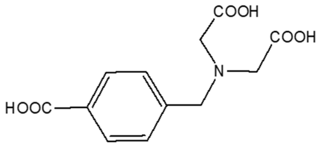
2.1. Materials

The ligand 4-((bis(carboxymethyl)amino)methyl)benzoic acid (H3L) used in this paper was synthesized according to the literature [39,40]. All reagents used in preparation were obtained from Macklin (Shanghai, China) and used without further purification.

2.2. Synthesis of [DyLH2O]n (1)

A mixture of Dy(NO3)3·6H2O (0.20 mmol, 0.0913 g), H3L (0.20 mmol, 0.0534 g) and 6 mL H2O was placed in a Teflon-lined stainless-steel reactor (20 mL). The reactor was heated at 140 °C for three days. After cooling to room temperature slowly, colorless diamond crystal of 1 suitable for X-ray diffraction was obtained. There was a yield of 40% (based on Dy3+).
Analysis calculations for C12H12NO7Dy (%): C 32.37, H 2.70, N 3.15; Found: C 32.32, H 2.80, N 3.20. FT-IR spectra data (KBr, cm−1): ~3400(bs), 2912(w), 1651(vs), 1585(vs), 1552(s), 1441(s), 1398(vs), 1324(s), 1245(m), 1178(m), 1106, 1005(m), 884(m), 874(m), 774(m), 733(m), 708(m), 561(w).

2.3. X-Ray Crystallography

Single-crystal X-ray diffraction data of 1 was collected on a Bruker SMART APEXII CCD diffractometer equipped with a graphite monochromate Mo radiation (λ = 0.71073 Å). The SADABS programs were used to perform empirical absorption correction. Direct methods and full-matrix least-squares methods on F2 were performed to solve and refine the structure using the program SHEXL-2014 [41,42]. All the nonhydrogen atoms were refined with anisotropic parameters while H atoms were placed in calculated positions and refined using a riding model. The crystallographic data and details of selected bond lengths and angles are summarized in Table 1 and Table S1, respectively.
The CCDC reference number 2,085,878 entry contains the supplementary crystallographic data for this paper. These data can be obtained free of charge from The Cambridge Crystallographic Data Center at https://www.ccdc.cam.ac.uk/structures/ (29 April 2025).

2.4. Fourier-Transform Infrared Spectroscopy (FT-IR)

The FT-IR spectra were measured on a Thermo Niclet IS50 FT-IR spectrometer using KBr Pellets (range of 4000–500 cm−1).

2.5. Thermogravimetric Analysis (TG)

A SHIMADZU DTG-60 thermo analyzer was used to perform the thermogravimetric analyses (TGA) under nitrogen at a heating rate of 5 °C min−1 from room temperature to 700 °C.

2.6. Elemental Analysis

Elemental analyses were carried out on an Elementar Vario Micro Cube analyzer for (C, H, N).

2.7. Powder X-Ray Diffraction (PXRD)

Powder X-ray diffraction (PXRD) data in the 2θ range 5–60° were recorded using Cu radiation (κ = 0.1542 nm) on a Bruker D8A A25 X-ray diffractometer, with the X-ray tube operated at 40 kV and 40 mA at room temperature.

2.8. Magnetic Measurements

Magnetic measurements on crystalline samples were carried out at an applied field of 1 kOe on a Quantum Design MPMS-XL7 superconducting quantum interference device (SQUID) magnetometer working in the temperature range of 300–1.8 K. The molar magnetic susceptibilities were corrected for diamagnetism estimated from Pascal’s tables and for the sample holder by previous calibration.

3. Results and Discussion

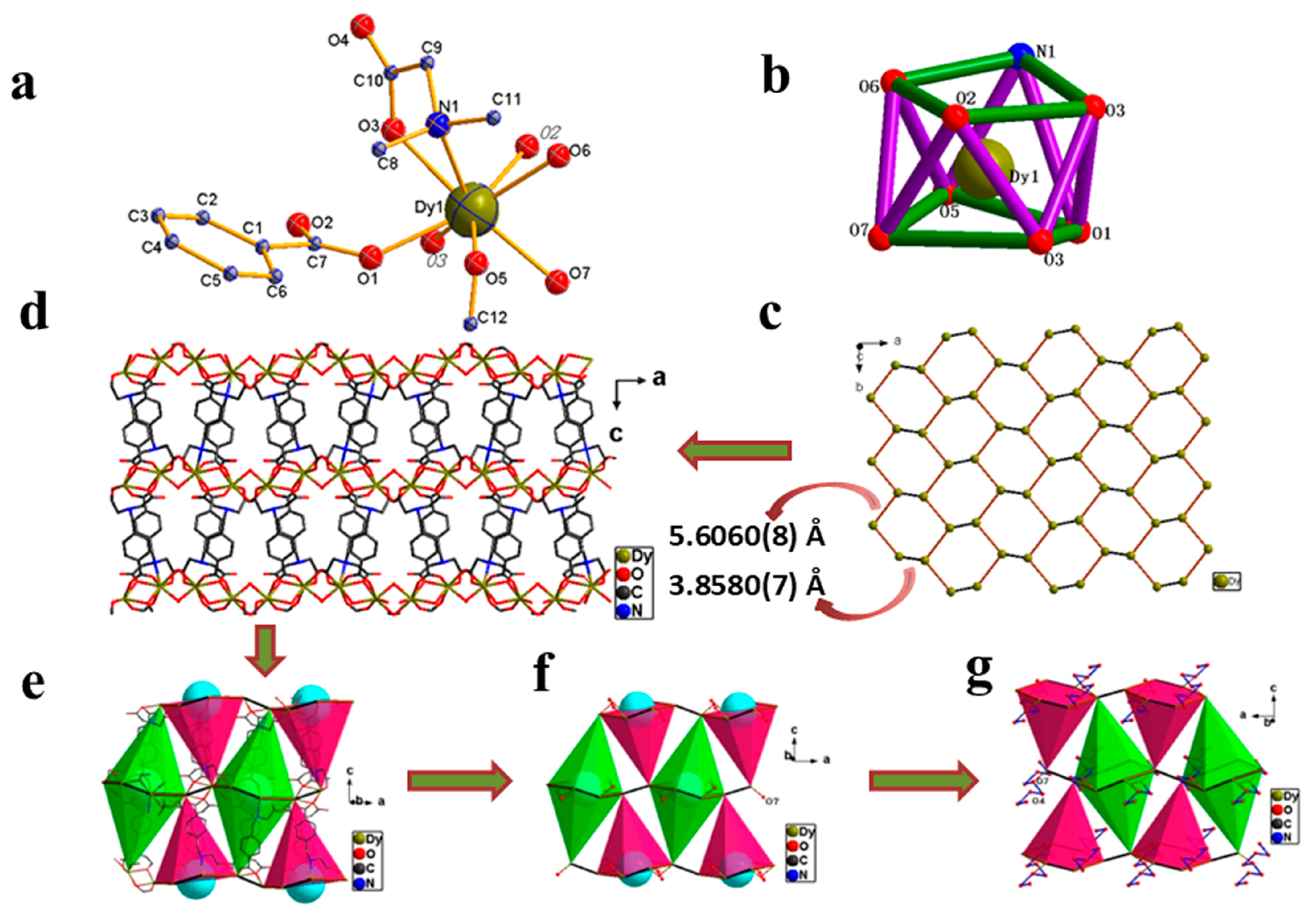
3.1. Crystal Structure

The reaction of H3L (H3L = 4-((bis(carboxymethyl)amino)-methyl)benzoic acid) with Dy(NO3)3·6H2O in water under hydrothermal conditions yields well-shaped crystals of 1. The X-ray diffraction structure analysis indicates that 1 crystallizes in the orthorhombic Pbca space group. The asymmetric unit [DyLH2O] is illustrated in Figure 1a. Each Dy3+ ion assumes an eight-coordinated geometry. Seven coordination sites of Dy3+ are occupied by one amino N atom and six O atoms (two from benzoic acid parts and four from four acetic groups) from five different L3−, and the remaining site is filled by a water molecule. As shown in Figure 1b, the DyNO7 moiety exhibits a distorted D4d-square antiprism (SAP) geometry [43,44]. The Dy-O distances range from 2.314(2) to 2.409(2) Å, and the Dy-N distance is 2.649(2) Å, resulting in two distorted square bases of SAP. All the values above fall in the normal Dy-N/O range [43,44]. The deprotonated L3− ligand adopts a μ5111211 coordination manner to connect five Dy3+ cations (Figure S1) by carboxyl groups with different linking modes: 220, syn-anti 211(for iminodiacetic groups) and syn-syn 211(for benzoic group) [45], respectively. Two adjacent Dy3+ ions are bridged by two acetic (κ-O) oxygen atoms and two syn-syn coordinated carboxylates, leading to the four-bridged Dy2 unit with the intermetallic distance of 3.8580(7) Å (Figure 1c). The bridge oxygen atoms form a perfect plane with the two metallic atoms of Dy2, and the bridging angle ∠Dy-O-Dy is 106.76°. Neighboring Dy2 units are linked by iminodiacetate groups in a syn-anti fashion. The Dy…Dy distance is 5.6060(8) Å (Figure 1c).
Each unit is bridged to 4 Dy2 units by sharing metal sites, ultimately leading to 44 connected 2D layer topologies (Figure 1c) containing chair-like Dy6 rings (Figure S1c). Furtherly, adjacent 2D layers were linked into a three-dimensional framework via the μ(L3−) moiety, as depicted in Figure 1d and Figure S1d. The corresponding Dy…Dy distances lie in the range of 11.4256(16) Å-13.6222(20) Å, which is marked in Figure S1b. The topological type of 1 was defined as a bcu body centered cubic (8/4/c1 and sqc3 (topos & RCSR.ttd) {424.64}). There are diverse tunnels along different directions in this 3D framework. Specifically, these cages (1-6-1) (Figure 1e–g) formed by neighboring Dy6 rings and L3− linkages (Figure 1f) are fascinating features. These cavities have a diameter of approximately 7.6 Å and a depth of 21.4 Å, potentially capable of accommodating some small guest molecule. However, the coordinated O7 hinders access to the cages (Figure 1g). Furthermore, O7…O4 (symmetry code: O4: (−0.5 + x, 1.5 − y, 1 − z, and 2 − x, 2 − y, 1 − z), O7(x, 1 + y, z)) (distance: 2.6898(50) Å and 2.8912(50) Å, angles: 161.819(262)° and 136.840(252)°) H-bond chains direct away from cages (Figure 1g). The volume of these voids and the solvent accessible volume are 9.7% and 271.7 Å, respectively, which is calculated by Platon.

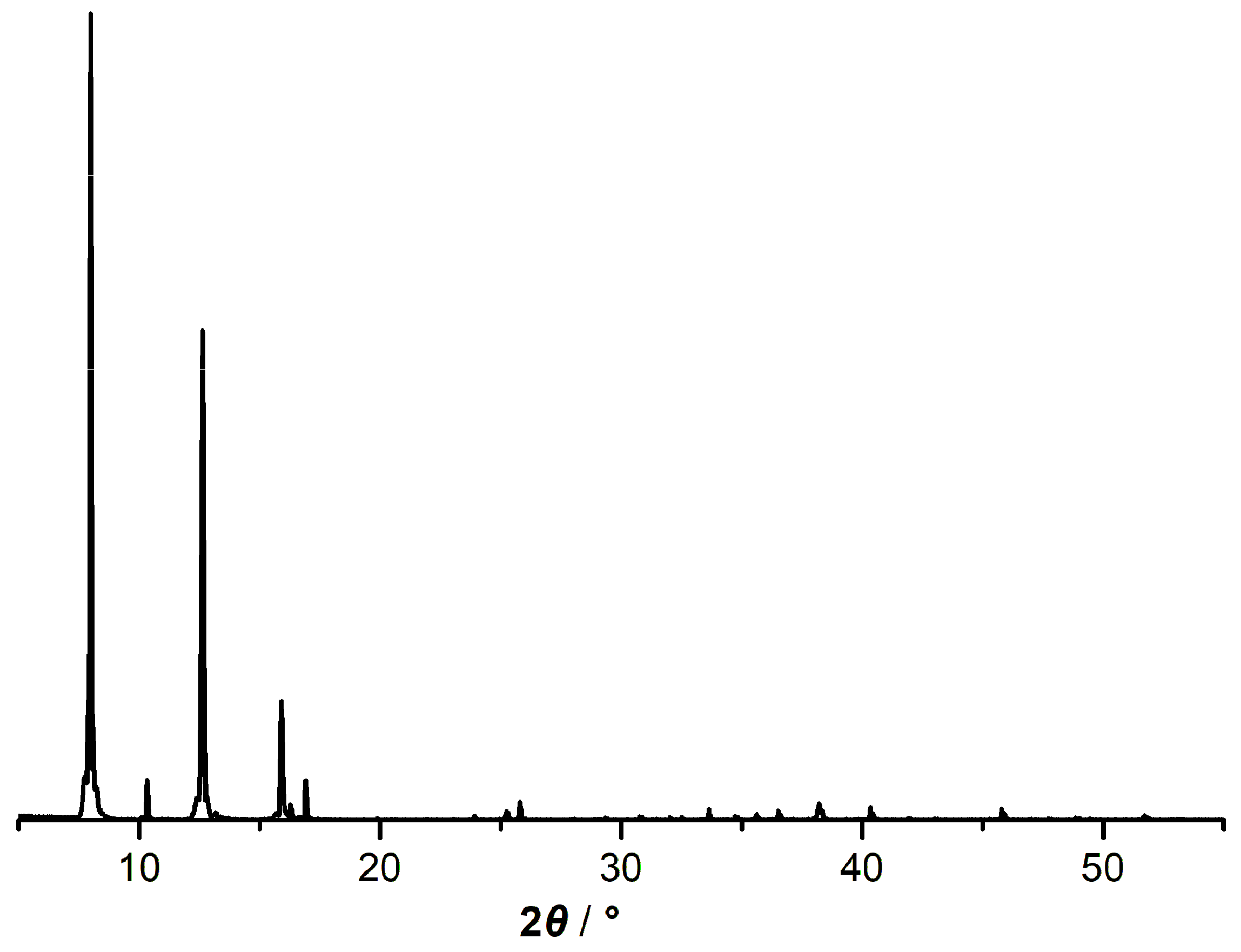
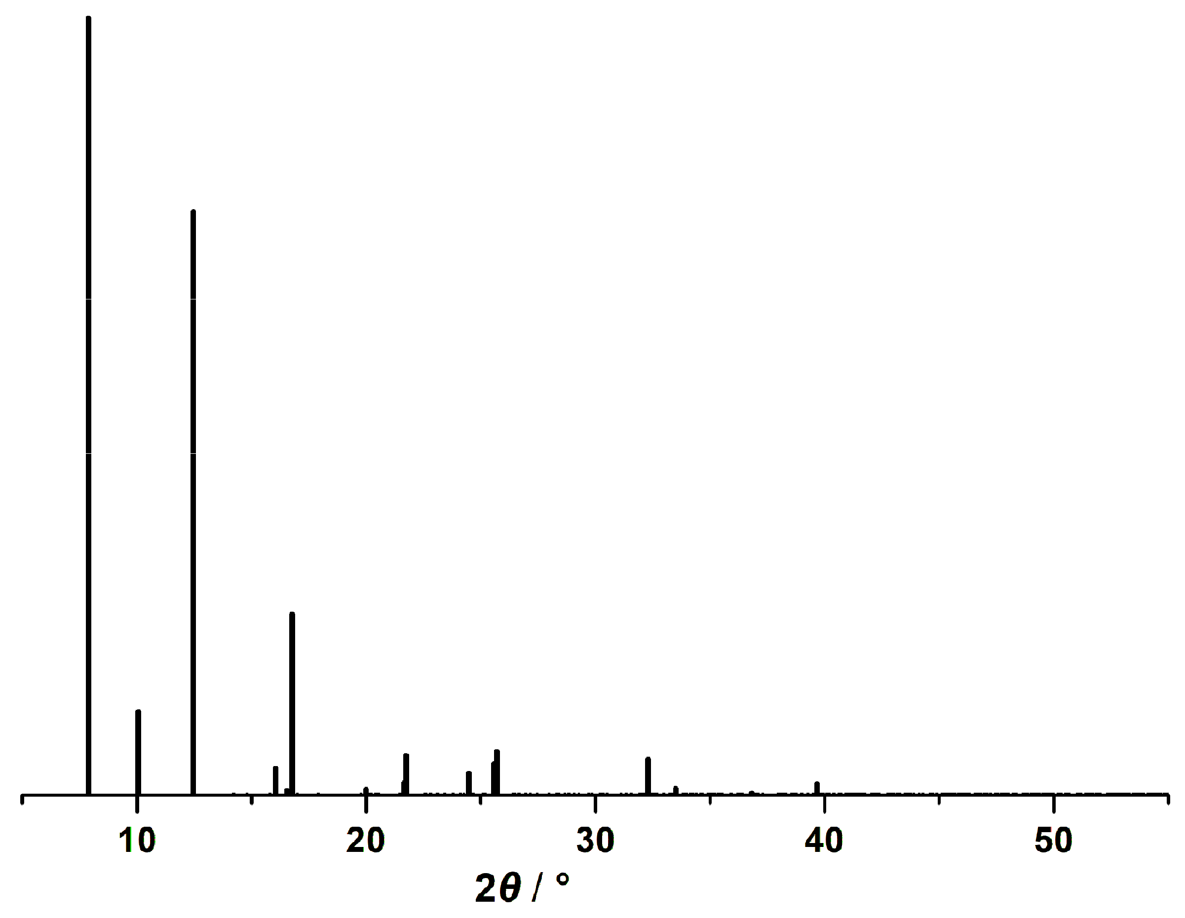
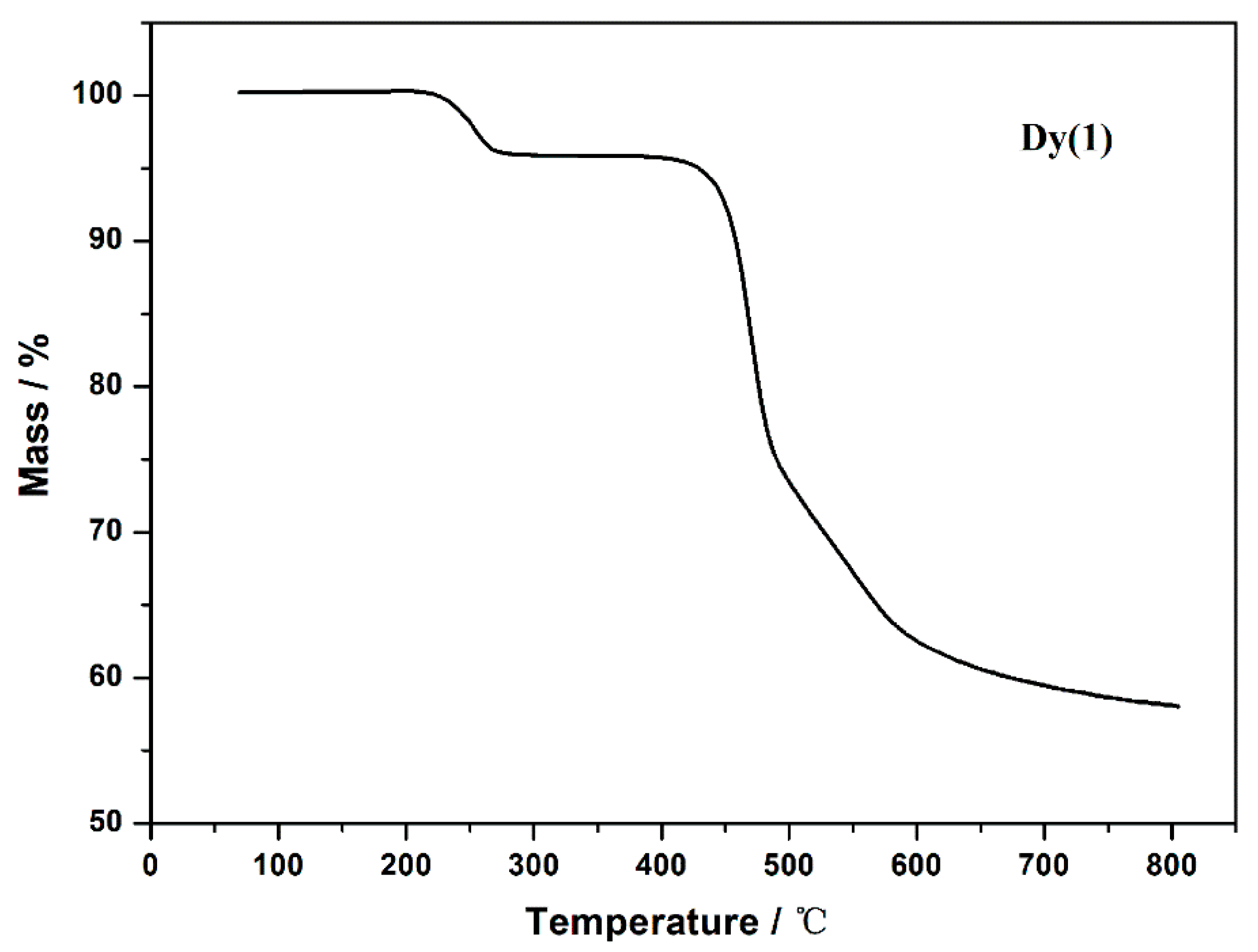
3.2. PXRD and TG Analysis

The as-synthesized crystal experimental PXRD patterns (Figure 2) of 1 closely match the corresponding calculated patterns (Figure 3), confirming the purity of the sample.
The thermal stability and chemical stability of 1 were investigated through thermo-gravimetric analysis (TGA). Two distinct mass-loss stages can be observed from the TGA curve (Figure 4). Sample 1 underwent two separate weight loss processes as indicated by the TGA curve. The first stage of weight loss occurred within the temperature range of 220–270 °C, with a mass loss of 3.9%, due to the loss of the coordinated H2O (Calc. 4.0%). The stability plateau from room temperature to 220 °C demonstrated high thermal stability of 1. The second broader stability plateau from 270 °C shows that the residual framework remained stable until 426 °C. Subsequently, the curve dropped sharply, which should be ascribed to the decomposition of the ligand, with a mass loss of 37.8% (Calc. 37%) in the temperature range of 426–695 °C.
Compared with the light rare earth MOFs [38] previously reported by our group, 1 exhibited remarkable stability. From a structural perspective, the reason for the absence of weight loss of coordinated H2O below approximately 200 °C should be attributed to the wiggly H-bond chains formed by coordinated H2O and neighboring O(-OCO). The more complicated coordination modes of L3− to Ln3+ not only shorten the intermetallic distance but also facilitate the fabrication of stable Dy6 rings. So, the thermostability of 1 achieves significant improvement.

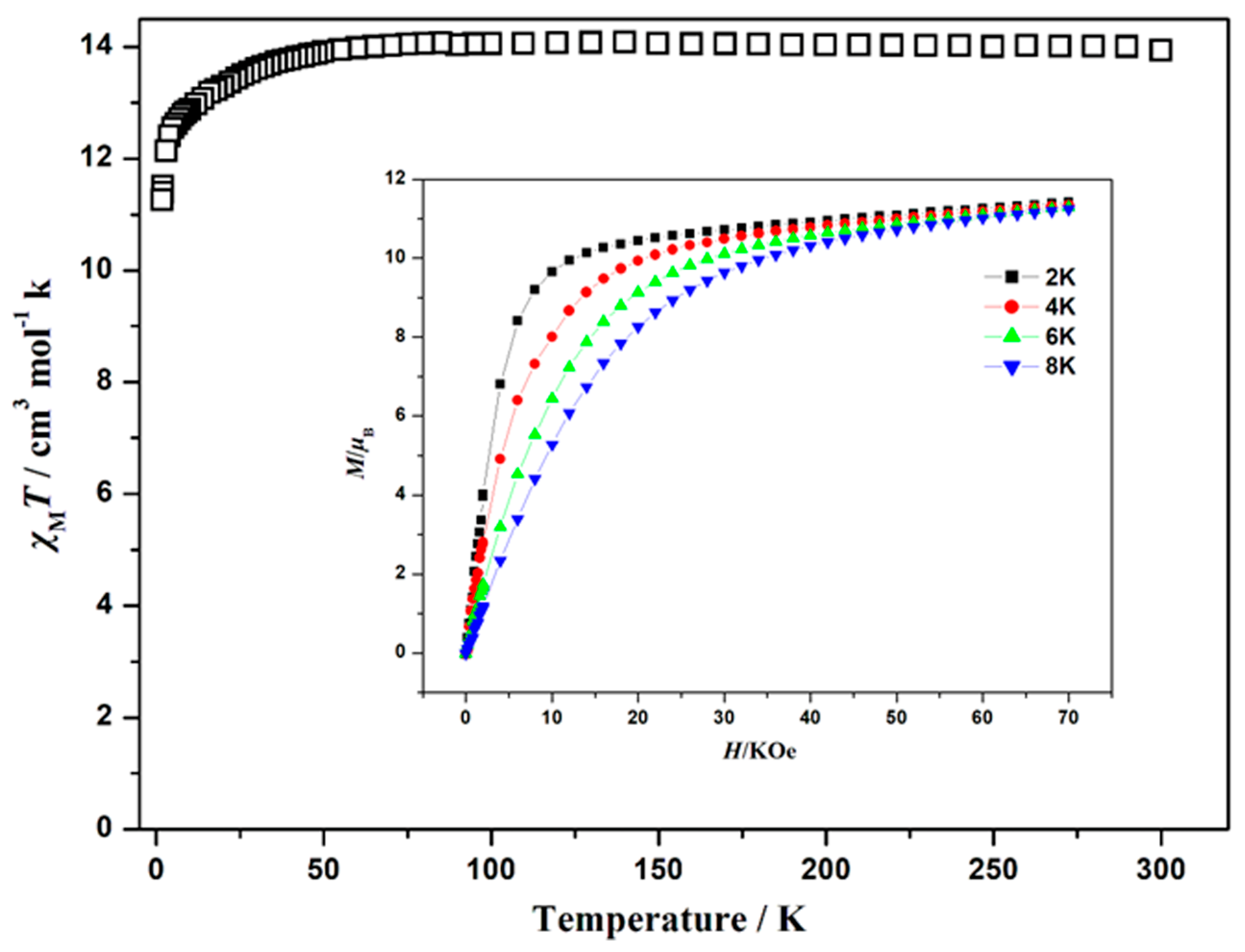
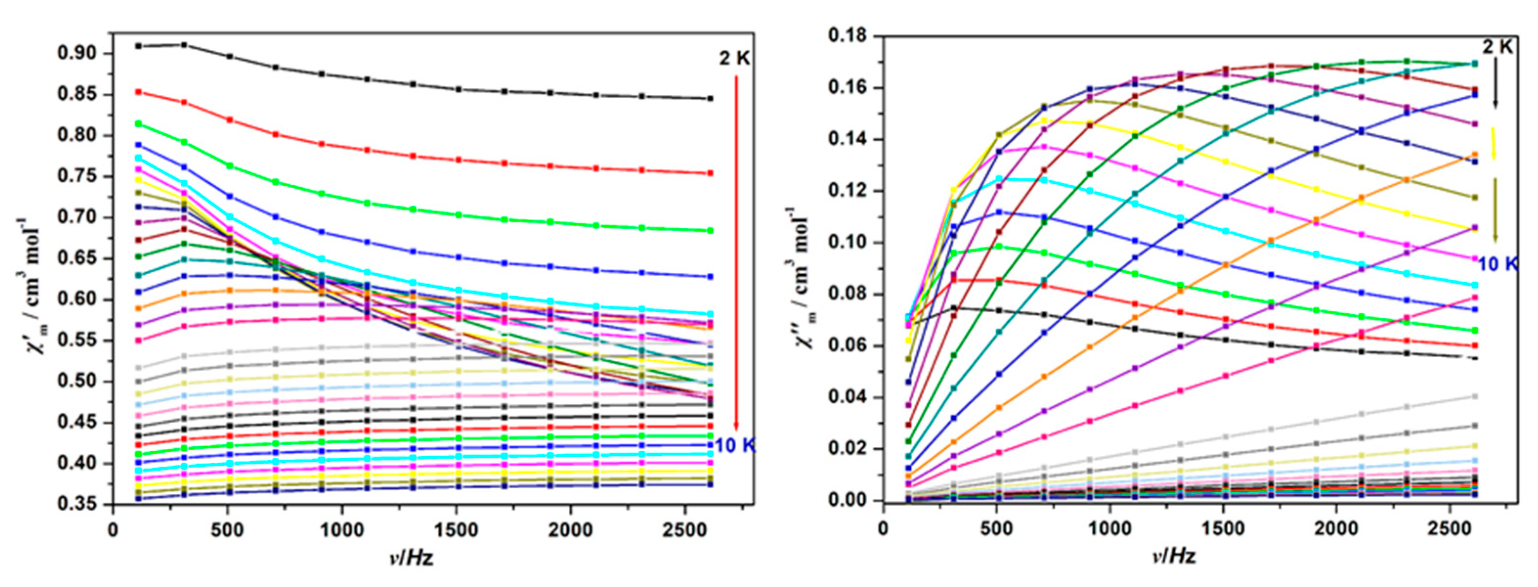
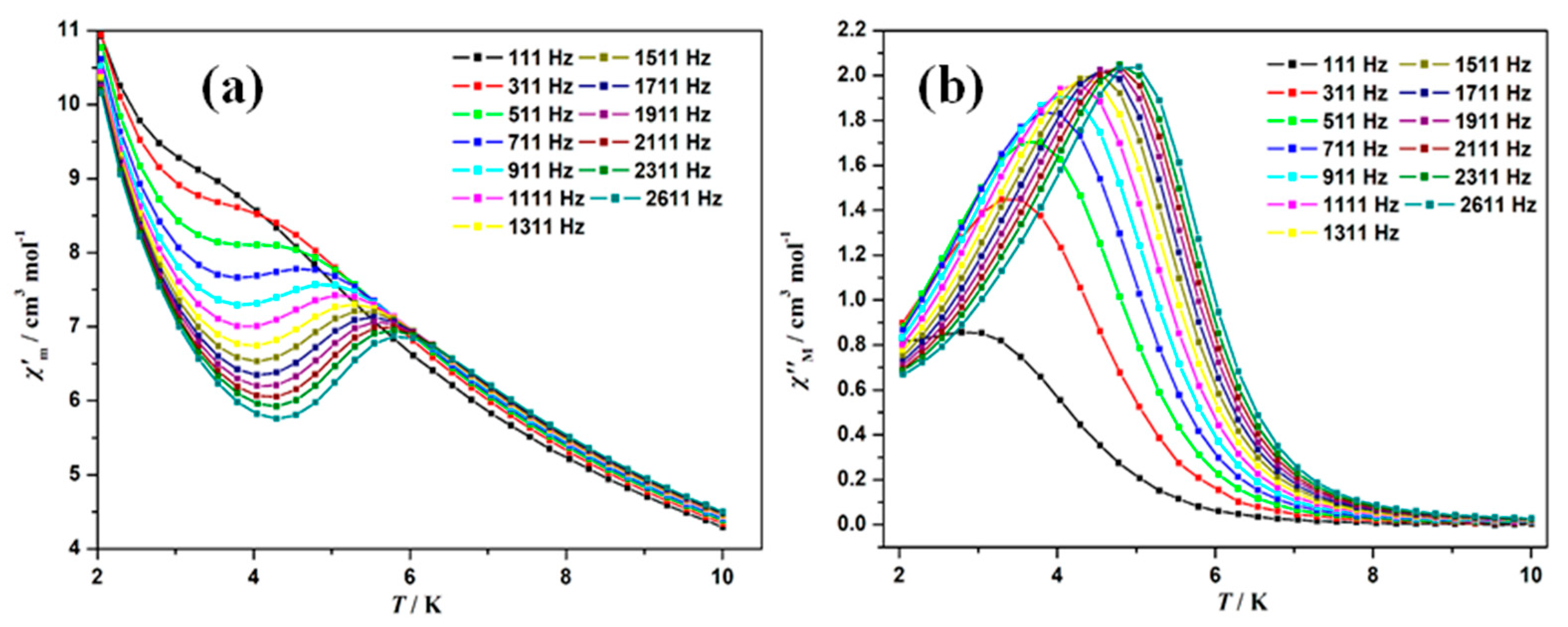
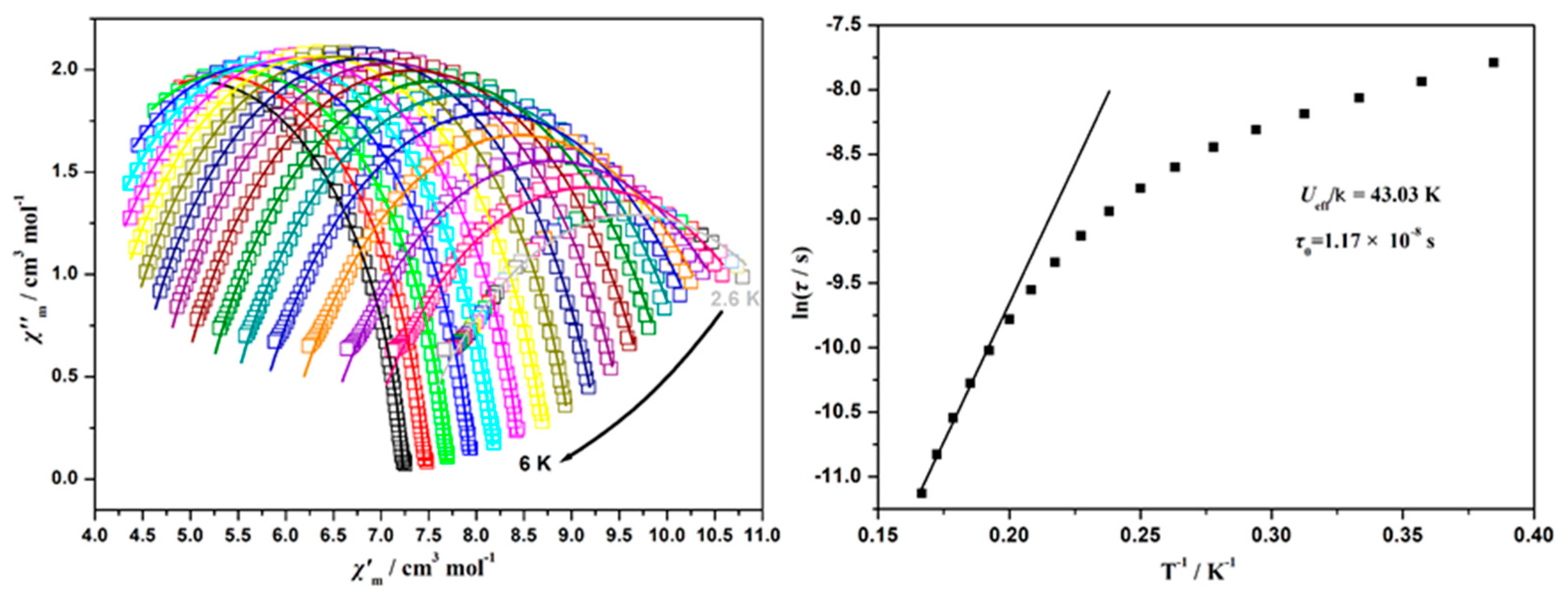
3.3. Magnetic Properties

The magnetic susceptibility of 1 was measured on polycrystalline samples within the temperature range of 2–300 K under an applied field of 1kOe. As shown in Figure 5, the observed χMT value of 14.0 cm3mol−1K at 300 K is slightly smaller than the expected value of 14.17 cm3mol−1K for a single Dy3+ ion (6H15/2, S = 5/2, L = 5, J = 15/2, g = 4/3). When the temperature is lowered, the χMT product for 1 has little change over a wide temperature range from 300 to 70 K. Upon further cooling, the χMT value decreases sharply to a minimum of 11.25 cm3 mol−1 K at 1.8 K, which might be due to crystal-field effects (i.e., thermal depopulation of the Dy3+ Stark sublevels) and/or the possible antiferromagnetic interaction [46]. The Dy-O-Dy bridging angle in Dy2 units is defined as 106.764°, which could account for the strong uniaxial anisotropy of the Dy3+ ions [47,48,49].
The field-dependent magnetization of 1 was measured at 2 K, 4 K, 6 K and 8 K within the field range of 0–7 T (Figure 5 inserted). The isothermal magnetization data exhibits a gradual increase with rising magnetic field. At the lowest investigated temperature (2 K), 1 reaches saturation with a magnetization value of 5.7 μB. Obviously, these values differ significantly from the expected magnetic moment of the isolated Dy3+ (10.0 μB) center. The experimental data of magnetization versus H at different temperatures cannot be superimposed, which indicates the existence of significant magnetic anisotropy and/or low-lying excited states of Dy3+ ions [50,51,52].
To explore the magnetization dynamics of 1, the temperature- and frequency-dependent alternating current (ac) susceptibility measurements were performed in a zero applied direct current (dc) field. In the absence of dc fields, the ac susceptibility measurements detected weak out-of-phase signals for 1 (Figure 6). Upon cooling, the values of χ′, χ″ of 1 increased, suggesting a fast quantum tunneling of magnetization (QTM). This phenomenon is typically associated with mononuclear Dy3+ units [53,54,55,56,57,58]. In order to study the ac susceptibility deeply, the measurement was carried out under an optimized dc field of 1000 Oe. As shown in Figure 7, a slow relaxation process can be observed, indicating the presence of field-induced slow magnetic relaxation. At selected frequencies, 1 exhibited a peak in χM′ and a maximum in χM″. With increasing frequency, the maximum of χM″ shifted to higher temperatures. The magnetization relaxation times τ obtained from the temperature-dependent measurements are plotted as a function of 1/T in Figure 8. In the high-temperature regime, the Arrhenius analysis [τ = τ0 exp(Ueff/T)] was utilized to process the linear fitting of ln τ versus T−1 plots. This analysis aimed to determine the anisotropy energy barrier Ueff with a pre-exponential factor value τ0. For 1, the Ueff value is 43.03 K ( τ 0 = 1.17 × 10 8 s ), consistent with the expected τ0 range of 10−6~10−11 s for a single-molecule magnet (SMM) [59,60,61]. Furthermore, ln (τ) values become weakly dependent on T−1 with decreasing temperature, indicating a transition from a predominant thermally activated Orbach mechanism at high temperature to a Raman process [3,62]. The Cole-Cole plots (χ″ vs. χ′) illustrated in Figure 8 (right) were fitted using a generalized Debye model [63]. The present nonsymmetric semicircle and the α value of 0.38–0.06 (Table S2) falling in the range of previously reported SMMs indicate the presence of a relatively moderate distribution of relaxation time [64].

4. Conclusions

In summary, a novel Dy3+-based metal–organic framework (MOF), derived from 4-((bis(carboxymethyl)amino)methyl) benzoic acid (H3L), has been successfully synthesized via the hydrothermal method. Single-crystal X-ray analysis was employed to characterize its crystal structure, revealing that this complex adopts diverse bridging modes. Specifically, Dy2 units, based on double μ-O and double syn-syn μ-OCO bridges, were interconnected into 44 topology layers through the single syn-anti μ-OCO groups. These layers are then further assembled into a three-dimensional framework via μ-L3− linkages. Notably, small cages are present within this framework, which may potentially accommodate some small molecules, unhindered by hydrogen bonds. Thermogravimetric (TG) analysis indicates that 1 can retain its stable framework upon losing its water molecules, within a broad temperature range (270–426 °C). Moreover, 1 displays slow magnetization relaxation, suggesting its potential as single-ion magnets (SIMs).

Supplementary Materials

The following supporting information can be downloaded at: https://www.mdpi.com/article/10.3390/cryst15060550/s1, Table S1: Important bond length (Å) and angles (°) for complex 1; Figure S1: Coordination mode of L3− (a), simplified ligand with the intermetallic distance (b), chair-like Dy6 ring formed by Dy2 units (c) and the three-dimensional framework (d) in 1; Figure S2: 3D framework of 1 along a direction; Table S2: Table of the values extracted from the fitting of χM″ vs frequency curves. CIF file of the structure was deposited by the Cambridge Crystallographic Data Centre: 2,085,878. The copy can be obtained free of charge on written application to CCDC, 12 Union Road, Cambridge, CB2 1EZ, UK (fax: +44-1223 336033); or by access to http://www.ccdc.cam.ac.uk (29 April 2025).

Author Contributions

Conceptualization, M.L.; methodology, M.L. and X.W.; software, M.L. and Z.M.; validation, M.L. and X.W.; formal analysis, M.L. and Y.C.; investigation, M.L.; resources, M.L. and Y.C.; data curation, M.L., Y.C. and Z.M.; writing—original draft preparation, M.L.; writing—review and editing, M.L.; visualization, M.L. and Z.M.; supervision, M.L. and X.W.; project administration, M.L.; funding acquisition, M.L. and X.W. All authors have read and agreed to the published version of the manuscript.

Funding

This work was supported by the National Natural Science Foundation of China (no.: 21501022, 21601028 and 21371028) and the Natural Science Fund of Shandong province, China (ZR2019QB026).

Data Availability Statement

The original contributions presented in this study are included in the article/Supplementary Material. Further inquiries can be directed to the corresponding author.

Conflicts of Interest

The authors declare no conflicts of interest.

References

  1. Wang, X.X.; Yue, L.L.; Zhou, P.; Fan, L.H.; He, Y.B. Lanthanide–Organic Frameworks Featuring Three-Dimensional Inorganic Connectivity for Multipurpose Hydrocarbon Separation. Inorg. Chem. 2021, 60, 17249–17257. [Google Scholar] [CrossRef] [PubMed]
  2. Wang, X.; Ma, F.Y.; Xiong, S.S.; Bai, Z.L.; Zhang, Y.G.; Li, G.D.; Chen, J.C.; Yuan, M.J.; Wang, Y.L.; Dai, X.; et al. Efficient Xe/Kr Separation Based on a Lanthanide-Organic Framework with One-Dimensional Local Positively Charged Rhomboid Channels. ACS Appl. Mater. Interfaces 2022, 14, 22233–22241. [Google Scholar] [CrossRef]
  3. Zhang, X.J.; Vieru, V.; Feng, X.W.; Liu, J.L.; Zhang, Z.J.; Na, B.; Shi, W.; Wang, B.W.; Powell, A.K.; Chibotaru, L.F.; et al. Influence of Guest Exchange on the Magnetization Dynamics of Dilanthanide Single-Molecule-Magnet Nodes within a Metal–Organic Framework. Angew. Chem. Int. Ed. 2015, 54, 9861–9865. [Google Scholar] [CrossRef]
  4. Wu, S.Y.; Lin, Y.N.; Liu, J.W.; Shi, W.; Yang, G.M.; Cheng, P. Rapid Detection of the Biomarkers for Carcinoid Tumors by a Water Stable Luminescent Lanthanide Metal–Organic Framework Sensor. Adv. Funct. Mater. 2018, 28, 1707169. [Google Scholar] [CrossRef]
  5. Echenique-Errandonea, E.; Mendes, R.F.; Figueira, F.; Choquesillo-Lazarte, D.; Beobide, G.; Cepeda, J.; Ananias, D.; Rodríguez-Diéguez, A.; Paz, F.A.A.; Seco, J.M. Multifunctional Lanthanide-Based Metal-Organic Frameworks Derived from 3-Amino-4-hydroxybenzoate: Single-Molecule Magnet Behavior, Luminescent Properties for Thermometry, and CO2 Adsorptive Capacity. Inorg. Chem. 2022, 61, 12977–12990. [Google Scholar] [CrossRef]
  6. Sahoo, J.; Arunachalam, R.; Subramanian, P.S.; Suresh, E.; Valkonen, A.; Rissanen, K.; Albrecht, M. Coordinatively Unsaturated Lanthanide(III) Helicates: Luminescence Sensors for Adenosine Monophosphate in Aqueous Media. Angew. Chem. Int. Ed. 2016, 55, 9625–9629. [Google Scholar] [CrossRef] [PubMed]
  7. Wang, X.Q.; Zhang, L.L.; Yang, J.; Liu, F.L.; Dai, F.; Wang, R.M.; Sun, D.F. Lanthanide metal–organic frameworks containing a novel flexible ligand for luminescence sensing of small organic molecules and selective adsorption. J. Mater. Chem. A 2015, 3, 12777–12785. [Google Scholar] [CrossRef]
  8. Zhao, T.H.; Han, J.L.; Jin, X.; Liu, Y.; Liu, M.H.; Duan, P.F. Enhanced Circularly Polarized Luminescence from Reorganized Chiral Emitters on the Skeleton of a Zeolitic Imidazolate Framework. Angew. Chem. Int. Ed. 2019, 58, 4978–4982. [Google Scholar] [CrossRef] [PubMed]
  9. Senkovska, I.; Bon, V.; Abylgazina, L.; Mendt, M.; Berger, J.; Kieslich, G.; Petkov, P.; Fiorio, J.L.; Joswig, J.O.; Schaper, L.; et al. Understanding MOF Flexibility: An Analysis Focused on Pillared Layer MOFs as a Model System. Angew. Chem. Int. Ed. 2023, 62, e202218076. [Google Scholar] [CrossRef]
  10. Seth, S.; Savitha, G.; Moorthy, J.N. Diverse isostructural MOFs by postsynthetic metal node metathesis: Anionic-to-cationic framework conversion, luminescence and separation of dyes. J. Mater. Chem. A 2015, 3, 22915–22922. [Google Scholar] [CrossRef]
  11. Leoi, M.W.N.; Zheng, X.T.; Yu, Y.; Gao, J.J.; Ong, D.H.S.; Koh, C.Z.H.; Chen, P.; Yang, L. Redefining Metal Organic Frameworks in Biosensors: Where Are We Now? ACS Appl. Mater. Interfaces 2025, 17, 13246–13278. [Google Scholar] [CrossRef]
  12. Meng, S.Y.; Li, G.; Wang, P.; He, M.; Sun, X.H.; Li, Z.X. Rare earth-based MOFs for photo/electrocatalysis. Mater. Chem. Front. 2023, 7, 806–827. [Google Scholar] [CrossRef]
  13. Allain, C.; Faulkner, S. Photophysical Approaches to Responsive Optical Probes. Future Med. Chem. 2010, 2, 339–350. [Google Scholar] [CrossRef] [PubMed]
  14. Li, C.L.; Pang, Y.D.; Xu, Y.L.; Lu, M.J.; Tu, L.; Li, Q.; Sharma, A.; Guo, Z.Z.; Li, X.Y.; Sun, Y. Near-infrared metal agents assisting precision medicine: From strategic design to bioimaging and therapeutic applications. Chem. Soc. Rev. 2023, 52, 4392–4442. [Google Scholar] [CrossRef] [PubMed]
  15. Wei, S.X.; Li, Z.; Lu, W.; Liu, H.; Zhang, J.W.; Chen, T.; Tang, B.Z. Multicolor Fluorescent Polymeric Hydrogels. Angew. Chem. Int. Ed. 2020, 60, 8608–8624. [Google Scholar] [CrossRef]
  16. Krishnaraj, C.; Kaczmarek, A.M.; Jena, H.S.; Leus, K.; Chaoui, N.; Schmidt, J.; Deun, R.V.; Voort, P.V.D. Triggering White-Light Emission in a 2D Imine Covalent Organic Framework Through Lanthanide Augmentation. ACS Appl. Mater. Interfaces 2019, 11, 27343–27352. [Google Scholar] [CrossRef] [PubMed]
  17. Hao, J.N.; Niu, D.C.; Gu, J.L.; Lin, S.L.; Li, Y.S.; Shi, J.L. Structure Engineering of a Lanthanide-Based Metal–Organic Framework for the Regulation of Dynamic Ranges and Sensitivities for Pheochromocytoma Diagnosis. Adv. Mater. 2020, 32, 2000791. [Google Scholar] [CrossRef]
  18. Carlos, L.D.; Ferreira, R.A.S.; Bermudez, V.S.; Lopez, B.J.; Escribano, P. Progress on lanthanide-based organic–inorganic hybrid phosphors. Chem. Soc. Rev. 2011, 40, 536–549. [Google Scholar] [CrossRef]
  19. Yang, Y.W.; Su, P.R.; Tang, Y. Stimuli-Responsive Lanthanide-Based Smart Luminescent Materials for Optical Encoding and Bio-applications. ChemNanoMat 2018, 4, 1097–1120. [Google Scholar] [CrossRef]
  20. Pan, M.; Liao, W.M.; Yin, S.Y.; Sun, S.S.; Su, C.Y. Single-Phase White-Light-Emitting and Photoluminescent Color-Tuning Coordination Assemblies. Chem. Rev. 2018, 118, 8889–8935. [Google Scholar] [CrossRef]
  21. Thorarinsdottir, A.E.; David Harris, T. Metal–Organic Framework Magnets. Chem. Rev. 2020, 120, 8716–8789. [Google Scholar] [CrossRef] [PubMed]
  22. Layfield, R.A.; Murugesu, M. Lanthanides and Actinides in Molecular Magnetism; Wiley-VCH: Weinheim, Germany, 2015. [Google Scholar] [CrossRef]
  23. Bartolomé, E.; Arauzo, A.; Luzón, J.; Bartolomé, J.; Bartolom, É.F. Chapter 1—Magnetic Relaxation of Lanthanide-Based Molecular Magnets. In Handbook of Magnetic Materials; Brück, E., Ed.; Elsevier: Amsterdam, The Netherlands, 2017; pp. 1–289. [Google Scholar] [CrossRef]
  24. Ishikawa, N.; Sugita, M.; Ishikawa, T.; Koshihara, S.Y.; Kaizu, Y. Lanthanide Double-Decker Complexes Functioning as Magnets at the Single-Molecular Level. J. Am. Chem. Soc. 2003, 125, 8694–8695. [Google Scholar] [CrossRef]
  25. Zhang, P.; Guo, Y.N.; Tang, J.K. Recent advances in dysprosium-based single molecule magnets: Structural overview and synthetic strategies. Coord. Chem. Rev. 2013, 257, 1728–1763. [Google Scholar] [CrossRef]
  26. Wang, W.M.; Wang, S.Y.; Cui, J.Z.; Zhao, B. Modulating single-molecule magnet behaviour of phenoxo-O bridged lanthanide(III) dinuclear complexes by using different β-diketonate coligands. Inorg. Chem. Front. 2016, 3, 133–141. [Google Scholar] [CrossRef]
  27. Krylov, D.S.; Liu, F.; Avdoshenko, S.M.; Spree, L.; Weise, B.; Waske, A.; Wolter, A.U.B.; Buchner, B.; Popov, A.A. Record-high thermal barrier of the relaxation of magnetization in the nitride clusterfullerene Dy2ScN@C80-Ih. Chem. Commun. 2017, 53, 7901–7904. [Google Scholar] [CrossRef]
  28. Guo, F.S.; Day, B.M.; Chen, Y.C.; Tong, M.L.; Mansikkamäki, A.; Layfield, R.A. Magnetic hysteresis up to 80 kelvin in a dysprosium metallocene single-molecule magnet. Science 2018, 362, 1400–1403. [Google Scholar] [CrossRef]
  29. Zhang, J.; Wu, T.; Chen, S.M.; Feng, P.Y.; Bu, X.H. Versatile Structure-Directing Roles of Deep-Eutectic Solvents and Their Implication in the Generation of Porosity and Open Metal Sites for Gas Storage. Angew. Chem. Int. Ed. 2009, 48, 3486–3490. [Google Scholar] [CrossRef] [PubMed]
  30. Wei, Y.; Sa, R.; Wu, K. A highly stable and white-light-emitting Eu(III) MOF. Dalton Trans. 2016, 45, 18661–18667. [Google Scholar] [CrossRef]
  31. Gu, Z.G.; Chen, Z.; Fu, W.Q.; Wang, F.; Zhang, J. Liquid-Phase Epitaxy Effective Encapsulation of Lanthanide Coordination Compounds into MOF Film with Homogeneous and Tunable White-Light Emission. ACS Appl. Mater. Interfaces 2015, 7, 28585–28590. [Google Scholar] [CrossRef]
  32. Du, B.B.; Zhu, Y.X.; Pan, M.; Yue, M.Q.; Hou, Y.J.; Wu, K.; Zhang, L.Y.; Chen, L.; Yin, S.Y.; Fan, Y.N.; et al. Direct white-light and a dual-channel barcode module from Pr(iii)-MOF crystals. Chem. Commun. 2015, 51, 12533–12536. [Google Scholar] [CrossRef]
  33. Dong, J.; Zhao, D.; Lu, Y.; Sun, W.Y. Photoluminescent metal–organic frameworks and their application for sensing biomolecules. J. Mater. Chem. A 2019, 7, 22744–22767. [Google Scholar] [CrossRef]
  34. Tang, Q.; Liu, S.; Liu, Y.; He, D.; Miao, J.; Wang, X.; Ji, Y.; Zheng, Z. Color Tuning and White Light Emission via in Situ Doping of Luminescent Lanthanide Metal–Organic Frameworks. Inorg. Chem. 2014, 53, 289–293. [Google Scholar] [CrossRef] [PubMed]
  35. Wang, Z.X.; Wu, Q.F.; Liu, H.J.; Shao, M.; Xiao, H.P.; Li, M.X. 2D and 3D lanthanide coordination polymers constructed from benzimidazole-5,6-dicarboxylic acid and sulfate bridged secondary building units. CrystEngComm 2010, 12, 1139–1146. [Google Scholar] [CrossRef]
  36. Lu, W.G.; Jiang, L.; Lu, T.B. Lanthanide Contraction and Temperature-Dependent Structures of Lanthanide Coordination Polymers with Imidazole-4,5-Dicarboxylate and Oxalate. Cryst. Growth Des. 2010, 10, 4310–4318. [Google Scholar] [CrossRef]
  37. Zhan, C.H.; Wang, F.; Kang, Y.; Zhang, J. Lanthanide-Thiophene-2,5-dicarboxylate Frameworks: Ionothermal Synthesis, Helical Structures, Photoluminescent Properties, and Single-Crystal-to-Single-Crystal Guest Exchange. Inorg. Chem. 2012, 51, 523–530. [Google Scholar] [CrossRef]
  38. Liu, M.L.; Shi, Q.; Liu, L.F.; Li, W.B. Lanthanide-Aromatic Iminodiacetate Frameworks with Helical Tubes: Structure, Properties, and Low-Temperature Heat Capacity. ACS Omega 2021, 6, 10475–10485. [Google Scholar] [CrossRef] [PubMed]
  39. Lymperopoulou, S.; Papastergiou, M.; Louloudi, M.; Raptopoulou, C.; Psycharis, V.; Milios, C.J.; Plakatouras, J.C. Synthesis, Characterization, Magnetic and Catalytic Properties of a Ladder-Shaped MnII Coordination Polymer. Eur. J. Inorg. Chem. 2014, 2014, 3638–3644. [Google Scholar] [CrossRef]
  40. Liu, M.L.; Shi, W.; Song, H.B.; Cheng, P.; Liao, D.Z.; Yan, S.P. Open and closed copper chain coordination polymers with alternating ferromagnetic and antiferromagnetic interactions. CrystEngComm 2009, 11, 102–108. [Google Scholar] [CrossRef]
  41. Sheldrick, G.M. A short history of SHELX. Acta Crystallogr. Sect. A 2008, 64, 112–122. [Google Scholar] [CrossRef]
  42. Sheldrick, G.M. Crystal structure refinement with SHELX. Acta Crystallogr. Sect. C 2015, 71, 3–8. [Google Scholar] [CrossRef]
  43. Lin, S.Y.; Wang, C.; Zhao, L.; Tang, J.K. Enantioselective Self-Assembly of Triangular Dy3 Clusters with Single-Molecule Magnet Behavior. Chem. Asian J. 2014, 9, 3558–3564. [Google Scholar] [CrossRef] [PubMed]
  44. Zheng, X.Y.; Zhang, H.; Cao, L.Y.; Kong, X.J.; Long, L.S.; Zheng, L.S. Chirality detection of two enantiomorphic 3D lanthanide coordination polymers by vibrational circular dichroism spectra. Dalton Trans. 2015, 44, 5299–5302. [Google Scholar] [CrossRef]
  45. Xu, Y.Q.; Yuan, D.Q.; Wu, B.L.; Han, L.; Wu, M.Y.; Jiang, F.L.; Hong, M.C. 1D Tube, 2D Layer, and 3D Framework Derived from a New Series of Metal(II)−5-Aminodiacetic Isophthalate Coordination Polymers. Cryst. Growth Des. 2006, 6, 1168–1174. [Google Scholar] [CrossRef]
  46. Sun, W.B.; Han, B.L.; Lin, P.H.; Li, H.F.; Chen, P.; Tian, Y.M.; Murugesu, M.; Yan, P.F. Series of dinuclear and tetranuclear lanthanide clusters encapsulated by salen-type and β-diketionate ligands: Single-molecule magnet and fluorescence properties. Dalton Trans. 2013, 42, 13397–13403. [Google Scholar] [CrossRef]
  47. Chen, M.; Sauñudo, E.C.; Jiménez, E.; Fang, S.M.; Liu, C.S.; Du, M. Lanthanide–Organic Coordination Frameworks Showing New 5-Connected Network Topology and 3D Ordered Array of Single-Molecular Magnet Behavior in the Dy Case. Inorg. Chem. 2014, 53, 6708–6714. [Google Scholar] [CrossRef] [PubMed]
  48. Yao, R.X.; Xu, X.; Zhang, X.M. Ferromagnetically coupled chiral dysprosium hydroxysulfate and centrosymmetric dysprosium hydroxysulfate-oxalate: Dy4(OH)4 cubane based high-connected networks via in situ conversion of organosulfur to sulfate. RSC Adv. 2014, 4, 53954–53959. [Google Scholar] [CrossRef]
  49. Liu, S.J.; Zhao, J.P.; Song, W.C.; Han, S.D.; Liu, Z.Y.; Bu, X.H. Slow Magnetic Relaxation in Two New 1D/0D DyIII Complexes with a Sterically Hindered Carboxylate Ligand. Inorg. Chem. 2013, 52, 2103–2109. [Google Scholar] [CrossRef] [PubMed]
  50. Sethi, W.; Sanz, S.; Pedersen, K.S.; Sørensen, M.A.; Nichol, G.S.; Lorusso, G.; Evangelisti, M.; Brechin, E.K.; Piligkos, S. Magnetic and magnetocaloric properties of an unusual family of carbonate-panelled [Ln III6Zn II2] cages. Dalton Trans. 2015, 44, 10315–10320. [Google Scholar] [CrossRef]
  51. Ren, M.; Pinkowicz, D.; Yoon, M.; Kim, K.; Zheng, L.M.; Breedlove, B.K.; Yamashita, M. Dy(III) Single-Ion Magnet Showing Extreme Sensitivity to (De)hydration. Inorg. Chem. 2013, 52, 8342–8348. [Google Scholar] [CrossRef]
  52. Zhao, X.X.; Li, G.; Ma, J.J.; Liu, W.S. Two Octanuclear {Cu4Ln4} (Ln = Dy or Tb) Complexes with a Butterfly-Shaped Unit Exhibiting Zero-Field Single-Molecule Magnet Behavior. Inorg. Chem. 2020, 59, 2328–2336. [Google Scholar] [CrossRef]
  53. Sun, L.; Zhang, S.; Qiao, C.F.; Chen, S.P.; Yin, B.; Wang, W.Y.; Wei, W.; Xie, G.; Gao, S.L. Fine-Tuning of the Coordination Environment To Regulate the Magnetic Behavior in Solvent/Anion-Dependent DyIII Compounds: Synthesis, Structure, Magnetism, and Ab Initio Calculations. Inorg. Chem. 2016, 55, 10587–10596. [Google Scholar] [CrossRef] [PubMed]
  54. Chen, Y.C.; Liu, J.L.; Ungur, L.; Liu, J.; Li, Q.W.; Wang, L.F.; Ni, Z.P.; Chibotaru, L.F.; Chen, X.M.; Tong, M.L. Symmetry-Supported Magnetic Blocking at 20 K in Pentagonal Bipyramidal Dy(III) Single-Ion Magnets. J. Am. Chem. Soc. 2016, 138, 2829–2837. [Google Scholar] [CrossRef]
  55. Zhang, S.; Ke, H.S.; Sun, L.; Li, X.; Shi, Q.; Xie, G.; Wei, Q.; Yang, D.S.; Wang, W.Y.; Chen, S.P. Magnetization Dynamics Changes of Dysprosium(III) Single-Ion Magnets Associated with Guest Molecules. Inorg. Chem. 2016, 55, 3865–3871. [Google Scholar] [CrossRef] [PubMed]
  56. Gregson, M.; Chilton, N.F.; Ariciu, A.M.; Tuna, F.; Crowe, I.F.; Lewis, W.; Blake, A.J.; Collison, D.; McInnes, E.J.L.; Winpenny, R.E.P.; et al. A monometallic lanthanide bis (methanediide) single molecule magnet with a large energy barrier and complex spin relaxation behaviour. Chem. Sci. 2016, 7, 155–165. [Google Scholar] [CrossRef]
  57. Wu, J.F.; Jung, J.L.; Zhang, P.; Zhang, H.X.; Tang, J.K.; Le Guennic, B. Cis−trans isomerism modulates the magnetic relaxation of dysprosium single-molecule magnets. Chem. Sci. 2016, 7, 3632–3639. [Google Scholar] [CrossRef]
  58. Pugh, T.; Tuna, F.; Ungur, L.; Collison, D.; McInnes, E.J.; Chibotaru, L.F.; Layfield, R.A. Influencing the properties of dysprosium single-molecule magnets with phosphorus donor ligands. Nat. Commun. 2015, 6, 7492. [Google Scholar] [CrossRef] [PubMed]
  59. Cole, K.S.; Cole, R.H. Dispersion and absorption in dielectrics I. Alternating current characteristics. J. Chem. Phys. 1941, 9, 341–351. [Google Scholar] [CrossRef]
  60. Ishii, N.; Okamura, Y.; Chiba, S.; Nogami, T.; Ishida, T. Giant coercivity in a one-dimensional cobalt-radical coordination magnet. J. Am. Chem. Soc. 2008, 130, 24–25. [Google Scholar] [CrossRef]
  61. Aubin, S.M.; Sun, Z.; Pardi, L.; Krzystek, J.; Folting, K.; Brunel, L.C.; Rheingold, A.L.; Christou, G.; Hendrickson, D.N. Reduced anionic Mn12 molecules with half-integer ground states as single-molecule magnets. Inorg. Chem. 1999, 38, 5329–5340. [Google Scholar] [CrossRef]
  62. Joarder, B.; Chaudhari, A.K.; Rogez, G.; Ghosh, S.K. A carboxylate-based dinuclear dysprosium(III) cluster exhibiting slow magnetic relaxation behaviour. Dalton Trans. 2012, 41, 7695–7699. [Google Scholar] [CrossRef]
  63. Guo, Y.N.; Xu, G.F.; Tang, J.K. Relaxation dynamics of dysprosium(III) single molecule magnets. Dalton Trans. 2011, 40, 9953–9963. [Google Scholar] [CrossRef] [PubMed]
  64. Sun, L.; Zhang, S.; Zhang, Z.J.; Yang, Q.; Chen, S.P.; Zhang, Y.Q.; Wang, W.Y.; Wei, Q.; Xie, G. Interchange between coordinated and lattice solvents generates the highest energy barrier within nine-coordinated DyIII single molecule magnets. Dalton Trans. 2017, 46, 11159–11165. [Google Scholar] [CrossRef] [PubMed]
Scheme 1. The diagram of the ligand H3L.
Crystals 15 00550 sch001
Figure 1. Structure of 1: (a) Molecule fragment, (b) coordination polyhedron around Dy3+, (c) 2D topology (black lines represent quadruple bridges inner Dy2 units and red lines replace single –OCO bridges between Dy2 units), (d) 3D framework along b direction, (e) cages formed in 1, (f) distribution of coordinated H2O molecules in framework, and (g) O-H…O H-bond chains formed in framework.
Crystals 15 00550 g001
Figure 2. The experiment XRD diffractograms for 1.
Crystals 15 00550 g002
Figure 3. The calculated XRD diffractograms for 1.
Crystals 15 00550 g003
Figure 4. The TG curve plot of 1.
Crystals 15 00550 g004
Figure 5. Temperature dependence of χMT for 1 at 1kOe applied field over the temperature of 2–300 K, and M vs. H plots for 1 at different temperatures (inserted).
Crystals 15 00550 g005
Figure 6. Frequency-dependence of the in-phase (χM′) (left) and out-of-phase ac susceptibility (χM″) (right) signals for 1.
Crystals 15 00550 g006
Figure 7. Temperature-dependence of the in-phase (χM′) (a) and out-of-phase ac susceptibility (χM″) (b) signals under a 1000 Oe dc field for 1.
Crystals 15 00550 g007
Figure 8. (Left) Magnetization relaxation time, ln(τ) versus T−1, plots under a 1000 Oe dc field for 1. The solid line is fitted with the Arrhenius law, and (Right) Cole-Cole plots at 2.6-6 K of 1.
Crystals 15 00550 g008
Table 1. X-ray diffraction single crystallographic data and structure refinement details.
Parameter[DyLH2O]n (1)
FormulaC12H12NO7Dy
Formula weight444.73
T [K] 298(2)
Crystal system
space group
Orthorhombic,
Pbca
a [Å]14.1380(12)
b [Å]8.8120(6)
c [Å]22.416(2)
α [°]90
β [°]90
γ [°]90
V3]2792.6(4)
Z8
ρcalc. [gcm−3]2.116
µ [mm−1]5.384
F(000)1704
θ range [°]2.32 to 25.01
Limiting indices−16 ≤ h ≤ 15,
−9 ≤ k ≤ 10,
−26 ≤ l ≤ 25
Reflections collected
/unique
12,364/2468
[Rint = 0.0326]
Data/
restraints/para.
2468/0/190
GoF1.055
R1
wR2 [I > 2σ(I)]
0.0220, 0.0493
R1
wR2 (all data)
0.0315, 0.0525
Largest diff. peak and hole [e.A−3]0.716, −0.582
Disclaimer/Publisher’s Note: The statements, opinions and data contained in all publications are solely those of the individual author(s) and contributor(s) and not of MDPI and/or the editor(s). MDPI and/or the editor(s) disclaim responsibility for any injury to people or property resulting from any ideas, methods, instructions or products referred to in the content.

