Article

Structural Analysis of PlyKp104, a Novel Phage Endoysin

Department of Food Science and Biotechnology, Kyungsung University, Busan 48434, Republic of Korea
Crystals 2025, 15(5), 448; https://doi.org/10.3390/cryst15050448
Submission received: 7 April 2025 / Revised: 7 May 2025 / Accepted: 7 May 2025 / Published: 9 May 2025
(This article belongs to the Special Issue Crystallography of Enzymes)

Abstract

:
Antibiotic resistance has emerged as a critical global public health challenge, prompting increased interest in non-antibiotic antimicrobial strategies such as bacteriophage-derived endolysins. Although endolysins possess strong lytic potential, their application to Gram-negative bacteria remains limited due to the outer membrane barrier. PlyKp104 is a recently identified phage-derived endolysin that exhibits lytic activity against Gram-negative bacteria without the aid of membrane permeabilizers. In this study, the crystal structure of PlyKp104 was determined at a resolution of 1.85 Å. PlyKp104 consists solely of a catalytic SLT domain, and structure-based analysis revealed a putative active site and key structural features associated with substrate binding. Comparative analysis with homologous structures suggested that PlyKp104 belongs to lytic transglycosylase family 1. B-factor analysis and hydrophobic interaction mapping indicated that the domain exhibits high structural stability, supported by conserved hydrophobic residues clustered in motifs I and II. During structure determination, an unidentified electron density was consistently observed near a neutral, hydrophobic surface region. Its shape and environment suggest the presence of a lipid-like molecule, implying a potential lipid-binding site. These findings provide structural insight into PlyKp104 and contribute to the understanding of endolysin mechanisms against Gram-negative bacteria, with implications for future protein engineering efforts.

1. Introduction

Since the discovery of penicillin by Alexander Fleming in 1929, antibiotics have served as a cornerstone in the treatment of bacterial infections [1]. These agents target essential physiological and biochemical processes in bacteria, thereby killing the cells or inhibiting their growth [2]. Because such targets are structurally or functionally distinct—or even absent—in eukaryotic cells, including those of humans, antibiotics are generally considered safe and selective treatments [3].
However, when exposed to antibiotics, bacteria exhibit high genetic plasticity through spontaneous mutations, genome rearrangements, and horizontal gene transfer. This adaptability is a major driver of antibiotic resistance and is considered an inevitable outcome of bacterial evolution [4,5]. As long as antibiotics are in use, bacteria will continue to evolve and develop resistance strategies [5]. Moreover, the widespread misuse and overuse of antibiotics in both healthcare and agriculture have accelerated the emergence of multidrug-resistant (MDR) bacteria, defined as those resistant to at least three different classes of antibiotics [6].
For several decades, the development of new classes of antibiotics has helped mitigate the resistance crisis [4]. However, in recent years, the pace of antibiotic discovery has slowed considerably [7]. Developing new antibiotics is both time-consuming and costly, and resistance to newly introduced drugs often emerges rapidly [7]. As a result, antibiotic resistance has become a significant global public health threat, undermining the effectiveness of infection treatment and prevention strategies [7].
This growing challenge has led to increasing interest in alternative antimicrobial strategies [8]. Among these, non-antibiotic antimicrobial therapies are actively being explored [9]. These therapies operate through mechanisms distinct from those of conventional antibiotics and hold significant potential for synergy when used in combination with them [9].
Endolysins are bacteriophage-derived enzymes that degrade the peptidoglycan layer of bacterial cell walls during the lytic cycle, resulting in cell lysis [10]. As they target the highly conserved peptidoglycan structure, endolysins exhibit broad-spectrum yet specific activity and have a low likelihood of inducing resistance. Consequently, they are regarded as promising antimicrobial agents against MDR bacteria [11], and are emerging as strong candidates for non-antibiotic antibacterial therapies [10].
Endolysins are particularly effective against Gram-positive bacteria, where the peptidoglycan is exposed on the cell surface. In contrast, their efficacy against Gram-negative bacteria is limited due to the presence of an outer membrane that shields the peptidoglycan [10]. Overcoming this membrane barrier remains a major challenge in the development of endolysins targeting Gram-negative pathogens. Fortunately, bacteriophages that infect Gram-negative bacteria do exist, and ongoing research aims to identify and characterize endolysins derived from these phages [12]. Although progress has been made, endolysins effective against Gram-negative bacteria remain scarce, and structural and biochemical studies elucidating their mechanisms are still limited [10]. Among the few characterized examples, the structures of LysECD7 and Enc34 have been elucidated [13,14]. These endolysins exhibit activity against Gram-negative bacteria and are reported to traverse the outer membrane via a C-terminal antimicrobial peptide-like structure [13,14].
Recently, a novel endolysin, PlyKp104, was identified from lysogenic phages infecting Gram-negative bacteria [15]. This protein was discovered through sequence similarity analyses with two putative endolysins, PlyPa101 (accession number: YP_009285812) and PlyPa102 (accession number: AMQ76165), derived from the genomes of phages NP1 and NP3 [15]. PlyKp104 demonstrated lytic activity against Pseudomonas aeruginosa and Klebsiella pneumoniae, both of which are Gram-negative members of the ESKAPE group (Enterococcus faecium, Staphylococcus aureus, K. pneumoniae, Acinetobacter baumannii, P. aeruginosa, and Enterobacter) that are classified as high-priority MDR pathogens [15]. Notably, this activity was observed without the use of membrane permeabilizers or protein modification [15]. Therefore, further structural and biochemical studies of PlyKp104 are expected to provide valuable insights into the lytic mechanism of phage endolysins against Gram-negative bacteria.
In this study, the crystal structure of PlyKp104 was determined at a resolution of 1.85 Å using X-ray crystallography and its structural features were characterized to provide insights into its bacteriolytic mechanism against Gram-negative pathogens.

2. Materials and Methods

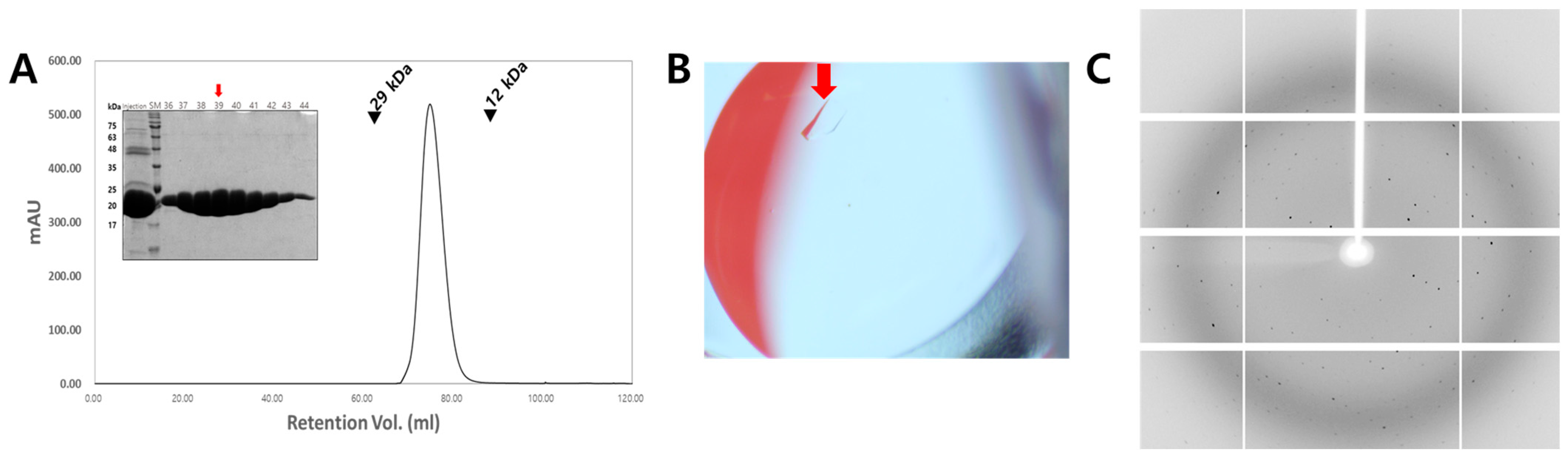
2.1. Cloning, Protein Expression, and Purification

The gene encoding PlyKp104 in the genome of Klebsiella variicola (PlyKp104, GenBank, Bethesda, MD, USA, No. MBC5100607) was synthesized (IDT Inc., Coralville, IA, USA) and cloned into the pET-28a expression vector (Novagen, Madison, WI, USA) using the NdeI and XhoI restriction sites. The sequence of the cloned gene was verified by DNA sequencing (Macrogen, Seoul, Republic of Korea). The verified plasmid was transformed into Escherichia coli BL21 (DE3) cells. The transformed cells were cultured in LB medium at 37 °C until the optical density at 600 nm (OD600) reached 0.5. Protein expression was then induced by adding 1 mM isopropyl β-D-1-thiogalactopyranoside (IPTG), followed by further incubation at 18 °C overnight. After induction, the cultured cells were harvested by centrifugation at 7000 rpm for 30 min at 4 °C. The cell pellet was resuspended in 15 mL of lysis buffer containing 20 mM Tris-HCl (pH 7.4) and 400 mM NaCl. The cells were lysed using sonication, and the lysate was centrifuged again at 17,000 rpm for 1 h at 4 °C to collect the supernatant. Ni-NTA affinity chromatography was performed to purify the recombinant protein from the collected supernatant using the N-terminal 6xHis-tag of PlyKp104. The supernatant was applied to a Ni-NTA resin (Bio-Works, Uppsala, Sweden), and the resin was washed with a buffer containing 20 mM Tris-HCl (pH 7.4) and 400 mM NaCl. The protein was eluted with an elution buffer containing 50 mM Tris-HCl (pH 7.4), 400 mM NaCl, and 250 mM imidazole. The final purification step involved size exclusion chromatography with a HiLoad Superdex 75 pg column (Cytiva, Marlborough, MA, USA), during which, the buffer was exchanged to 5 mM Tris-HCl (pH 7.4) containing 50 mM NaCl. The purity of the eluted protein solution was assessed using 15% sodium dodecyl sulfate–polyacrylamide gel electrophoresis (SDS-PAGE), and the protein concentration was determined using the Bradford assay. The purified protein was concentrated to 10 mg/mL, aliquoted, and stored at −70 °C until crystallization.

2.2. Crystallization

Crystallization screening was performed using the sitting drop vapor diffusion method at 20 °C using a commercial crystallization kit, including the Crystal Screen 1, 2, MembFac, and Natrix (Hampton Research, Aliso Viejo, CA, USA) and Wizard Classic 1, 2, 3, and 4 (Rigaku, The Woodlands, TX, USA). The protein solution (1 µL) was mixed with the crystallization screen solution (1 µL) and equilibrated with a reservoir solution (50 µL). Crystalline-like material was first observed approximately 5 days after setting up the crystallization trials and four different microcrystals were obtained from the initial screening within 4 weeks. The dimensions of the microcrystals were approximately 30–50 µm in length and 5–10 µm in width. Through a preliminary diffraction experiment, suitable crystals for X-ray diffraction were obtained from one condition.

2.3. X-Ray Diffraction Data Collection

X-ray diffraction data were collected at beamline 7A at Pohang Light Source II (PLS-II, Pohang, Republic of Korea) [16]. The PlyKp104 crystal was cryoprotected using a reservoir solution supplemented with 25% (v/v) glycerol. The crystal was mounted on a goniometer under a 100 K nitrogen gas stream. Diffraction data were recorded using a Eiger2 S 9M detector (DECTRIS, Baden, Switzerland). Diffraction data were indexed, integrated, and scaled using HKL2000 [17]. The X-ray diffraction images were visualized using Dials [18].

2.4. Structure Determination

The phase problem was solved by molecular replacement using Phaser in PHENIX [19]. A three-dimensional molecular model generated with AlphaFold2 was used as the search model [20]. Coot was used for manual model building based on the electron density map [21]. Structure refinement was conducted with phenix.refine in PHENIX [19].

2.5. Bioinformatics and Structure Analysis

A structural similarity search was performed using the DALI server [22]. Amino acid sequence alignment was performed using Clustal Omega [23]. The structure-based multiple sequence alignment was visualized with ESPript 3.0 [24]. The protein structures were visualized using PyMOL (https://pymol.org, accessed on 1 March 2025).

3. Results

3.1. Structure Determination

The molecular mass of PlyKp104 was approximately 17 kDa, determined using size exclusion chromatography, which closely matches the theoretical monomer mass of 18 kDa, indicating that PlyKp104 exists as a monomer in solution (Figure 1A). The initial crystallization trials yielded crystals under the following four conditions: (i) 100 mM HEPES sodium (pH 7.5) and 1.5 M lithium sulfate monohydrate; (ii) 200 mM ammonium acetate, 100 mM sodium citrate tribasic dihydrate (pH 5.6), and 30% (w/v) polyethylene glycol (PEG) 4000; (iii) 200 mM lithium sulfate, 100 mM Tris–HCl (pH 8.5), and 30% (w/v) PEG 4000; and (iv) 100 mM imidazole (pH 6.5) and 1.0 M sodium acetate trihydrate. Among these, the crystal obtained from condition (i) showed relatively good diffraction, reaching a resolution of ~5 Å. The crystals from the other three conditions exhibited either poor or no diffraction. Therefore, condition (i) was selected for further optimization, resulting in crystals suitable for high-resolution data collection (Figure 1B). The optimized crystal diffracted to a resolution of ~1.8 Å (Figure 1C). The PlyKp104 crystal belonged to the orthorhombic space group C2221, with unit cell dimensions of a = 67.73 Å, b = 89.58 Å, and c = 67.74 Å (Table 1). The structure of PlyKp104 was determined at a resolution of 1.85 Å, with Rwork and Rfree values of 19% and 22%, respectively (Table 1).

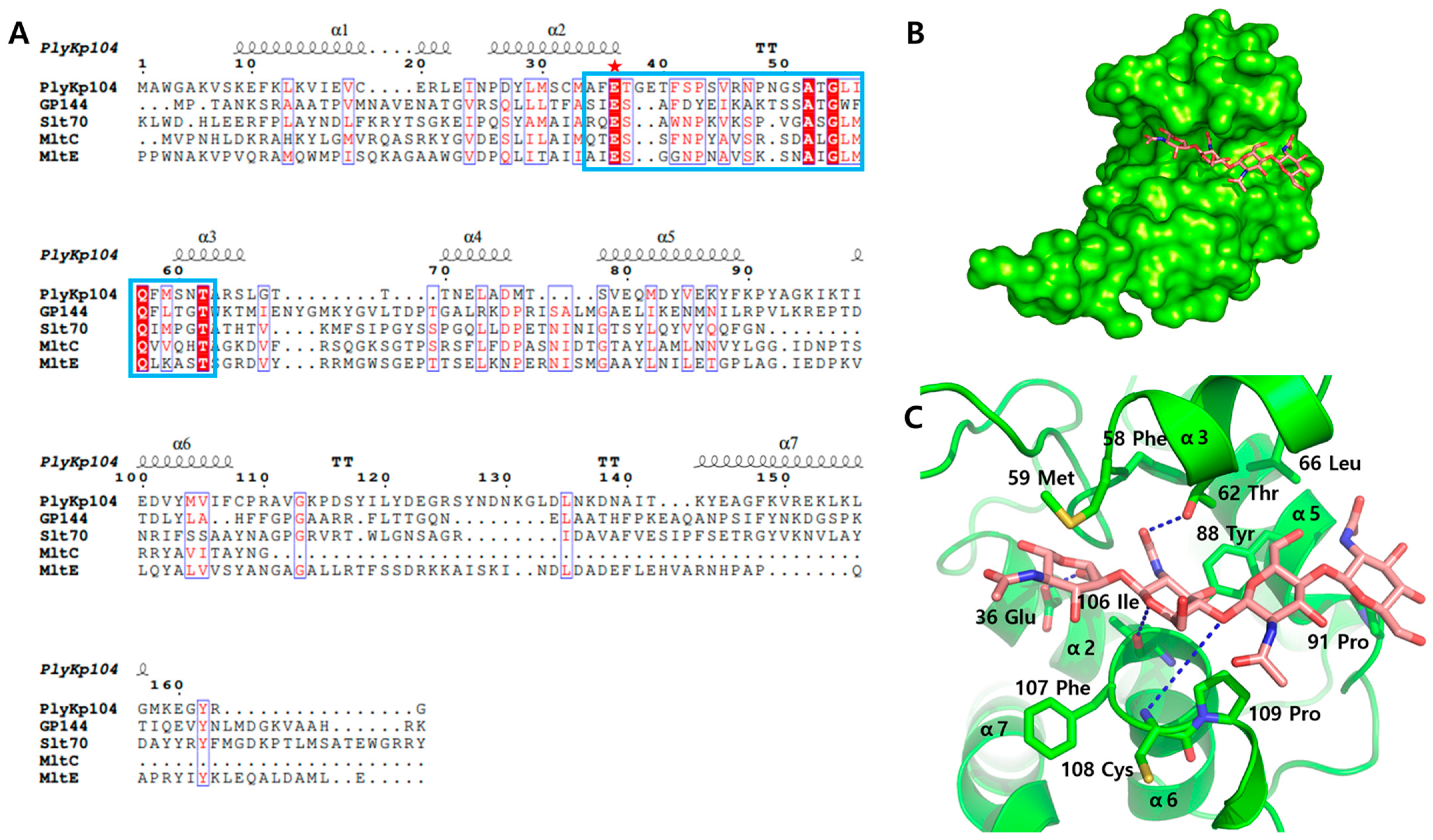
3.2. Crystal Structure of PlyKp104

PlyKp104 was present as a monomer within an asymmetric unit, consistent with the size exclusion chromatography results. The crystal structure revealed a small, single-domain fold composed entirely of seven α-helices (Figure 2A).
To determine the identity of this domain, a structural similarity search was performed using the crystal structure. This analysis identified four homologous proteins, GP144, Slt70, MltC, and MltE, all belonging to the lytic transglycosylase family 1, suggesting that the domain of PlyKp104 is a transglycosidase domain.
This finding is consistent with that of a previous study, which predicted a putative lytic transglycosylase domain in PlyKp104 through sequence-based domain analysis [15].
Although the AlphaFold2-predicted model used for molecular replacement showed an overall structural similarity to the crystal structure of PlyKp104, some notable differences were observed (Figure 2B). Specifically, an additional helix located between α6 and α7 in the predicted model was absent in the crystal structure. Instead, this region formed an extended and flexible loop that significantly deviated from the predicted conformation. In the crystal structure, this loop region lacked an electron density for several residues, indicating a high degree of flexibility in this segment (Figure 2B).

3.3. Active Site Structure of PlyKp104

Sequence alignment with structural homologs identified through the structural similarity search revealed a highly conserved catalytic residue, Glu36, located at the C-terminus of α2 (Figure 3A). The alignment also indicated the presence of several conserved residues both upstream and downstream of Glu36. This region (Ala34–Ala63 in PlyKp104) corresponds to conserved motifs I and II, which are characteristic of soluble lytic transglycosylase (SLT) catalytic domains in lytic transglycosylase family 1 [25]. The SLT domain is a catalytic module found in enzymes that cleave the glycosidic bonds of peptidoglycan, a major component of bacterial cell walls [26]. Structurally, SLT domains typically adopt an α/β fold and contain conserved acidic residues, such as glutamic acid or aspartic acid, that coordinate catalysis [26]. Unlike typical hydrolases, SLT domains catalyze a non-hydrolytic cleavage, generating a 1,6-anhydro ring at the MurNAc residue [27]. These domains play essential roles in bacterial cell wall remodeling and degradation and are also found in certain phage-derived endolysins that target Gram-negative bacteria [28].
By superimposing the structure of PlyKp104 onto that of GP144 (PDB ID: 3BKV) [25], a potential substrate-binding groove in PlyKp104 was identified based on the binding position of chitotetraose in GP144 (Figure 3B). To gain further structural insight into the substrate-binding mode of PlyKp104, the chitotetraose molecule, initially positioned within the putative groove by structural superposition, was manually adjusted to avoid steric clashes, guided by the known substrate-interacting residues of GP144. This modeling enabled the identification of potential substrate-interacting residues in PlyKp104 (Figure 3C).
As a result, although only a few sequence-conserved residues were identified, the substrate-binding interactions were found to be largely conserved. Specifically, hydrophobic interactions with the substrate were observed for Phe58, Met59, Tyr88, Phe107, and Pro109 in PlyKp104, which correspond to Phe135, Leu139, Leu175, Phe201, and Pro204 in GP144. Similarly, potential hydrogen bonds with the substrate were observed involving Thr62, Ile106, and Cys108 in PlyKp104, corresponding to Thr139, His200, and Gly203 in GP144 (Figure 3C).

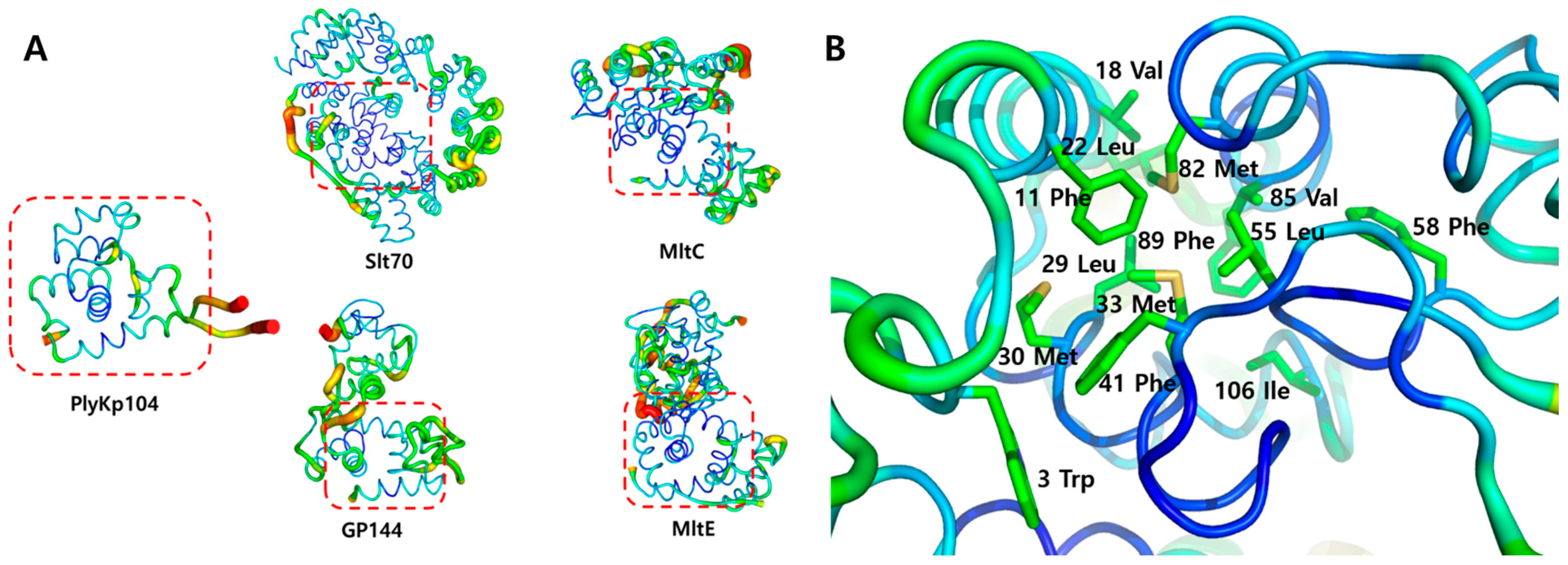
3.4. Structural Stability Analysis of PlyKp104

To evaluate the structural stability of PlyKp104, B-factor values were mapped onto its crystal structure as well as those of four homologous lytic transglycosylases that were identified through structural homology searches. B-factors are known to reflect structural flexibility and thermal motion; therefore, lower B-factor values indicate reduced atomic displacement due to thermal fluctuations and, consequently, higher structural stability [29].
Overall, in all five structures, the SLT catalytic domain exhibited low B-factor values, indicating that this domain is relatively stable (Figure 4A). In contrast, additional domains not present in PlyKp104 showed higher B-factor values, suggesting reduced structural stability in those regions (Figure 4A). To investigate the interactions contributing to the structural stability of the SLT domain, residue–residue interactions were analyzed. This analysis revealed a hydrophobic interaction network primarily formed by large hydrophobic residues such as Met, Phe, and Leu, located between the α1, α2, and α5 helices, which constitute the structural core of the domain (Figure 4B). Interestingly, many of these bulky hydrophobic residues are located within or adjacent to motifs I and II (Ala34–Ala63 in PlyKp104), which are highly conserved regions in the SLT domains of lytic transglycosylase family 1 (Figure 3A and Figure 4B). This observation may account for the consistently low B-factor values observed in the SLT domains across all five structures.
In summary, the hydrophobic interaction network formed by conserved, bulky hydrophobic residues within motifs I and II appears to play a critical role in stabilizing the core structure of the SLT domain, thereby enhancing its overall structural stability.

3.5. Potential Lipid-Binding Site of PlyKp104

During the structural determination of PlyKp104, a relatively large and unidentified electron density blob was consistently observed. To assess whether this was due to model bias, a composite omit map was generated; however, the density remained present (Figure 5A), indicating that it is not an artifact of model bias. This density did not correspond to the shape of any molecule introduced during the crystallization process, and thus no specific molecule could be reliably modeled into it. However, as shown in Figure 5, the elongated and narrow shape of the density suggests that it may represent a hydrocarbon chain-like molecule, such as a lipid.
To explore this possibility, a palmitic acid molecule was used as a surrogate based on the shape and size of the observed electron density. When placed into the density, it fit well with the observed shape, supporting the hypothesis that the density may indeed originate from a lipid-like hydrocarbon molecule (Figure 5B). Electrostatic surface potential mapping of the PlyKp104 structure revealed that the region surrounding the electron density is electrically neutral (Figure 5B), suggesting that the interaction between PlyKp104 and the molecule responsible for the density is not electrostatic in nature. Further analysis of the surface residues around the density showed a predominance of neutral and hydrophobic amino acids (Figure 5C). This supports the hypothesis that the region might serve as a potential lipid-binding site, where these residues could interact with lipid molecules originating from the bacterial outer membrane.

4. Discussion

In this study, the structure of the novel phage endolysin PlyKp104 was determined using X-ray crystallography, and its structural features were analyzed in detail. Sequence and structural comparisons with four known lytic transglycosylases—identified through homology searches based on the PlyKp104 crystal structure—revealed that PlyKp104 belongs to lytic transglycosylase family 1. Notably, PlyKp104 is phage-derived and consists solely of the catalytic SLT domain, suggesting that it can be classified within subgroup 1H, the same subgroup as the phage-derived enzyme GP144 [30]. This makes PlyKp104 the second known enzyme in this subgroup that acts on Gram-negative bacteria.
A notable structural deviation was observed in the loop between α6 and α7 when compared to the AlphaFold2-predicted model. This discrepancy may reflect functional flexibility in this region, potentially associated with dynamic interactions with membrane components or substrates. Although no molecular dynamics (MD) simulations were performed in the present study, future MD-based analyses could provide further insight into the conformational dynamics and functional relevance of this loop.
Structural superimposition with GP144, which was crystallized in complex with the substrate chitotetraose, confirmed the presence of a conserved catalytic residue and a putative substrate-binding groove in PlyKp104. Although the sequence conservation of the substrate-interacting residues is low, PlyKp104 appears to engage the substrate through a binding mechanism that closely resembles that of GP144.
The B-factor analysis of PlyKp104 and the four homologous lytic transglycosylases showed that the conserved SLT domain exhibits greater structural stability compared to the additional domains present in the other enzymes. This stability is likely supported by a hydrophobic interaction network formed between the α-helices at the core of the domain. Consequently, PlyKp104, which comprises only the SLT domain, may possess inherently higher structural stability than other lytic transglycosylases with additional domains [25,31,32].
During the structural analysis, a previously unidentified electron density blob was observed in the PlyKp104 crystal. Based on its elongated shape, electrostatic surface potential, and the distribution of surrounding residues, the density was hypothesized to correspond to a lipid-like hydrocarbon molecule. This putative lipid-binding site, inferred solely from structural features, remains hypothetical and requires further validation. Nevertheless, its presence may offer important clues as to how PlyKp104, despite consisting only of a catalytic domain, could interact with or penetrate the outer lipid membrane of Gram-negative bacteria. Future biochemical and biophysical studies will be essential to confirming the identity of the bound molecule and clarifying its potential role in membrane interaction.
Although this study could not provide definitive structural evidence for PlyKp104’s ability to penetrate the outer membrane, previously reported experimental results indicate that it can bypass this barrier without the aid of permeabilizers and exhibit potent lytic activity against multidrug-resistant Gram-negative bacteria both in vitro and in a murine infection model [15]. These findings suggest that PlyKp104 possesses intrinsic membrane-translocating ability, although the underlying mechanism remains unclear.
To date, the study of outer membrane permeabilization and lytic mechanisms of endolysins targeting Gram-negative bacteria has been limited due to a lack of structural and biochemical data. The structural characterization of PlyKp104, including its conserved catalytic features, substrate-binding interactions, high core stability, and a putative lipid-binding region, provides a valuable foundation for the rational design of engineered endolysins with improved efficacy against Gram-negative bacteria. These insights may guide future efforts to enhance outer membrane penetration and enzymatic activity, thereby expanding the therapeutic potential of phage-derived endolysins.

Funding

This research received no external funding.

Data Availability Statement

The coordinates and structure factor amplitudes for both structures have been deposited in the PDB under the accession code 9UDH (https://www.rcsb.org/structure/9UDH).

Acknowledgments

I would like to thank the beamline staff at the 7A beamline at the Pohang Accelerator Laboratory for their assistance with the data collection.

Conflicts of Interest

The author declares no conflict of interest.

References

  1. Breijyeh, Z.; Jubeh, B.; Karaman, R. Resistance of Gram-Negative Bacteria to Current Antibacterial Agents and Approaches to Resolve It. Molecules 2020, 25, 1340. [Google Scholar] [CrossRef] [PubMed]
  2. Wright, G.D. Q&A: Antibiotic resistance: Where does it come from and what can we do about it? BMC Biol. 2010, 8, 123. [Google Scholar]
  3. Baran, A.; Kwiatkowska, A.; Potocki, L. Antibiotics and bacterial resistance—A short story of an endless arms race. Int. J. Mol. Sci. 2023, 24, 5777. [Google Scholar] [CrossRef] [PubMed]
  4. Uddin, T.M.; Chakraborty, A.J.; Khusro, A.; Zidan, B.R.M.; Mitra, S.; Emran, T.B.; Dhama, K.; Ripon, M.K.H.; Gajdács, M.; Sahibzada, M.U.K. Antibiotic resistance in microbes: History, mechanisms, therapeutic strategies and future prospects. J. Infect. Public Health 2021, 14, 1750–1766. [Google Scholar] [CrossRef]
  5. Baharoglu, Z.; Garriss, G.; Mazel, D. Multiple pathways of genome plasticity leading to development of antibiotic resistance. Antibiotics 2013, 2, 288–315. [Google Scholar] [CrossRef]
  6. Pulingam, T.; Parumasivam, T.; Gazzali, A.M.; Sulaiman, A.M.; Chee, J.Y.; Lakshmanan, M.; Chin, C.F.; Sudesh, K. Antimicrobial resistance: Prevalence, economic burden, mechanisms of resistance and strategies to overcome. Eur. J. Pharm. Sci. 2022, 170, 106103. [Google Scholar] [CrossRef]
  7. Salam, M.A.; Al-Amin, M.Y.; Salam, M.T.; Pawar, J.S.; Akhter, N.; Rabaan, A.A.; Alqumber, M.A.A. Antimicrobial Resistance: A Growing Serious Threat for Global Public Health. Healthcare 2023, 11, 1946. [Google Scholar] [CrossRef]
  8. Murugaiyan, J.; Kumar, P.A.; Rao, G.S.; Iskandar, K.; Hawser, S.; Hays, J.P.; Mohsen, Y.; Adukkadukkam, S.; Awuah, W.A.; Jose, R.A.M.; et al. Progress in Alternative Strategies to Combat Antimicrobial Resistance: Focus on Antibiotics. Antibiotics 2022, 11, 200. [Google Scholar] [CrossRef]
  9. Gupta, R.; Sharma, S. Role of alternatives to antibiotics in mitigating the antimicrobial resistance crisis. Indian J. Med. Res. 2022, 156, 464–477. [Google Scholar] [CrossRef]
  10. Schmelcher, M.; Donovan, D.M.; Loessner, M.J. Bacteriophage endolysins as novel antimicrobials. Future Microbiol. 2012, 7, 1147–1171. [Google Scholar] [CrossRef]
  11. Lu, Y.; Wang, Y.; Wang, J.; Zhao, Y.; Zhong, Q.; Li, G.; Fu, Z.; Lu, S. Phage endolysin LysP108 showed promising antibacterial potential against methicillin-resistant Staphylococcus aureus. Front. Cell. Infect. Microbiol. 2021, 11, 668430. [Google Scholar] [CrossRef] [PubMed]
  12. Sisson, H.M.; Jackson, S.A.; Fagerlund, R.D.; Warring, S.L.; Fineran, P.C. Gram-negative endolysins: Overcoming the outer membrane obstacle. Curr. Opin. Microbiol. 2024, 78, 102433. [Google Scholar] [CrossRef]
  13. Vasina, D.V.; Antonova, N.P.; Gushchin, V.A.; Aleshkin, A.V.; Fursov, M.V.; Fursova, A.D.; Gancheva, P.G.; Grigoriev, I.V.; Grinkevich, P.; Kondratev, A.V.; et al. Development of novel antimicrobials with engineered endolysin LysECD7-SMAP to combat Gram-negative bacterial infections. J. Biomed. Sci. 2024, 31, 75. [Google Scholar] [CrossRef]
  14. Cernooka, E.; Rumnieks, J.; Zrelovs, N.; Tars, K.; Kazaks, A. Diversity of the lysozyme fold: Structure of the catalytic domain from an unusual endolysin encoded by phage Enc34. Sci. Rep. 2022, 12, 5005. [Google Scholar] [CrossRef]
  15. Euler Chad, W.; Raz, A.; Hernandez, A.; Serrano, A.; Xu, S.; Andersson, M.; Zou, G.; Zhang, Y.; Fischetti Vincent, A.; Li, J. PlyKp104, a Novel Phage Lysin for the Treatment of Klebsiella pneumoniae, Pseudomonas aeruginosa, and Other Gram-Negative ESKAPE Pathogens. Antimicrob. Agents Chemother. 2023, 67, e01519-22. [Google Scholar] [CrossRef]
  16. Park, S.-Y.; Ha, S.-C.; Kim, Y.-G. The protein crystallography beamlines at the pohang light source II. Biodesign 2017, 5, 30–34. [Google Scholar]
  17. Otwinowski, Z.; Minor, W. Processing of X-ray diffraction data collected in oscillation mode. In Methods Enzymol; Academic Press: Cambridge, MA, USA, 1997; Volume 276, pp. 307–326. [Google Scholar]
  18. Winter, G.; Waterman, D.G.; Parkhurst, J.M.; Brewster, A.S.; Gildea, R.J.; Gerstel, M.; Fuentes-Montero, L.; Vollmar, M.; Michels-Clark, T.; Young, I.D. DIALS: Implementation and evaluation of a new integration package. Biol. Crystallogr. 2018, 74, 85–97. [Google Scholar] [CrossRef]
  19. Liebschner, D.; Afonine, P.V.; Baker, M.L.; Bunkóczi, G.; Chen, V.B.; Croll, T.I.; Hintze, B.; Hung, L.-W.; Jain, S.; McCoy, A.J. Macromolecular structure determination using X-rays, neutrons and electrons: Recent developments in Phenix. Biol. Crystallogr. 2019, 75, 861–877. [Google Scholar] [CrossRef]
  20. Jumper, J.; Evans, R.; Pritzel, A.; Green, T.; Figurnov, M.; Ronneberger, O.; Tunyasuvunakool, K.; Bates, R.; Žídek, A.; Potapenko, A.; et al. Highly accurate protein structure prediction with AlphaFold. Nature 2021, 596, 583–589. [Google Scholar] [CrossRef]
  21. Emsley, P.; Lohkamp, B.; Scott, W.G.; Cowtan, K. Features and development of Coot. Biol. Crystallogr. 2010, 66, 486–501. [Google Scholar] [CrossRef]
  22. Holm, L.; Laiho, A.; Törönen, P.; Salgado, M. DALI shines a light on remote homologs: One hundred discoveries. Protein Sci. 2023, 32, e4519. [Google Scholar] [CrossRef] [PubMed]
  23. Sievers, F.; Higgins, D.G. Clustal Omega for making accurate alignments of many protein sequences. Protein Sci. 2018, 27, 135–145. [Google Scholar] [CrossRef] [PubMed]
  24. Robert, X.; Gouet, P. Deciphering key features in protein structures with the new ENDscript server. Nucleic Acids Res. 2014, 42, W320–W324. [Google Scholar] [CrossRef] [PubMed]
  25. Fokine, A.; Miroshnikov, K.A.; Shneider, M.M.; Mesyanzhinov, V.V.; Rossmann, M.G. Structure of the bacteriophage φKZ lytic transglycosylase gp144. J. Biol. Chem. 2008, 283, 7242–7250. [Google Scholar] [CrossRef]
  26. Dik, D.A.; Marous, D.R.; Fisher, J.F.; Mobashery, S. Lytic transglycosylases: Concinnity in concision of the bacterial cell wall. Crit. Rev. Biochem. Mol. Biol. 2017, 52, 503–542. [Google Scholar] [CrossRef]
  27. Vermassen, A.; Leroy, S.; Talon, R.; Provot, C.; Popowska, M.; Desvaux, M. Cell Wall Hydrolases in Bacteria: Insight on the Diversity of Cell Wall Amidases, Glycosidases and Peptidases Toward Peptidoglycan. Front. Microbiol. 2019, 10, 331. [Google Scholar] [CrossRef]
  28. Alcorlo, M.; Martínez-Caballero, S.; Molina, R.; Hermoso, J.A. Carbohydrate recognition and lysis by bacterial peptidoglycan hydrolases. Curr. Opin. Struct. Biol. 2017, 44, 87–100. [Google Scholar] [CrossRef]
  29. Sun, Z.; Liu, Q.; Qu, G.; Feng, Y.; Reetz, M.T. Utility of B-Factors in Protein Science: Interpreting Rigidity, Flexibility, and Internal Motion and Engineering Thermostability. Chem. Rev. 2019, 119, 1626–1665. [Google Scholar] [CrossRef]
  30. Broendum, S.S.; Buckle, A.M.; McGowan, S. Catalytic diversity and cell wall binding repeats in the phage-encoded endolysins. Mol. Microbiol. 2018, 110, 879–896. [Google Scholar] [CrossRef]
  31. Artola-Recolons, C.; Lee, M.; Bernardo-García, N.; Blázquez, B.; Hesek, D.; Bartual, S.; Mahasenan, K.; Lastochkin, E.; Pi, H.; Boggess, B. Structure and cell wall cleavage by modular lytic transglycosylase MltC of Escherichia coli. ACS Chem. Biol. 2014, 9, 2058–2066. [Google Scholar] [CrossRef]
  32. Artola-Recolons, C.; Carrasco-López, C.; Llarrull, L.I.; Kumarasiri, M.; Lastochkin, E.; Martínez de Ilarduya, I.; Meindl, K.; Usón, I.; Mobashery, S.; Hermoso, J.A. High-Resolution Crystal Structure of MltE, an Outer Membrane-Anchored Endolytic Peptidoglycan Lytic Transglycosylase from Escherichia coli. Biochemistry 2011, 50, 2384–2386. [Google Scholar] [CrossRef]
Figure 1. Purification, crystallization, and diffraction of PlyKp104. (A) Gel filtration profile of PlyKp104 indicating monomeric state. (Insert) Purity of PlyKp104 on SDS-PAGE gel. (B) Photo of optimized PlyKp104 crystal from condition. Crystal is indicated with red arrow. (C) Diffraction pattern of PlyKp104 from condition (i).
Crystals 15 00448 g001
Figure 2. Crystal structure of PlyKp104. (A) Overall structure of PlyKp104 with assigned secondary structures. (B) Superimposition of PlyKp104 (green) crystal structure and AlphaFold2-predicted model (purple). The most different part between the crystal structure and predicted model is denoted by the red dashed box.
Crystals 15 00448 g002
Figure 3. Potential active site of PlyKp104. (A). Multiple sequence alignment of lytic transglycosylases. Catalytic residue and conserved motif I and II are indicated red star and blue box, respectively. (B). Surface representation of potential substrate binding groove in PlyKp104. Chitotetraose is presented in stick model (salmon). (C). Substrate binding model of PlyKp104. Potential substrate binding residues are presented. Chitotetraose(substrate) is colored in salmon. H-bonds are presented in blue dotted lines.
Crystals 15 00448 g003
Figure 4. Analysis of the PlyKp104’s structural stability. (A) The B-factor derived from the diffraction data of each crystal is denoted on the structures with different colors and radii of the tube structure. The B-factor values are indicated by blue with a small radius (low) to red with a large radius (high). (B) Close-up view of PlyKp104’s SLT domain core. Residues participating in hydrophobic interaction network are presented in stick model.
Crystals 15 00448 g004
Figure 5. Composite omit map of unidentified molecule in crystal structure of PlyKp104. (A) Surface representation of PlyKp104 with composite omit map around unidentified molecule (mesh in red). (B) Surface charge representation of PlyKp104 with electron density blob of unidentified molecule (mesh in red). Negative charges and positive charges are indicated by red and blue. Palmitic acid modeled in the electron density with stick model in yellow. (C) Close-up view of potential interacting residues around the unidentified electron density.
Crystals 15 00448 g005
Table 1. Data and refinement statistics for PlyKp104.
Parameter
X-ray source7A beamline, PLS-II
Wavelength (Å)0.9793
Space groupC2221
Cell dimension
a, b, c (Å)67.73, 89.58, 67.74
α, β, γ (°)90, 90, 90
Resolution (Å)50.00–1.85 (1.88–1.85)
Unique reflections18,434 (710)
Completeness (%)98.7 (77.8)
Redundancy11.8 (6.5)
I/σ22.3 (1.3)
Rmerge0.123 (0.694)
Rmeas0.129 (0.747)
CC1/20.996 (0.867)
CC*0.999 (0.964)
Refinement
Resolution (Å)30.29–1.85
Rwork a0.19 (0.26)
Rfree b0.22 (0.30)
R.m.s. deviations
Bonds (Å)0.007
Angles (°)0.877
B factors (Å2)
Protein27.02
Ramachandran plot
Favored (%)98.70
Allowed (%)1.30
Disallowed (%)0.00
Values for the outer shell are noted in parentheses. a Rwork = Σ||Fobs|Σ|Fcalc||/Σ|Fobs|, where Fobs and Fcalc are the observed and calculated structure factor amplitudes, respectively. b Rfree was calculated as Rwork using a and a randomly selected subset of unique reflections not used for structural refinement.
Disclaimer/Publisher’s Note: The statements, opinions and data contained in all publications are solely those of the individual author(s) and contributor(s) and not of MDPI and/or the editor(s). MDPI and/or the editor(s) disclaim responsibility for any injury to people or property resulting from any ideas, methods, instructions or products referred to in the content.

