Article

Crystal Structure of Anthranilate Phosphoribosyltransferase from Methanocaldococcus jannaschii

Department of Food Science and Biotechnology, Kyungsung University, Busan 48434, Republic of Korea
Crystals 2025, 15(8), 702; https://doi.org/10.3390/cryst15080702 (registering DOI)
Submission received: 9 April 2025 / Revised: 28 July 2025 / Accepted: 29 July 2025 / Published: 31 July 2025
(This article belongs to the Special Issue Crystallography of Enzymes)

Abstract

Tryptophan is synthesized in microorganisms via a five-step enzymatic pathway originating from chorismate, which is a product of the shikimate pathway. As a biosynthetic precursor to a wide range of high-value compounds such as indole-3-acetic acid, indigo, indirubin, and violacein, this pathway has been a central target for metabolic engineering to enhance microbial production. Anthranilate phosphoribosyltransferase (AnPRT) catalyzes the second step of the pathway by transferring a phosphoribosyl group from PRPP to anthranilate, forming phosphoribosyl anthranilate (PRA). AnPRT, the sole member of class IV phosphoribosyltransferases, adopts a unique fold and functions as a homodimer. While the structural basis of AnPRT activity has been elucidated in several organisms, thermostable variants remain underexplored despite their relevance for high-temperature bioprocessing. In this study, the crystal structure of AnPRT from the thermophilic archaeon Methanocaldococcus jannaschii (MjAnPRT) was determined at a 2.16 Å resolution. The enzyme exhibits a conserved dimeric architecture and key catalytic motifs. Comparative structural analysis with mesophilic and hyper thermophilic homologs revealed that MjAnPRT possesses enhanced local stability in catalytically important regions and strengthened inter-subunit interactions. These features likely contribute to its thermostability and provide a valuable framework for the rational design of robust AnPRTs for industrial and synthetic biology applications.

1. Introduction

Tryptophan is one of the 20 standard amino acids that constitute proteins and is biosynthesized by a wide range of bacteria and plants. In microorganisms, the biosynthesis of tryptophan proceeds through a five-step enzymatic pathway, beginning with chorismate, a product of the shikimate pathway, which is converted to anthranilate and subsequently processed into tryptophan through a series of enzymatic reactions [1].
Beyond its role in protein biosynthesis, tryptophan serves as a precursor for a variety of biologically active compounds. It is the biosynthetic origin of the neurohormones melatonin and serotonin [2], and functions as a precursor to the plant hormone auxin (indole-3-acetic acid) [3]. Additionally, tryptophan is a key precursor in the biosynthesis of several high-value natural products, including the blue pigment indigo [4], the antileukemic agent indirubin [5], and the antibiotics violacein [6] and pyrrolnitrin [7].
Owing to the increasing demand for such tryptophan-derived compounds, extensive metabolic engineering efforts have been directed toward optimizing the tryptophan biosynthetic pathway [8,9]. These strategies have included the enhancement of enzymatic activity, the elimination of feedback inhibition, and the improvement of thermal and structural stability of pathway enzymes [10]. As a result, microbial production of a range of tryptophan derivatives has been achieved. For instance, engineered Escherichia coli strains have been developed for the efficient production of indole-3-acetic acid and violacein through overexpression or modification of key biosynthetic enzymes [11]. Similarly, production of indirubin and indigo has been demonstrated in Corynebacterium glutamicum and Escherichia coli using synthetic biology approaches to redirect metabolic flux toward tryptophan-derived pathways [5,12].
Among the enzymes in this pathway, anthranilate phosphoribosyl transferase (AnPRT) catalyzes the second step, transferring a phosphoribosyl group from 5′-phosphoribosyl-1′-pyrophosphate (PRPP) to anthranilate to form phosphoribosyl anthranilate (PRA), which is a central intermediate in tryptophan biosynthesis. AnPRT is a member of the phosphoribosyl transferase (PRT) functional superfamily, which is involved in a variety of nucleotide and amino acid metabolic processes [13,14]. Based on structural features, PRTs are classified into four distinct classes [15]. AnPRT represents the sole member of class IV, characterized by a unique PRT fold and a homodimeric architecture. Each monomer comprises a small N-terminal α-helical domain connected via a hinge region to a larger C-terminal α/β domain [16].
Although crystal structures of AnPRTs from various species have provided important insights into their catalytic mechanisms and inhibition, structural studies of thermostable AnPRTs, enzymes with enhanced stability suitable for protein engineering, remain limited. To date, only three thermostable AnPRTs have been structurally characterized, including those from Sulfolobus solfataricus (SsAnPRT) [17], Thermus thermophilus (TtAnPRT), and Thermococcus kodakarensis (TkAnPRT) [13].
In the present study, the crystal structure of AnPRT from the thermophilic archaeon M. jannaschii DSM 2661 [18] was determined at a resolution of 2.16 Å. Structural analysis identified key features associated with its thermostability. These findings offer a structural basis for the potential application of thermostable AnPRTs in high-temperature bioprocessing and synthetic biology.

2. Materials and Methods

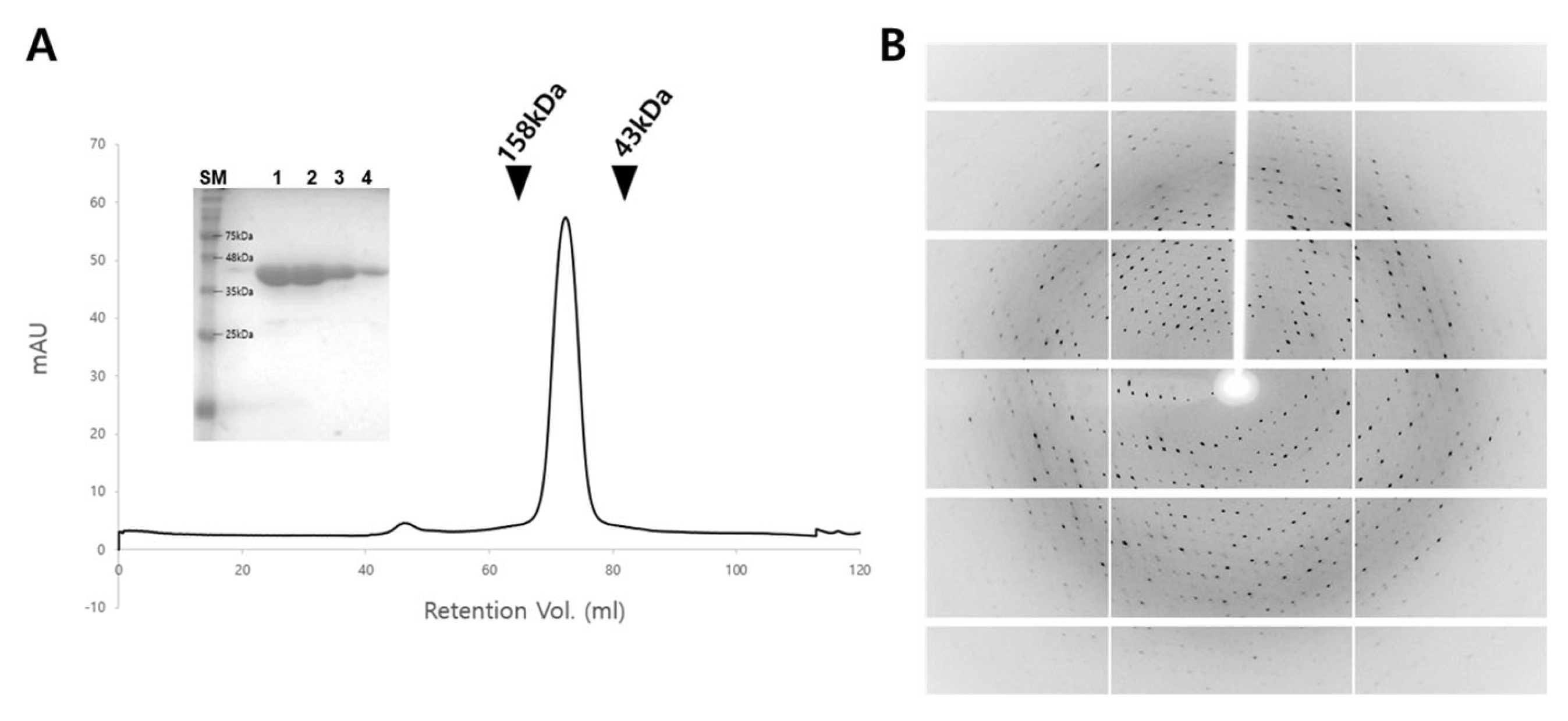
2.1. Cloning, Protein Expression and Purification

The gene encoding anthranilate phosphoribosyl-transferase from M. jannaschii DSM 2661 (MjAnPRT, Genebank No. AAB98221.1) was synthesized by IDT Inc. (San Diego, CA, USA) and cloned into the pET-28a expression vector (Novagen, Madison, WI, USA) using the NdeI and XhoI restriction sites. The sequence of the cloned gene was verified by DNA sequencing (Macrogen, Seoul, Republic of Korea). The verified plasmid was transformed into Escherichia coli BL21(DE3) cells. The transformed cells were cultured in lysogeny broth (LB) medium at 37 °C until the optical density at 600 nm (OD600) reached 0.5. Protein expression was then induced by adding 1 mM IPTG, followed by further incubation at 37 °C for 4 h. After induction, the cultured cells were harvested by centrifugation at 7000 rpm for 30 min at 4 °C. The cell pellet was resuspended in 15 mL of lysis buffer containing 20 mM Tris-HCl (pH 7.4) and 400 mM NaCl. The cells were lysed using sonication, and the lysate was centrifuged again at 17,000 rpm for 1 h at 4 °C to collect supernatant. To selectively denature E. coli proteins while retaining MjAnPRT activity, the supernatant was heat-treated at 60 °C for 30 min. Following heat treatment, the sample was centrifuged again at 17,000 rpm for 1 h at 4 °C, and the resulting supernatant was collected as the crude MjAnPRT fraction. Further purification was carried out using Ni-NTA affinity chromatography, exploiting the N-terminal 6×His-tag of MjAnPRT. The final purification step involved size exclusion chromatography with Superdex 200 16/600 column (Cytiva, Marlborough, MA, USA), during which the buffer was exchanged to 5 mM Tris-HCl (pH 7.4) containing 50 mM NaCl. The molecular weight of MjAnPRT was estimated using a standard calibration curve constructed from the elution volumes and known molecular weights of gel filtration standards (200–12 kDa; Sigma, Kanagawa, Japan). The purity of the eluted protein solution was assessed using 15% sodium dodecyl sulfate–polyacrylamide gel electrophoresis (SDS-PAGE), and protein concentration was determined using the Bradford assay. The purified protein was concentrated to 10 mg/mL, aliquoted and stored at –70 °C until crystallization.

2.2. Crystallization

Crystallization screening was performed using the sitting drop vapor diffusion method at 20 °C using a commercial crystallization kit, including the Crystal Screen 1, 2, MembFac and Natrix (Hampton Research, Aliso Viejo, CA, USA) and Wizard Classic 1, 2, 3 and 4 (Rigaku, The Woodlands, TX, USA). The protein solution (1 µL) was mixed with the crystallization screen solution (1 µL) and equilibrated with a reservoir solution (50 µL). 10 different microcrystals were obtained from the initial screening and reproduced for X-ray diffraction experiment within 4 weeks. Among them, crystals obtained from the following conditions used for data collection: 0.1 M of Tris-HCl (pH 8.5) and 8% (w/v) polyethylene glycol 8000.

2.3. X-Ray Diffraction Data Collection

X-ray diffraction data were collected at beamline 7A at Pohang Light Source II (PLS-II, Pohang, Republic of Korea) [19]. The MjAnPRT crystal was cryoprotected using the reservoir solution supplemented with 25% (v/v) glycerol and mounted on a goniometer under a nitrogen gas stream at 100 K. Diffraction images were recorded using an Eiger2 S 9M detector (DECTRIS, Baden, Switzerland). The data were indexed, integrated, and scaled using HKL3000 [20].

2.4. Structure Determination

The phase problem was solved by molecular replacement using Phaser in PHENIX 1.20.1 [21], with a three-dimensional model generated by AlphaFold2 2.0 used as the search model [22]. Manual model building based on the electron density map was performed using Coot 0.8.9.2 [23]. Structure refinement was conducted with phenix.refine in PHENIX [21].

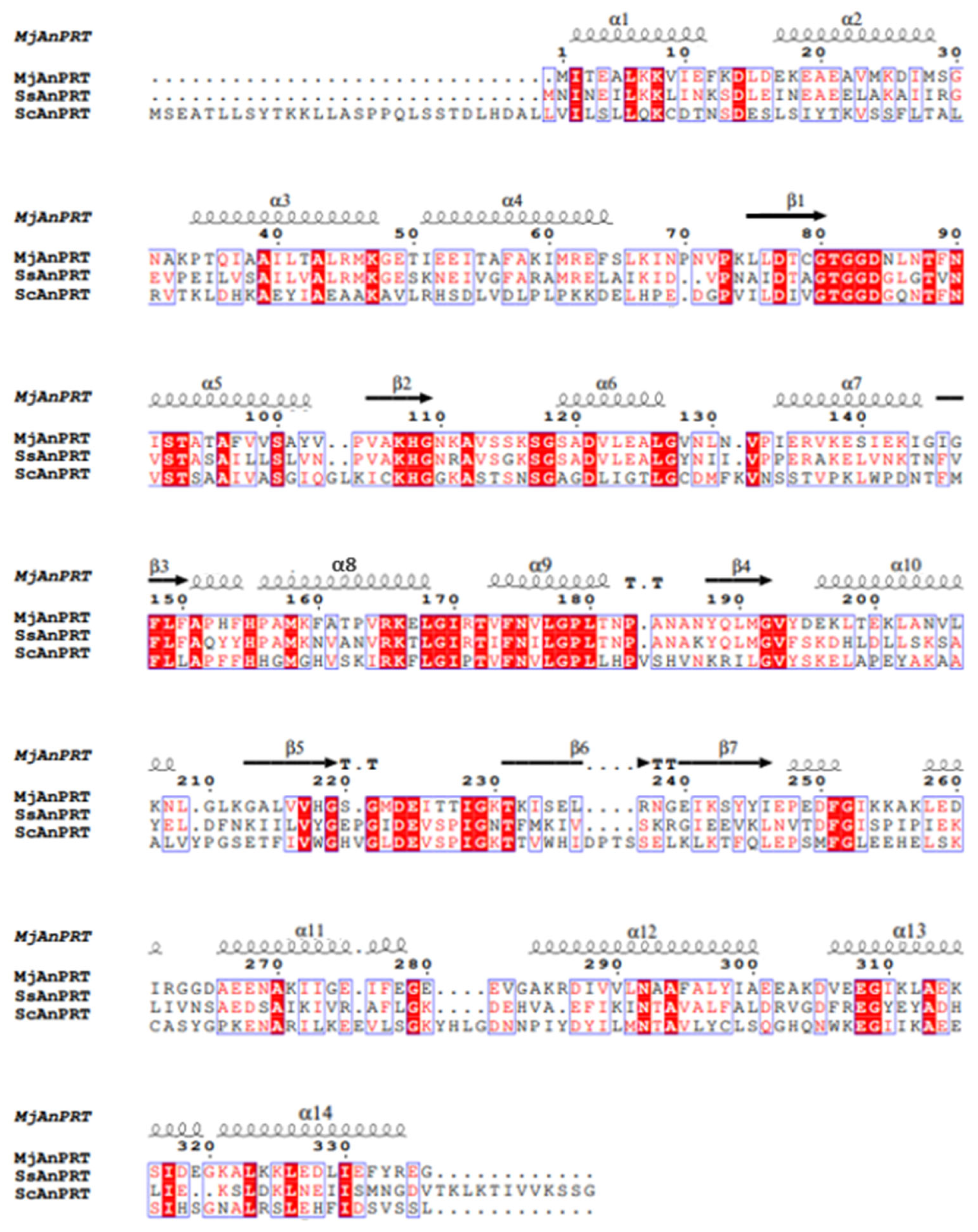
2.5. Bioinformatics and Structure Analysis

Structural similarity searches were performed using the DALI server [24]. Amino acid sequence alignment was proceeded using Clustal Omega [25]. Structure-based multiple sequence alignments were visualized with ESPript 3.0 [26]. Protein structures were visualized using PyMOL 1.8.0.2 (https://pymol.org, accessed on 1 March 2025).

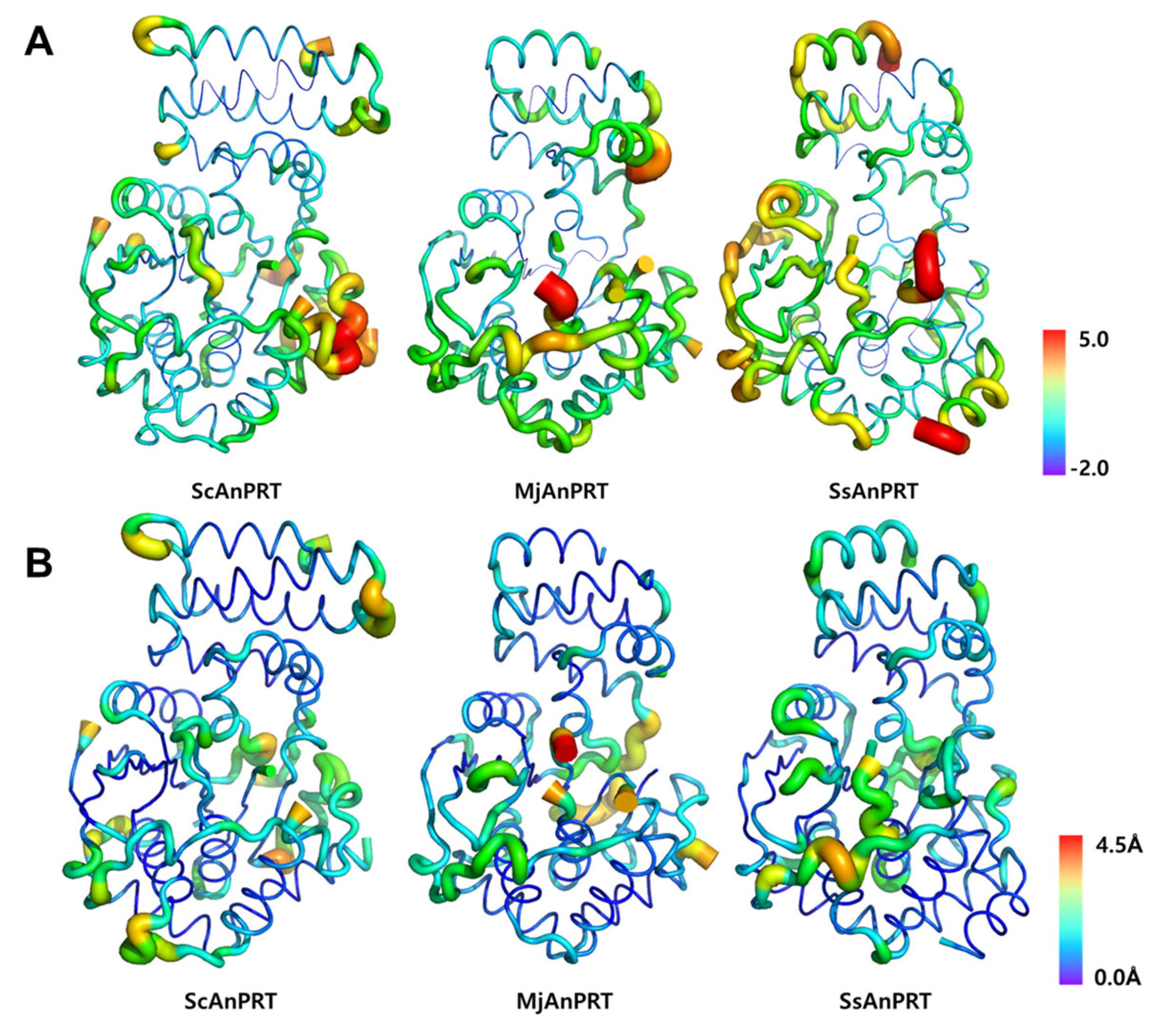
2.6. Preparation of B-Factor Analysis

To minimize the influence of radiation damage and modeling errors on B-factor values, the monomer with the lowest overall B-factor among the chains in each crystal structure was selected for comparative analysis; in all cases, this corresponded to chain A. To enable direct comparison between structures with varying resolutions and refinement statistics, atomic B-factors were normalized using Z-score normalization via BANΔIT [27]. Coarse-grained molecular dynamics (MD) simulations were performed using CABS-flex 2.0 [28] with default parameters [29] to evaluate structural flexibility and to assess and mitigate potential reductions in B-factor caused by crystallographic artifacts. Structural flexibility was evaluated based on Cα root mean square fluctuation (RMSF) values derived from the simulations.

3. Results

3.1. Structure Determination of MjAnPRT

The final yield of purified recombinant MjAnPRT was approximately 40 mg per liter of culture, with a purity exceeding 90% as estimated by SDS-PAGE analysis (Figure 1A), indicating that the sample was suitable for crystallization. The size exclusion chromatography elution profile suggested that MjAnPRT exists as a dimer in solution, consistent with the theoretical dimeric mass of 72 kDa. SDS-PAGE analysis confirmed that the monomeric unit has a molecular weight of approximately 40 kDa (Figure 1A). Initial crystallization trials yielded crystals under the 10 conditions. Among them, 3 out of 10 showed possible protein crystal-like diffractions in preliminary X-ray diffraction test. Overall, after optimization, crystals obtained from the following conditions showed good diffraction: 0.1 M of Tris-HCl (pH 8.5) and 8% (w/v) polyethylene glycol 8000 (Figure 1B). So, the high-resolution data collection proceeded with this crystal. The MjAnPRT crystal was determined to belong to the space group P21, with unit cell dimensions of a = 60.99 Å, b = 82.66 Å, and c = 73.87 Å, and angles α = 90°, β = 112.74°, and γ = 90°. (Table 1). The structure of MjAnPRT was determined at a 2.16 Å resolution, with Rwork and Rfree values of 21.51% and 25.75%, respectively (Table 1).

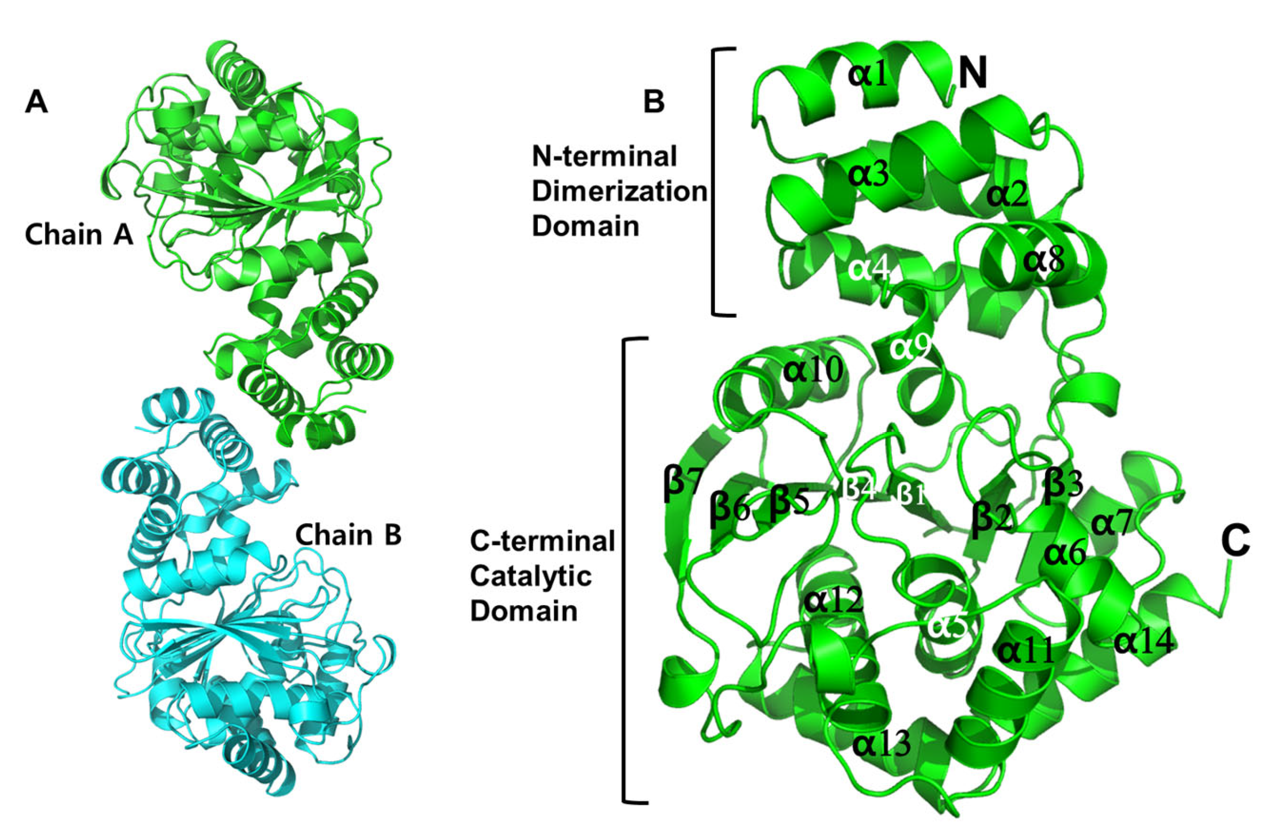
3.2. Crystal Structure of MjAnPRT

The crystal structure of MjAnPRT, determined by X-ray crystallography, revealed that the enzyme forms a homodimer. This oligomeric state is consistent with the molecular size observed in solution as determined by size exclusion chromatography (Figure 1A and Figure 2A). Each monomer of MjAnPRT adopts a fold characteristic of other AnPRTs, comprising a small N-terminal domain formed by four α-helices (α1-α4), which is further extended by helices α8 and α9 (Figure 2B). The large C-terminal domain features a central β-sheet composed of seven β-strands (β1–β7), flanked by eight α-helices (α5–α7, α10–α14), forming an α/β domain. This architecture represents a distinctive phosphoribosyl transferase (PRT) fold characteristic of AnPRTs, which are the sole members of class IV PRTs [16,30]. The two domains are connected by hinge regions spanning α4–β1, β3–α8, and α9–β4 (Figure 2B). The N-terminal domain is responsible for mediating dimerization of the AnPRT enzyme, while the C-terminal domain is known to harbor the active site [30] (Figure 2B).

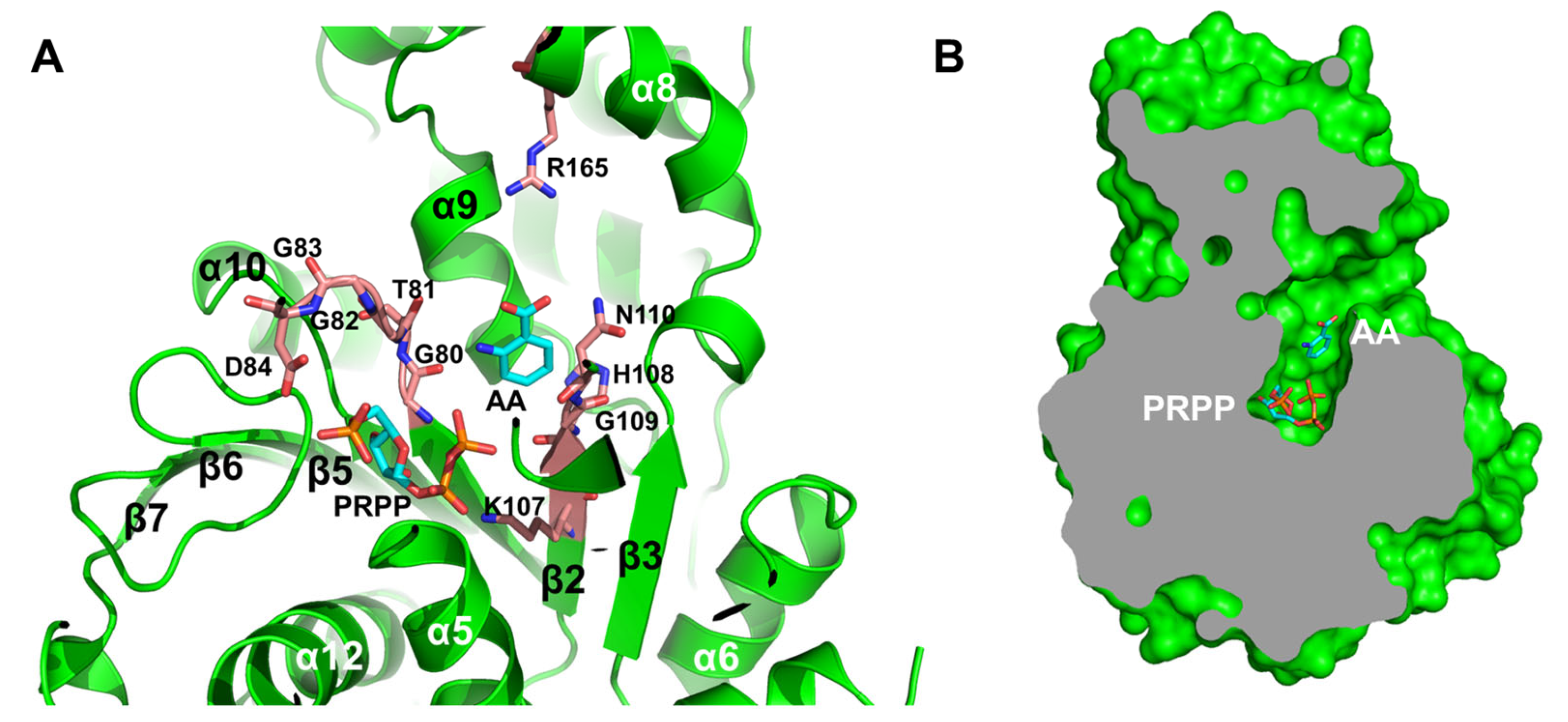
3.3. Active Site Structure of MjAnPRT

SsAnPRT and AnPRT from Saccharomyces cerevisiae (ScAnPRT) were selected for sequence and structural comparison with MjAnPRT, as both possess experimentally determined crystal structures, allowing for detailed comparative analysis. Structure-based homology searches using the determined crystal structure, combined with sequence alignment and structural superposition (Figure 3), identified two conserved motifs near the active site of MjAnPRT. One is a conserved Gly-rich sequence, GTGGD (residues 80–84), located in the β1–α5 loop. This motif is a signature sequence of the AnPRT family and is known to contribute to PRPP binding [31]. The other is a highly conserved anthranilate-binding motif, KHGN (residues 107–110), located in the β2–α6 loop [17].
To further investigate the spatial relationship between these motifs and their respective substrates, the substrate-bound structure of SsAnPRT (PDB ID: 1ZYK) was superimposed onto MjAnPRT. Based on this alignment, the two substrates, anthranilate (AA) and PRPP, were modeled into the active site of MjAnPRT. Due to the absence of interpretable electron density for Gly83 and Asp84 of the GTGGD motif, and Asn110 of the KHGN motif in the experimental structure, these residues were modeled using coordinates from the AlphaFold2-predicted structure, which also served as the initial search model for molecular replacement. The resulting model revealed that the GTGGD motif is positioned near PRPP, supporting its proposed role in substrate coordination (Figure 4A). In addition, Arg165, located on helix α8, lies adjacent to the carboxyl group of anthranilate and has been reported to form a hydrogen bond essential for catalytic activity [17] (Figure 4A).
Consistently, the KHGN motif is situated near both the pyrophosphate moiety of PRPP and the anthranilate molecule. Within this motif, Lys107 is implicated in anthranilate binding, while the remaining residues (HGN; 108–110) are involved in PRPP interaction [17].
Finally, a surface representation of the MjAnPRT structure reveals a narrow substrate channel positioned between the N-terminal and C-terminal domain. This channel corresponds to the hinge regions spanning α4–β1, β3–α8, and α9–β4 (Figure 4A,B), providing structural insights into substrate recognition and access to the active site.

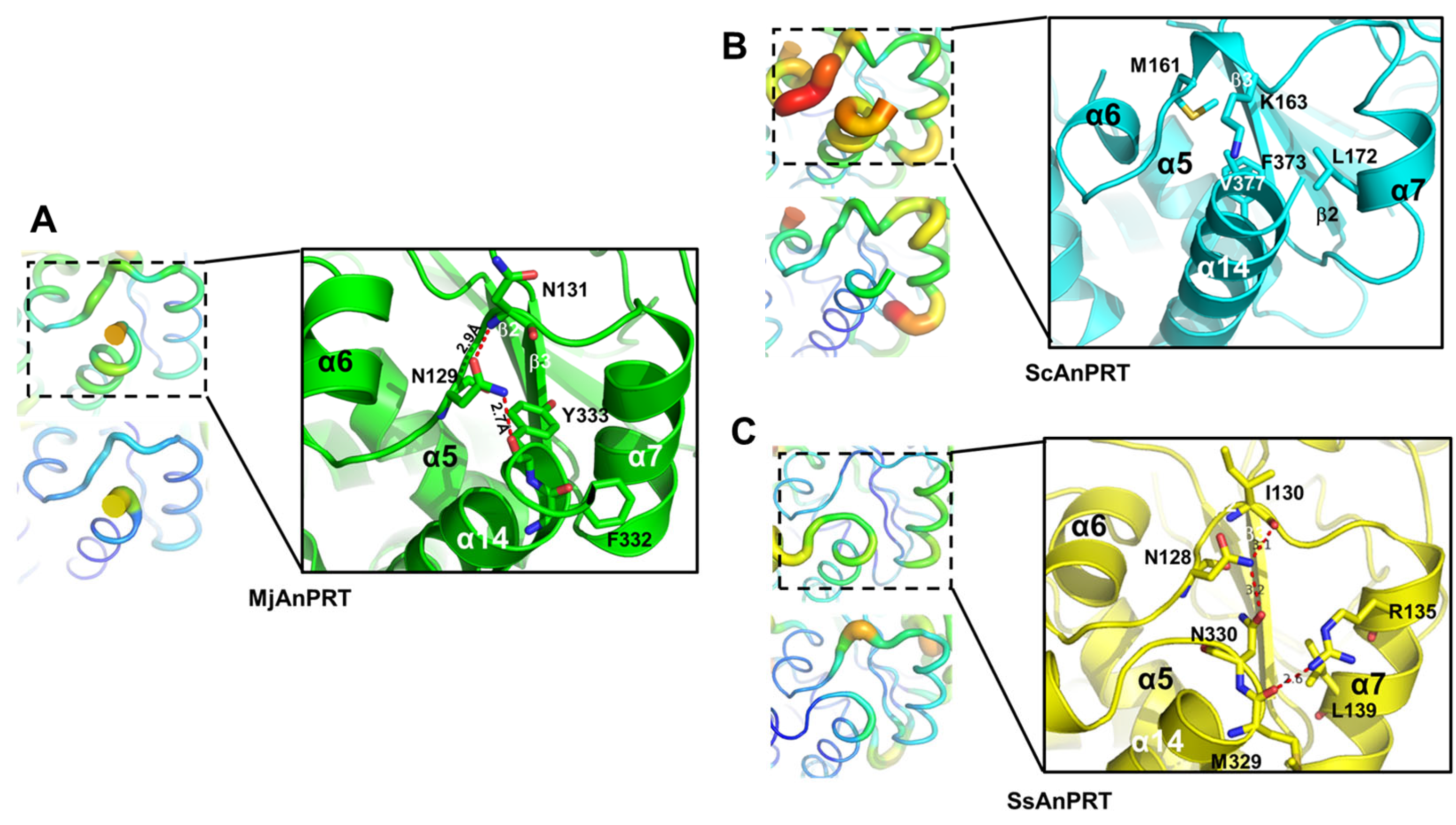
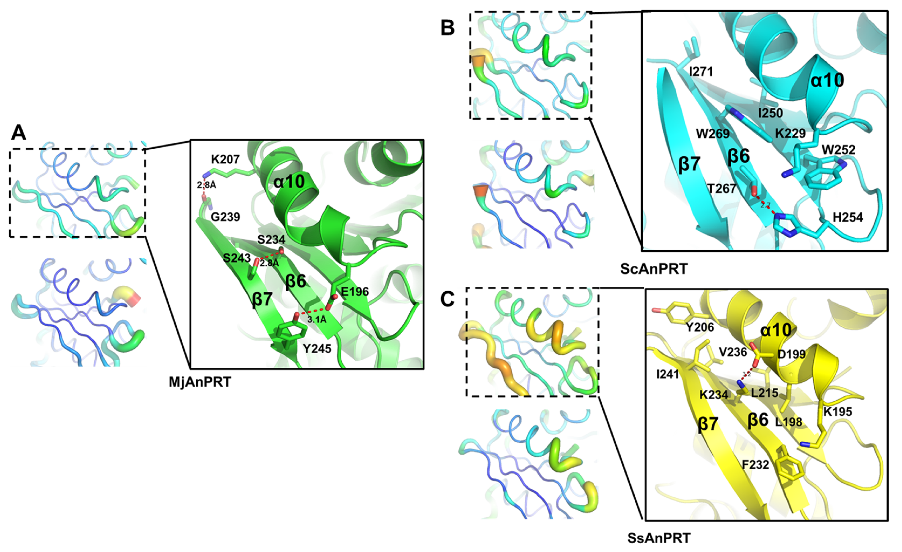
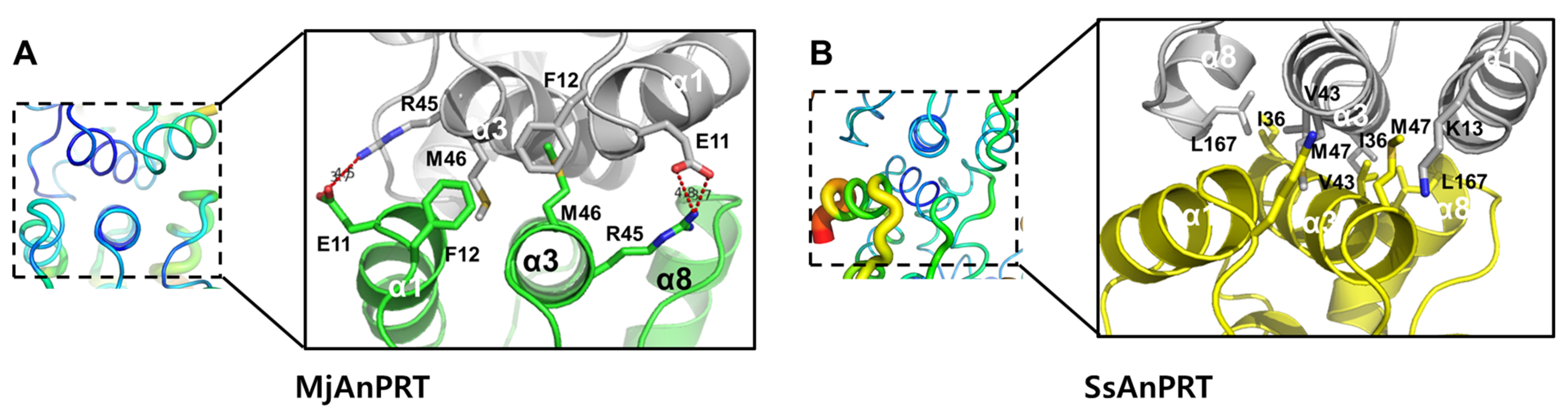
3.4. Structural Analysis of the Thermostability of MjAnPRT

Two homologous structures were selected based on both structural similarity and known thermostability: ScAnPRT (PDB ID: 7DSM) and SsAnPRT (PDB ID: 2GVQ). The root-mean-square deviations (RMSDs) from MjAnPRT were 2.1 Å and 1.4 Å, with sequence identities of 34.4% and 45.5%, respectively. ScAnPRT was expected to be relatively less thermostable than MjAnPRT, whereas SsAnPRT was selected as a representative thermostable AnPRT. Although the melting temperature (Tm) of ScAnPRT has not been reported, the average Tm of S. cerevisiae proteins is approximately 52 °C [32], and most proteins from this organism typically exhibit Tm values within the range of 40–60 °C [33]. Therefore, the Tm of ScAnPRT is presumed to fall within this range.
In contrast, SsAnPRT has a reported Tm of 92 °C [34], indicating exceptional thermostability. While the Tm of MjAnPRT has not been directly measured, its source organism, M. jannaschii, grows optimally at 85 °C [18]. Additionally, MjAnPRT remained stable during heat treatment at 60 °C throughout the purification process. Taken together, these observations suggest that MjAnPRT is likely stable within a temperature range of 60–85 °C.
To assess local flexibility and infer thermal stability, the B-factors of three structures were compared. B-factors in crystallographic structures reflect atomic displacement and are commonly used to evaluate structural rigidity; lower B-factor values indicate reduced thermal motion and greater conformational stability [18,35]. The average B-factors derived from Wilson plots were as follows: MjAnPRT: 48.4 Å2, SsAnPRT: 42.4 Å2, and ScAnPRT: 39.1 Å2.
However, B-factors can also be affected by several non-structural factors, including diffraction quality, resolution, refinement and modeling errors, radiation damage, and crystal packing [36]. As a result, even identical monomers within the same crystal structure may exhibit variations in B-factor distributions [37]. To account for these potential artifacts, B-factor distributions were examined for equivalent monomer chains within each structure.
In the crystal structure of ScAnPRT, the two chains exhibited similar B-factor distributions. In contrast, substantial variation was observed between the two chains of MjAnPRT (Figure S1). SsAnPRT contains four chains in the asymmetric unit, and notable variation in B-factor distributions was also observed among them (Figure S1).
To minimize the influence of radiation damage and modeling errors, the monomer with the lowest overall B-factor among the chains in each crystal structure was selected for comparative analysis; in all cases, this corresponded to chain A (Figure S1). To enable direct comparison between structures with different resolutions and refinement statistics, atomic B-factors were normalized using Z-score normalization and visualized on the corresponding structures (Figure 5A).
Coarse-grained MD simulations were also performed to evaluate structural flexibility and to assess and mitigate potential reductions in B-factors caused by crystallographic artifacts, including crystal packing, in the selected chains. Structural flexibility was quantified using Cα RMSF values obtained from the simulations and mapped onto the corresponding structures (Figure 5B). Although some local differences were observed between the B-factor and RMSF distributions, the overall patterns were similar, indicating that the B-factors derived from the crystal structures reliably reflect the intrinsic flexibility of the proteins (Figure S2).
Across all three structures, the central α/β fold within the C-terminal catalytic domain consistently exhibited low B-factor values, indicating relatively high intrinsic stability (Figure 5A). In contrast, peripheral helices and loop regions displayed relatively elevated B-factor distributions, indicating greater structural flexibility and reduced stability (Figure 5A).
Detailed comparisons revealed that the region α6–α7 loop and α7 helix showed decreasing B-factor and RMSF distributions in correlation with increased thermostability (Figure 6). This region surrounds the anthranilate-binding motif, KHGN, and forms part of the outer surface of the substrate channel (Figure 4A,B). Therefore, its low structural stability is presumed to affect enzymatic activity.
At the residue level, MjAnPRT’s α6–α7 loop is stabilized by a hydrogen bonding network involving Asn129, Asn131, and the backbone of Tyr333, along with additional hydrophobic interactions between Phe332 and the α7 helix (Figure 6A). Such hydrogen bonding networks are known to be synergistic, providing greater stabilization than individual hydrogen bonds [36,38]. Similarly to MjAnPRT, Asn128 in SsAnPRT also forms a hydrogen bond network involving Asn330 and the backbone of Ile130 (Figure 6C). The α7 helix in SsAnPRT is further stabilized by a hydrogen bond between Arg138 and the backbone of Met329, and a hydrophobic interaction involving Leu139 (Figure 6C). In ScAnPRT, no such hydrogen bonding network is observed in the α6–α7 loop, but a hydrophobic interaction involving Leu172 helps stabilize the α7 helix (Figure 6B).
Additionally, the region α10 helix and β7 strand exhibited relatively elevated B-factor and RMSF distribution in both ScAnPRT and SsAnPRT than in MjAnPRT (Figure 7). These regions may reflect reduced overall structural stability as observed in the ScAnPRT and SsAnPRT crystal structures. Notably, β7 and α10 are key components of the β-sheet core in the C-terminal domain where the substrates anthranilate and PRPP bind, suggesting that decreased stability in these regions could compromise substrate-binding site integrity.
In MjAnPRT, β7 forms a stabilizing hydrogen bond with β6 through Ser234 and Ser243, and is further stabilized through hydrogen bonds between Lys207 and the backbone of Gly239, as well as Tyr245 and Glu196, contributing to the anchoring of α10 (Figure 7A). In contrast, ScAnPRT possesses a bulky Trp269 residue on β6, which imposes steric hindrance that prevents close packing between β7 and α10, thereby disrupting their interaction (Figure 7B). Similarly, in SsAnPRT, Lys234 on β6 also introduces steric interference that impairs the β7–α10 interaction, although it forms a compensatory hydrogen bond with Asp199 on α10 (Figure 7C).
Additionally, the α1 helix in SsAnPRT exhibited elevated B-factor and RMSF distribution compared to that of MjAnPRT (Figure 5A and Figure 8). As this region is involved in dimer formation, and oligomerization is generally associated with enhanced thermostability and structural integrity, the relatively high flexibility of α1 in SsAnPRT suggests weakened dimer interactions. This hypothesis is supported by previous studies showing that dimer dissociation in SsAnPRT was achieved with only two mutations [14]. Structural comparison revealed that MjAnPRT forms more stable dimeric interactions through dual hydrogen bonds and a salt bridge between Glu11 of one subunit and Arg45 of the opposite subunit, as well as additional hydrophobic π-interactions involving Phe12 (Figure 8A). Notably, salt-bridged hydrogen bonds are known to exhibit particularly high binding energy due to their synergistic nature [39,40].

4. Discussion

The high-resolution crystal structure of anthranilate phosphoribosyl transferase (AnPRT) from the thermophilic archaeon M. jannaschii (MjAnPRT) was determined and compared with previously reported structures of AnPRTs from S. solfataricus (SsAnPRT) and S. cerevisiae (ScAnPRT) to investigate structural determinants of thermostability. MjAnPRT adopts a conserved class IV phosphoribosyl transferase fold, comprising an N-terminal α-helical dimerization domain and a C-terminal α/β catalytic domain, consistent with previously characterized AnPRTs.
Structural analysis of the active site revealed the presence of two conserved catalytic motifs, GTGGD and KHGN, essential for PRPP and anthranilate binding, respectively [31]. The architecture of substrate-binding channel, inferred through modeling based on the SsAnPRT structure, suggests a narrow and well-ordered path likely to facilitate efficient substrate access and alignment. Arg165, located near the carboxyl group of anthranilate, appears to play a crucial role in stabilizing substrate binding via hydrogen bonding [17].
Normalized B-factor and RMSF analyses identified regions of differential structural flexibility among MjAnPRT, ScAnPRT, and SsAnPRT. MjAnPRT displayed reduced B-factors in the α6–α7 loop, α7 helix, β7 strand, and α10 helix, indicating enhanced local stability. Stabilizing interactions in MjAnPRT, including hydrogen bonds involving Asn129, Asn131, and Tyr333, as well as hydrophobic contacts by Phe332, were either less pronounced or absent in homologous structures. These interactions likely contribute to the structural reinforcement of the anthranilate-binding region.
The β7–α10 region of MjAnPRT was further stabilized by a hydrogen bonding network involving Ser234, Ser243, Lys207, Gly239, Tyr245, and Glu196. In contrast, in ScAnPRT and SsAnPRT, similar interactions were disrupted by bulky or charged residues such as Trp269 or Lys234, suggesting compromised core stability of the substrate-binding region in those homologs.
Dimer interface analysis revealed additional inter-subunit stabilization in MjAnPRT, evidenced by lower B-factors in the α1 helix and the presence of synergistic hydrogen bonds, salt bridges (Glu11–Arg45), and π-stacking interactions (Phe12). These interactions were either weakened or absent in SsAnPRT and ScAnPRT, correlating with previously reported tendencies for dimer dissociation in SsAnPRT.
Collectively, these data indicate that MjAnPRT possesses distinct structural features contributing to its presumed thermostability, including locally rigidified peripheral regions and a stabilized dimeric interface. Previous studies on the reaction mechanism of AnPRTs have demonstrated that binding of PRPP induces conformational rearrangements of active-site loops, including conserved catalytic motifs [30]. These findings imply that excessive rigidification at the active site may hinder the dynamic flexibility required for catalysis, potentially leading to reduced or abolished enzymatic activity. Therefore, efforts to enhance the structural stability of AnPRT should focus on regions that are structurally important but spatially distant from the active site, such as the α6–α7 loop, α7 helix, β7 strand, α10 helix, or the dimer interface, to avoid impairing catalytic function. Stabilization of these peripheral regions may improve global protein stability while preserving essential catalytic dynamics.
Although the thermostability of MjAnPRT was inferred based on the optimal growth temperature of M. jannaschii and the enzyme’s thermal tolerance during purification, this remains a broad approximation. Therefore, experimental determination of the melting temperature using techniques such as differential scanning fluorimetry (DSF) or circular dichroism (CD) spectroscopy will be necessary to accurately characterize its thermostability.
These findings provide a structural framework for the rational design of thermostable AnPRT variants, highlighting MjAnPRT as a promising candidate for applications in high-temperature biocatalysis and synthetic biology. Further studies involving mutational analysis and substrate-bound structural characterization will be essential to deepen the understanding of structure–function relationships in thermostable AnPRTs.

5. Conclusions

In this study, the crystal structure of anthranilate phosphoribosyl transferase (AnPRT) from the thermophilic archaeon M. jannaschii (MjAnPRT) was determined and analyzed in detail. The enzyme adopts a characteristic class IV phosphoribosyl transferase fold, featuring conserved catalytic motifs and a homodimeric architecture. Comparative structural analyses with mesophilic and hyper thermophilic homologs revealed that MjAnPRT possesses enhanced local stability in key functional regions, including the substrate-binding loops and the dimer interface. These stabilizing features are supported by hydrogen bonding networks, hydrophobic interactions, and salt bridges, all of which likely contribute to its presumed thermostability. The structural insights gained from this study enhance our understanding of thermostable AnPRTs and provide a foundation for the rational engineering of robust enzymes for use in high-temperature biocatalysis and synthetic biology.

Supplementary Materials

The following supporting information can be downloaded at: https://www.mdpi.com/article/10.3390/cryst15080702/s1, Figure S1: B factor comparisons between the chains in crystal. Figure S2: Plots of normalized B-factors and CaRMSF of each structure according to the residue numbers.

Funding

This research was supported by the BB21plus, funded by Busan Metropolitan City and Busan Techno Park (The funding number is 202301130002).

Data Availability Statement

Coordinate and structure factor amplitude for MjAnPRT structure has been deposited in the PDB under the accession code 9UCW (https://www.rcsb.org/structure/unreleased/9UCW (accessed on 8 April 2025)).

Acknowledgments

I would like to thank the beamline staff at the 7A beamline at the Pohang Accelerator Laboratory for their assistance with data collection.

Conflicts of Interest

The author declares no conflicts of interest.

References

  1. Maeda, H.; Dudareva, N. The shikimate pathway and aromatic amino acid biosynthesis in plants. Annu. Rev. Plant Biol. 2012, 63, 73–105. [Google Scholar] [CrossRef] [PubMed]
  2. Arnao, M.B.; Hernández-Ruiz, J. Melatonin and its relationship to plant hormones. Ann. Bot. 2018, 121, 195–207. [Google Scholar] [CrossRef] [PubMed]
  3. Mano, Y.; Nemoto, K. The pathway of auxin biosynthesis in plants. J. Exp. Bot. 2012, 63, 2853–2872. [Google Scholar] [CrossRef] [PubMed]
  4. Ham, S.; Cho, D.H.; Oh, S.J.; Hwang, J.H.; Kim, H.J.; Shin, N.; Ahn, J.; Choi, K.Y.; Bhatia, S.K.; Yang, Y.H. Enhanced production of bio-indigo in engineered Escherichia coli, reinforced by cyclopropane-fatty acid-acyl-phospholipid synthase from psychrophilic Pseudomonas sp. B14-6. J. Biotechnol. 2023, 366, 1–9. [Google Scholar] [CrossRef]
  5. Yin, H.; Chen, H.; Yan, M.; Li, Z.; Yang, R.; Li, Y.; Wang, Y.; Guan, J.; Mao, H.; Wang, Y. Efficient bioproduction of indigo and indirubin by optimizing a novel terpenoid cyclase XiaI in Escherichia coli. ACS Omega 2021, 6, 20569–20576. [Google Scholar] [CrossRef]
  6. Zhou, Y.; Fang, M.-Y.; Li, G.; Zhang, C.; Xing, X.-H. Enhanced production of crude violacein from glucose in Escherichia coli by overexpression of rate-limiting key enzyme(s) involved in violacein biosynthesis. Appl. Biochem. Biotechnol. 2018, 186, 909–916. [Google Scholar] [CrossRef]
  7. Pawar, S.; Chaudhari, A.; Prabha, R.; Shukla, R.; Singh, D.P. Microbial pyrrolnitrin: Natural metabolite with immense practical utility. Biomolecules 2019, 9, 443. [Google Scholar] [CrossRef]
  8. Dong, Y.; Chen, Z. Systems metabolic engineering of Corynebacterium glutamicum for efficient l-tryptophan production. Synth. Syst. Biotechnol. 2025, 10, 511–522. [Google Scholar] [CrossRef]
  9. Ramos-Valdovinos, M.A.; Martínez-Antonio, A. Optimizing Fermentation Strategies for Enhanced Tryptophan Production in Escherichia coli: Integrating Genetic and Environmental Controls for Industrial Applications. Processes 2024, 12, 2422. [Google Scholar] [CrossRef]
  10. Guo, L.; Ding, S.; Liu, Y.; Gao, C.; Hu, G.; Song, W.; Liu, J.; Chen, X.; Liu, L. Enhancing tryptophan production by balancing precursors in Escherichia coli. Biotechnol. Bioeng. 2022, 119, 983–993. [Google Scholar] [CrossRef]
  11. Rodrigues, A.L.; Trachtmann, N.; Becker, J.; Lohanatha, A.F.; Blotenberg, J.; Bolten, C.J.; Korneli, C.; de Souza Lima, A.O.; Porto, L.M.; Sprenger, G.A. Systems metabolic engineering of Escherichia coli for production of the antitumor drugs violacein and deoxyviolacein. Metab. Eng. 2013, 20, 29–41. [Google Scholar] [CrossRef]
  12. Du, J.; Yang, D.; Luo, Z.W.; Lee, S.Y. Metabolic engineering of Escherichia coli for the production of indirubin from glucose. J. Biotechnol. 2018, 267, 19–28. [Google Scholar] [CrossRef] [PubMed]
  13. Perveen, S.; Rashid, N.; Tang, X.F.; Imanaka, T.; Papageorgiou, A.C. Anthranilate phosphoribosyltransferase from the hyperthermophilic archaeon Thermococcus kodakarensis shows maximum activity with zinc and forms a unique dimeric structure. FEBS Open Bio 2017, 7, 1217–1230. [Google Scholar] [CrossRef] [PubMed]
  14. Schwab, T.; Skegro, D.; Mayans, O.; Sterner, R. A rationally designed monomeric variant of anthranilate phosphoribosyltransferase from Sulfolobus solfataricus is as active as the dimeric wild-type enzyme but less thermostable. J. Mol. Biol. 2008, 376, 506–516. [Google Scholar] [CrossRef] [PubMed]
  15. Schramm, V.L.; Grubmeyer, C. Phosphoribosyltransferase mechanisms and roles in nucleic acid metabolism. Prog. Nucleic Acid Res. Mol. Biol. 2004, 78, 261–304. [Google Scholar] [CrossRef]
  16. Kim, C.; Xuong, N.H.; Edwards, S.; Madhusudan; Yee, M.C.; Spraggon, G.; Mills, S.E. The crystal structure of anthranilate phosphoribosyltransferase from the enterobacterium Pectobacterium carotovorum. FEBS Lett. 2002, 523, 239–246. [Google Scholar] [CrossRef]
  17. Marino, M.; Deuss, M.; Svergun, D.I.; Konarev, P.V.; Sterner, R.; Mayans, O. Structural and Mutational Analysis of Substrate Complexation by Anthranilate Phosphoribosyltransferase from Sulfolobus solfataricus. J. Biol. Chem. 2006, 281, 21410–21421. [Google Scholar] [CrossRef]
  18. Jones, W.J.; Leigh, J.A.; Mayer, F.; Woese, C.R.; Wolfe, R.S. Methanococcus jannaschii sp. nov., an extremely thermophilic methanogen from a submarine hydrothermal vent. Arch. Microbiol. 1983, 136, 254–261. [Google Scholar] [CrossRef]
  19. Park, S.-Y.; Ha, S.-C.; Kim, Y.-G. The protein crystallography beamlines at the pohang light source II. Biodesign 2017, 5, 30–34. [Google Scholar]
  20. Minor, W.; Cymborowski, M.; Otwinowski, Z.; Chruszcz, M. HKL-3000: The integration of data reduction and structure solution—from diffraction images to an initial model in minutes. Acta Crystallogr. Sect. D Biol. Crystallogr. 2006, 62 Pt 8, 859–866. [Google Scholar] [CrossRef]
  21. Liebschner, D.; Afonine, P.V.; Baker, M.L.; Bunkóczi, G.; Chen, V.B.; Croll, T.I.; Hintze, B.; Hung, L.-W.; Jain, S.; McCoy, A.J. Macromolecular structure determination using X-rays, neutrons and electrons: Recent developments in Phenix. Biol. Crystallogr. 2019, 75, 861–877. [Google Scholar] [CrossRef] [PubMed]
  22. Jumper, J.; Evans, R.; Pritzel, A.; Green, T.; Figurnov, M.; Ronneberger, O.; Tunyasuvunakool, K.; Bates, R.; Žídek, A.; Potapenko, A.; et al. Highly accurate protein structure prediction with AlphaFold. Nature 2021, 596, 583–589. [Google Scholar] [CrossRef] [PubMed]
  23. Emsley, P.; Lohkamp, B.; Scott, W.G.; Cowtan, K. Features and development of Coot. Biol. Crystallogr. 2010, 66, 486–501. [Google Scholar] [CrossRef]
  24. Holm, L. Dali server: Structural unification of protein families. Nucleic Acids Res. 2022, 50, W210–W215. [Google Scholar] [CrossRef]
  25. Sievers, F.; Higgins, D.G. Clustal Omega for making accurate alignments of many protein sequences. Protein Sci. 2018, 27, 135–145. [Google Scholar] [CrossRef]
  26. Robert, X.; Gouet, P. Deciphering key features in protein structures with the new ENDscript server. Nucleic Acids Res. 2014, 42, W320–W324. [Google Scholar] [CrossRef]
  27. Barthels, F.; Schirmeister, T.; Kersten, C. BANΔIT: B’-Factor Analysis for Drug Design and Structural Biology. Mol. Inform. 2021, 40, 2000144. [Google Scholar] [CrossRef]
  28. Kuriata, A.; Gierut, A.M.; Oleniecki, T.; Ciemny, M.P.; Kolinski, A.; Kurcinski, M.; Kmiecik, S. CABS-flex 2.0: A web server for fast simulations of flexibility of protein structures. Nucleic Acids Res. 2018, 46, W338–W343. [Google Scholar] [CrossRef]
  29. Jamroz, M.; Orozco, M.; Kolinski, A.; Kmiecik, S. Consistent View of Protein Fluctuations from All-Atom Molecular Dynamics and Coarse-Grained Dynamics with Knowledge-Based Force-Field. J. Chem. Theory Comput. 2013, 9, 119–125. [Google Scholar] [CrossRef]
  30. Scully, T.W.; Jiao, W.; Mittelstädt, G.; Parker, E.J. Structure, mechanism and inhibition of anthranilate phosphoribosyltransferase. Philos. Trans. R. Soc. Lond. B Biol. Sci. 2023, 378, 20220039. [Google Scholar] [CrossRef]
  31. Cookson, T.V.M. Probing the Active Site of Anthranilate Phosphoribosyltransferase from Mycobacterium tuberculosis to Facilitate Novel Drug Development; University of Canterbury Chemistry: Christchurch, New Zealand, 2013. [Google Scholar]
  32. Leuenberger, P.; Ganscha, S.; Kahraman, A.; Cappelletti, V.; Boersema, P.J.; von Mering, C.; Claassen, M.; Picotti, P. Cell-wide analysis of protein thermal unfolding reveals determinants of thermostability. Science 2017, 355, eaai7825. [Google Scholar] [CrossRef]
  33. Caspeta, L.; Nielsen, J. Thermotolerant Yeast Strains Adapted by Laboratory Evolution Show Trade-Off at Ancestral Temperatures and Preadaptation to Other Stresses. mBio 2015, 6, e00431-15. [Google Scholar] [CrossRef]
  34. Schwab, T.; Sterner, R. Stabilization of a metabolic enzyme by library selection in Thermus thermophilus. ChemBioChem 2011, 12, 1581–1588. [Google Scholar] [CrossRef] [PubMed]
  35. Sun, Z.; Liu, Q.; Qu, G.; Feng, Y.; Reetz, M.T. Utility of B-factors in protein science: Interpreting rigidity, flexibility, and internal motion and engineering thermostability. Chem. Rev. 2019, 119, 1626–1665. [Google Scholar] [CrossRef]
  36. Mlynek, G.; Djinović-Carugo, K.; Carugo, O. B-Factor Rescaling for Protein Crystal Structure Analyses. Crystals 2024, 14, 443. [Google Scholar] [CrossRef]
  37. Shelley, K.L.; Garman, E.F. Identifying and avoiding radiation damage in macromolecular crystallography. Acta Crystallogr. Sect. D 2024, 80, 314–327. [Google Scholar] [CrossRef]
  38. Ribeiro, F.d.S.; Lima, L.M.T.R. Temperature-Resolved Crystallography Reveals Rigid-Body Dominance Over Local Flexibility in B-Factors. bioRxiv 2025. [Google Scholar] [CrossRef]
  39. Hung, C.-L.; Kuo, Y.-H.; Lee, S.W.; Chiang, Y.-W. Protein stability depends critically on the surface hydrogen-bonding network: A case study of Bid protein. J. Phys. Chem. B 2021, 125, 8373–8382. [Google Scholar] [CrossRef]
  40. Xu, D.; Tsai, C.-J.; Nussinov, R. Hydrogen bonds and salt bridges across protein-protein interfaces. Protein Eng. 1997, 10, 999–1012. [Google Scholar] [CrossRef]
Figure 1. Purification, crystallization, and diffraction of MjAnPRT. (A) Gel filtration profile of MjAnPRT indicating monomeric state. The molecular masses indicated on the top were gel filtration standards of IgG (158 kDa) and ovalbumin (43 kDa). (Insert) Purity of MjAnPRT on SDS-PAGE. Lane1–4: peak fractions of gel-filtration from highest to tail. (B) Diffraction pattern of MjAnPRT in data collection.
Crystals 15 00702 g001
Figure 2. Overall structure of MjAnPRT. (A) Homo-dimeric form of MjAnPRT found in the asymmetric unit of crystal. Chains are indicated. (B) Structure of monomeric MjAnPRT. Annotation of N-, C-terminal and each secondary structures were assigned on the structure. N-terminal dimerization domain and C-terminal catalytic domain are indicated.
Crystals 15 00702 g002
Figure 3. Multiple sequence alignment of AnPRTs. The PDB IDs corresponding to the representative AnPRT structures used for sequence alignment are as follows: MjAnPRT (PDB ID:9UCW), SsAnPRT (PDB ID: 2GVQ) and ScAnPRT (PDB ID: 7DSM).
Crystals 15 00702 g003
Figure 4. Substrate binding model of MjAnPRT at the active site. (A) Coordination of the substrates and conserved catalytic motifs. Key residues are shown as stick models in salmon. Two substrates, anthranilate (AA) and PRPP, were modeled based on structural superposition with the substrate-bound structure of SsAnPRT (PDB ID: 1ZYK) and are depicted as stick models in cyan. (B) Surface representation of the substrate-binding channel in MjAnPRT. The orientation is the same as in (A). Substrates are shown as stick models in cyan.
Crystals 15 00702 g004
Figure 5. Structural flexibility of ScAnPRT, MjAnPRT and SsAnPRT. (A) Normalized B-factor derived from the diffraction data of each crystal structure are represented by the color and radius of tubes. (B) Normalized Cα RMSF from MD simulations of each structure are similarly represented by the color and radius of tubes. In both panels, B-factor and RMSF values increase from blue (narrow radius) to red (wide radius). Scale bars indicate the normalized B-factor and RMSF values.
Crystals 15 00702 g005
Figure 6. B-factor and RMSF differences in the structures of the α6-α7 loop and α7 region. B-factor (upper left panel) and RMSF (lower left panel) of (A) MjAnPRT, (B) ScAnPRT, and (C) SsAnPRT are presented with a close-up view of interacting residues. H-bonds are presented as dotted red lines.
Crystals 15 00702 g006
Figure 7. B-factor and RMSF differences in the structures of β7 and α10 region. B-factor (left panel up) and RMSF (left panel down) of (A) MjAnPRT, (B) ScAnPRT and (C) SsAnPRT are presented with close-up view of interacting residues. H-bonds are presented as dotted red lines.
Crystals 15 00702 g007
Figure 8. B-factor differences in the structures of α1 region. (A) MjAnPRT, (B) SsAnPRT with close-up view of interacting residues.
Crystals 15 00702 g008
Table 1. Data collection and refinement statistics for MjanPRT.
Data CollectionMjanPRT (PDB ID: 9UCW)
X-ray source7A beamline, PLS-II
Wavelength (Å)0.9793
Space groupP21
Cell dimension
a, b, c (Å)60.99, 82.66, 73.87
α, β, γ (◦)90, 112.74, 90
Resolution (Å)30.00–2.16 (2.23–2.16)
Unique reflections35,499 (3417)
Completeness (%)97.40 (88.20)
Redundancy6.7 (6.0)
I/σ13.9 (2.1)
Rmerge0.007 (0.087)
Rmeas0.008 (0.095)
CC1/20.998 (0.734)
Refinement
Resolution (Å)28.12–2.16
Rwork a0.2144
Rfree b0.2580
R.m.s. deviations
Bonds (Å)0.009
Angles (◦)1.037
No. macromolecules2
protein residues614
water96
B factors (Å2)
Protein54.25
water54.58
Rotamer outliers (%)1.55
Clashcore9.37
Ramachandran plot
Favored (%)97.72
Allowed (%)2.28
Disallowed (%)0.00
Values for the outer shell are noted in parentheses. a Rwork = Σ||Fobs | Σ|Fcalc ||/Σ|Fobs |, where Fobs and Fcalc are the observed and calculated structure factor amplitudes, respectively. b Rfree was calculated as Rwork using a randomly selected subset of unique reflections not used for structural refinement.
Disclaimer/Publisher’s Note: The statements, opinions and data contained in all publications are solely those of the individual author(s) and contributor(s) and not of MDPI and/or the editor(s). MDPI and/or the editor(s) disclaim responsibility for any injury to people or property resulting from any ideas, methods, instructions or products referred to in the content.

