Review

The B30.2/SPRY-Domain: A Versatile Binding Scaffold in Supramolecular Assemblies of Eukaryotes

by
Peer R. E. Mittl
1,* and
Hans-Dietmar Beer
2,*
1
Department of Biochemistry, University Zurich, Winterthurer Str. 190, 8057 Zurich, Switzerland
2
Department of Dermatology, University Zurich, Wagistrasse 18, 8952 Schlieren, Switzerland
*
Authors to whom correspondence should be addressed.
Crystals 2025, 15(3), 281; https://doi.org/10.3390/cryst15030281
Submission received: 25 February 2025 / Revised: 13 March 2025 / Accepted: 14 March 2025 / Published: 19 March 2025
(This article belongs to the Special Issue Protein Crystallography: The State of the Art)

Abstract

:
B30.2 domains, sometimes referred to as PRY/SPRY domains, were originally identified by sequence profiling methods at the gene level. The B30.2 domain comprises a concanavalin A-like fold consisting of two twisted seven-stranded anti-parallel β-sheets. B30.2 domains are present in about 150 human and 700 eukaryotic proteins, usually fused to other domains. The B30.2 domain represents a scaffold, which, through six variable loops, binds different unrelated peptides or endogenous low-molecular-weight compounds. At the cellular level, B30.2 proteins engage in supramolecular assemblies with important signaling functions. In humans, B30.2 domains are often found in E3-ligases, such as tripartite motif (Trim) proteins, SPRY domain-containing SOCS box proteins, Ran binding protein 9 and −10, Ret-finger protein-like, and Ring-finger proteins. The B30.2 protein recognizes the target and recruits the E2-conjugase by means of the fused domains, often involving specific adaptor proteins. Further well-studied B30.2 proteins are the methyltransferase adaptor protein Ash2L, some butyrophilins, and Ryanodine Receptors. Although the affinity of an isolated B30.2 domain to its ligand might be weak, it can increase strongly due to avidity effects upon recognition of oligomeric targets or in the context of macromolecular machines.

Graphical Abstract

1. Introduction

The selective interaction of macromolecules is a governing principle in biochemistry and crucial for the development of living organisms. Because this process is of such central importance, specialized structural entities have emerged to take on this task. Some of these structures are very specific, whereas others are extremely versatile. The concanavalin A-like superfamily (SCOP ID: 3000120) is an example from the second category, because it involves over 30 protein families with similar three-dimensional structures that seem to be unrelated on the sequence and functional level.
The B30.2 domain, which is also called the PRY/SPRY domain, belongs to the concanavalin A-like superfamily. Both names are often used synonymously in the literature. The name B30.2 was derived from the observation that some genes from the human MHC class 1 region have sequences that are similar to the B30-2 exon [1]. These regions encode B30.2 domains, which are approximately 170 amino acids in size. The B30.2 domain is only found in eukaryotes (Prosite ID: PS50188). Almost simultaneously, a similar domain with unknown function was identified in the dual-specificity kinase SPlA and in Ryanodine Receptors using sequence profiling methods. This domain, with a poorly defined N-terminus, was called the SPRY domain [2]. In comparison to B30.2 domains, SPRY domains are truncated at the N-terminus because they are missing the PRY motif [3].
In the following text, we are not distinguishing between SPRY domains with or without the PRY motif. Instead, we are using the name B30.2 domain because this name is the most ancient. Starting as a molecular entity that was initially characterized on the sequence level, B30.2 domains have gained significant recognition over the past 30 years because they seem to play important roles in many central regulatory processes in eukaryotes, but despite detailed functional and structural reports, the molecular functions of B30.2 domains often remain enigmatic. The purpose of this review is to summarize the current knowledge of B30.2 proteins, focusing on the structure and function of this domain.

2. The B30.2 Domain

The B30.2 domain was initially characterized on the sequence level comprising three blocks of conserved residues (sequence motifs LDPD, WEVE, and LDYE), which were predicted to fold into a β-sheet rich domain [2,3,4]. Crystal structures of the B30.2 domains from Ret-finger protein-like 4A (RFPL4) and GUSTAVUS, as well as the NMR structure of the SPRY domain-containing SOCS box proteins (SPSB) 2 (PDB: 2FBE, 2FNJ, 2IHS, 2AFJ), basically confirmed this prediction [5,6,7,8]. The B30.2 domain was described as a twisted β-sandwich structure with 13 to 15 antiparallel β-strands that fold into two sheets. The exact number of strands depends on the individual protein. Figure 1a shows the ribbon diagram of the Trim20 B30.2 domain (PDB: 2WL1) [9] with 14 strands (designated A to N). In each of the β-sheets A and –B, seven strands are connected in the order ADMFGHI and BCNEJKL, respectively (Figure 1b). The connectivity of strands is preserved in more distantly related proteins from the concanavalin A-like superfamily. The N- and C-termini of strands A and N are spatially adjacent and often extend into additional secondary structural elements [10].
The three conserved sequence blocks correspond to β-strands A, E, and J, and several conserved hydrophobic side chains participate in the formation of the hydrophobic core between the sheets. The tryptophan side chain from the WEVE block in strand E forms an H-bond with an integral water molecule that is buried in the hydrophobic core (W76 in Figure 1c, residue numbering according to the sequence logo in Figure 1d). This water molecule forms additional H-bonds with carbonyl oxygens from residues at positions 90 and 177 in strands F and M, respectively. Residue G90 is conserved because strands G, F, and M are kinked, and any larger side chain at position 90 in strand F would clash with strand G. Residues F153 and P176 are also well conserved. The side chain of F153 is at van der Waals distance to G90. Residue P176 is located at the kink of strand M and interrupts the H-bond pattern of β-sheet A.
The pattern of conserved residues is not strict. In the B30.2 domain from human SPRY domain-containing protein 7, residues G90, F153, and P176 are conserved, but W76 is mutated to phenylalanine so that the buried water molecule is displaced (PDB: 7CCB; Cα rmsd with 2WL1: 2.31 Å) [11]. Due to the weak conservation of the profile, some B30.2 domains remain undetected on the sequence level. For example, in the more distantly related B30.2 domain from cytokine receptor-like factor 3 (CRLF3) (PDB: 6RPX, Cα rmsd with 2WL1: 2.80 Å), only G90 is conserved, but the strands still show the pronounced kink [12]. The kinks of strands G, F, and M are not seen in other proteins from the concanavalin-A superfamily, e.g., isolectin 1 from L. ochrus (PDB: 1LOE, Cα rmsd with 2WL1: 3.43 Å).
The β-strands are connected by loops that are crucial for the recognition of the target. The loop length varies considerably among B30.2 domains. The smallest variation is seen for loop 4, which is six residues long in many B30.2 domains (Figure 1a,b). In contrast to that, loops 2, 3, 5, and 6 are extremely variable. For example, more than 50 residues, most of which are negatively charged, are inserted in Trim26 loop 5, whereas loop 3 is exceptionally short in SPRY7 [11].

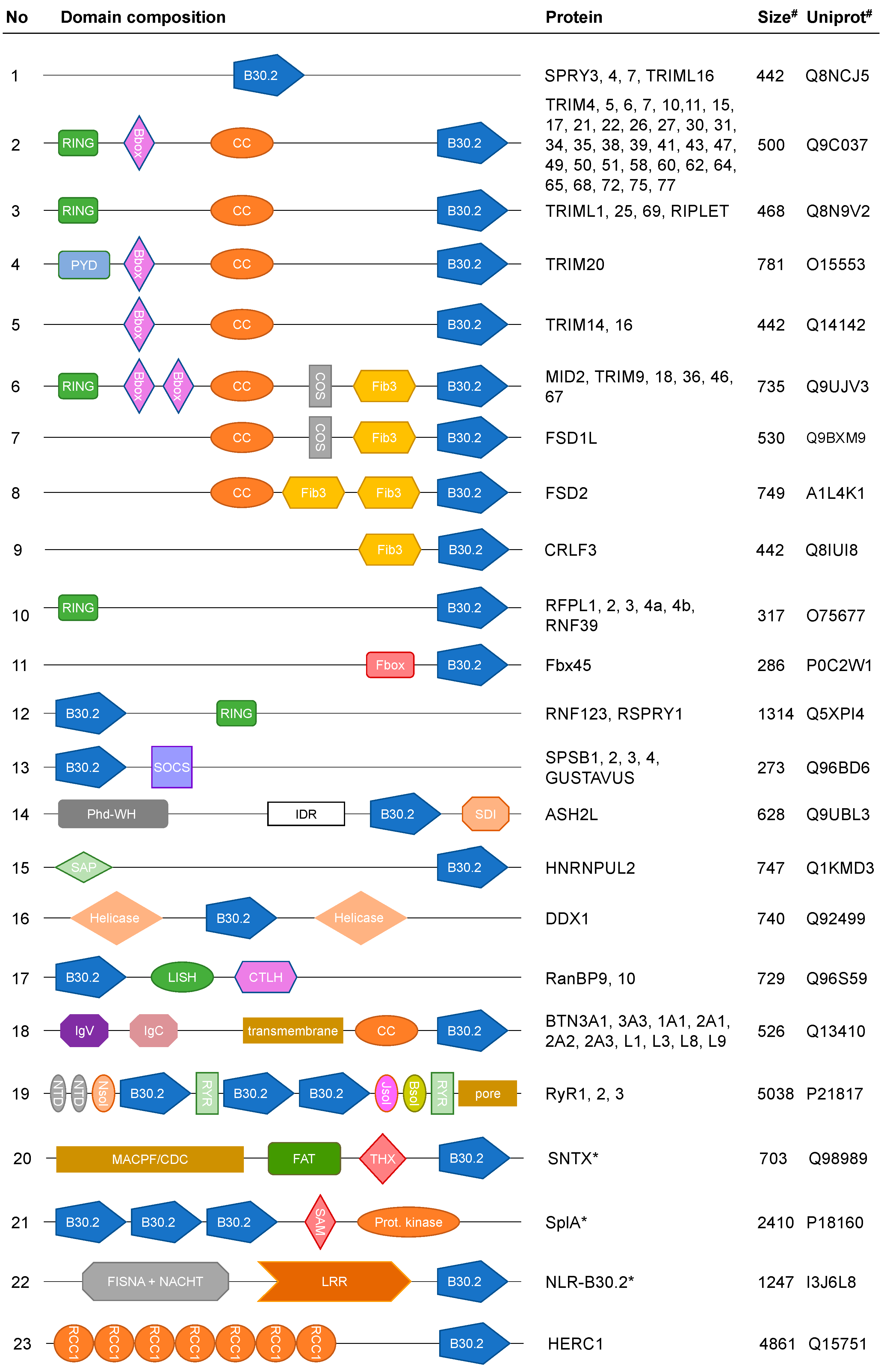
3. B30.2 Multidomain Proteins

B30.2 domains are often fused to other domains (Figure 2). Single-domain B30.2 proteins, such as SPRY3 and −7, are the exception (No. 1 in Figure 2) [11]. The largest family of B30.2 multidomain proteins contains the tripartite (Trim) motif. The Trim motif comprises a Really Interesting New Gene (RING) domain at the N-terminus, a B-box, and a Coiled Coil (CC) domain. The Trim motif is often fused to a B30.2 domain at the C-terminus [13,14]. Many Trim proteins function as E3 ubiquitin (Ub) ligases, where the 40–54 residue RING domain is responsible for recognizing the E2 Ub-conjugating enzyme. The B-box (32–42 residues) and CC domains (30–110 residues) act as oligomerization modules. RING and B-box domains are connected by a well-conserved 25–37 residue RING-B-box linker. The CC domain causes dimerization of Trim proteins and the B-box domain generates higher order oligomers of Trim dimers. RING and B-box domains typically bind two zinc ions each [15].
There is substantial variation within the family of Trim proteins, and according to the domain composition, the Trim family was subdivided into nine subfamilies [16]. In some Trim proteins, either the RING or the B-box domain is missing. In Trim20, the RING domain is replaced by the 90 residue PYD domain (No. 4 in Figure 2) [17]. Some Trim proteins (No. 6 to 8 in Figure 2) contain additional C-terminal subgroup One Signature (COS) and fibronectin type-III domains (130 residue). The 60 residue COS domain confers binding to microtubules [16]. CRLF3 comprises one fibronectin type-III domain, but no COS, B-box or CC domain (No. 9 in Figure 2). Deficiency in CRLF3 leads to increased microtubule stability [12].
Several B30.2 proteins without B-box or CC oligomerization domains are involved in ubiquitination reactions. The Ret-finger protein-like (RFPL) and F-box/SPRY domain-containing protein families are characterized by C-terminal B30.2 domains that are fused directly either to RING- or F-box domains (50 residues), respectively (No. 10 and 11 in Figure 2) [18,19]. F-box domain proteins serve as substrate receptors in Cullin-RING E3-ligases [20]. In contrast to that, the sequential order of domains is inverted in the families of Ring-finger (e.g., Rnf123, also known as Kip1 ubiquitination-promoting complex subunit 1) and SPSB proteins (No. 12 and 13 in Figure 2). SPSB and Rnf proteins contain N-terminal B30.2 domains and C-terminal Suppressor of Cytokine Signaling (SOCS) and RING domains, respectively [21,22].
Ash2l comprises an N-terminal plant homeodomain finger and winged helix (Phd-WH) DNA binding domain (110 residues), intrinsically disordered regions (IDR), a B30.2 domain, and a C-terminal Sdc1-DPY-30 interacting (SDI) motif (No. 14 in Figure 2). These IDRs are necessary for stimulating the MLL histone methyltransferase activity [23]. The Heterogeneous Nuclear Ribonucleoprotein U-like protein 2 (HNRNPUL2) is involved in the DNA damage response and carries a 35 residue SAP domain (SAF-A/B, Acinus and PIAS motif) at the N-terminus of the B30.2 domain (No. 15 in Figure 2) [24]. Several RNA helicases that are responsible for RNA unwinding share the DEAD tetrapeptide. These DEAD box helicases (DDX) contain several additional sequence motifs that are responsible for ATP- and RNA binding [25]. In some DDX proteins (e.g., DDX1), a B30.2 domain is inserted into the helicase domain [26], whereas DDX4 requires binding of a B30.2 containing adaptor protein, such as Ran-binding protein (RanBP) 9, for activity [27]. The LISH (Lis1-homology, 33 residues) and CTLH (C-terminal to LISH) domains of RanBPs regulate interactions with the microtubule (No. 16 and 17 in Figure 2) [28]. The yeast Gid1 protein also contains B30.2-, LISH- and CTLH domains and serves as a scaffold subunit in the chelator-GIDSR4 E3-ligase [29].
B30.2 domains also play important roles in membrane proteins. Proteins from the butyrophilin family are single-pass membrane proteins, where the N-terminus of the B30.2 domain is fused to a CC domain, a transmembrane helix, and two extracellular Ig-like domains from the B7 protein superfamily (No. 18 in Figure 2) [30]. Ryanodine Receptors (RyR) are large tetrameric receptors in the sarcoplasmic reticulum membrane. The N-terminal cytoplasmic shell of RyRs contains several solenoid domains and three B30.2 domains and extends into the C-terminal transmembrane pore domain (No. 19 in Figure 2). The Stonustoxin subunit alpha (SNTX) and the dual specificity protein kinase SplA from Dictyostelium discoideum are two examples of B30.2 proteins that have no homologues in humans (No. 20 and 21 in Figure 2). The SplA and RyR genes were among the first genes to be discovered, where B30.2 domains were also discovered. NACHT-domain and Leucine-Rich-Repeat-containing proteins (NLR, also known as NOD-like receptors) are pathogen recognition receptors in innate immunity. In vertebrates that lack an adaptive immune system, such as fish, NLRs are particularly abundant. Members from the NLR family C possess B30.2 domains at the C-terminus of the Leucine-Rich-Repeat (LRR) domain (No. 22 in Figure 2) [31,32].
The phylogenetic tree of B30.2 domain sequences can be broadly subdivided into two families [3]. The family of extended B30.2 domains comprises the Trim and butyrophilin sequences. The closely related butyrophilins form one branch of this family, suggesting that some Trim proteins are more closely related to the butyrophilins than to other Trim proteins. All other B30.2 domains belong to the second family [13]. Interestingly, the intermolecular sequence identity between different RyR isoforms is higher than the intramolecular sequence identity between the three B30.2 domains within the same RyR [33].

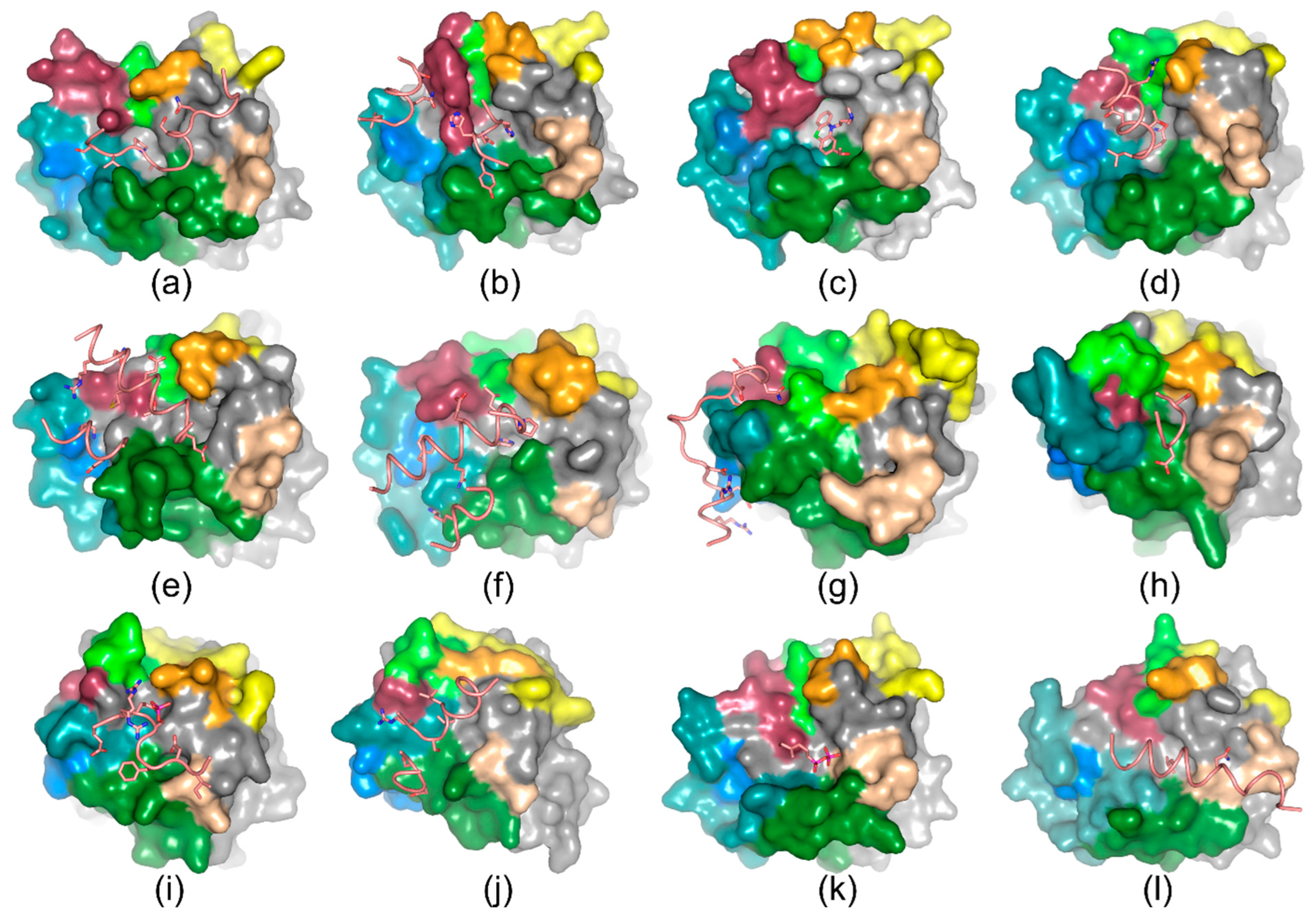
4. Ligand Recognition

B30.2 domains serve as scaffold domains and for the recognition of ligands. A canonical ligand binding site is located at the concave side of sheet A, just above the kinked β-strands G, F, and M. Initially, this site was predicted because it forms a peculiar crystal contact with the extended C-terminal peptide in the crystal structure of the RFPL4A B30.2 domain (Figure 3a) [5]. However, the first experimental proof that this site is functionally relevant came from the crystal structure of the Trim21 B30.2 domain in complex with an IgG Fc fragment (Figure 3b) [34]. The shape of this binding site is reminiscent of the complementarity-determining region seen in antibodies and, consequently, the Trim21 loops that surround the IgG Fc binding site were defined as variable loops similar to antibodies. The variable loops 1, 2, 3, 4, 5a, 5b, and 6 defined by James and coworkers (2007) correspond to loops 3, 5, 6, 7, 8, 9, and 13 in Figure 1a [34]. Since different loop nomenclatures are used in the literature [5,7,9,34,35,36,37], we are using the nomenclature from Figure 1a in the following text. The Trim21/IgG Fc interface was probed by alanine scanning, isothermal titration calorimetry (ITC), and stopped-flow kinetics, revealing a specific and preformed interface with Kd values of 37 nM and 437 nM for the human and mouse proteins, respectively (Figure 3b) [34,38]. The low molecular weight compound (S)-ACE-OH (2-(S1-hydroxyethyl) promazine) binds to the lower pocket of the IgG Fc binding site of the Trim21 D355A mutant (Figure 3c). Although ITC revealed just a modest Kd value of 17.9 µM for the binary interaction, binding of (S)-ACE-OH was mandatory for the recognition of NUP98 [39]. The Trim21/IgG Fc binding site coincides with the binding site for the glycogenin-1 C-terminal peptide on Trim7 (Figure 3d). Kd values between 2.3 µM and 21.1 µM have been determined depending on the length of the glycogenin-1 peptide [40]. The Trim7 B30.2 domain specifically recognizes the C-terminal glutamine and the preceding leucine side chains. Almost identical binding modes were observed between Trim7 and other viral proteins with C-terminal glutamine residues [41,42].
Trim65 and the Trim-like protein RIPLET (also known as RNF135) are E3-ligases that recognize the RNA helicases MDA5 and RIG-I, respectively. The cryo-EM structures of the Trim65/MDA5/dsRNA and RIPLET/RIG-I/dsRNA ternary complexes show that the B30.2 domains of Trim65 and RIPLET recognize the C-terminus of helix 1 in the RNA helicase, albeit at slightly different orientations (Figure 3e,f). The affinity of Trim65 for MDA5 in the absence of dsRNA is low. The Kd value of the Trim65 CC-B30.2 fragment for the MDA5 filament in the presence of dsRNA is 170.6 nM, suggesting that binding depends crucially on avidity effects [43]. SPSB proteins, including the Drosophila protein GUSTAVUS, serve as adaptor proteins between E3-ligases and their cognate target proteins, e.g., human prostate apoptosis response protein-4 (Par-4) or the DEAD-box RNA helicase VASA. The SOCS box and the B30.2 domains recognize the E3-ligase and an asparagine-rich loop region from the target, respectively. Human SPSB1 binds the VASA peptide with a Kd value of 40 nM. Particularly loops 5 and 13 from the B30.2 domain form H-bonds with the conserved asparagine side chains (Figure 3g) [44]. Similar interactions are seen in other SPSB/peptide complexes [7,45,46,47,48]. RanBP9 is a scaffolding protein that binds the DEAD-box RNA helicase DDX4 (also known as mouse VASA homolog) [27,49]. Human RanBP9 binds DDX4 residues 228 to 247 with a Kd value of 12.7 µM, but not the VASA peptide from Drosophila. The human DDX4 peptide lacks the asparagine residues and binds in a different region on the RanBP9 B30.2 domain compared to SPSB1. Human RanBP9 involves loops 3, 6, 7, and 8 for binding the DDX4 peptide (Figure 3h) [35].
Together with the WDR5, RbBP5, and DPY-30 subunits, the mammalian B30.2 protein Ash2L forms the WRAD complex. The WRAD complex binds to the SET domain of a lysine (K) methyltransferase from the mixed-lineage leukemia (MLL) family and modulates lysine 4 methylation of histone 3 [50]. The Ash2L B30.2 domain binds RbBP5 with a Kd value of 3.3 µM, which is reduced to 0.24 µM upon phosphorylation of S350. The phosphorylation generates an intramolecular salt bridge that stabilizes the favorable binding conformation of E349. The binding mode is independent of S350 phosphorylation and similar to the binding mode of the unphosphorylated peptide (Figure 3i) [51]. Structures in complex with MLL, the nucleosome core particle (NCP), or other components from the WRAD complex revealed similar peptide conformations [52,53,54,55,56,57,58,59,60,61,62]. In the 6.9 Å resolution cryo-EM structure of the class1 MLL1-WRAD-NCP complex without DPY-30, the Ash2L B30.2 domain contacts MLL1 directly and not via RbBP5 (Figure 3j) [23].
B30.2 domains bind not only peptides but also low molecular weight metabolites. The human butyrophilins BTN3A1 and BTN3A3, but not BTN3A2, contain intracellular B30.2 domains. These intracellular B30.2 domains bind isoprenoid diphosphates, known as phosphoantigens (pAgs). The synthetic pAgs molecule hydroxy-methyl-butyl-pyrophosphonate (cHDMAPP) binds into a positively charged pocket of butyrophilin 3A1 (BTN3A1) with a Kd value of 0.51 µM [63]. Several other pAgs and chemically related compounds bind to butyrophilins in a similar way, as shown in Figure 3k [64,65,66]. Trim58 forms a domain-swapped dimer in the crystal lattice, but in contrast to RFPL4A, it recognizes its own N-terminus (Figure 3l) [67].

5. Tripartite Motif Proteins with B30.2 Domains

Since the first discovery of a Trim protein in 1991 (Xenopus laevis nuclear factor 7 [68]), approximately 80 members of this protein family have been identified in the human genome [69]. The B30.2 domain is not present in all Trim proteins. It can be replaced by other peptide binding domains, such as Bromo- (e.g., Trim24, O15164), Math- (e.g., Trim37, Q6PCX9), NHL-repeat (e.g., Trim2, Q9C040) or cyclophilin domains (e.g., TrimCyp, F7HKF6) [14,16]. Many Trim proteins confer substrate specificity to the ubiquitination cascade, which involves the Ub-activating enzyme (E1), the Ub-conjugating enzyme (E2), and the Ub-ligase (E3). There are more than 600 E3-ligases in the human genome that have been grouped into three families: (i) RING E3-ligases, (ii) HECT E3-ligases, and (iii) RING-between-RING E3-ligases [70].
Many Trim proteins, e.g., Trim5α or Trim72, act as homodimeric RING E3-ligases [70,71]. Trims bind the E2 enzyme by means of their RING domain and form an isopeptide bond between the Ub C-terminus and a lysine side chain from the target protein. The Ub itself comprises several lysine residues, allowing E3-ligases to synthesize mono- and poly-ubiquitinated substrates as well as unanchored poly-Ub chains. Poly-K48 chains of Ub proteins lead to substrate degradation via the proteasome and poly-K63 chains to the activation of protein kinases, DNA repair, and lysosomal import [72,73]. Trim proteins are also involved in the transfer of other peptides to the target, such as the small ubiquitin modifier SUMO and interferon-induced 15-KDa protein [74,75]. All three kinds of ubiquitination activities have been observed in the C-IV family of Trim proteins, which is characterized by C-terminal B30.2 domains. It is generally believed that the B30.2 domain recognizes the target for ubiquitination. The CC domain is responsible for dimerization, and the B-box domain forms higher order oligomers and/or supports the function of the RING domain [15,76]. The functional analysis of Trim proteins is complicated by the avidity-driven recognition of substrates, that assemble under well-defined cellular states, by the ability to form homo- and hetero-oligomers [72], and the loss of terminal domains by either alternative splicing events or proteolytic digestion [77,78,79].
High-resolution structures of isolated B30.2 domains from Trim1, −5α, −7, −10, −14, −15, −16, −20, −21, −25, −58, −65, −70 and −72 have been determined by X-ray crystallography (PDB: 2IWG, 2WL1, 3KB5, 3UV9, 4B3N, 6FLM, 6JBM, 7JL4, 6SJH, 6UMA, 7QRZ, 7QS0, 7QS2, 8A8X, 8PD4) [34,36,40,43,67,80,81,82,83,84]. In Trim proteins, the B30.2 domain is fused to the C-terminus of a central CC domain (No. 2 to 9 in Figure 2). Crystal structures of CC-B30.2 fragments from Trim20, −25, and −72 revealed dimers that are reminiscent of a coat hanger (Figure 4a) [83,85,86]. The B30.2 domains are located on the convex side of the curved CC dimer. Different crossing angles between the CC domain and the B30.2 domain linker position the B30.2 domain on opposite sides of the CC dimer (Figure 4b). Small-angle X-ray scattering data from Trim20, −21 and −25 indicate substantial flexibility in solution and a weak CC/B30.2 domain interface [71,83,87,88]. Defined open and closed conformations of the CC-B30.2 assembly have been proposed [85,86]. Structures of supramolecular assemblies involving Trim proteins have been determined by cryo-electron microscopy (cryo-EM) for Trim5α, −21, −65, −72 and RIPLET [37,43,89,90].
Trim proteins are involved in innate immune responses by restricting viruses from various families, modulating the release of IL-1β and pathways involving Toll-like receptors, retinoic acid-induced gene-I-like receptors (RLR), and stimulators of IFN genes (STING) [14,17,73,91,92,93]. In an almost comprehensive screen, 20 out of 55 Trim proteins affected the early and late stages of the retroviral life cycle [94]. The relationship between viruses and Trim proteins is complex. In addition to viral restriction, some Trims, e.g., Trim7, −11 and −26, can enhance viral replication [95,96]. Trim5α restricts certain retroviruses, such as HIV1, in a species-specific manner by recognizing the capsid lattice, which causes premature capsid disassembly and the activation of innate immune response pathways [91]. The monomeric Trim5α B30.2 domain binds very weakly to the retroviral capsid and fails to disrupt the tubular assembly, whereas full-length Trim5α binds the retroviral capsid by avidity-driven lattice matching [82,97]. The Trim5α B30.2 domain dimer recognizes the capsid protein and guides the formation of a hexagonal semi-crystalline lattice (Figure 4c) [89,98]. The HIV1 capsid serves as a scaffold to force the assembly of an asymmetric RING domain trimer, where two RING domains activate the E2-conjugase to poly-ubiquitinate the disordered N-terminus of the third RING domain [99].
The Trim7 B30.2 domain binds helical peptides with C-terminal FQ or LQ dipeptide motifs that can be mimicked by small molecules, such as malonate [36,40,41,42]. The carboxy terminus and the glutamine side chain form polar interactions with R385 and Q436 at the bottom of the canonical Trim7 binding site, respectively, and the leucine side chain interacts with loops 2, 3, and 6 (Figure 3d). Trim7 recognizes endogenous proteins, such as glycogenin-1, RACO-1, and STING, and proteins originating from viral polyproteins. Since peptides with C-terminal glutamine residues are generated by viral 3C proteases, it was suggested that Trim7 could serve as a broadly-acting restriction factor, but phylogenetic data, tissue expression and gene regulation argue against this hypothesis [40,95]. Finally, Trim7 itself can be cleaved by 3C proteases at Q24, and the resulting fragment inhibits the E3-ligase activity of Trim7 [79]. Trim14 reveals a B30.2 domain with a positively charged canonical binding site, similar to Trim72 and butyrophilin BTN3A1. The Trim14 B30.2 domain interacts with negatively charged poly(Glu:Tyr) peptides [84].
Trim20 (also known as Pyrin or Marenostrin) is an atypical member of the Trim family because the RING domain is replaced by a PYD domain (No. 4 in Figure 2). Hence, Trim20 does not transfer Ub chains; instead, it forms PYD-PYD homotypic interactions with the apoptosis-associated speck-like protein containing a caspase recruitment domain (ASC), which finally leads to caspase-1 activation, IL-1β release, and fever. Trim20 is regulated by phosphorylation in the disordered region on S208 and S242 [100,101]. The function of the B30.2 domain, which is absent in murine Trim20, is uncertain. It could keep Trim20 in a repressed state (autoinhibitory function), or it could interact with a yet unknown ligand (proinflammatory function) [17]. The proline-rich stretch in the CC domain can interact with a secondary binding site on the B30.2 domain [85]. The inherited disease Familial Mediterranean Fever (FMF) is characterized by excessive inflammasome activation [102,103]. FMF mutations cluster in the B30.2 domain and those with the most severe disease outcome in loops 6 and 7 (Figure 1a) [9]. FMF mutations decrease the activation threshold of Trim20 either by attenuating the autoinhibitory function or enhancing the proinflammatory function [104]. Perhaps FMF mutations provided a selection advantage against Y. pestis infection during historic plague pandemics [105].
Trim21 serves as a cytosolic antibody receptor. Although antibodies normally do not penetrate into the cytoplasm, some of them remain attached to the capsid of incoming pathogens [106]. The Trim21 B30.2 domain effectively recognizes the Fc domain of attached IgG molecules (Figure 3b) [34]. Trim21 is activated upon substrate-induced clustering, but in contrast to Trim5α, it does not require a regular structural network [88]. Using cryo-EM and other biophysical techniques, a mechanism for Trim21 activation was postulated [90]. To achieve full activity, two RING dimers need to form a complex with the E2-conjugase [107]. Trim21 also recognizes other cellular substrates via its B30.2 domain besides IgG [108]. The molecular glue (S)-ACE-OH enables the recruitment of Trim21 to the autoproteolytic domain of the nuclear pore subunit NUP98 [39]. Trim21 allows the selective degradation of endogenous target proteins either by administering target-specific IgG molecules in the Trim-Away technology or the design of bifunctional proteolysis-targeting chimers (PROTACs) [39,88].
The Trim22 B30.2 domain recognizes endogenous AKT phosphatase PHLPP2 and Zika virus nonstructural proteins and labels them for degradation [109,110]. Trim72 is a critical component for the Ca2+-independent repair of plasma membranes by enabling vesicular transport to the site of injury [111]. It binds negatively charged phospholipids, such as phosphatidylserine (PS) and phosphatidylcholine, via its positively charged B30.2 domain (Figure 4d) [37,86]. The proper orientation of the B30.2 domain is crucial for avidity-driven binding because the monomeric Trim72 B30.2 domain, in contrast to the artificial bivalent Trim72-B30.2 construct, showed impaired binding on liposomes (Kd for Trim72-B30.2-GST dimer and Trim72 is 76.7 nM and 9.9 nM, respectively) [37]. A working model for Trim72 activation was proposed on the basis of the cryo-EM structure, liposome sedimentation, and cross-linking data. Soluble Trim72 dimers do not bind the E2-conjugase, whereas PS-enriched phospholipids generate a Trim72 network, where the reduced mobility of the RING domain enables the transfer of Ub chains [37].
Upon binding to viral double-stranded (ds) RNA, innate immune receptors from the RLR family activate interferon pathways via an N-terminal Caspase Activation and Recruitment Domain (CARD). The tetramerization of the CARD domain is driven by poly-K63 Ub chains that are assembled by E3-ligases from the Trim family [112,113]. The Trim proteins RIPLET and Trim65 recognize dsRNA-bound forms of oligomeric RIG-I and MDA5, respectively. RIG-I (DDX58) and MDA5 belong to the family of ATP-dependent DEAD box RNA helicases. The ternary complex structures of dsRNA/MDA5ΔCARD/GST-Trim65B30.2, dsRNA/RIG-IΔCARD/GST-RIPLETB30.2, and dsRNA/RIG-I2/RIPLET2 were determined by cryo-EM (Figure 4e) [43,114]. To avoid the unwanted activation of IFN pathways in the absence of dsRNA, monovalent B30.2 domains have poor affinities, whereas bivalent RIPLET and Trim65 recognize the dsRNA-bound forms of RLRs. The B30.2 domains of RIPLET and Trim65 interact with a conserved epitope involving helices 1 and −3 from the Hel2 domains of the RLRs. Despite this, the spatial orientation of the helices differs by approximately 60° (Figure 3e,f). Similar epitopes have been observed in the helicases Dicer and DDX41 that were recognized in pull-down assays by bivalent B30.2 constructs of Trim25, −26, and −41 [43]. Trim65 recognizes other cellular targets, such as the transcription factor TOX4, by means of its B30.2 domain and transfers poly-K48 Ub chains [115].
Some DDX proteins, such as DDX4, DDX41 and DDX58, are recognized by B30.2 domain proteins via non-covalent interactions. In contrast to that, DDX1 carries a B30.2 insertion between the I- and Ia motifs in the N-terminal helicase domain (No. 16 in Figure 2). DDX1 senses viral nucleic acids by forming a complex with DDX21 and DHX36 that recognizes poly(I:C) oligonucleotides [116]. The structure of the DDX1 B30.2 domain was determined by X-ray crystallography, but the function of this domain is currently unknown [117].

6. SPRY Domain-Containing SOCS Box Proteins

The crystal structures of the Drosophila protein GUSTAVUS and the NMR structure of mouse SPSB2 were among the first experimental B30.2 domain structures [6,7,8]. SPSB proteins recruit target proteins to the ECS complex, comprising Elongin-B, -C, Cullin-5, SPSB, and the RING-finger containing protein Rbx2. This ECS complex, which is also called Cullin-RING ubiquitin ligase 5 (CRL5), belongs to the Cullin-RING E3-ligase subfamily [70]. The Elongin-B/C heterodimer recognizes the SOCS box of SPSB proteins [6]. The Rbx2 subunit interacts with the E2-conjugase. In some SOCS box proteins, the B30.2 domain is replaced by other peptide-binding domains, such as SH2, WD40, LRR, or GTPase domains [118], very similar to what has been observed for Trim proteins.
The four mammalian SPSB proteins bind various cellular targets by means of their B30.2 domains. SPSB1, −2, and −4 bind inducible nitric oxide synthase (iNOS). SPSB2 is a negative regulator of iNOS and thereby modulates the level of reactive nitrogen species. It interacts with the N-terminal region of iNOS and marks it for proteasomal degradation by polyubiquitination [21,46,47,48]. Other targets are c-MET and Par-4 [8,44,45]. Targets for SPSB3 are the zinc finger transcription factor SNAIL and cyclic GMP-AMP synthase (cGAS) [119,120].
There is no exact orthologue of the Drosophila protein GUSTAVUS in vertebrates. The closest homologues are SPSB1 and −4 with 75% and 70% sequence identity, respectively [121]. GUSTAVUS interacts with the ATP-dependent DEAD box RNA helicase VASA. This interaction is essential for the localization of VASA at the posterior pole of the oocyte [122,123]. The VASA binding site on GUSTAVUS coincides with the binding site for iNOS, Par-4, and cGAS on SPSB proteins (Figure 5a). This binding site differs from the canonical binding site because it only involves residues from loops 3, 5, 6, and 13 (Figure 3g). Loop 13 is exceptionally short and loop 6 is significantly longer in SPSB proteins compared to other B30.2 domains.
GUSTAVUS, SPSB1, −2, and −4 recognize proteins with D(I,L)NNN consensus sequences, which can be mimicked by cyclic peptides (Figure 5a) [47,124]. The asparagine side chains are recognized by H-bonds involving main- and side-chain atoms from loops 3, 5, and 13. The aspartic acid side chain interacts with a conserved tyrosine side chain from loop 6. The 3.5 Å resolution cryo-EM structure of the CRL5 core (comprising cGAS/SPSB3/Elongin-B/-C) in complex with an NCP revealed a minimal NN degron sequence (Figure 5b,c). SPSB3 recognizes a degraded consensus sequence at the C-terminus of cGAS. Modeling studies showed that the cGAS/SPSB3 interface positions the RING-between-RING E3-ligase ARIH2 adjacent to lysine residues from cGAS for direct Ub transfer [120].

7. Heterodimeric RING E3-Ligases

The germline cell-specific ATP-dependent DEAD box RNA helicase DDX4 is the VASA homologue in mice. DDX4 binds to the atypical RanBP9. In contrast to classical RanBPs, the Ran binding domain is replaced by a B30.2 domain in RanBP9 and −10 [27]. RanBP9 is a core component of the CTLH E3-ligase, which belongs to the family of heterodimeric RING E3-ligases [125]. The name of this E3-ligase refers to the high frequency of CTLH repeats in this complex. The binding mode of RanBP9 and DDX4 was revealed by the crystal structures of human RanBP9, mouse RanBP10 and human RanBP9 in complex with mouse DDX4 residues 201–220 (PDB: 5JI7, 5JI9, 5JIA, 5JIU) [35]. The target binding mode of RanBP9 differs from the binding mode seen in SPSB proteins (Figure 3g,h). Residues 208ESSDSQ213 from DDX4 are recognized by H-bonds with residues from the RanBP9 B30.2 domain loops 6, 7, and 8.
The glucose-induced degradation (Gid) E3-ligase is the CTLH homologue in yeast. The Gid E3-ligase is not a structurally unique complex. Depending on the task, Gid exists in different subunit compositions because it primes the cell to respond properly to environmental changes. Yeast can grow on various different carbon sources, and depending on the source, glycolytic or gluconeogenic enzymes need to be expressed or degraded [126]. Gid degrades oligomeric enzymes, such as tetrameric fructose-1,6-bisphosphatase (Fbp1), to shut down unnecessary metabolic pathways. The 3.4 Å resolution cryo-EM structure of the yeast chelator GIDSR4 particle shows that this supramolecular assembly comprises three modules: the substrate receptor scaffolding module (SRS), the supramolecular assembly module (SA), and the catalytic module (Cat). Subunit Gid1, which contains the B30.2 domain, is part of the SRS and SA modules [29].
In the yeast Gid and human CTLH E3-ligase the SRS modules consist of subunits Gid1/Gid5/Gid8/Gid4 (PDB: 6SWY) and subunits RanBP9/ArmC8/TWA1/hGid4 (PDB: 7NSC), respectively [29,127]. The SRS module binds the target receptor Gid4 (or Gid10) that recognizes the N-degron of the target and presents it to the Cat module (subunits Gid2/Gid9). The B30.2 domain of Gid1 (RanBP9 in human CTLH) has a scaffolding function (Figure 5d). It tightly interacts with Gid5 (ArmC8 in human CTLH). The Gid1 B30.2 domain is not directly involved in recognizing the targeted Fbp1 [29]. Instead, the N-terminal Fbp1 degron is recruited into the complex via the target receptor Gid4, which primarily interacts with Gid5 but also with loop 5 of the Gid1 B30.2 domain. However, the interaction between Gid4 and the Gid1 B30.2 domain seems to be dispensable [127]. In yeast, the Gid1 B30.2 domain loop 6 folds into a short helix, that partially occludes the canonical binding site (PDB: 7NS3, 7NSB), whereas in the human CTLH SRS module, the canonical binding site of RanBP9 is empty and positioned in close proximity to ArmC8 (PDB: 7NSC) (Figure 5d) [29].
The SA module comprises Gid1, Gid7, and Gid8 (PDB: 7NSB). In the yeast chelator GIDSR4 particle structure, intra- and intermodular interactions are dominated by LISH-, CTLH, and CRA-domains. In the SA module, the Gid1 B30.2 domain is located in proximity to the C-terminal β-propeller domain of Gid7, but there are no direct contacts between those domains [29].

8. Set1/COMPASS Methyltransferase Complexes

Methylation of lysine 4 of histone H3 (H3K4) from the NCP is a hallmark of actively transcribed genes. Mono-, di- and trimethylation of H3K4 is catalyzed by methyltransferases from the Set1/COMPASS family, which in many cases depend on mono-ubiquitination of histone H2B K120 (H2B-K120Ub). The MLL1 catalytic domain is sufficient for monomethylation, whereas di- and trimethylation require the incorporation of MLL1 into a complex with WDR5, RbBP5, Ash2L, and DPY-30 (MLL1-WRAD complex) [128]. The core of this complex is formed by the B30.2 domain protein Ash2L, WDR5, RbBP5, and the C-terminal region of MLL1, whereas DPY-30 is bound transiently to modulate trimethylation of H3K4 [129]. The B30.2 domain has a 40 amino acid insertion in loop 10. In addition, Ash2L comprises a PHD-WH domain, an IDR region, and a SDI motif (No. 14 in Figure 2).
The Ash2L B30.2 domain was analyzed free (PDB: 3TOJ) and in complex with RbBP5-derived peptides (PDB: 4X8N, 4X8P) (Figure 3i) [10,51]. The RbBP5 C-terminal peptide (residues 347–354) binds to the canonical binding site of the B30.2 domain and allows the recruitment of different MLL methyltransferase isoforms into the core complex. Crystal structures of the quaternary core complexes involving the Ash2L B30.2 domain, the RbBP5 C-terminus, the catalytic methyltransferase domain from either MLL1, −2 or −3, and the S-adenosyl homocysteine reaction product have been determined by X-ray crystallography (PDB: 5F6L, 5F6K, 7BRE). In the MLL2 core complex, an arginine side chain from the MLL2 N-terminus blocks the H3K4 binding site [52,58]. The rate constants for different H3K4 methylation states of the six mammalian methyltransferases from the Set1/COMPASS family have been determined by mass-spectrometry. It was proposed that MLL1 residues Y3858 and Y3942 form a F/Y switch that governs the methylation rate constants (PDB: 7W6L, 7W6A, 7W67, 7W6I, 7W6J) (Figure 6a) [60]. The methylation rate constants are also affected by DPY-30, which binds to the SDI segment at the C-terminus of the Ash2L B30.2 domain (PDB: 6E2H) [130].
Structures of the mammalian MLL1- and MLL3-WRAD core complexes, together with the H2B-K120Ub modified NCP from Xenopus laevis, have been determined by cryo-EM at resolutions between 3.2 Å and 4.5 Å. They reveal different binding modes of the MLL1-WRAD complex on the NCP depending on the ubiquitination state of H2B K120 (PDB: 6KIU, 6KIV, 6KIW, 6KIX, 6KIZ) [57]. The MLL1-WRAD-NCP complex was also analyzed with and without DPY-30 in the absence of the H2B-K120Ub modification at 4.4 Å and 6.2 Å resolution, respectively (PDB: 6PWW, 6PWV) [55]. The MLL1-WRAD-NCP complex is a very dynamic supramolecular assembly. Particularly in the absence of the H2B-K120Ub modification, it can adopt several different structural states. The DPY-30 subunit interacts with the Ash2L IDR region and restricts the mobility of the MLL1-WRAD-NCP complex, which has an impact on the formation of higher methylation states of H3K4. (PDB: 6W5I, 6W5M, 6W5N, 7MBM, 7MBN) [23,61]. The 4.25 Å resolution cryo-EM structure of the H2B-K120Ub-modified MLL1-WRAD-NCP with DPY-30 (PDB: 7UD5) is similar to the structure in the absence of DPY-30. The H2B-K120Ub modification positions the MLL1-WRAD complex in an active conformation on the NCP (Figure 6b) [62].
The Complex of Proteins Associated with Set1 (COMPASS) is the yeast homologue of the mammalian MLL1-WRAD complex. The yeast subunits Set1/Swd3/Swd1/Bre2/Sdc1 correspond to the human MLL1/WDR5/RbBP5/Ash2L/DPY-30 subunits. The COMPASS structure was determined by EM, either free or in complex with the H2B-K120Ub-modified Xenopus laevis NCP at 4.3 Å and 3.37 Å resolution, respectively (PDB: 6BX3, 6VEN) [54,59]. The B30.2 domain proteins human Ash2L and yeast Bre2 have different domain compositions. Bre2 lacks the N-terminal PHD-WH domain and IDR region but carries a 110-residue insertion in loop 10, which is partially resolved in the COMPASS-NCP structure. The positioning of the Ub chain is strikingly different in the MLL1-WRAD-NCP and COMPASS-NCP complexes (Figure 6b,c).

9. Butyrophilins

Cytotoxic gamma delta T cells (γδT cells) do not require antigen processing and are independent of peptide epitopes presented by MHC complexes. Instead, they interact with stress-induced molecules, e.g., on the surface of tumor- or infected cells, and secrete cytotoxic molecules. Cells from the most prevalent Vγ9Vδ2 lineage are activated by an inside-out signaling process that depends on pAgs, such as (E)-4-hydroxy-3-methyl-but-2-enyl diphosphate (HMBPP), isopentenyl diphosphate (IPP), and dimethylallyl diphosphate (DMAPP). IPP/DMAPP and HMBPP are produced by the mevalonate- and mevalonate-independent (DOXP/MEP) pathways in humans and pathogens, respectively. HMBPP, DMAPP, and IPP are recognized by BTN3A1, but HMBPP is much more potent than IPP. In humans, there are three BTN3A isoforms. BTN3A1 and −3 have similar domain compositions and share 87% sequence identity, whereas BTN3A2 has no B30.2 domain at all [131,132].
The intracellular B30.2 domains of butyrophilins recognize pAgs and forward the signal to the ectodomains (No. 18 in Figure 2) [64,66]. The B30.2 domain of BTN3A isoform-1 was crystallized free (PDB: 4N7I, 4V1P, 5HM7, 6ISM), in complex with HMBPP (PDB: 5ZXK) or HMBPP analogues (PDB: 4N7U, 6J06) (Figure 7a). The structures of the apo BTN3A isoform-3 (PDB: 5ZZ3) and its R351H mutant (PDB: 6J0L, 6J0K, 6J0G) are very similar to the structures of isoform-1 [63,65,133,134]. In both isoforms, arginine, lysine, and histidine side chains from loops 3, 6, and 8 and strands F, H, and M form a positively charged groove at the canonical binding site (Figure 7a). BTN3A1 and the BTN3A3 R351H mutant, but not wild-type BTN3A3 or BTN2A1, recognize negatively charged pAgs molecules (Figure 3k). Organic anions, such as citrate and malonate, can mimic the pAgs (PDB: 5LYG, 5LYK). It was suggested that pAgs-induced conformational changes in the B30.2 domains are propagated to the extracellular Ig-like domains [64].
For a long time, the activation of BTN3A1 was enigmatic because the cellular activities of pAgs did not correlate with their binding affinities to the isolated BTN3A1 B30.2 domain in vitro [135]. It was shown recently that BTN3A1 requires the accessory protein BTN2A1 to activate Vγ9Vδ2T cells upon pAgs stimulation. HMBPP serves as a molecular glue that enables the association between B30.2 domains of BTN3A1 and BTN2A1 with a Kd of 611nM [66]. BTN2A1 has no positively charged groove. Hence, it does not bind pAgs. The B30.2 domains form a (BTN3A1/BTN2A1)2 heterotetramer that harbors two pAgs molecules (PDB: 8JYC, 8JYE) (Figure 7b). In the tetramer, BTN2A1 B30.2 domains form a symmetric dimer that is almost identical to the BTN2A1 dimer in the absence of BTN3A1 (PDB: 8IGT). The mutagenesis and NMR data of the isolated BTN2A1 B30.2 domain confirm that it forms a homodimer, even in the absence of the N-terminal domains [136]. The extended C-terminal BTN2A1 tail forms a short antiparallel β-sheet with strand L from the second subunit. The extended C-terminal tail of BTN2A1 interacts with the pAgs molecule bound to the canonical binding site and recruits BTN3A1 into the complex (Figure 7b). Thus, pAgs molecules activate Vγ9Vδ2T cells by triggering the oligomerization of BTN3A1 and BTN2A1 proteins [66]. Progress on the understanding of butyrophilin activation by pAgs has been reviewed recently [137].

10. Ryanodine Receptors

Ryanodine Receptors (RyR) are homotetramers with a molecular weight of over 2 MDa. They are named after the natural diterpene ryanodine from plants that locks the receptor in a closed conformation. RyRs release Ca2+ ions from the sarcoplasmic reticulum upon activation. Many small molecules and accessory proteins, such as FK506-binding proteins (FKBP), S100A1, and calmodulin, allosterically affect the gating properties of RyRs. There are three RyRs in the human proteome (RyR1, −2, and −3) and each of them comprises three B30.2 domains that are named SPRY1, −2 and −3 in the literature. Isolated B30.2 domains of RyR1 and −2 have been analyzed by X-ray crystallography [138,139,140,141]. The full-length RyR structures in different gating states and in complex with various ligands have been intensively studied by EM, leading to more than 100 structures for RyR1 [142,143,144,145,146,147,148,149,150,151,152,153,154,155,156,157,158], more than 50 structures for RyR2 [153,157,159,160,161,162,163,164,165], and 2 structures for RyR3 [166]. The RyR tetramer has a mushroom-shaped structure with a large cytoplasmic shell and a narrow pore embedded in the sarcoplasmic membrane (Figure 8a). The structural interplay of domains in the cytoplasmic shell of RyR1 that leads to the different gating states of the pore has been reviewed [167].
In all full-length RyR structures, the three B30.2 domains tightly interact with each other (Figure 8a). In some SPRY domains, the B30.2 fold has been reshuffled by the circular permutation of β-strands and exceptionally large insertions in loop regions. The RyR1 SPRY1 domain starts at strand D. Strands A, B, and C have been moved from the N-terminus to the C-terminus of the B30.2 fold, so that strand A follows strand N. Approximately 800 residues, including the SPRY2 and −3 domains, have been inserted between strands A and B (Figure 8b). The B30.2 fold is maintained in the SPRY2 domain, but approximately 200 residues that are forming the ryanodine repeats 1 and 2 are inserted in loop 3. The RyR1 SPRY3 domain is truncated. The β-turn formed by strands H and I has been replaced by a loop that directly connects strands G and J. Approximately 140 residues, most of them are disordered in the cryo-EM structures, have been inserted in loop 4. Loop 6 folds into an elongated β-turn that extends the antiparallel β-sheet at strand L from the SPRY1 domain (Figure 8b).
The B30.2 domains of RyRs participate in binding accessory proteins. Domains SPRY1 and −3 recognize FKBP12, involving residues from the canonical binding site of SPRY1, and loops 4 and 12 from SPRY3. FKBP12 has a lower affinity for RyR3, compared to RyR1, which has been explained by point mutations of interacting residues in these domains [166]. Residues 1076–1112 from the RyR1 SPRY2 domain interact specifically with the peptide between transmembrane repeats II and -III of the dihydropyridine receptor (II-III loop), whereas the equivalent residues from RyR2 do not [168]. Using the isolated RyR1 SPRY2 domain alone, the binding of the flexible II-III loop peptide could not be confirmed by ITC [138]. Several mutations of RyRs cause skeletal and cardiac muscle-related diseases, and many of them cluster in the B30.2 domains [169,170].

11. Conclusions

B30.2 domains are often fused to other domains such as B-box, CC, COS, CTLH, fibronectin, LRR, RING, or SOCS box domains. These multidomain proteins are involved in the formation of large multi-chain molecular machines, with the B30.2 domain serving as a scaffold domain or for ligand recognition. In humans, B30.2 domains are particularly abundant in E3-ligases. The ligands range from low molecular weight compounds, such as pAgs in the case of butyrophilins, to large macromolecular assemblies, such as NCP in the case of SPSB3. B30.2 domains recognize their ligands either directly or via adaptor proteins. In some of these multidomain proteins, the B30.2 domains can be replaced by other peptide-binding domains. The affinities of binary B30.2/ligand interactions are often weak but can be enhanced by avidity effects when multiple B30.2 domains simultaneously recognize a ligand network. The ligands or adaptor proteins are often recognized at a canonical binding pocket formed by several variable loop regions. This principle is reminiscent of antibodies. In this respect, B30.2 domains can serve for the design of target-specific binders, e.g., in the Trim-Away, PROTACS, and host lattice display technologies [39,88,171].

Author Contributions

All authors contributed equally to all aspects of this article. All authors have read and agreed to the published version of the manuscript.

Funding

The research received no funding.

Conflicts of Interest

The authors declare no conflict of interest.

References

  1. Vernet, C.; Boretto, J.; Mattei, M.G.; Takahashi, M.; Jack, L.J.; Mather, I.H.; Rouquier, S.; Pontarotti, P. Evolutionary study of multigenic families mapping close to the human MHC class I region. J. Mol. Evol. 1993, 37, 600–612. [Google Scholar] [CrossRef] [PubMed]
  2. Ponting, C.; Schultz, J.; Bork, P. SPRY domains in ryanodine receptors (Ca2+-release channels). Trends Biochem. Sci. 1997, 22, 193–194. [Google Scholar] [CrossRef] [PubMed]
  3. Rhodes, D.A.; de Bono, B.; Trowsdale, J. Relationship between SPRY and B30.2 protein domains. Evolution of a component of immune defence? Immunology 2005, 116, 411–417. [Google Scholar] [CrossRef]
  4. Henry, J.; Mather, I.H.; McDermott, M.F.; Pontarotti, P. B30.2-like domain proteins: Update and new insights into a rapidly expanding family of proteins. Mol. Biol. Evol. 1998, 15, 1696–1705. [Google Scholar] [CrossRef]
  5. Grütter, C.; Briand, C.; Capitani, G.; Mittl, P.R.; Papin, S.; Tschopp, J.; Grütter, M.G. Structure of the PRYSPRY-domain: Implications for autoinflammatory diseases. FEBS Lett. 2006, 580, 99–106. [Google Scholar] [CrossRef]
  6. Woo, J.S.; Imm, J.H.; Min, C.K.; Kim, K.J.; Cha, S.S.; Oh, B.H. Structural and functional insights into the B30.2/SPRY domain. EMBO J. 2006, 25, 1353–1363. [Google Scholar] [CrossRef]
  7. Woo, J.S.; Suh, H.Y.; Park, S.Y.; Oh, B.H. Structural basis for protein recognition by B30.2/SPRY domains. Mol. Cell 2006, 24, 967–976. [Google Scholar] [CrossRef] [PubMed]
  8. Masters, S.L.; Yao, S.; Willson, T.A.; Zhang, J.G.; Palmer, K.R.; Smith, B.J.; Babon, J.J.; Nicola, N.A.; Norton, R.S.; Nicholson, S.E. The SPRY domain of SSB-2 adopts a novel fold that presents conserved Par-4-binding residues. Nat. Struct. Mol. Biol. 2006, 13, 77–84. [Google Scholar] [CrossRef]
  9. Weinert, C.; Grütter, C.; Roschitzki-Voser, H.; Mittl, P.R.; Grütter, M.G. The crystal structure of human pyrin b30.2 domain: Implications for mutations associated with familial Mediterranean fever. J. Mol. Biol. 2009, 394, 226–236. [Google Scholar] [CrossRef]
  10. Chen, Y.; Cao, F.; Wan, B.; Dou, Y.; Lei, M. Structure of the SPRY domain of human Ash2L and its interactions with RbBP5 and DPY30. Cell Res. 2012, 22, 598–602. [Google Scholar] [CrossRef]
  11. Yang, J.; Guan, X.; Zhang, D.; Zhao, P.; Guo, S.; Kuang, Z. Crystal structure of the SPRY domain-containing protein 7 reveals unique structural features. Biochem. Biophys. Res. Commun. 2020, 531, 350–356. [Google Scholar] [CrossRef]
  12. Bennett, C.; Lawrence, M.; Guerrero, J.A.; Stritt, S.; Waller, A.K.; Yan, Y.; Mifsud, R.W.; Ballester-Beltran, J.; Baig, A.; Mueller, A.; et al. CRLF3 plays a key role in the final stage of platelet genesis and is a potential therapeutic target for thrombocythemia. Blood 2022, 139, 2227–2239. [Google Scholar] [CrossRef] [PubMed]
  13. Perfetto, L.; Gherardini, P.F.; Davey, N.E.; Diella, F.; Helmer-Citterich, M.; Cesareni, G. Exploring the diversity of SPRY/B30.2-mediated interactions. Trends Biochem. Sci. 2013, 38, 38–46. [Google Scholar] [CrossRef] [PubMed]
  14. Koepke, L.; Gack, M.U.; Sparrer, K.M. The antiviral activities of TRIM proteins. Curr. Opin. Microbiol. 2021, 59, 50–57. [Google Scholar] [CrossRef]
  15. Gushchina, L.V.; Kwiatkowski, T.A.; Bhattacharya, S.; Weisleder, N.L. Conserved structural and functional aspects of the tripartite motif gene family point towards therapeutic applications in multiple diseases. Pharmacol. Ther. 2018, 185, 12–25. [Google Scholar] [CrossRef]
  16. Short, K.M.; Cox, T.C. Subclassification of the RBCC/TRIM superfamily reveals a novel motif necessary for microtubule binding. J. Biol. Chem. 2006, 281, 8970–8980. [Google Scholar] [CrossRef] [PubMed]
  17. Schnappauf, O.; Chae, J.J.; Kastner, D.L.; Aksentijevich, I. The Pyrin Inflammasome in Health and Disease. Front. Immunol. 2019, 10, 1745. [Google Scholar] [CrossRef]
  18. Bonnefont, J.; Laforge, T.; Plastre, O.; Beck, B.; Sorce, S.; Dehay, C.; Krause, K.H. Primate-specific RFPL1 gene controls cell-cycle progression through cyclin B1/Cdc2 degradation. Cell Death Differ. 2011, 18, 293–303. [Google Scholar] [CrossRef]
  19. Desbois, M.; Crawley, O.; Evans, P.R.; Baker, S.T.; Masuho, I.; Yasuda, R.; Grill, B. PAM forms an atypical SCF ubiquitin ligase complex that ubiquitinates and degrades NMNAT2. J. Biol. Chem. 2018, 293, 13897–13909. [Google Scholar] [CrossRef]
  20. Nguyen, K.M.; Busino, L. The Biology of F-box Proteins: The SCF Family of E3 Ubiquitin Ligases. Adv. Exp. Med. Biol. 2020, 1217, 111–122. [Google Scholar] [CrossRef]
  21. Kuang, Z.; Lewis, R.S.; Curtis, J.M.; Zhan, Y.; Saunders, B.M.; Babon, J.J.; Kolesnik, T.B.; Low, A.; Masters, S.L.; Willson, T.A.; et al. The SPRY domain-containing SOCS box protein SPSB2 targets iNOS for proteasomal degradation. J. Cell Biol. 2010, 190, 129–141. [Google Scholar] [CrossRef] [PubMed]
  22. Kravtsova-Ivantsiv, Y.; Goldhirsh, G.; Tomuleasa, C.; Pikarsky, E.; Ciechanover, A. The NF-kB p50 subunit generated by KPC1-mediated ubiquitination and limited proteasomal processing, suppresses tumor growth. Cancer Cell Int. 2023, 23, 67. [Google Scholar] [CrossRef]
  23. Lee, Y.T.; Ayoub, A.; Park, S.H.; Sha, L.; Xu, J.; Mao, F.; Zheng, W.; Zhang, Y.; Cho, U.S.; Dou, Y. Mechanism for DPY30 and ASH2L intrinsically disordered regions to modulate the MLL/SET1 activity on chromatin. Nat. Commun. 2021, 12, 2953. [Google Scholar] [CrossRef] [PubMed]
  24. Polo, S.E.; Blackford, A.N.; Chapman, J.R.; Baskcomb, L.; Gravel, S.; Rusch, A.; Thomas, A.; Blundred, R.; Smith, P.; Kzhyshkowska, J.; et al. Regulation of DNA-end resection by hnRNPU-like proteins promotes DNA double-strand break signaling and repair. Mol. Cell 2012, 45, 505–516. [Google Scholar] [CrossRef] [PubMed]
  25. Linder, P.; Lasko, P.F.; Ashburner, M.; Leroy, P.; Nielsen, P.J.; Nishi, K.; Schnier, J.; Slonimski, P.P. Birth of the D-E-A-D box. Nature 1989, 337, 121–122. [Google Scholar] [CrossRef]
  26. Godbout, R.; Hale, M.; Bisgrove, D. A human DEAD box protein with partial homology to heterogeneous nuclear ribonucleoprotein U. Gene 1994, 138, 243–245. [Google Scholar] [CrossRef]
  27. Shibata, N.; Tsunekawa, N.; Okamoto-Ito, S.; Akasu, R.; Tokumasu, A.; Noce, T. Mouse RanBPM is a partner gene to a germline specific RNA helicase, mouse vasa homolog protein. Mol. Reprod. Dev. 2004, 67, 1–7. [Google Scholar] [CrossRef]
  28. Emes, R.D.; Ponting, C.P. A new sequence motif linking lissencephaly, Treacher Collins and oral-facial-digital type 1 syndromes, microtubule dynamics and cell migration. Hum. Mol. Genet. 2001, 10, 2813–2820. [Google Scholar] [CrossRef]
  29. Sherpa, D.; Chrustowicz, J.; Qiao, S.; Langlois, C.R.; Hehl, L.A.; Gottemukkala, K.V.; Hansen, F.M.; Karayel, O.; von Gronau, S.; Prabu, J.R.; et al. GID E3 ligase supramolecular chelate assembly configures multipronged ubiquitin targeting of an oligomeric metabolic enzyme. Mol. Cell 2021, 81, 2445–2459.e2413. [Google Scholar] [CrossRef]
  30. Abeler-Dorner, L.; Swamy, M.; Williams, G.; Hayday, A.C.; Bas, A. Butyrophilins: An emerging family of immune regulators. Trends Immunol. 2012, 33, 34–41. [Google Scholar] [CrossRef]
  31. Howe, K.; Schiffer, P.H.; Zielinski, J.; Wiehe, T.; Laird, G.K.; Marioni, J.C.; Soylemez, O.; Kondrashov, F.; Leptin, M. Structure and evolutionary history of a large family of NLR proteins in the zebrafish. Open Biol. 2016, 6, 160009. [Google Scholar] [CrossRef] [PubMed]
  32. Suurvali, J.; Garroway, C.J.; Boudinot, P. Recurrent expansions of B30.2-associated immune receptor families in fish. Immunogenetics 2022, 74, 129–147. [Google Scholar] [CrossRef] [PubMed]
  33. D’Cruz, A.A.; Babon, J.J.; Norton, R.S.; Nicola, N.A.; Nicholson, S.E. Structure and function of the SPRY/B30.2 domain proteins involved in innate immunity. Protein Sci. 2013, 22, 1–10. [Google Scholar] [CrossRef]
  34. James, L.C.; Keeble, A.H.; Khan, Z.; Rhodes, D.A.; Trowsdale, J. Structural basis for PRYSPRY-mediated tripartite motif (TRIM) protein function. Proc. Natl. Acad. Sci. USA 2007, 104, 6200–6205. [Google Scholar] [CrossRef]
  35. Hong, S.K.; Kim, K.H.; Song, E.J.; Kim, E.E. Structural Basis for the Interaction between the IUS-SPRY Domain of RanBPM and DDX-4 in Germ Cell Development. J. Mol. Biol. 2016, 428, 4330–4344. [Google Scholar] [CrossRef] [PubMed]
  36. Munoz Sosa, C.J.; Issoglio, F.M.; Carrizo, M.E. Crystal structure and mutational analysis of the human TRIM7 B30.2 domain provide insights into the molecular basis of its binding to glycogenin-1. J. Biol. Chem. 2021, 296, 100772. [Google Scholar] [CrossRef]
  37. Park, S.H.; Han, J.; Jeong, B.C.; Song, J.H.; Jang, S.H.; Jeong, H.; Kim, B.H.; Ko, Y.G.; Park, Z.Y.; Lee, K.E.; et al. Structure and activation of the RING E3 ubiquitin ligase TRIM72 on the membrane. Nat. Struct. Mol. Biol. 2023, 30, 1695–1706. [Google Scholar] [CrossRef]
  38. Keeble, A.H.; Khan, Z.; Forster, A.; James, L.C. TRIM21 is an IgG receptor that is structurally, thermodynamically, and kinetically conserved. Proc. Natl. Acad. Sci. USA 2008, 105, 6045–6050. [Google Scholar] [CrossRef]
  39. Lu, P.; Cheng, Y.; Xue, L.; Ren, X.; Xu, X.; Chen, C.; Cao, L.; Li, J.; Wu, Q.; Sun, S.; et al. Selective degradation of multimeric proteins by TRIM21-based molecular glue and PROTAC degraders. Cell 2024, 187, 7126–7142.e7120. [Google Scholar] [CrossRef]
  40. Luptak, J.; Mallery, D.L.; Jahun, A.S.; Albecka, A.; Clift, D.; Ather, O.; Slodkowicz, G.; Goodfellow, I.; James, L.C. TRIM7 Restricts Coxsackievirus and Norovirus Infection by Detecting the C-Terminal Glutamine Generated by 3C Protease Processing. Viruses 2022, 14, 1610. [Google Scholar] [CrossRef]
  41. Liang, X.; Xiao, J.; Li, X.; Liu, Y.; Lu, Y.; Wen, Y.; Li, Z.; Che, X.; Ma, Y.; Zhang, X.; et al. A C-terminal glutamine recognition mechanism revealed by E3 ligase TRIM7 structures. Nat. Chem. Biol. 2022, 18, 1214–1223. [Google Scholar] [CrossRef] [PubMed]
  42. Ru, Y.; Yan, X.; Zhang, B.; Song, L.; Feng, Q.; Ye, C.; Zhou, Z.; Yang, Z.; Li, Y.; Zhang, Z.; et al. C-terminal glutamine acts as a C-degron targeted by E3 ubiquitin ligase TRIM7. Proc. Natl. Acad. Sci. USA 2022, 119, e2203218119. [Google Scholar] [CrossRef] [PubMed]
  43. Kato, K.; Ahmad, S.; Zhu, Z.; Young, J.M.; Mu, X.; Park, S.; Malik, H.S.; Hur, S. Structural analysis of RIG-I-like receptors reveals ancient rules of engagement between diverse RNA helicases and TRIM ubiquitin ligases. Mol. Cell 2021, 81, 599–613.e598. [Google Scholar] [CrossRef] [PubMed]
  44. Filippakopoulos, P.; Low, A.; Sharpe, T.D.; Uppenberg, J.; Yao, S.; Kuang, Z.; Savitsky, P.; Lewis, R.S.; Nicholson, S.E.; Norton, R.S.; et al. Structural basis for Par-4 recognition by the SPRY domain- and SOCS box-containing proteins SPSB1, SPSB2, and SPSB4. J. Mol. Biol. 2010, 401, 389–402. [Google Scholar] [CrossRef]
  45. Kuang, Z.; Yao, S.; Xu, Y.; Lewis, R.S.; Low, A.; Masters, S.L.; Willson, T.A.; Kolesnik, T.B.; Nicholson, S.E.; Garrett, T.J.; et al. SPRY domain-containing SOCS box protein 2: Crystal structure and residues critical for protein binding. J. Mol. Biol. 2009, 386, 662–674. [Google Scholar] [CrossRef]
  46. You, T.; Wang, Y.; Li, K.; Zhang, D.; Wei, H.; Luo, Y.; Li, H.; Lu, Y.; Su, X.; Kuang, Z. Crystal structure of SPSB2 in complex with a rational designed RGD-containing cyclic peptide inhibitor of SPSB2-iNOS interaction. Biochem. Biophys. Res. Commun. 2017, 489, 346–352. [Google Scholar] [CrossRef]
  47. Sadek, M.M.; Barlow, N.; Leung, E.W.W.; Williams-Noonan, B.J.; Yap, B.K.; Shariff, F.M.; Caradoc-Davies, T.T.; Nicholson, S.E.; Chalmers, D.K.; Thompson, P.E.; et al. A Cyclic Peptide Inhibitor of the iNOS-SPSB Protein-Protein Interaction as a Potential Anti-Infective Agent. ACS Chem. Biol. 2018, 13, 2930–2938. [Google Scholar] [CrossRef]
  48. Li, K.; You, T.; Zhao, P.; Luo, Y.; Zhang, D.; Wei, H.; Wang, Y.; Yang, J.; Guan, X.; Kuang, Z. Structural basis for the regulation of inducible nitric oxide synthase by the SPRY domain-containing SOCS box protein SPSB2, an E3 ubiquitin ligase. Nitric Oxide 2021, 113–114, 1–6. [Google Scholar] [CrossRef]
  49. Suresh, B.; Ramakrishna, S.; Baek, K.H. Diverse roles of the scaffolding protein RanBPM. Drug Discov. Today 2012, 17, 379–387. [Google Scholar] [CrossRef]
  50. Van, H.T.; Xie, G.; Dong, P.; Liu, Z.; Ge, K. KMT2 Family of H3K4 Methyltransferases: Enzymatic Activity-dependent and -independent Functions. J. Mol. Biol. 2024, 436, 168453. [Google Scholar] [CrossRef]
  51. Zhang, P.; Chaturvedi, C.P.; Tremblay, V.; Cramet, M.; Brunzelle, J.S.; Skiniotis, G.; Brand, M.; Shilatifard, A.; Couture, J.F. A phosphorylation switch on RbBP5 regulates histone H3 Lys4 methylation. Genes. Dev. 2015, 29, 123–128. [Google Scholar] [CrossRef]
  52. Li, Y.; Han, J.; Zhang, Y.; Cao, F.; Liu, Z.; Li, S.; Wu, J.; Hu, C.; Wang, Y.; Shuai, J.; et al. Structural basis for activity regulation of MLL family methyltransferases. Nature 2016, 530, 447–452. [Google Scholar] [CrossRef] [PubMed]
  53. Hsu, P.L.; Li, H.; Lau, H.T.; Leonen, C.; Dhall, A.; Ong, S.E.; Chatterjee, C.; Zheng, N. Crystal Structure of the COMPASS H3K4 Methyltransferase Catalytic Module. Cell 2018, 174, 1106–1116.e1109. [Google Scholar] [CrossRef] [PubMed]
  54. Qu, Q.; Takahashi, Y.H.; Yang, Y.; Hu, H.; Zhang, Y.; Brunzelle, J.S.; Couture, J.F.; Shilatifard, A.; Skiniotis, G. Structure and Conformational Dynamics of a COMPASS Histone H3K4 Methyltransferase Complex. Cell 2018, 174, 1117–1126.e1112. [Google Scholar] [CrossRef]
  55. Park, S.H.; Ayoub, A.; Lee, Y.T.; Xu, J.; Kim, H.; Zheng, W.; Zhang, B.; Sha, L.; An, S.; Zhang, Y.; et al. Cryo-EM structure of the human MLL1 core complex bound to the nucleosome. Nat. Commun. 2019, 10, 5540. [Google Scholar] [CrossRef]
  56. Hsu, P.L.; Shi, H.; Leonen, C.; Kang, J.; Chatterjee, C.; Zheng, N. Structural Basis of H2B Ubiquitination-Dependent H3K4 Methylation by COMPASS. Mol. Cell 2019, 76, 712–723.e714. [Google Scholar] [CrossRef]
  57. Xue, H.; Yao, T.; Cao, M.; Zhu, G.; Li, Y.; Yuan, G.; Chen, Y.; Lei, M.; Huang, J. Structural basis of nucleosome recognition and modification by MLL methyltransferases. Nature 2019, 573, 445–449. [Google Scholar] [CrossRef] [PubMed]
  58. Li, Y.; Zhao, L.; Tian, X.; Peng, C.; Gong, F.; Chen, Y. Crystal Structure of MLL2 Complex Guides the Identification of a Methylation Site on P53 Catalyzed by KMT2 Family Methyltransferases. Structure 2020, 28, 1141–1148.e1144. [Google Scholar] [CrossRef]
  59. Worden, E.J.; Zhang, X.; Wolberger, C. Structural basis for COMPASS recognition of an H2B-ubiquitinated nucleosome. Elife 2020, 9, e53199. [Google Scholar] [CrossRef]
  60. Li, Y.; Zhao, L.; Zhang, Y.; Wu, P.; Xu, Y.; Mencius, J.; Zheng, Y.; Wang, X.; Xu, W.; Huang, N.; et al. Structural basis for product specificities of MLL family methyltransferases. Mol. Cell 2022, 82, 3810–3825.e3818. [Google Scholar] [CrossRef]
  61. Ayoub, A.; Park, S.H.; Lee, Y.T.; Cho, U.S.; Dou, Y. Regulation of MLL1 Methyltransferase Activity in Two Distinct Nucleosome Binding Modes. Biochemistry 2022, 61, 1–9. [Google Scholar] [CrossRef]
  62. Rahman, S.; Hoffmann, N.A.; Worden, E.J.; Smith, M.L.; Namitz, K.E.W.; Knutson, B.A.; Cosgrove, M.S.; Wolberger, C. Multistate structures of the MLL1-WRAD complex bound to H2B-ubiquitinated nucleosome. Proc. Natl. Acad. Sci. USA 2022, 119, e2205691119. [Google Scholar] [CrossRef] [PubMed]
  63. Sandstrom, A.; Peigne, C.M.; Leger, A.; Crooks, J.E.; Konczak, F.; Gesnel, M.C.; Breathnach, R.; Bonneville, M.; Scotet, E.; Adams, E.J. The intracellular B30.2 domain of butyrophilin 3A1 binds phosphoantigens to mediate activation of human Vgamma9Vdelta2 T cells. Immunity 2014, 40, 490–500. [Google Scholar] [CrossRef] [PubMed]
  64. Salim, M.; Knowles, T.J.; Baker, A.T.; Davey, M.S.; Jeeves, M.; Sridhar, P.; Wilkie, J.; Willcox, C.R.; Kadri, H.; Taher, T.E.; et al. BTN3A1 Discriminates gammadelta T Cell Phosphoantigens from Nonantigenic Small Molecules via a Conformational Sensor in Its B30.2 Domain. ACS Chem. Biol. 2017, 12, 2631–2643. [Google Scholar] [CrossRef]
  65. Yang, Y.; Li, L.; Yuan, L.; Zhou, X.; Duan, J.; Xiao, H.; Cai, N.; Han, S.; Ma, X.; Liu, W.; et al. A Structural Change in Butyrophilin upon Phosphoantigen Binding Underlies Phosphoantigen-Mediated Vgamma9Vdelta2 T Cell Activation. Immunity 2019, 50, 1043–1053.e1045. [Google Scholar] [CrossRef]
  66. Yuan, L.; Ma, X.; Yang, Y.; Qu, Y.; Li, X.; Zhu, X.; Ma, W.; Duan, J.; Xue, J.; Yang, H.; et al. Phosphoantigens glue butyrophilin 3A1 and 2A1 to activate Vgamma9Vdelta2 T cells. Nature 2023, 621, 840–848. [Google Scholar] [CrossRef] [PubMed]
  67. Hoegenauer, K.; An, S.; Axford, J.; Benander, C.; Bergsdorf, C.; Botsch, J.; Chau, S.; Fernandez, C.; Gleim, S.; Hassiepen, U.; et al. Discovery of Ligands for TRIM58, a Novel Tissue-Selective E3 Ligase. ACS Med. Chem. Lett. 2023, 14, 1631–1639. [Google Scholar] [CrossRef]
  68. Reddy, B.A.; Kloc, M.; Etkin, L. The cloning and characterization of a maternally expressed novel zinc finger nuclear phosphoprotein (xnf7) in Xenopus laevis. Dev. Biol. 1991, 148, 107–116. [Google Scholar] [CrossRef]
  69. Qiu, S.; Liu, H.; Jian, Z.; Fan, Z.; Liu, S.; Xing, J.; Li, J. Characterization of the primate TRIM gene family reveals the recent evolution in primates. Mol. Genet. Genom. 2020, 295, 1281–1294. [Google Scholar] [CrossRef]
  70. Morreale, F.E.; Walden, H. Types of Ubiquitin Ligases. Cell 2016, 165, 248.e241. [Google Scholar] [CrossRef]
  71. Niu, Y.; Chen, G.; Lv, F.; Xiao, R.P.; Hu, X.; Chen, L. Cryo-EM structure of human MG53 homodimer. Biochem. J. 2022, 479, 1909–1916. [Google Scholar] [CrossRef]
  72. Napolitano, L.M.; Meroni, G. TRIM family: Pleiotropy and diversification through homomultimer and heteromultimer formation. IUBMB Life 2012, 64, 64–71. [Google Scholar] [CrossRef] [PubMed]
  73. Versteeg, G.A.; Benke, S.; Garcia-Sastre, A.; Rajsbaum, R. InTRIMsic immunity: Positive and negative regulation of immune signaling by tripartite motif proteins. Cytokine Growth Factor. Rev. 2014, 25, 563–576. [Google Scholar] [CrossRef] [PubMed]
  74. Chu, Y.; Yang, X. SUMO E3 ligase activity of TRIM proteins. Oncogene 2011, 30, 1108–1116. [Google Scholar] [CrossRef]
  75. Zou, W.; Zhang, D.E. The interferon-inducible ubiquitin-protein isopeptide ligase (E3) EFP also functions as an ISG15 E3 ligase. J. Biol. Chem. 2006, 281, 3989–3994. [Google Scholar] [CrossRef] [PubMed]
  76. Napolitano, L.M.; Jaffray, E.G.; Hay, R.T.; Meroni, G. Functional interactions between ubiquitin E2 enzymes and TRIM proteins. Biochem. J. 2011, 434, 309–319. [Google Scholar] [CrossRef]
  77. Zhai, L.; Dietrich, A.; Skurat, A.V.; Roach, P.J. Structure-function analysis of GNIP, the glycogenin-interacting protein. Arch. Biochem. Biophys. 2004, 421, 236–242. [Google Scholar] [CrossRef] [PubMed]
  78. Versteeg, G.A.; Rajsbaum, R.; Sanchez-Aparicio, M.T.; Maestre, A.M.; Valdiviezo, J.; Shi, M.; Inn, K.S.; Fernandez-Sesma, A.; Jung, J.; Garcia-Sastre, A. The E3-ligase TRIM family of proteins regulates signaling pathways triggered by innate immune pattern-recognition receptors. Immunity 2013, 38, 384–398. [Google Scholar] [CrossRef]
  79. Fan, W.; McDougal, M.B.; Schoggins, J.W. Enterovirus 3C Protease Cleaves TRIM7 To Dampen Its Antiviral Activity. J. Virol. 2022, 96, e0133222. [Google Scholar] [CrossRef]
  80. Park, S.H.; Kwon, O.B.; Jeong, B.C.; Yi, J.S.; Lee, C.S.; Ko, Y.G.; Song, H.K. Crystal structure of PRY-SPRY domain of human TRIM72. Proteins 2009, 78, 790–795. [Google Scholar] [CrossRef]
  81. Biris, N.; Yang, Y.; Taylor, A.B.; Tomashevski, A.; Guo, M.; Hart, P.J.; Diaz-Griffero, F.; Ivanov, D.N. Structure of the rhesus monkey TRIM5alpha PRYSPRY domain, the HIV capsid recognition module. Proc. Natl. Acad. Sci. USA 2012, 109, 13278–13283. [Google Scholar] [CrossRef]
  82. Yang, H.; Ji, X.; Zhao, G.; Ning, J.; Zhao, Q.; Aiken, C.; Gronenborn, A.M.; Zhang, P.; Xiong, Y. Structural insight into HIV-1 capsid recognition by rhesus TRIM5alpha. Proc. Natl. Acad. Sci. USA 2012, 109, 18372–18377. [Google Scholar] [CrossRef] [PubMed]
  83. Koliopoulos, M.G.; Lethier, M.; van der Veen, A.G.; Haubrich, K.; Hennig, J.; Kowalinski, E.; Stevens, R.V.; Martin, S.R.; Reis e Sousa, C.; Cusack, S.; et al. Molecular mechanism of influenza A NS1-mediated TRIM25 recognition and inhibition. Nat. Commun. 2018, 9, 1820. [Google Scholar] [CrossRef] [PubMed]
  84. Yu, Y.; Liang, L.; Jin, Y.; Yin, Y. The TRIM14 PRYSPRY domain mediates protein interaction via its basic interface. FEBS Lett. 2019, 593, 1122–1129. [Google Scholar] [CrossRef] [PubMed]
  85. Weinert, C.; Morger, D.; Djekic, A.; Grütter, M.G.; Mittl, P.R. Crystal structure of TRIM20 C-terminal coiled-coil/B30.2 fragment: Implications for the recognition of higher order oligomers. Sci. Rep. 2015, 5, 10819. [Google Scholar] [CrossRef]
  86. Ma, Y.; Ding, L.; Li, Z.; Zhou, C. Structural basis for TRIM72 oligomerization during membrane damage repair. Nat. Commun. 2023, 14, 1555. [Google Scholar] [CrossRef]
  87. D’Cruz, A.A.; Kershaw, N.J.; Hayman, T.J.; Linossi, E.M.; Chiang, J.J.; Wang, M.K.; Dagley, L.F.; Kolesnik, T.B.; Zhang, J.G.; Masters, S.L.; et al. Identification of a second binding site on the TRIM25 B30.2 domain. Biochem. J. 2018, 475, 429–440. [Google Scholar] [CrossRef]
  88. Zeng, J.; Santos, A.F.; Mukadam, A.S.; Osswald, M.; Jacques, D.A.; Dickson, C.F.; McLaughlin, S.H.; Johnson, C.M.; Kiss, L.; Luptak, J.; et al. Target-induced clustering activates Trim-Away of pathogens and proteins. Nat. Struct. Mol. Biol. 2021, 28, 278–289. [Google Scholar] [CrossRef]
  89. Skorupka, K.A.; Roganowicz, M.D.; Christensen, D.E.; Wan, Y.; Pornillos, O.; Ganser-Pornillos, B.K. Hierarchical assembly governs TRIM5alpha recognition of HIV-1 and retroviral capsids. Sci. Adv. 2019, 5, eaaw3631. [Google Scholar] [CrossRef]
  90. Reusch, J.; Franken, L.E.; Then, J.; Ringler, P.; Butzer, J.; Juroschek, T.; Klein, C.; Schlothauer, T.; Lariviere, L. TRIM21 and Fc-engineered antibodies: Decoding its complex antibody binding mode with implications for viral neutralization. Front. Immunol. 2024, 15, 1401471. [Google Scholar] [CrossRef]
  91. Shen, Z.; Wei, L.; Yu, Z.B.; Yao, Z.Y.; Cheng, J.; Wang, Y.T.; Song, X.T.; Li, M. The Roles of TRIMs in Antiviral Innate Immune Signaling. Front. Cell. Infect. Microbiol. 2021, 11, 628275. [Google Scholar] [CrossRef]
  92. Wang, H.T.; Hur, S. Substrate recognition by TRIM and TRIM-like proteins in innate immunity. Semin. Cell Dev. Biol. 2021, 111, 76–85. [Google Scholar] [CrossRef] [PubMed]
  93. Ahsan, N.; Shariq, M.; Surolia, A.; Raj, R.; Khan, M.F.; Kumar, P. Multipronged regulation of autophagy and apoptosis: Emerging role of TRIM proteins. Cell. Mol. Biol. Lett. 2024, 29, 13. [Google Scholar] [CrossRef]
  94. Uchil, P.D.; Quinlan, B.D.; Chan, W.T.; Luna, J.M.; Mothes, W. TRIM E3 ligases interfere with early and late stages of the retroviral life cycle. PLoS Pathog. 2008, 4, e16. [Google Scholar] [CrossRef] [PubMed]
  95. Liu, Y.; Jiang, L.; Sun, X.; Song, Y.; Liu, Y.; Zhang, L. Interplay between TRIM7 and antiviral immunity. Front. Cell. Infect. Microbiol. 2023, 13, 1256882. [Google Scholar] [CrossRef] [PubMed]
  96. Nakaya, Y.; Nishizawa, T.; Nishitsuji, H.; Morita, H.; Yamagata, T.; Onomura, D.; Murata, K. TRIM26 positively affects hepatitis B virus replication by inhibiting proteasome-dependent degradation of viral core protein. Sci. Rep. 2023, 13, 13584. [Google Scholar] [CrossRef]
  97. Ganser-Pornillos, B.K.; Chandrasekaran, V.; Pornillos, O.; Sodroski, J.G.; Sundquist, W.I.; Yeager, M. Hexagonal assembly of a restricting TRIM5alpha protein. Proc. Natl. Acad. Sci. USA 2011, 108, 534–539. [Google Scholar] [CrossRef]
  98. Wagner, J.M.; Roganowicz, M.D.; Skorupka, K.; Alam, S.L.; Christensen, D.; Doss, G.; Wan, Y.; Frank, G.A.; Ganser-Pornillos, B.K.; Sundquist, W.I.; et al. Mechanism of B-box 2 domain-mediated higher-order assembly of the retroviral restriction factor TRIM5alpha. Elife 2016, 5, e16309. [Google Scholar] [CrossRef]
  99. Herkules, F.; Yu, C.H.; Taylor, A.B.; Dougherty, V.; Weintraub, S.T.; Ivanov, D.N. Structural and functional asymmetry of RING trimerization controls priming and extension events in TRIM5alpha autoubiquitylation. Nat. Commun. 2022, 13, 7104. [Google Scholar] [CrossRef]
  100. Park, Y.H.; Wood, G.; Kastner, D.L.; Chae, J.J. Pyrin inflammasome activation and RhoA signaling in the autoinflammatory diseases FMF and HIDS. Nat. Immunol. 2016, 17, 914–921. [Google Scholar] [CrossRef]
  101. Gao, W.; Yang, J.; Liu, W.; Wang, Y.; Shao, F. Site-specific phosphorylation and microtubule dynamics control Pyrin inflammasome activation. Proc. Natl. Acad. Sci. USA 2016, 113, E4857–E4866. [Google Scholar] [CrossRef] [PubMed]
  102. The International FMF Consortium. Ancient missense mutations in a new member of the RoRet gene family are likely to cause familial Mediterranean fever. The International FMF Consortium. Cell 1997, 90, 797–807. [Google Scholar] [CrossRef]
  103. Bernot, A.; Clepet, C.; Dasilva, C.; Devaud, C.; Petit, J.L.; Caloustian, C.; Cruaud, C.; Samson, D.; Pulcini, F.; Weissenbach, J.; et al. A candidate gene for familial Mediterranean fever. Nat. Genet. 1997, 17, 25–31. [Google Scholar] [CrossRef]
  104. Jamilloux, Y.; Lefeuvre, L.; Magnotti, F.; Martin, A.; Benezech, S.; Allatif, O.; Penel-Page, M.; Hentgen, V.; Seve, P.; Gerfaud-Valentin, M.; et al. Familial Mediterranean fever mutations are hypermorphic mutations that specifically decrease the activation threshold of the Pyrin inflammasome. Rheumatology 2018, 57, 100–111. [Google Scholar] [CrossRef]
  105. Park, Y.H.; Remmers, E.F.; Lee, W.; Ombrello, A.K.; Chung, L.K.; Shilei, Z.; Stone, D.L.; Ivanov, M.I.; Loeven, N.A.; Barron, K.S.; et al. Ancient familial Mediterranean fever mutations in human pyrin and resistance to Yersinia pestis. Nat. Immunol. 2020, 21, 857–867. [Google Scholar] [CrossRef] [PubMed]
  106. Mallery, D.L.; McEwan, W.A.; Bidgood, S.R.; Towers, G.J.; Johnson, C.M.; James, L.C. Antibodies mediate intracellular immunity through tripartite motif-containing 21 (TRIM21). Proc. Natl. Acad. Sci. USA 2010, 107, 19985–19990. [Google Scholar] [CrossRef]
  107. Kiss, L.; Clift, D.; Renner, N.; Neuhaus, D.; James, L.C. RING domains act as both substrate and enzyme in a catalytic arrangement to drive self-anchored ubiquitination. Nat. Commun. 2021, 12, 1220. [Google Scholar] [CrossRef]
  108. Huang, Y.; Gao, X.; He, Q.Y.; Liu, W. A Interacting Model: How TRIM21 Orchestrates with Proteins in Intracellular Immunity. Small Methods 2024, 8, e2301142. [Google Scholar] [CrossRef]
  109. Kang, D.; Hwang, H.J.; Baek, Y.; Sung, J.Y.; Kim, K.; Park, H.J.; Ko, Y.G.; Kim, Y.N.; Lee, J.S. TRIM22 induces cellular senescence by targeting PHLPP2 in hepatocellular carcinoma. Cell Death Dis. 2024, 15, 26. [Google Scholar] [CrossRef]
  110. Zu, S.; Li, C.; Li, L.; Deng, Y.Q.; Chen, X.; Luo, D.; Ye, Q.; Huang, Y.J.; Li, X.F.; Zhang, R.R.; et al. TRIM22 suppresses Zika virus replication by targeting NS1 and NS3 for proteasomal degradation. Cell Biosci. 2022, 12, 139. [Google Scholar] [CrossRef]
  111. Cai, C.; Masumiya, H.; Weisleder, N.; Matsuda, N.; Nishi, M.; Hwang, M.; Ko, J.K.; Lin, P.; Thornton, A.; Zhao, X.; et al. MG53 nucleates assembly of cell membrane repair machinery. Nat. Cell Biol. 2009, 11, 56–64. [Google Scholar] [CrossRef] [PubMed]
  112. Cadena, C.; Ahmad, S.; Xavier, A.; Willemsen, J.; Park, S.; Park, J.W.; Oh, S.W.; Fujita, T.; Hou, F.; Binder, M.; et al. Ubiquitin-Dependent and -Independent Roles of E3 Ligase RIPLET in Innate Immunity. Cell 2019, 177, 1187–1200.e1116. [Google Scholar] [CrossRef] [PubMed]
  113. Hayman, T.J.; Hsu, A.C.; Kolesnik, T.B.; Dagley, L.F.; Willemsen, J.; Tate, M.D.; Baker, P.J.; Kershaw, N.J.; Kedzierski, L.; Webb, A.I.; et al. RIPLET, and not TRIM25, is required for endogenous RIG-I-dependent antiviral responses. Immunol. Cell Biol. 2019, 97, 840–852. [Google Scholar] [CrossRef] [PubMed]
  114. Wang, W.; Gotte, B.; Guo, R.; Pyle, A.M. The E3 ligase Riplet promotes RIG-I signaling independent of RIG-I oligomerization. Nat. Commun. 2023, 14, 7308. [Google Scholar] [CrossRef]
  115. Huang, Y.; Chen, T.; Jiang, M.; Xiong, C.; Mei, C.; Nie, J.; Zhang, Q.; Zhu, Q.; Huang, X.; Zhang, X.; et al. E3 ligase TRIM65 alleviates intestinal ischemia/reperfusion injury through inhibition of TOX4-mediated apoptosis. Cell Death Dis. 2024, 15, 29. [Google Scholar] [CrossRef]
  116. Zhang, Z.; Kim, T.; Bao, M.; Facchinetti, V.; Jung, S.Y.; Ghaffari, A.A.; Qin, J.; Cheng, G.; Liu, Y.J. DDX1, DDX21, and DHX36 helicases form a complex with the adaptor molecule TRIF to sense dsRNA in dendritic cells. Immunity 2011, 34, 866–878. [Google Scholar] [CrossRef]
  117. Kellner, J.N.; Meinhart, A. Structure of the SPRY domain of the human RNA helicase DDX1, a putative interaction platform within a DEAD-box protein. Acta Crystallogr. F Struct. Biol. Commun. 2015, 71, 1176–1188. [Google Scholar] [CrossRef]
  118. Kamura, T.; Maenaka, K.; Kotoshiba, S.; Matsumoto, M.; Kohda, D.; Conaway, R.C.; Conaway, J.W.; Nakayama, K.I. VHL-box and SOCS-box domains determine binding specificity for Cul2-Rbx1 and Cul5-Rbx2 modules of ubiquitin ligases. Genes Dev. 2004, 18, 3055–3065. [Google Scholar] [CrossRef]
  119. Liu, Y.; Zhou, H.; Zhu, R.; Ding, F.; Li, Y.; Cao, X.; Liu, Z. SPSB3 targets SNAIL for degradation in GSK-3beta phosphorylation-dependent manner and regulates metastasis. Oncogene 2018, 37, 768–776. [Google Scholar] [CrossRef]
  120. Xu, P.; Liu, Y.; Liu, C.; Guey, B.; Li, L.; Melenec, P.; Ricci, J.; Ablasser, A. The CRL5-SPSB3 ubiquitin ligase targets nuclear cGAS for degradation. Nature 2024, 627, 873–879. [Google Scholar] [CrossRef]
  121. Xing, Y.; Gosden, R.; Lasko, P.; Clarke, H. Murine homologues of the Drosophila gustavus gene are expressed in ovarian granulosa cells. Reproduction 2006, 131, 905–915. [Google Scholar] [CrossRef] [PubMed]
  122. Carrera, P.; Johnstone, O.; Nakamura, A.; Casanova, J.; Jackle, H.; Lasko, P. VASA mediates translation through interaction with a Drosophila yIF2 homolog. Mol. Cell 2000, 5, 181–187. [Google Scholar] [CrossRef]
  123. Styhler, S.; Nakamura, A.; Lasko, P. VASA localization requires the SPRY-domain and SOCS-box containing protein, GUSTAVUS. Dev. Cell 2002, 3, 865–876. [Google Scholar] [CrossRef]
  124. Li, K.; Luo, Y.; Hu, W.; Yang, J.; Zhang, D.; Wei, H.; You, T.; Lin, H.S.; Kuang, Z. Subtle Structural Differences Affect the Inhibitory Potency of RGD-Containing Cyclic Peptide Inhibitors Targeting SPSB Proteins. Int. J. Mol. Sci. 2024, 25, 6764. [Google Scholar] [CrossRef]
  125. Francis, O.; Han, F.; Adams, J.C. Molecular phylogeny of a RING E3 ubiquitin ligase, conserved in eukaryotic cells and dominated by homologous components, the muskelin/RanBPM/CTLH complex. PLoS ONE 2013, 8, e75217. [Google Scholar] [CrossRef] [PubMed]
  126. Santt, O.; Pfirrmann, T.; Braun, B.; Juretschke, J.; Kimmig, P.; Scheel, H.; Hofmann, K.; Thumm, M.; Wolf, D.H. The yeast GID complex, a novel ubiquitin ligase (E3) involved in the regulation of carbohydrate metabolism. Mol. Biol. Cell 2008, 19, 3323–3333. [Google Scholar] [CrossRef]
  127. Qiao, S.; Langlois, C.R.; Chrustowicz, J.; Sherpa, D.; Karayel, O.; Hansen, F.M.; Beier, V.; von Gronau, S.; Bollschweiler, D.; Schafer, T.; et al. Interconversion between Anticipatory and Active GID E3 Ubiquitin Ligase Conformations via Metabolically Driven Substrate Receptor Assembly. Mol. Cell 2020, 77, 150–163.e159. [Google Scholar] [CrossRef] [PubMed]
  128. Wysocka, J.; Myers, M.P.; Laherty, C.D.; Eisenman, R.N.; Herr, W. Human Sin3 deacetylase and trithorax-related Set1/Ash2 histone H3-K4 methyltransferase are tethered together selectively by the cell-proliferation factor HCF-1. Genes Dev. 2003, 17, 896–911. [Google Scholar] [CrossRef]
  129. Jiang, H.; Shukla, A.; Wang, X.; Chen, W.Y.; Bernstein, B.E.; Roeder, R.G. Role for Dpy-30 in ES cell-fate specification by regulation of H3K4 methylation within bivalent domains. Cell 2011, 144, 513–525. [Google Scholar] [CrossRef]
  130. Haddad, J.F.; Yang, Y.; Takahashi, Y.H.; Joshi, M.; Chaudhary, N.; Woodfin, A.R.; Benyoucef, A.; Yeung, S.; Brunzelle, J.S.; Skiniotis, G.; et al. Structural Analysis of the Ash2L/Dpy-30 Complex Reveals a Heterogeneity in H3K4 Methylation. Structure 2018, 26, 1594–1603.e4. [Google Scholar] [CrossRef]
  131. Wiemer, A.J. Structure-Activity Relationships of Butyrophilin 3 Ligands. ChemMedChem 2020, 15, 1030–1039. [Google Scholar] [CrossRef] [PubMed]
  132. Herrmann, T.; Karunakaran, M.M. Butyrophilins: Gammadelta T Cell Receptor Ligands, Immunomodulators and More. Front. Immunol. 2022, 13, 876493. [Google Scholar] [CrossRef] [PubMed]
  133. Rhodes, D.A.; Chen, H.C.; Price, A.J.; Keeble, A.H.; Davey, M.S.; James, L.C.; Eberl, M.; Trowsdale, J. Activation of human gammadelta T cells by cytosolic interactions of BTN3A1 with soluble phosphoantigens and the cytoskeletal adaptor periplakin. J. Immunol. 2015, 194, 2390–2398. [Google Scholar] [CrossRef] [PubMed]
  134. Gu, S.; Sachleben, J.R.; Boughter, C.T.; Nawrocka, W.I.; Borowska, M.T.; Tarrasch, J.T.; Skiniotis, G.; Roux, B.; Adams, E.J. Phosphoantigen-induced conformational change of butyrophilin 3A1 (BTN3A1) and its implication on Vgamma9Vdelta2 T cell activation. Proc. Natl. Acad. Sci. USA 2017, 114, E7311–E7320. [Google Scholar] [CrossRef]
  135. Wang, H.; Henry, O.; Distefano, M.D.; Wang, Y.C.; Raikkonen, J.; Monkkonen, J.; Tanaka, Y.; Morita, C.T. Butyrophilin 3A1 plays an essential role in prenyl pyrophosphate stimulation of human Vgamma2Vdelta2 T cells. J. Immunol. 2013, 191, 1029–1042. [Google Scholar] [CrossRef]
  136. Nguyen, K.; Jin, Y.; Howell, M.; Hsiao, C.C.; Wiemer, A.J.; Vinogradova, O. Mutations to the BTN2A1 Linker Region Impact Its Homodimerization and Its Cytoplasmic Interaction with Phospho-Antigen-Bound BTN3A1. J. Immunol. 2023, 211, 23–33. [Google Scholar] [CrossRef]
  137. Herrmann, T.; Karunakaran, M.M. Phosphoantigen recognition by Vgamma9Vdelta2 T cells. Eur. J. Immunol. 2024, 54, e2451068. [Google Scholar] [CrossRef]
  138. Lau, K.; Van Petegem, F. Crystal structures of wild type and disease mutant forms of the ryanodine receptor SPRY2 domain. Nat. Commun. 2014, 5, 5397. [Google Scholar] [CrossRef]
  139. Yuchi, Z.; Yuen, S.M.; Lau, K.; Underhill, A.Q.; Cornea, R.L.; Fessenden, J.D.; Van Petegem, F. Crystal structures of ryanodine receptor SPRY1 and tandem-repeat domains reveal a critical FKBP12 binding determinant. Nat. Commun. 2015, 6, 7947. [Google Scholar] [CrossRef]
  140. Alvarado, F.J.; Bos, J.M.; Yuchi, Z.; Valdivia, C.R.; Hernandez, J.J.; Zhao, Y.T.; Henderlong, D.S.; Chen, Y.; Booher, T.R.; Marcou, C.A.; et al. Cardiac hypertrophy and arrhythmia in mice induced by a mutation in ryanodine receptor 2. JCI Insight 2019, 5, e126544. [Google Scholar] [CrossRef]
  141. Touat-Hamici, Z.; Blancard, M.; Ma, R.; Lin, L.; Iddir, Y.; Denjoy, I.; Leenhardt, A.; Yuchi, Z.; Guicheney, P. A SPRY1 domain cardiac ryanodine receptor variant associated with short-coupled torsade de pointes. Sci. Rep. 2021, 11, 5243. [Google Scholar] [CrossRef]
  142. Yan, Z.; Bai, X.; Yan, C.; Wu, J.; Li, Z.; Xie, T.; Peng, W.; Yin, C.; Li, X.; Scheres, S.H.W.; et al. Structure of the rabbit ryanodine receptor RyR1 at near-atomic resolution. Nature 2015, 517, 50–55. [Google Scholar] [CrossRef] [PubMed]
  143. Efremov, R.G.; Leitner, A.; Aebersold, R.; Raunser, S. Architecture and conformational switch mechanism of the ryanodine receptor. Nature 2015, 517, 39–43. [Google Scholar] [CrossRef]
  144. Bai, X.C.; Yan, Z.; Wu, J.; Li, Z.; Yan, N. The Central domain of RyR1 is the transducer for long-range allosteric gating of channel opening. Cell Res. 2016, 26, 995–1006. [Google Scholar] [CrossRef] [PubMed]
  145. des Georges, A.; Clarke, O.B.; Zalk, R.; Yuan, Q.; Condon, K.J.; Grassucci, R.A.; Hendrickson, W.A.; Marks, A.R.; Frank, J. Structural Basis for Gating and Activation of RyR1. Cell 2016, 167, 145–157.e117. [Google Scholar] [CrossRef]
  146. Willegems, K.; Efremov, R.G. Influence of Lipid Mimetics on Gating of Ryanodine Receptor. Structure 2018, 26, 1303–1313.e1304. [Google Scholar] [CrossRef]
  147. Iyer, K.A.; Hu, Y.; Nayak, A.R.; Kurebayashi, N.; Murayama, T.; Samso, M. Structural mechanism of two gain-of-function cardiac and skeletal RyR mutations at an equivalent site by cryo-EM. Sci. Adv. 2020, 6, eabb2964. [Google Scholar] [CrossRef] [PubMed]
  148. Ma, R.; Haji-Ghassemi, O.; Ma, D.; Jiang, H.; Lin, L.; Yao, L.; Samurkas, A.; Li, Y.; Wang, Y.; Cao, P.; et al. Structural basis for diamide modulation of ryanodine receptor. Nat. Chem. Biol. 2020, 16, 1246–1254. [Google Scholar] [CrossRef]
  149. Woll, K.A.; Haji-Ghassemi, O.; Van Petegem, F. Pathological conformations of disease mutant Ryanodine Receptors revealed by cryo-EM. Nat. Commun. 2021, 12, 807. [Google Scholar] [CrossRef]
  150. Melville, Z.; Kim, K.; Clarke, O.B.; Marks, A.R. High-resolution structure of the membrane-embedded skeletal muscle ryanodine receptor. Structure 2022, 30, 172–180.e173. [Google Scholar] [CrossRef]
  151. Nayak, A.R.; Samso, M. Ca2+ inactivation of the mammalian ryanodine receptor type 1 in a lipidic environment revealed by cryo-EM. Elife 2022, 11, e75568. [Google Scholar] [CrossRef] [PubMed]
  152. Melville, Z.; Dridi, H.; Yuan, Q.; Reiken, S.; Wronska, A.; Liu, Y.; Clarke, O.B.; Marks, A.R. A drug and ATP binding site in type 1 ryanodine receptor. Structure 2022, 30, 1025–1034.e1024. [Google Scholar] [CrossRef]
  153. Iyer, K.A.; Hu, Y.; Klose, T.; Murayama, T.; Samso, M. Molecular mechanism of the severe MH/CCD mutation Y522S in skeletal ryanodine receptor (RyR1) by cryo-EM. Proc. Natl. Acad. Sci. USA 2022, 119, e2122140119. [Google Scholar] [CrossRef] [PubMed]
  154. Cholak, S.; Saville, J.W.; Zhu, X.; Berezuk, A.M.; Tuttle, K.S.; Haji-Ghassemi, O.; Alvarado, F.J.; Van Petegem, F.; Subramaniam, S. Allosteric modulation of ryanodine receptor RyR1 by nucleotide derivatives. Structure 2023, 31, 790–800.e794. [Google Scholar] [CrossRef]
  155. Haji-Ghassemi, O.; Chen, Y.S.; Woll, K.; Gurrola, G.B.; Valdivia, C.R.; Cai, W.; Li, S.; Valdivia, H.H.; Van Petegem, F. Cryo-EM analysis of scorpion toxin binding to Ryanodine Receptors reveals subconductance that is abolished by PKA phosphorylation. Sci. Adv. 2023, 9, eadf4936. [Google Scholar] [CrossRef]
  156. Weninger, G.; Miotto, M.C.; Tchagou, C.; Reiken, S.; Dridi, H.; Brandenburg, S.; Riedemann, G.C.; Yuan, Q.; Liu, Y.; Chang, A.; et al. Structural insights into the regulation of RyR1 by S100A1. Proc. Natl. Acad. Sci. USA 2024, 121, e2400497121. [Google Scholar] [CrossRef]
  157. Li, C.; Willegems, K.; Uchanski, T.; Pardon, E.; Steyaert, J.; Efremov, R.G. Rapid small-scale nanobody-assisted purification of ryanodine receptors for cryo-EM. J. Biol. Chem. 2024, 300, 107734. [Google Scholar] [CrossRef]
  158. Lin, L.; Wang, C.; Wang, W.; Jiang, H.; Murayama, T.; Kobayashi, T.; Hadiatullah, H.; Chen, Y.S.; Wu, S.; Wang, Y.; et al. Cryo-EM structures of ryanodine receptors and diamide insecticides reveal the mechanisms of selectivity and resistance. Nat. Commun. 2024, 15, 9056. [Google Scholar] [CrossRef] [PubMed]
  159. Peng, W.; Shen, H.; Wu, J.; Guo, W.; Pan, X.; Wang, R.; Chen, S.R.; Yan, N. Structural basis for the gating mechanism of the type 2 ryanodine receptor RyR2. Science 2016, 354, aah5324. [Google Scholar] [CrossRef]
  160. Chi, X.; Gong, D.; Ren, K.; Zhou, G.; Huang, G.; Lei, J.; Zhou, Q.; Yan, N. Molecular basis for allosteric regulation of the type 2 ryanodine receptor channel gating by key modulators. Proc. Natl. Acad. Sci. USA 2019, 116, 25575–25582. [Google Scholar] [CrossRef]
  161. Gong, D.; Chi, X.; Wei, J.; Zhou, G.; Huang, G.; Zhang, L.; Wang, R.; Lei, J.; Chen, S.R.W.; Yan, N. Modulation of cardiac ryanodine receptor 2 by calmodulin. Nature 2019, 572, 347–351. [Google Scholar] [CrossRef] [PubMed]
  162. Miotto, M.C.; Weninger, G.; Dridi, H.; Yuan, Q.; Liu, Y.; Wronska, A.; Melville, Z.; Sittenfeld, L.; Reiken, S.; Marks, A.R. Structural analyses of human ryanodine receptor type 2 channels reveal the mechanisms for sudden cardiac death and treatment. Sci. Adv. 2022, 8, eabo1272. [Google Scholar] [CrossRef]
  163. Kobayashi, T.; Tsutsumi, A.; Kurebayashi, N.; Saito, K.; Kodama, M.; Sakurai, T.; Kikkawa, M.; Murayama, T.; Ogawa, H. Molecular basis for gating of cardiac ryanodine receptor explains the mechanisms for gain- and loss-of function mutations. Nat. Commun. 2022, 13, 2821. [Google Scholar] [CrossRef]
  164. Miotto, M.C.; Reiken, S.; Wronska, A.; Yuan, Q.; Dridi, H.; Liu, Y.; Weninger, G.; Tchagou, C.; Marks, A.R. Structural basis for ryanodine receptor type 2 leak in heart failure and arrhythmogenic disorders. Nat. Commun. 2024, 15, 8080. [Google Scholar] [CrossRef]
  165. Dhindwal, S.; Lobo, J.; Cabra, V.; Santiago, D.J.; Nayak, A.R.; Dryden, K.; Samso, M. A cryo-EM-based model of phosphorylation- and FKBP12.6-mediated allosterism of the cardiac ryanodine receptor. Sci. Signal. 2017, 10, eaai8842. [Google Scholar] [CrossRef]
  166. Chen, Y.S.; Garcia-Castaneda, M.; Charalambous, M.; Rossi, D.; Sorrentino, V.; Van Petegem, F. Cryo-EM investigation of ryanodine receptor type 3. Nat. Commun. 2024, 15, 8630. [Google Scholar] [CrossRef] [PubMed]
  167. Samso, M. A guide to the 3D structure of the ryanodine receptor type 1 by cryoEM. Protein Sci. 2017, 26, 52–68. [Google Scholar] [CrossRef]
  168. Cui, Y.; Tae, H.S.; Norris, N.C.; Karunasekara, Y.; Pouliquin, P.; Board, P.G.; Dulhunty, A.F.; Casarotto, M.G. A dihydropyridine receptor alpha1s loop region critical for skeletal muscle contraction is intrinsically unstructured and binds to a SPRY domain of the type 1 ryanodine receptor. Int. J. Biochem. Cell Biol. 2009, 41, 677–686. [Google Scholar] [CrossRef] [PubMed]
  169. Hadiatullah, H.; He, Z.; Yuchi, Z. Structural Insight Into Ryanodine Receptor Channelopathies. Front. Pharmacol. 2022, 13, 897494. [Google Scholar] [CrossRef]
  170. Iyer, K.A.; Barnakov, V.; Samso, M. Three-dimensional perspective on ryanodine receptor mutations causing skeletal and cardiac muscle-related diseases. Curr. Opin. Pharmacol. 2023, 68, 102327. [Google Scholar] [CrossRef]
  171. Ernst, P.; Plückthun, A.; Mittl, P.R.E. Structural analysis of biological targets by host:guest crystal lattice engineering. Sci. Rep. 2019, 9, 15199. [Google Scholar] [CrossRef] [PubMed]
Figure 1. B30.2 domain architecture. (a) Trim20 B30.2 domain (PDB: 2WL1 [9]) shown as a ribbon diagram. β-sheets A and B are shown in dark and light grey, respectively. Loops are shown as a color ramp from dark blue (N-terminus) to dark red (C terminus). β-strands and loops are labeled in italics. (b) Connectivity diagram colored as before. (c) Conserved residues are shown as sticks with magenta carbons and the Cα-atom of G90 as a sphere. H-bonds and van der Waals distances are shown as dotted lines in orange and grey, respectively, and the conserved water molecule as a small red sphere. The kinked parts of strands G, F, and M are colored in light grey. (d) Sequence logo for B30.2 domains (Prosite Release 2023_05 entry PS50188). β-Strands are boxed and labelled as above. Strand A corresponds to the PRY- and strands E and J to the SPRY motif [3].
Crystals 15 00281 g001
Figure 2. Domain compositions of B30.2 proteins. # The number of amino acids and the Uniprot entry numbers refer to the first specified human gene from the list. * Genes that have no equivalent in humans are marked with an asterisk.
Crystals 15 00281 g002
Figure 3. Ligands bound to the canonical binding site of B30.2 domains (shown in surface representation, coloring, and domain orientation like in Figure 1a (left)). Binding partners are color-coded with carbons shown in wheat. Large complexes are truncated to focus on the epitope. (a) RFPL4A crystal contact (PDB: 2FBE), (b) Trim21/IgG Fc (PDB: 2IWG), (c) Trim21 D355A mutant/(S)-ACE-OH (PDB: 8Y59), (d) Trim7/glycogenin-1 peptide (PDB: 7OVX), (e) Trim65/Interferon-induced helicase C domain-containing protein 1 (MDA5) (PDB: 7JL0), (f) E3 ubiquitin-protein ligase RNF135 (RIPLET)/Antiviral innate immune response receptor RIG-I (PDB: 7JL1), (g) SPSB1/VASA peptide (PDB: 3F2O), (h) Ran-binding protein 9/RNA helicase DDX4 peptide (PDB: 5JIU), (i) Ash2L/Retinoblastoma-binding protein 5 peptide (PDB: 4X8N), (j) Ash2L/Histone-lysine N-methyltransferase 2A (PDB: 6W5I), (k) BTN3A1/[(E)-4-methyl-5-oxidanyl-pent-3-enyl]-phosphonooxy-phosphinic acid (PDB: 4N7U), and (l) Trim58 crystal contact (PDB: 8PD4).
Crystals 15 00281 g003
Figure 4. Structures of Trim proteins. (a) Trim72 Bbox-CC-B30.2 domain structure (PDB: 7XT2). Bbox-, CC-, and B30.2 domains are colored magenta, blue, and orange, respectively. The second subunit of the dimer is shown in grey. (b) The crossing angle defines the spatial orientation of the B30.2 domain in the dimer. Top: Trim20 (PDB: 4CG4), middle: Trim25 (PDB: 6FLN), bottom: Trim72 (PDB: 7XT2). The swapped B30.2 domain orientation in Trim72 was observed independently in different crystal lattices and cryo-EM [37,71,86]. (c) Cryo-electron tomography map of Trim5α/HIV1 capsid complex at 32 Å resolution (EMD-20565). Density belonging to the capsid and Trim5α are shown in light and dark grey, respectively [89]. (d) Cryo-electron tomography map of Trim72 on a lipid bilayer at 25 Å resolution [37]. (e) Cryo-EM structure of RIPLET/RIG-I/dsRNA complex (PDB: 8G7T). The dsRNA and RIPLET B30.2 domains are shown as surfaces, and RIG-I as a cartoon. The dsRNA is colored green and the RIPLET2:RIG-I2 heterotetramer in blue and grey. The cognate RIG-I helix, which is also shown in Figure 3f, is highlighted in wheat.
Crystals 15 00281 g004
Figure 5. B30.2 domains in Cullin-RING- and heterodimeric RING E3-ligases. (a) Superposition of D(I,L)NNN degrons in iNOS/SPSB2 (sticks with carbons in wheat, PDB: 6KEY), VASA/GUSTAVUS (pink, PDB: 2IHS) and Par-4/SPSB1 (green, PDB: 2JK9). GUSTAVUS and SPSB1 have been omitted for clarity. (b) NN degron in cGas/SPSB3 (cGas, blue carbons). SPSB2 and SPSB3 are shown as transparent surfaces. H-bonds are indicated by dotted lines. (c) cryo-EM structure of the human NCP (histone proteins, wheat surface, dsDNA, light and dark green surface), cGAS (blue cartoon), SPSB3 (B30.2 domain in grey, SOCS box in orange) and Elongin-B/-C (pink cartoon) complex (PDB: 8OL1). The area shown in (b) is boxed. (d) Superposition of the crystal structure of RanBP9/DDX4 peptide complex (RanBP9, light grey cartoon; DDX4 peptide, spheres with grey carbons, PDB: 5JIU) on the cryo-EM structure of the SRS module from the human CTLH E3-ligase (RanBP9, dark grey cartoon; hGid4, cyan cartoon; ArmC8, pink surface; TWA1, yellow surface, PDB: 7NSC). The RanBP9 B30.2 domain loop 5 is highlighted in green.
Crystals 15 00281 g005
Figure 6. B30.2 domains in Set1/COMPASS methyl transferases. (a) Complex comprising Ash2L B30.2 domain (grey), RbBP5 residues 336–354 (wheat), catalytic domain of MLL1 (blue), and H3 peptide (yellow) (PDB: 7W67). MLL1 residues Y3858 and Y3942 (cyan carbons), RbBP5 residues E349 and D353, Ash2L residues Y313 and R343, H3 residue K4, and S-adenosyl homocysteine are shown as sticks. Polar interactions are shown as yellow dotted lines, and the insertion in loop 10 is indicated by small grey spheres. (b) MLL1-WRAD-NCP complex (PDB: 7UD5). (c) Set1-COMPASS-NCP complex (PDB: 6VEN). Subunits are shown as surfaces with Ash2L (Bre2) in grey, MLL1 (Set1) in blue, RbBP5 (Swd1) in wheat, DPY-30 (Sdc1) in light green, WDR5 (Swd3) in magenta, Spp1 in teal, and ubiquitin in red. The insertion in Bre2 loop 10 is highlighted in dark grey, and the NCP as a transparent cartoon.
Crystals 15 00281 g006
Figure 7. Butyrophilin B30.2 domains. (a) Cluster of positively charged residues in the BTN3A1/HMBPP complex (PDB: 5ZXK). Loops 3 (blue), 6 (green), and 8 (wheat) are colored as in Figure 1a. (b) (BTN2A1/BTN3A1/HMBPP)2 complex (PDB: 8JYE). The BTN2A1 dimer is shown as a cartoon with light- and dark green subunits and BTN3A1 as light- and dark grey surfaces. HMBPP is shown as spheres.
Crystals 15 00281 g007
Figure 8. Cryo-EM structure of RyR1 at 2.45 Å resolution (PDB: 7TZC). (a) The RyR1 tetramer is shown as a grey transparent surface. FKBP1A and calmodulin-1 are shown as light green and blue surfaces, respectively. The B30.2 domains SPRY1, −2, and −3 are indicated as cartoons in orange, magenta, and deep teal, respectively. (b) Closeup view of B30.2 domains shown as cartoons. SPRY1 (residues 640–829, orange; residues 1615–1633, yellow), SPRY2 (residues 830–845, light pink; residues 1066–1243, magenta), and SPRY3 (residues 1246–1305, light blue; residues 1432–1572, deep teal). The strands and loops discussed in the main text are labeled. The approximate number of inserted residues is indicated by dashed lines.
Crystals 15 00281 g008
Disclaimer/Publisher’s Note: The statements, opinions and data contained in all publications are solely those of the individual author(s) and contributor(s) and not of MDPI and/or the editor(s). MDPI and/or the editor(s) disclaim responsibility for any injury to people or property resulting from any ideas, methods, instructions or products referred to in the content.

