


Nanoarchitectonics and Molecular Docking of 4-(Dimethylamino)Pyridin-1-Ium 2-3 Methyl-4-Oxo-Pyri-Do[1,2-a]Pyrimidine-3-Carboxylate

 , , ,
, , ,
Abstract
:1. Introduction
2. Results and Discussion
2.1. Mechanism of the Synthesis of 4-(Dimethylamino)Pyridin-1-Ium 2-Methyl-4-Oxo-Pyrido[1,2-a Pyrimidine-3-Carboxylate
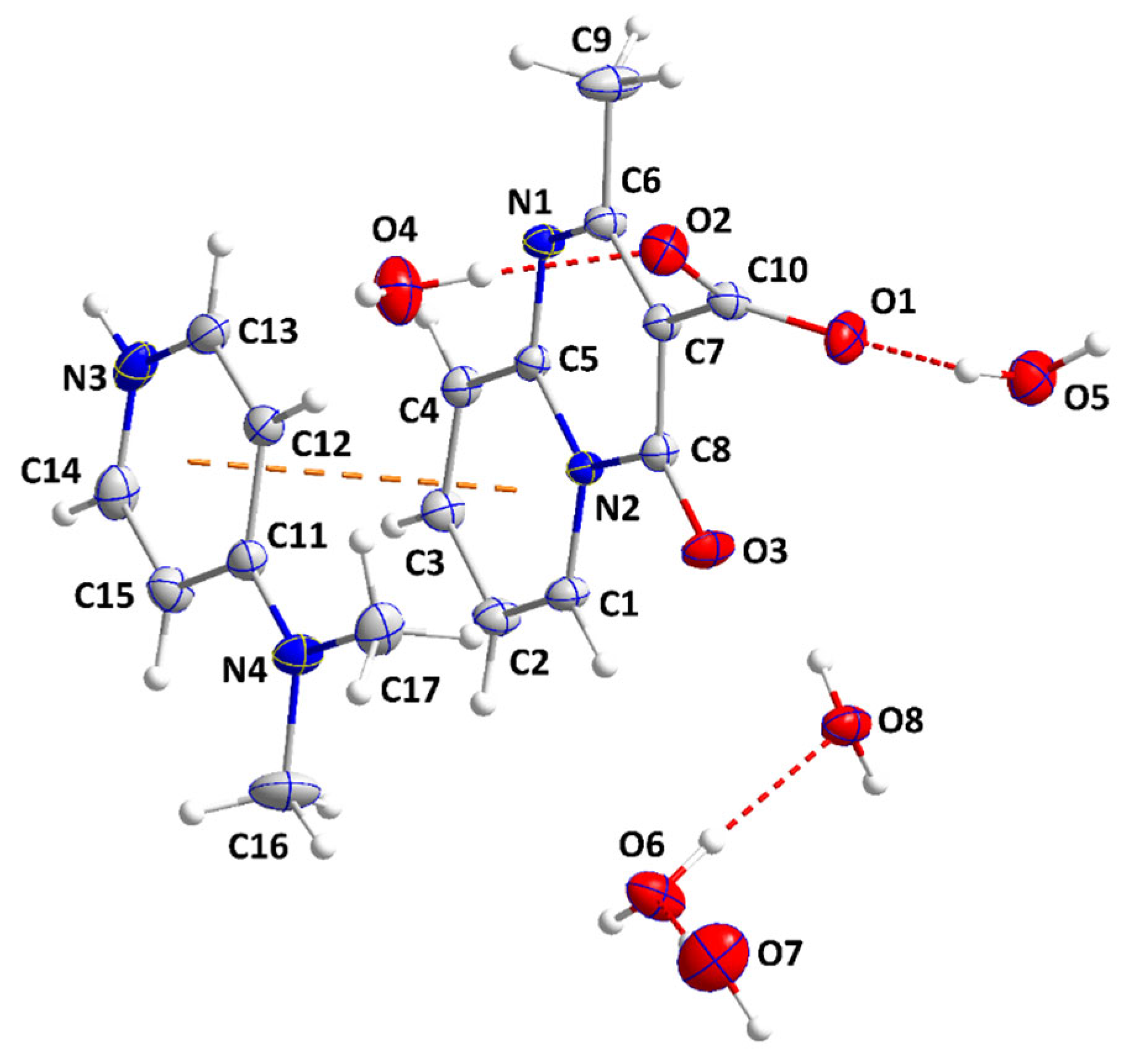
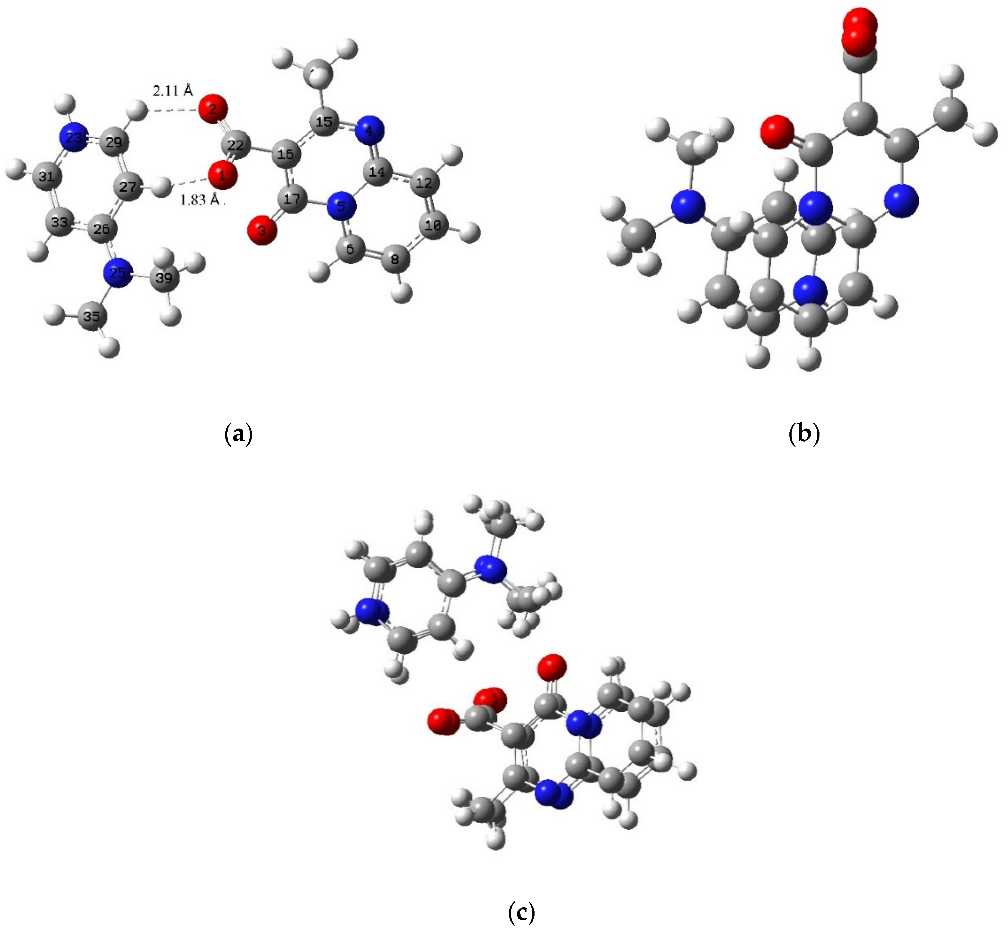
2.2. X-ray Analysis
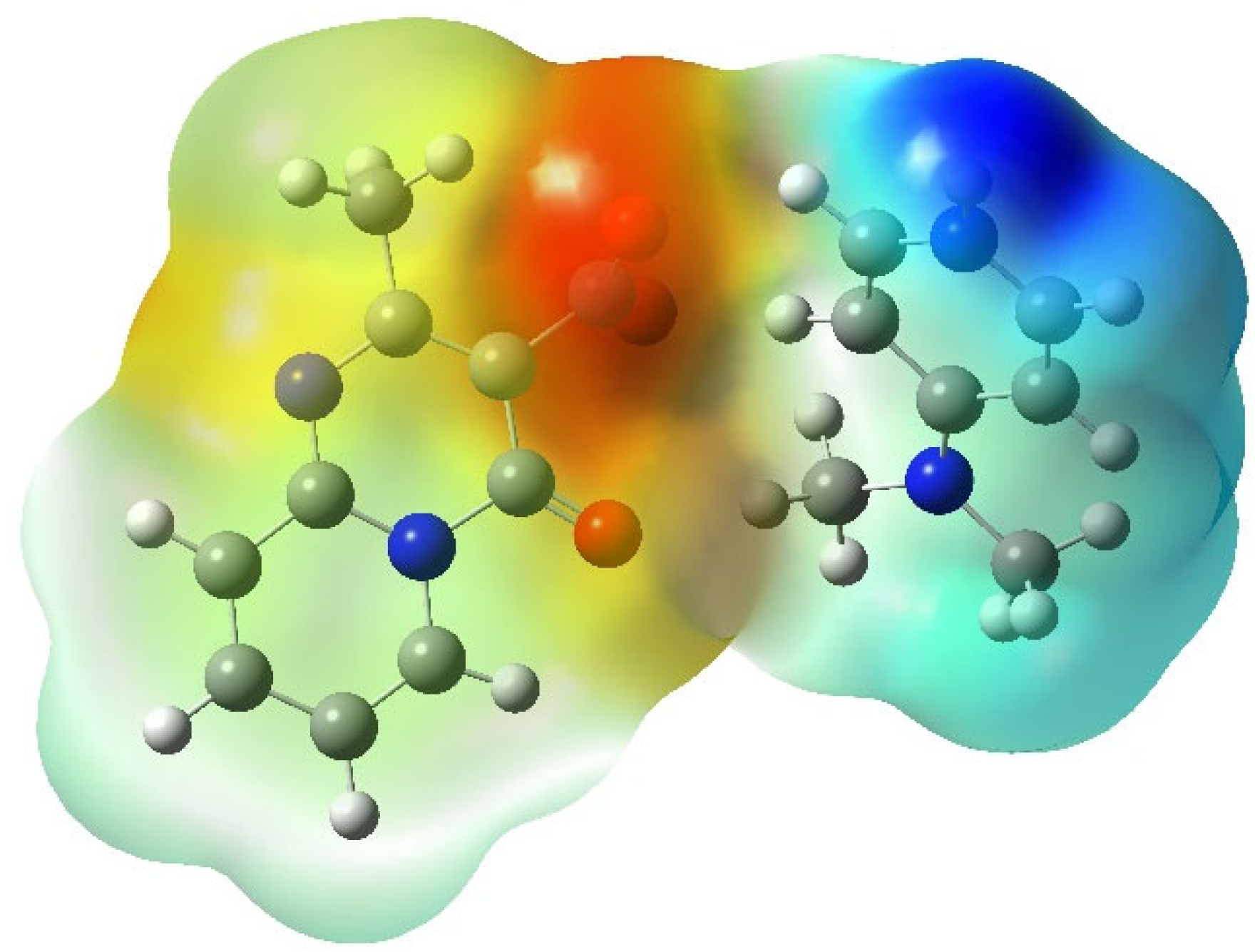
2.3. DFT Results
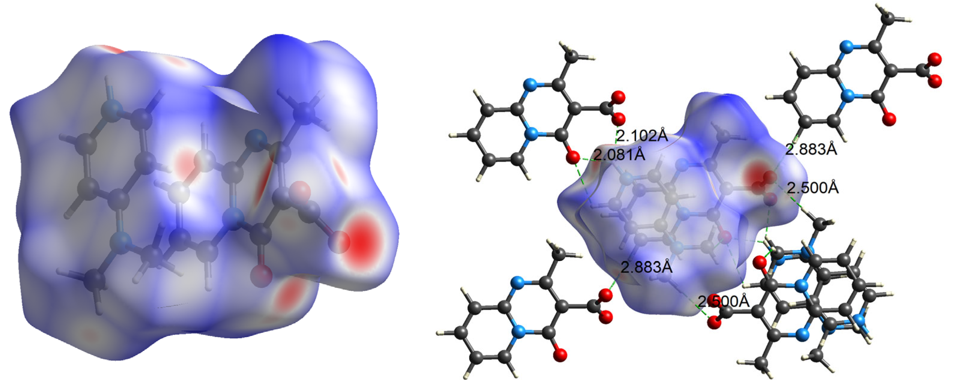
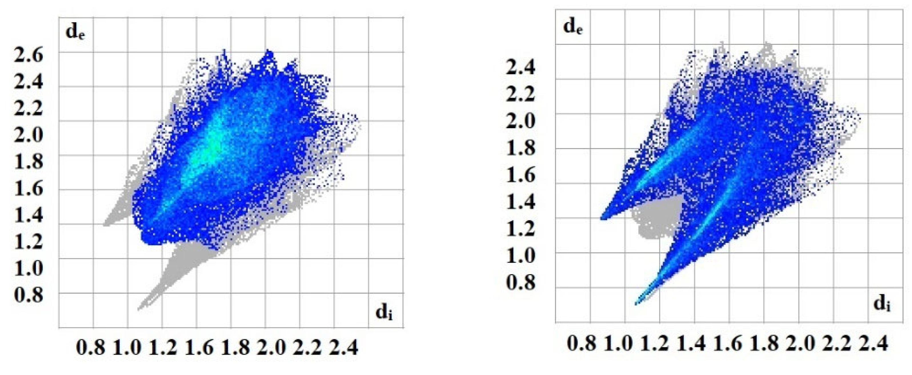
2.4. Hirshfeld Surface Analysis
2.5. ADMET and Druglikeness Prediction of 5
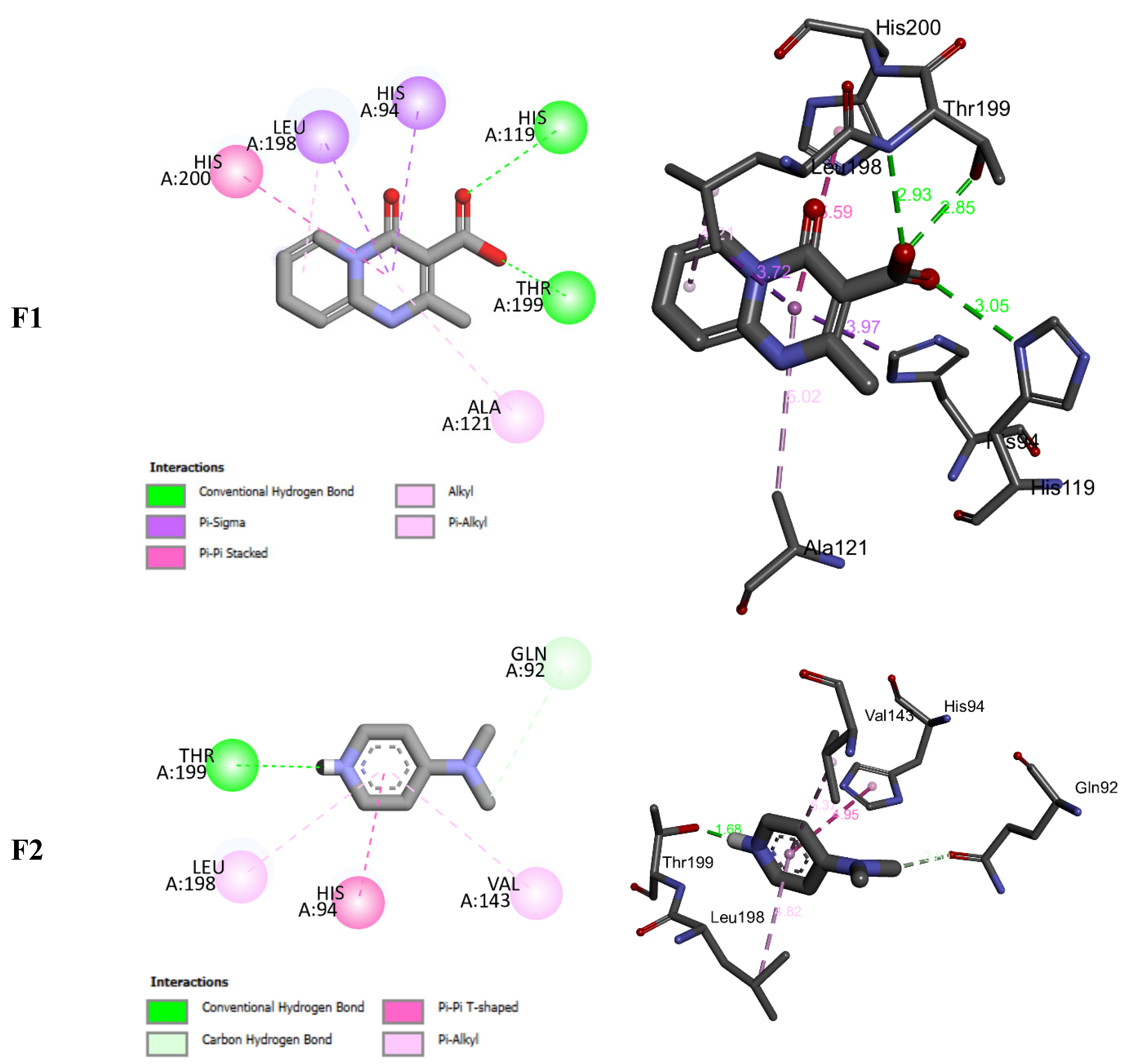
2.6. Molecular Docking Study
3. Materials and Methods
3.1. General Procedure
3.2. X-ray Crystallography
3.3. Hirshfeld Surface Analysis
3.4. DFT Calculations
3.5. In Silico ADMET and Druglikeness Properties
3.6. Molecular Docking Study
4. Conclusions
Supplementary Materials
Author Contributions
Funding
Data Availability Statement
Acknowledgments
Conflicts of Interest
Sample Availability
References
- Eftekhari-Sis, B.; Zirak, M.; Akbari, A. Arylglyoxals in synthesis of heterocyclic compounds. Chem. Rev. 2013, 113, 2958–3043. [Google Scholar] [CrossRef] [PubMed]
- Kerru, N.; Maddila, S.; Jonnalagadda, S.B. Design of carbon-carbon and carbon-heteroatom bond formation reactions under green conditions. Curr. Org. Chem. 2019, 23, 3154–3190. [Google Scholar] [CrossRef]
- Ju, Y.; Varma, R.S. Aqueous N-heterocyclization of primary amines and hydrazines with dihalides: Microwave-assisted syntheses of N-azacycloalkanes, isoindole, pyrazole, pyrazolidine, and phthalazine derivatives. J. Org. Chem. 2006, 71, 135–141. [Google Scholar] [CrossRef] [PubMed]
- Zárate-Zárate, D.; Aguilar, R.; Hernández-Benitez, R.I.; Labarrios, E.M.; Delgado, F.; Tamariz, J. Synthesis of α-ketols by functionalization of captodative alkenes and divergent preparation of heterocycles and natural products. Tetrahedron 2015, 71, 6961–6978. [Google Scholar] [CrossRef]
- Leeson, P.D.; Springthorpe, B. The influence of drug-like concepts on decision-making in medicinal chemistry. Nat. Rev. Drug Discov. 2007, 6, 881–890. [Google Scholar] [CrossRef]
- Gallop, M.A.; Barrett, R.W.; Dower, W.J.; Fodor, S.P.; Gordon, E.M. Applications of combinatorial technologies to drug discovery. 1. Background and peptide combinatorial libraries. J. Med. Chem. 1994, 37, 1233–1251. [Google Scholar] [CrossRef]
- Walsh, C.T. Nature loves nitrogen heterocycles. Tetrahedron Lett. 2015, 56, 3075–3081. [Google Scholar] [CrossRef]
- Zhang, B.; Studer, A. Recent advances in the synthesis of nitrogen heterocycles via radical cascade reactions using isonitriles as radical acceptors. Chem. Soc. Rev. 2015, 44, 3505–3521. [Google Scholar] [CrossRef]
- Pettersson, A.; Gradin, K.; Hedner, T.; Persson, B. Antihypertensive mechanism of action of ketanserin and some ketanserin analogues in the spontaneously hypertensive rat. Naunyn-Schmiedebergs Arch. Pharmacol. 1985, 329, 394–397. [Google Scholar] [CrossRef]
- Awouters, F.; Vermeire, J.; Smeyers, F.; Vermote, P.; Van Beek, R.; Niemegeers, C.J. Oral antiallergic activity in ascaris hypersensitive dogs: A study of known antihistamines and of the new compounds ramastine (R 57 959) and levocabastine (R 50 547). Drug Dev. Res. 1986, 8, 95–102. [Google Scholar] [CrossRef]
- Yanagihara, Y.; Kasai, H.; Kawashima, T.; Shida, T. Immunopharmacological studies on TBX, a new antiallergic drug (1) Inhibitory effects on passive cutaneous anaphylaxis in rats and guinea pigs. Jpn. J. Pharmacol. 1988, 48, 91–101. [Google Scholar] [CrossRef]
- Smith, R.L.; Barrett, R.J.; Sanders-Bush, E. Neurochemical and behavioral evidence that quipazine-ketanserin discrimination is mediated by serotonin2A receptor. J. Pharmacol. Exp. Ther. 1995, 275, 1050–1057. [Google Scholar]
- Kennis, L.; Bischoff, F.; Mertens, C.; Love, C. FAFV d. Keybus, MBS Pieters, AAHP Megens and JE Leysen. Bioorg. Med. Chem. Lett. 2002, 10, 71. [Google Scholar] [CrossRef]
- Varga, M.; Kapui, Z.; Bátori, S.; Nagy, L.T.; Vasvári-Debreczy, L.; Mikus, E.; Urbán-Szabó, K.; Arányi, P. A novel orally active inhibitor of HLE. Eur. J. Med. Chem. 2003, 38, 421–425. [Google Scholar] [CrossRef] [PubMed]
- Jeste, D.V.; Okamoto, A.; Napolitano, J.; Kane, J.M.; Martinez, R.A. Low incidence of persistent tardive dyskinesia in elderly patients with dementia treated with risperidone. Am. J. Psychiatry 2000, 157, 1150–1155. [Google Scholar] [CrossRef] [PubMed]
- Yao, P.; Zhai, X.; Liu, D.; Qi, B.H.; Tan, H.L.; Jin, Y.C.; Gong, P. Synthesis and Antiproliferative Activitiy of Novel Diaryl Ureas Possessing a 4H-Pyrido [1, 2-a] pyrimidin-4-one Group. Arch. Der Pharm. Int. J. Pharm. Med. Chem. 2010, 343, 17–23. [Google Scholar] [CrossRef] [PubMed]
- Hussein, M.; Huynh, V.; Hommelsheim, R.; Koenigs, R.; Nguyen, T. An efficient method for retro-Claisen-type C–C bond cleavage of diketones with tropylium catalyst. Chem. Commun. 2018, 54, 12970–12973. [Google Scholar] [CrossRef] [PubMed]
- Jukic, M.; Sterk, D.; Casar, Z. Recent advances in the retro-Claisen reaction and its synthetic applications. Curr. Org. Synth. 2012, 9, 488–512. [Google Scholar] [CrossRef]
- Lahmidi, S.; Sert, Y.; Şen, F.; El Hafi, M.; Ettahiri, W.; Gökce, H.; Essassi, E.M.; Mague, J.T.; Ucun, F. Synthesis, crystal structure, Hirshfeld surface analysis, spectral characterizations and quantum computational assessments of 1-hydroxy-3-methyl-11H-pyrido [2, 1-b] quinazolin-11-one. J. Mol. Struct. 2022, 1249, 131592. [Google Scholar] [CrossRef]
- El Hafi, M.; Lahmidi, S.; Boulhaoua, M.; El Ghayati, L.; Albalwi, H.; Anouar, E.H.; Alharthi, A.I.; Mague, J.T.; Essassi, E.M.; Lai, C.H. A new synthetic route for the preparation of 2, 2′, 5′-trimethyl-7-oxo-4, 7-dihydro-[6, 7′-bipyrazolo [1, 5-a] pyrimidine]-3, 3′-dicarbonitrile, structural elucidation, Hirshfeld surface analysis, energy framework, density functional theory and molecular docking investigations. J. Chin. Chem. Soc. 2022, 69, 717–730. [Google Scholar]
- Elotmani, B.; Elmahi, M.; Essassi, E.; Pierrot, M. 2-Méthyl-3-(3-méthyl-1H-pyrazol-5-yl) pyrido [1, 2-a] pyrimidin-4-one. Acta Crystallogr. Sect. E Struct. Rep. Online 2002, 58, o388–o389. [Google Scholar] [CrossRef]
- Bruker AXS Inc. APEX 3; Bruker Advanced X-ray Solutions: Madison, WI, USA, 2016. [Google Scholar]
- Spackman, P.R.; Turner, M.J.; McKinnon, J.J.; Wolff, S.K.; Grimwood, D.J.; Jayatilaka, D.; Spackman, M.A. CrystalExplorer: A program for Hirshfeld surface analysis, visualization and quantitative analysis of molecular crystals. J. Appl. Crystallogr. 2021, 54, 1006–1011. [Google Scholar] [CrossRef] [PubMed]
- Turner, M.; McKinnon, J.; Wolff, S.; Grimwood, D.; Spackman, P.; Jayatilaka, D.; Spackman, M. CrystalExplorer17; University of Western Australia: Crawley WA, Australia, 2017. [Google Scholar]
- Frisch, M.; Trucks, G.; Schlegel, H.; Scuseria, G.; Robb, M.; Cheeseman, J.; Scalmani, G.; Barone, V.; Petersson, G.; Nakatsuji, H. Gaussian 16; Gaussian, Inc.: Wallingford, CT, USA, 2016. [Google Scholar]
- Andersson, M.P.; Uvdal, P. New scale factors for harmonic vibrational frequencies using the B3LYP density functional method with the triple-ζ basis set 6-311+ G (d, p). J. Phys. Chem. A 2005, 109, 2937–2941. [Google Scholar] [CrossRef] [PubMed]
- Tomasi, J.; Persico, M. Molecular Interactions in Solution: An Overview of Methods Based on Continuous Distributions of the Solvent. Chem. Rev. 1994, 94, 2027–2094. [Google Scholar] [CrossRef]
- Morris, G.M.; Huey, R.; Lindstrom, W.; Sanner, M.F.; Belew, R.K.; Goodsell, D.S.; Olson, A.J. AutoDock4 and AutoDockTools4: Automated docking with selective receptor flexibility. J. Comput. Chem. 2009, 30, 2785–2791. [Google Scholar] [CrossRef]
- Srivastava, D.; Jude, K.M.; Banerjee, A.L.; Haldar, M.; Manokaran, S.; Kooren, J.; Mallik, S.; Christianson, D.W. Structural analysis of charge discrimination in the binding of inhibitors to human carbonic anhydrases I and II. J. Am. Chem. Soc. 2007, 129, 5528–5537. [Google Scholar] [CrossRef]
- Karrouchi, K.; Fettach, S.; Anouar, E.H.; Bayach, I.; Albalwi, H.; Arshad, S.; Sebbar, N.K.; Tachalait, H.; Bougrin, K.; Faouzi, M.E.A.; et al. Synthesis, Spectroscopic Characterization, DFT, Molecular Docking and Antidiabetic Activity of N-Isonicotinoyl Arylaldehyde Hydrazones. Polycycl. Aromat. Compd. 2023, 43, 1469–1481. [Google Scholar] [CrossRef]


| D—H⋯A | D—H | H⋯A | D⋯A | D—H⋯A |
|---|---|---|---|---|
| C1—H1⋯O6 | 0.95 | 2.57 | 3.2568 (11) | 130 |
| C2—H2⋯O2 i | 0.95 | 2.51 | 3.3905 (10) | 154 |
| N3—H3A⋯O1 ii | 0.91 | 2.17 | 2.9118 (10) | 138 |
| N3—H3A⋯O3 ii | 0.91 | 2.15 | 2.8662 (10) | 135 |
| C12—H12⋯O4 | 0.95 | 2.44 | 3.3663 (11) | 164 |
| C13—H13⋯O6 iii | 0.95 | 2.58 | 3.5143 (11) | 168 |
| C14—H14⋯O7 ii | 0.95 | 2.54 | 3.2957 (14) | 137 |
| O4—H4A⋯O7 iv | 0.87 | 1.89 | 2.7427 (12) | 165 |
| O4—H4B⋯O2 | 0.87 | 1.90 | 2.7713 (10) | 175 |
| O5—H5A⋯O1 | 0.87 | 1.92 | 2.7831 (10) | 170 |
| O5—H5B⋯O8 v | 0.87 | 1.93 | 2.7944 (10) | 172 |
| O5—H5C⋯O5 v | 0.87 | 1.98 | 2.8259 (15) | 164 |
| O6—H6A⋯O8 | 0.87 | 1.93 | 2.7975 (10) | 176 |
| O6—H6B⋯O1 i | 0.87 | 1.91 | 2.7705 (9) | 172 |
| O7—H7A⋯O4 vi | 0.87 | 1.95 | 2.8018 (13) | 167 |
| O7—H7B⋯O6 | 0.87 | 1.84 | 2.7058 (10) | 173 |
| O8—H8A⋯N1 vii | 0.87 | 1.98 | 2.8457 (9) | 179 |
| O8—H8B⋯O8 viii | 0.87 | 1.91 | 2.7716 (13) | 170 |
| O8—H8C⋯O5 v | 0.87 | 1.93 | 2.7944 (10) | 176 |
| Bond Lengths (Å) | Bond Angles (°) | ||||||
|---|---|---|---|---|---|---|---|
| Cal | X-ray | ΔL(Å) | Cal | X-ray | ΔA (°) | ||
| O1-C22 | 1.265 | 1.259 (4) | 0.01 | O1-C22-O2 | 125.15 | 128.43 (8) | 3.29 |
| O2-C22 | 1.244 | 1.259 (2) | 0.01 | O1-C22-C16 | 125.15 | 114.59 (9) | 10.55 |
| C16-C22 | 1.514 | 1.530 (3) | 0.02 | O3-C17-C16 | 127.44 | 128.82 (6) | 1.39 |
| O3-C17 | 1.233 | 1.233 (5) | 0.00 | C17-N5-C14 | 121.18 | 121.31 (8) | 0.13 |
| N4-C14 | 1.330 | 1.318 (7) | 0.01 | N5-C14-N4 | 122.43 | 122.61 (9) | 0.19 |
| N5-C14 | 1.381 | 1.398 (5) | 0.02 | C26-N25-C39 | 123.00 | 119.66 (9) | 3.33 |
| N23-C29 | 1.351 | 1.365 (4) | 0.01 | O1-C22-C16-C15 | −124.14 | −118.98 (6) | 5.15 |
| N23-C31 | 1.346 | 1.359 (0) | 0.01 | O1-C22-C16-C17 | 57.03 | 60.86 (8) | 3.83 |
| N25-C26 | 1.338 | 1.346 (6) | 0.01 | O3-C17-C16-C22 | −2.97 | 3.78 (4) | 6.75 |
| N25-C35 | 1.462 | 1.458 (6) | 0.00 | C6-N5-C17-O3 | 3.41 | −2.28 (6) | 5.70 |
| N25-C39 | 1.459 | 1.477 (0) | 0.02 | C27-C26-N25-C39 | −1.99 | −1.99 (2) | 0.00 |
| C26-C27 | 1.424 | 1.422 (3) | 0.00 | C27-C26-N25-C35 | −176.34 | −176.34 (1) | 0.00 |
| C27-C29 | 1.367 | 1.366 (2) | 0.00 | C33-C26-N25-C35 | 4.16 | 4.15 (8) | 0.00 |
| Free Binding Energy (kcal/mol) | H-Bonds (HBs) | Number of Closest Residues to the Docked Ligand in the Active Site * | |
|---|---|---|---|
| F1 | −5.54 | 3 | 7 |
| F2 | −3.72 | 1 | 5 |
| Chemical Formula | C10H7N2O3·C7H11N2·5(H2O) |
|---|---|
| Mr | 416.43 |
| Crystal system, space group | Triclinic, |
| Temperature (K) | 150 |
| a, b, c (Å) | 9.4302 (3), 9.7094 (3), 12.9484 (4) |
| α, β, γ (°) | 77.686 (2), 71.945 (2), 66.414 (1) |
| V (Å3) | 1027.60 (6) |
| Z | 2 |
| Radiation type | Mo Kα |
| µ (mm−1) | 0.11 |
| Crystal size (mm) | 0.28 × 0.27 × 0.15 |
| Data collection | |
| Diffractometer | Bruker D8 QUEST PHOTON 3 diffractometer |
| Absorption correction | Numerical SADABS (Krause et al., 2015) |
| Tmin, Tmax | 0.94, 0.98 |
| No. of measured, independent andobserved [I > 2σ(I)] reflections | 68,565, 7857, 6719 |
| Rint | 0.026 |
| (sin θ/λ)max (Å−1) | 0.771 |
| Refinement | |
| R[F2 > 2σ(F2)], wR(F2), S | 0.042, 0.119, 1.03 |
| No. of reflections | 7857 |
| No. of parameters | 267 |
| H-atom treatment | H-atom parameters constrained |
| Δρmax, Δρmin (e Å−3) | 0.49, −0.25 |
Disclaimer/Publisher’s Note: The statements, opinions and data contained in all publications are solely those of the individual author(s) and contributor(s) and not of MDPI and/or the editor(s). MDPI and/or the editor(s) disclaim responsibility for any injury to people or property resulting from any ideas, methods, instructions or products referred to in the content. |
© 2023 by the authors. Licensee MDPI, Basel, Switzerland. This article is an open access article distributed under the terms and conditions of the Creative Commons Attribution (CC BY) license (https://creativecommons.org/licenses/by/4.0/).
Share and Cite
Lahmidi, S.; Anouar, E.H.; Ettahiri, W.; El Hafi, M.; Lazrak, F.; Alanazi, M.M.; Alanazi, A.S.; Hefnawy, M.; Essassi, E.M.; Mague, J.T. Nanoarchitectonics and Molecular Docking of 4-(Dimethylamino)Pyridin-1-Ium 2-3 Methyl-4-Oxo-Pyri-Do[1,2-a]Pyrimidine-3-Carboxylate. Crystals 2023, 13, 1333. https://doi.org/10.3390/cryst13091333
Lahmidi S, Anouar EH, Ettahiri W, El Hafi M, Lazrak F, Alanazi MM, Alanazi AS, Hefnawy M, Essassi EM, Mague JT. Nanoarchitectonics and Molecular Docking of 4-(Dimethylamino)Pyridin-1-Ium 2-3 Methyl-4-Oxo-Pyri-Do[1,2-a]Pyrimidine-3-Carboxylate. Crystals. 2023; 13(9):1333. https://doi.org/10.3390/cryst13091333
Chicago/Turabian StyleLahmidi, Sanae, El Hassane Anouar, Walid Ettahiri, Mohamed El Hafi, Fatima Lazrak, Mohammed M. Alanazi, Ashwag S. Alanazi, Mohamed Hefnawy, El Mokhtar Essassi, and Joel T. Mague. 2023. "Nanoarchitectonics and Molecular Docking of 4-(Dimethylamino)Pyridin-1-Ium 2-3 Methyl-4-Oxo-Pyri-Do[1,2-a]Pyrimidine-3-Carboxylate" Crystals 13, no. 9: 1333. https://doi.org/10.3390/cryst13091333
APA StyleLahmidi, S., Anouar, E. H., Ettahiri, W., El Hafi, M., Lazrak, F., Alanazi, M. M., Alanazi, A. S., Hefnawy, M., Essassi, E. M., & Mague, J. T. (2023). Nanoarchitectonics and Molecular Docking of 4-(Dimethylamino)Pyridin-1-Ium 2-3 Methyl-4-Oxo-Pyri-Do[1,2-a]Pyrimidine-3-Carboxylate. Crystals, 13(9), 1333. https://doi.org/10.3390/cryst13091333