Article

Bulk Polystyrene-BaF2 Composite Scintillators for Highly Efficient Radiation Detection

1
Shanghai Institute of Ceramics, Chinese Academy of Sciences, Shanghai 201899, China
2
Collaborative Innovation Center of IFSA, Shanghai Jiao Tong University, Shanghai 200240, China
*
Author to whom correspondence should be addressed.
Crystals 2023, 13(9), 1334; https://doi.org/10.3390/cryst13091334
Submission received: 8 August 2023 / Revised: 24 August 2023 / Accepted: 29 August 2023 / Published: 31 August 2023
(This article belongs to the Special Issue Photoelectric Functional Crystals)

Abstract

:
Organic–inorganic composite scintillators, demonstrating advantages of easy large-area preparation and a high detection efficiency, have shown enormous potential application prospects in radiation detection and imaging. In this study, bulk polystyrene (PS) composite scintillators were successfully prepared by embedding inorganic BaF2 particles with a loading amount of up to 80 wt% during the polymerization process of the plastic scintillator. The inorganic BaF2 particles were uniformly dispersed in the organic matrix. With the increase of the loading amounts of BaF2 particles, the X-ray-excited luminescence intensity of the PS-BaF2 composite scintillators was about eight times higher than that of the commercial pure plastic scintillator. The scintillation counts under the gamma ray (59.5 KeV) irradiation also showed that the detection efficiency was obviously enhanced by BaF2 particle loading. More importantly, their scintillation pulse retains the decay kinetics of the organic matrix without loading the slow-decay component of BaF2. This work provides a promising solution for the application of the PS-BaF2 composite scintillator in high-efficiency radiation detection and large-area imaging.

1. Introduction

Scintillators have been extensively investigated and widely used in a variety of fields, such as medical imaging, high-energy physics, security inspection, and geophysical exploration [1]. Technical and cost issues have become increasingly important when different scintillators are used. Among them, large-area scintillators with high stopping power demonstrate great potential for application in radiation detection and imaging [2,3,4,5,6]. Scintillators can be generally classified into organic scintillators and inorganic scintillators. Organic scintillators are attractive due to their fast response, low cost, and feasibility of large size preparation. However, their poor stopping power limits their applications in high-energy ionizing radiation [7,8]. Comparatively, inorganic scintillators have excellent light output, energy resolution, as well as a high detection efficiency for energetic ionizing radiation. Nevertheless, the growth of large crystals is difficult and costly, and the shape of crystals is limited by their growth habits [9,10,11].
Organic–inorganic composite scintillators are one of the most effective solutions to meet the needs of efficient, low-cost, and large-area radiation applications. Composite scintillators combine the high efficiency of inorganic scintillators and the cost-effectiveness of organic scintillators, ensuring an optimal balance between the good scintillation quality and the reasonable price of the scintillator [12]. Due to the loading of inorganic powder, composite scintillators usually have a better effective absorption capacity of high-energy rays and a higher luminous intensity than the pure organic scintillators [13,14,15,16,17]. In addition, the properties of organic–inorganic composite scintillators can be adjusted by controlling the composition, content, and morphology of the particle reinforcement, various processing techniques, or the polymer matrix. Incorporating inorganic particles in a polymer matrix and molding into a desired shape is the most common method to fabricate organic–inorganic composite scintillators. This method retains the functionality of both the constituents of the composite and has also been found to generate new properties that do not exist in either of the materials in a few cases.
Ensuring homogeneous mixing of inorganic particles into the organic matrix is a major challenge in preparing composite scintillators. It is difficult to prevent agglomeration in the polymer matrix while maintaining the transparency of the polymer. Agglomeration increases the light scattering and limits the loading amount of the inorganic particles into the organic matrix. Hamroun et al. prepared composites with a thickness of 200 µm by embedding Lu2SiO5:Ce3+ nanoparticles in the PS and polylactic acid polymer matrix [18]. Demkiv et al. added BaF2, SrF2, CeF3, and LaPO4-Pr particles to the PS plastic scintillator, and the luminous intensity was enhanced [19,20,21,22,23]. Rajakrishna et al. loaded B2O3, LiF, Gd2O2S:Tb, Gd2O3, and Gd(BO2)3:Tb into plastic scintillators and prepared PS sheets with a size of 200 × 150 mm2 and a thickness of 250 ± 50 µm by using multiple coatings [24]. The detection efficiency of low-energy γ-ray and thermal neutrons was improved.
However, they mainly used a co-soluble mixture of C2H4Cl2 + CCl4 or toluene solution to dissolve the plastic scintillator, and the organic–inorganic composite scintillator film was obtained after ultrasonic dispersion. Obviously, it is difficult to obtain a uniform composite scintillator in this way. When the loading amount of inorganic scintillation powder is high, or the organic matrix is viscous, ultrasonic dispersion is difficult to evenly disperse the powder in the organic matrix. When the viscosity of the matrix is very low, the powder is easy to precipitate. This is likely to cause powder agglomeration, which makes the light transmission of the composite scintillator worse, and thus reduces its performance. Further, bubbles easily form during the solidification process after the organic matrix has dissolved, and the film thickness is limited. Thus, these scintillators have lower stopping power against high-energy photons [25,26,27,28]. This is because the average free range of X-ray or gamma rays in solids varies between a few tens of micrometers and a few tens of centimeters, depending on the magnitude of their energy. Moreover, the luminescent center of plastic scintillators tends to be damaged, and then causes a reduction of the luminescence performance after the above-mentioned dissolving process.
In this work, we developed a novel method for loading inorganic BaF2 powder particles into PS plastic scintillators. The high-performance bulk organic–inorganic composite scintillators were prepared by combining in situ embedding of inorganic particles in the plastic scintillation polymerization process with a hybrid defoaming process. The loading amount of BaF2 particles was up to 80 wt% without destroying the original luminescence properties of the plastic scintillator. The inorganic BaF2 particles were uniformly dispersed in the organic matrix, characterized by a scanning electron microscope (SEM). In addition, the effects of the inorganic component amount and thickness on X-ray-excited luminescence (XEL) were studied in detail. The XEL intensity, detection efficiency, and decay kinetics curves of the composite scintillator were compared with those of the commercial pure plastic scintillator.

2. Experimental Process

2.1. Preparation of Composite Scintillators

Commercial BaF2 particles (Suzhou Putin Vacuum Technology Co., Ltd. (Suzhou, China), >99.99%) with high purity were sieved by a 200 mesh and used as raw materials. Then, the BaF2 powders were dried in a vacuum oven at 200 °C for 120 min to eliminate the moisture. Finally, the pellets were naturally cooled to room temperature before removal to minimize the effects of deliquescence.
The plastic scintillator was prepared by the polymerization of styrene monomers and the addition of luminous impurity p-terphenyl and wave shifter 1,4-bis (5-phenyloxazol-2-yl) benzene (POPOP). Commercial styrene monomers contain certain amounts of inhibitors, such as hydroquinone, and may also be mixed with small amounts of air, water, and other impurities. In order to remove them, the styrene monomer (Aladdin, >99.5%) was distilled under reduced pressure to remove the polymerization inhibitor, air, moisture, and other impurities.
Styrene monomers, p-terphenyl (Aladdin, >99%) and POPOP (Aladdin, >97%), in amounts of 1 wt% and 0.1 wt%, were added to the glass bottle. It was then sealed in a nitrogen atmosphere glove box to prevent oxygen from weakening the performance of the plastic scintillator. Then, ultrasonic treatment was performed for 30 min to ensure full dissolution of p-terphenyl and POPOP in the styrene monomers. The temperature was raised to 50 °C at a rate of 5 °C/h, and the reaction lasted for 15 h. At this time, the solution was pre-polymerized with very low fluidity. Then, BaF2 particles were added into the bottle, opened in the glove box mentioned above, and the bottle was sealed again. The above process keeps the BaF2 particles from settling to the bottom during subsequent curing. Of course, it is also not permissible to maintain the temperature for long periods of time to avoid excessive pre-polymerization, resulting in uneven dispersion of the powder in the organic matrix during the mixing process. Next, the mixture was transferred into a mixer with a defoaming function at a rotation speed of 2000~2500 r/min. After stirring for 2 min, the mixture was heated to 85 °C, and then held at that temperature for 2 days. Both the heating and cooling rates were 5 °C/h. These slow heating and cooling rates can prevent bubbles from appearing in the composite scintillator. PS-BaF2 composite scintillator samples were obtained after cutting and polishing. For comparison, we used a commercial plastic scintillator, HND-S2 from Beijing Gaonengkedi company (Beijing, China), as a comparison sample.

2.2. Characterization Methods

SEM measurements of BaF2 particles were carried out using a field-emission scanning electron microscope SU8220 (HITACHI) with an energy-dispersive X-ray spectroscopy (EDS) accessory. The morphology of composite scintillators has been carried out by using a high-resolution field-emission scanning electron microscope, Verios G (FEI). The crystalline structure of BaF2 particles was investigated by the X-ray diffraction (XRD) technique using a Germany Bruker D8 ADVANCE, with Cu Kα radiation operated at 40 kV and 30 mA, a scan range of 10~80°, and a scan step of 0.02°. The Fourier transform infrared spectroscopy (FTIR) spectrum was obtained using a Spotlight 400 spectrometer (Perkin Elmer, Waltham, MA, USA). Photoluminescence (PL) was measured using an Edinburgh Instruments (EI) FLS920 fluorescence spectrometer. For the XEL measurements, samples were placed in the sample compartment of an Edinburgh instrument FLS920 fluorescence spectrometer. X-rays generated by an Amptek Mini-X-OEM X-ray tube operated at 40 V and 80 µA were used to excite the sample. The X-ray was perpendicular to the sample surface. The scintillation light was collected by a R928P photomultiplier tube (PMT) and analyzed by an EI FLS920 fluorescence spectrometer. The UV-VIS optical transmittance was measured using a Perkin Elmer (PE) Lambda 950 spectrometer. Pulse height spectra (PHS) of composite scintillators were measured with a shaping time of 0.5 µs by using a Beijing Hamamatsu CR160-01 PMT, and γ-ray (241Am source, 59.5 KeV). The luminescence decay kinetics measurements of composite scintillators were conducted under the excitation of a 137Cs source. Scintillation pulses from a Hamamatsu R2059 PMT were recorded by the Tektronix oscilloscope, which have a sampling rate of 5 Gs/s and a 1 GHz bandwidth.

3. Results

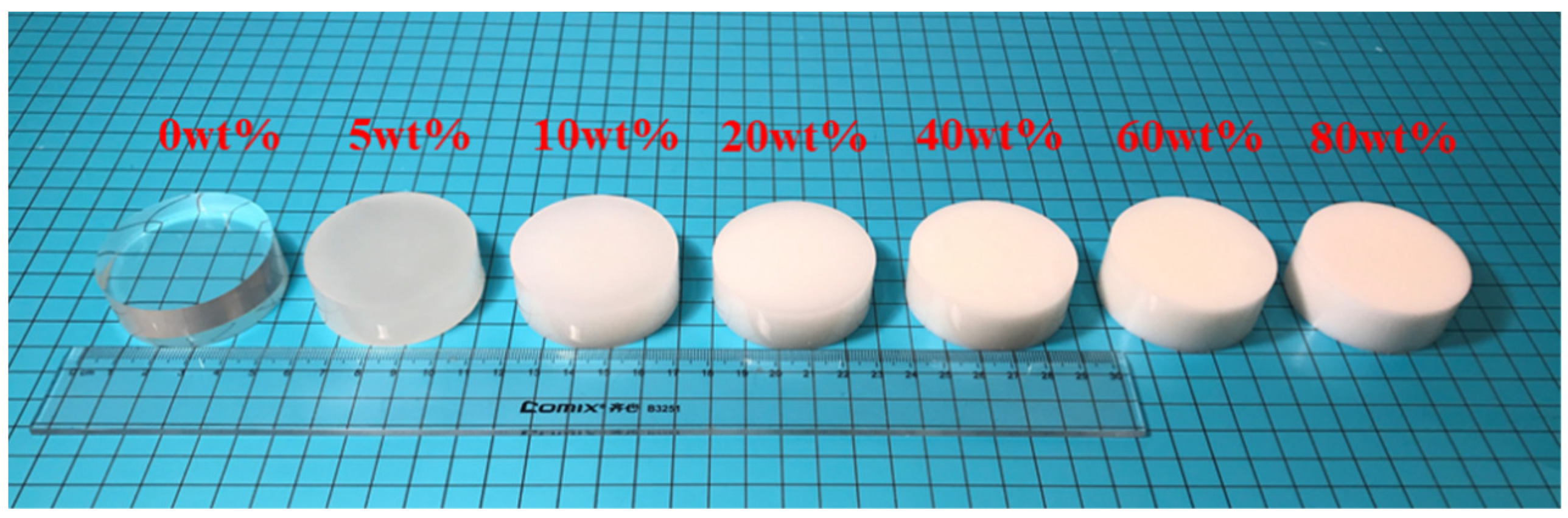
3.1. Composite Scintillators

Composite scintillators with different loading amounts of BaF2 particles were prepared, while the amounts of p-terphenyl and POPOP remained unchanged. Samples were polished into cylinders with a thickness of 20 mm and a diameter of 50 mm (Figure 1). It was found that these bulk composite scintillators possess excellent uniformity. Moreover, with the increase of the BaF2 addition, the transparency of the composite scintillator gradually decreased, changing from transparent to opaque. Figure 2 shows a series of square composite scintillators with different thicknesses, from 0.5 mm to 4 mm, and different loading amounts of BaF2 particles.
Figure S1 shows the XRD pattern of the BaF2 particles. The peak positions of the XRD pattern agree well with the data reported in the JCPDS standard card (85–1341) of pure BaF2. The SEM image of BaF2 particles is presented in Figure S2. The particle size was relatively uniform, about 2 µm. The results of the EDS analysis are shown in Figure S3 and Table S1. No other impurity elements were found, except Ba and F.
To demonstrate the dispersion uniformity of the BaF2 particles in our method, we performed SEM morphological characterization on composite scintillator samples with different BaF2 particle loading amounts. Figure 3a–d show the SEM images of composite scintillators loaded with BaF2 particles of 0 wt%, 5 wt%, 20 wt%, and 80 wt%, respectively. The dispersion of BaF2 particles in the organic matrix was relatively uniform. Even when the loading concentration reached 80 wt%, the dispersion of BaF2 particles in the PS matrix was still uniform. For comparison, we prepared PS-BaF2 composite scintillators with 20 wt% loading amounts under ultrasonic dispersion technology after pre-polymerization, and typical PS-BaF2 composite samples are shown in Figure S4. It can be seen that the powder was not evenly dispersed in the organic matrix and exhibited a severe agglomeration phenomenon (Figure 3e). This agglomeration phenomenon will seriously affect the uniformity of the scintillation performance, which limits its application. Figure 3f–i show the EDS element mapping corresponding to different loading concentrations, in which cyan, red, green, and yellow represent the elements C, O, Ba, and F, respectively. Figure 3j shows EDS mapping of PS-BaF2 composite scintillators with 20 wt% loading amounts under ultrasonic dispersion. It is clear that the BaF2 particles were uniformly embedded in the organic matrix using the new method, compared with the sample prepared through ultrasonic dispersion technology after pre-polymerization.
FTIR was performed to investigate the effect of inorganic powder embedding on matrix structure changes and polymerization, as shown in Figure 4. The peaks at 3100~3000 cm−1 were assigned to the C-H stretching vibration of olefins. However, there was no C=C stretching vibration of the olefin in the range of 1680~1620 cm−1, indicating that it may be a polymer. The absorption peaks at 3000~2800 cm−1 demonstrated the symmetric and asymmetric stretching vibrations of C-H in the structure. There were also obvious absorption peaks near 1495 cm−1 and 1453 cm−1, which can be inferred as the bending vibrations of C-H. Additionally, there was an absorption peak between 1190 cm−1 and 840 cm−1, indicating the vibration of the C-C skeleton, but it did not have strong specificity. There was an obvious absorption peak around 1600 cm−1, which can be inferred as the characteristic absorption peak of the benzene ring skeleton. The monadic substitution of the benzene ring was in the range of 770~650 cm−1, which is consistent with 752 cm−1 and 693 cm−1 in the diagram [29]. The above results have the same characteristics as polystyrene. Moreover, the characteristic peaks of the composite scintillator overlapped with those of the pure polystyrene plastic scintillator, and there was neither a peak shift nor new characteristic peaks. Therefore, we believe that the loading of BaF2 particles has no effect on the polymerization process of styrene.
The optical transmittance of the samples with the same dimensions in the normal direction are shown in Figure 5. It was observed that the transmittance decreased with an increase of the loading amount, ascribed to the light scattering with the increased amount of BaF2 particles. On the other hand, the refractive index of BaF2 (n = 1.47) is slightly different from that of the PS matrix (n = 1.59), which also led to a decrease in transparency due to the birefringent effect. The dashed line in Figure 5 shows the XEL spectrum of the PS-BaF2 composite scintillator with the 10 wt% loading amount of BaF2 particles, and the maximum of the emission peak is 420 nm.
The emission-weighted longitudinal transmittance (EWLT) at 420 nm is defined as:
E W L T = L T ( λ ) E m ( λ ) d λ E m ( λ ) d λ
where LT(λ) and Em(λ) are the longitudinal transmittance and the emission at wavelength (λ). The EWLT value decreased from 29.25% to 6.04% as the BaF2 addition increased from 5 wt% to 80 wt%.

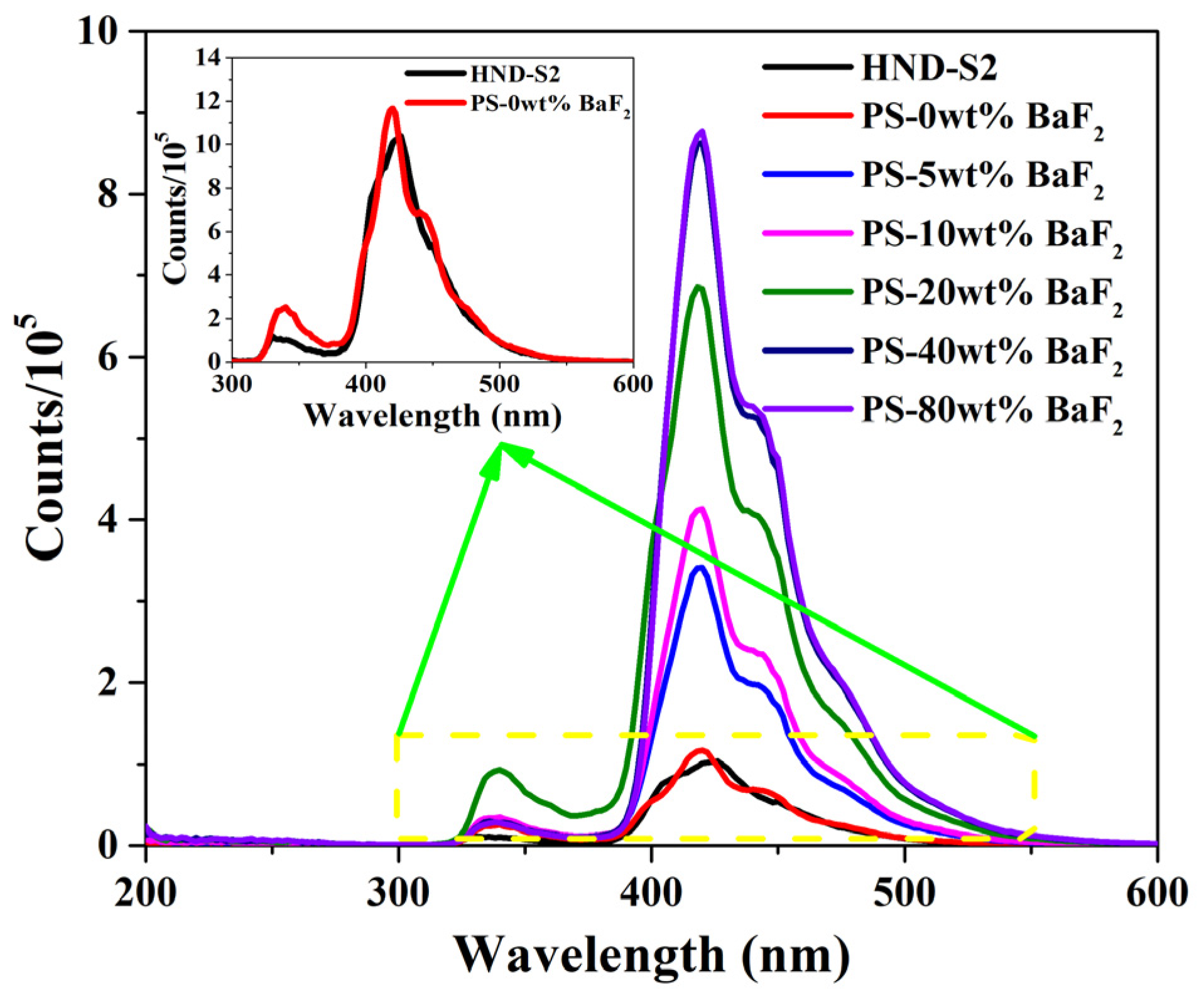
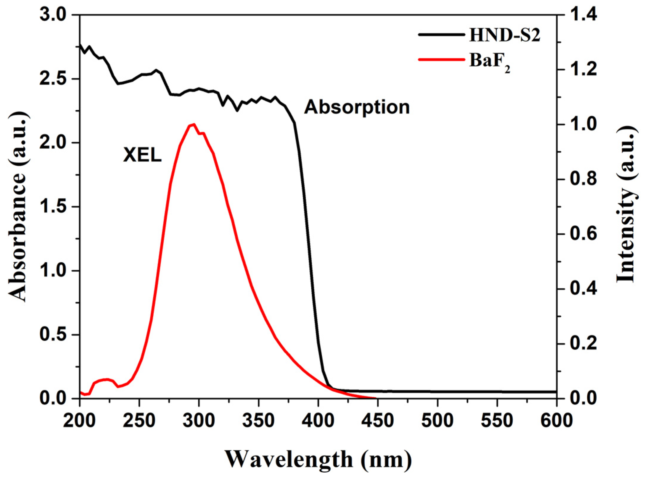
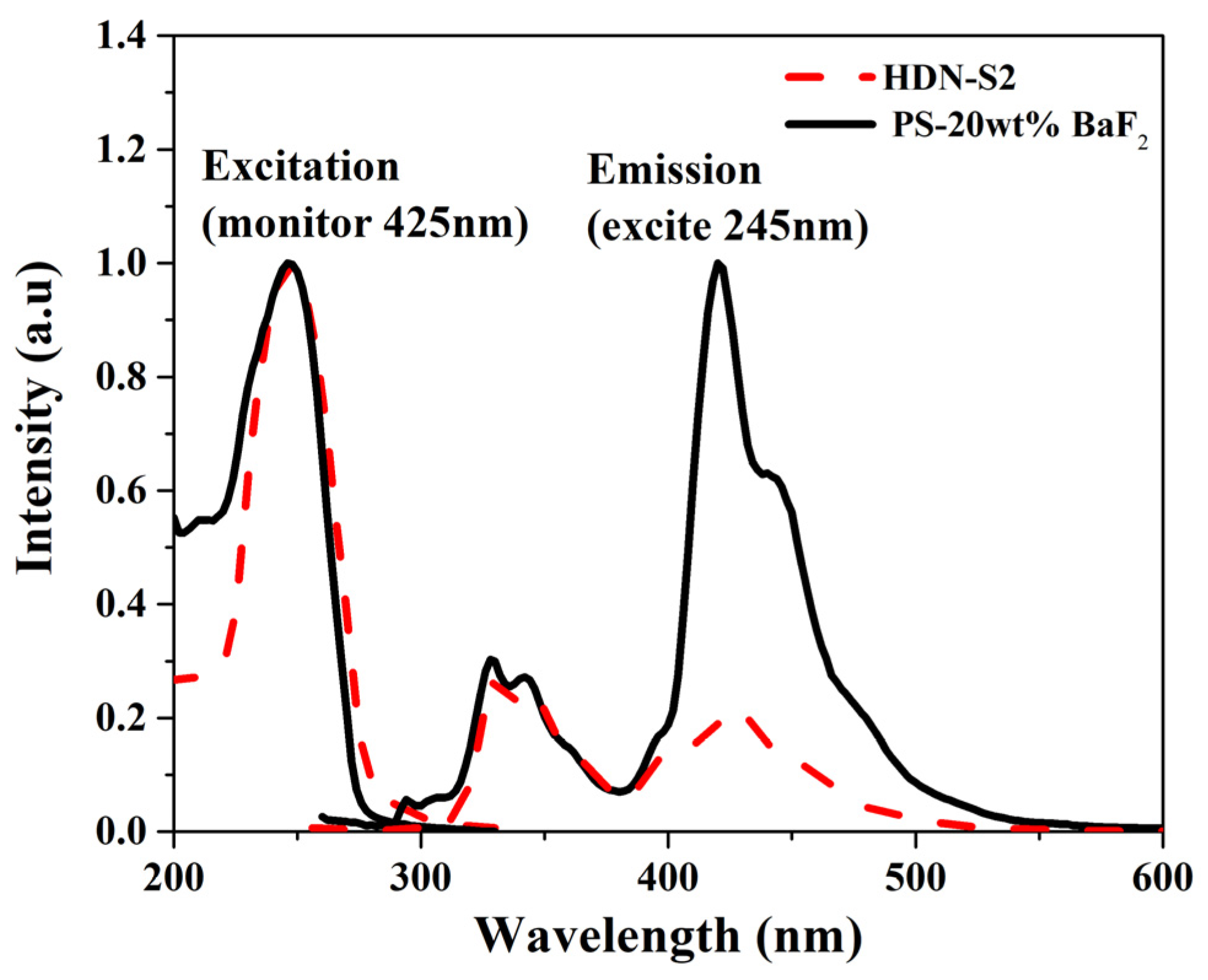
3.2. Luminescence Properties

To further investigate the luminescence properties of the prepared bulk composite scintillators, XEL and PL characterization were investigated. As the loading amount of BaF2 particles increased from 0 wt% to 80 wt%, the XEL intensity gradually increased (Figure 6). The luminescent intensity of the composite scintillator with 80 wt% BaF2 particles is about 8 times that of commercial HND-S2. The inset in Figure 6 shows that the luminescent intensity of the pure plastic scintillator without BaF2 loading is basically the same as that of HND-S2. Two emission bands with the maxima at 350 nm and 425 nm were observed in the XEL spectra of the composite scintillator, which are consistent with the pure plastic scintillator. Therefore, it is believed that the addition of BaF2 particles does not introduce deviation or change of the XEL luminescence peak, but the luminescence intensity significantly increases.
The PL and PLE spectra of the composite scintillator with 20 wt% BaF2 particles and the commercial pure plastic scintillator are shown in Figure 7. The position of the emission and excitation peaks was not changed by adding BaF2 powder to the plastic scintillator. Though the composite scintillator showed similar emission characteristics to the pure plastic scintillator, the emission intensity at 425 nm of the composite scintillator was significantly enhanced. The sensitive wavelength maximum of common photodetectors, such as PMT with the Bialkali cathode, is close to 425 nm. Compared with the emission of pure BaF2 at 310 nm and 220 nm, the enhancement is very beneficial for highly efficient radiation detection.

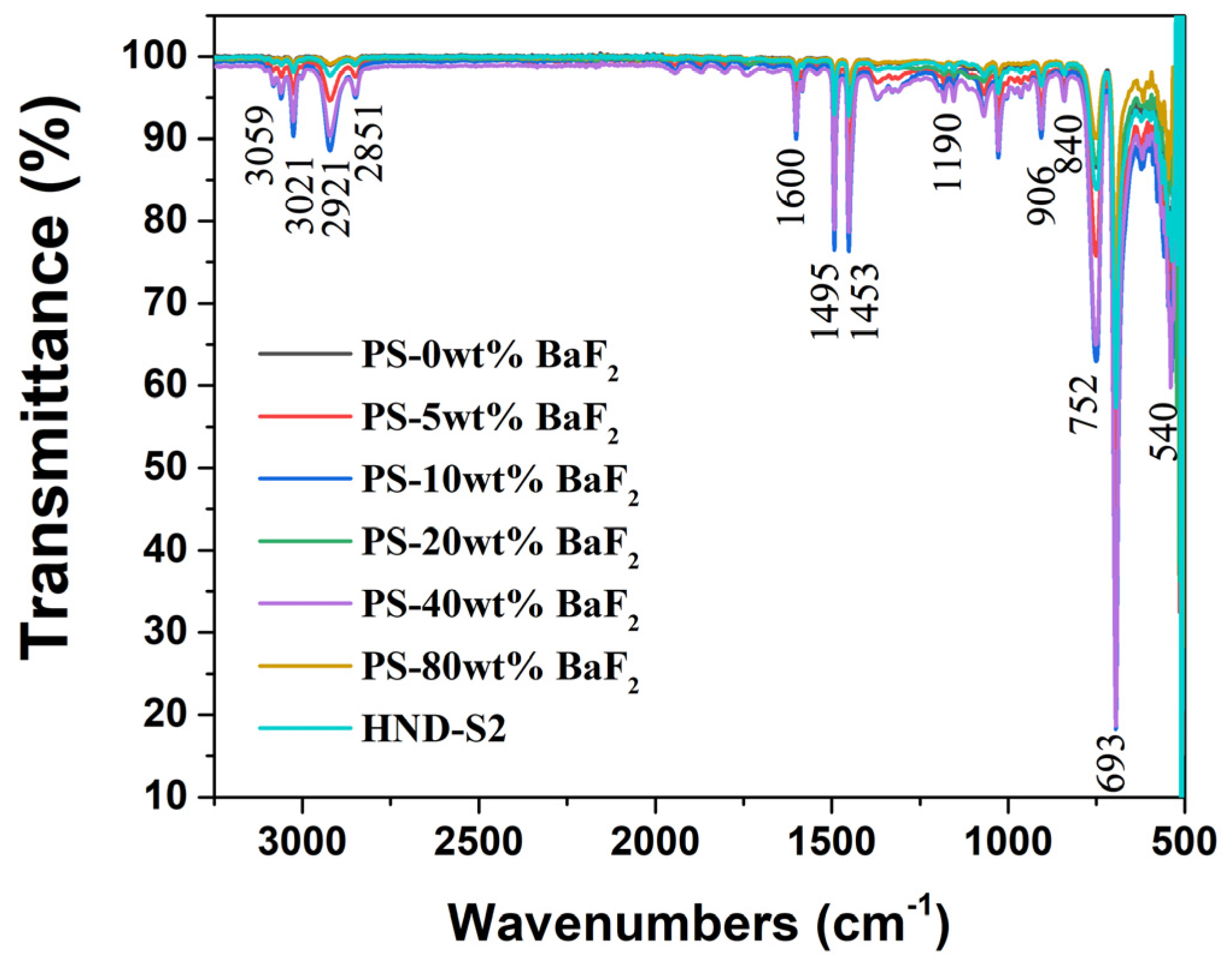
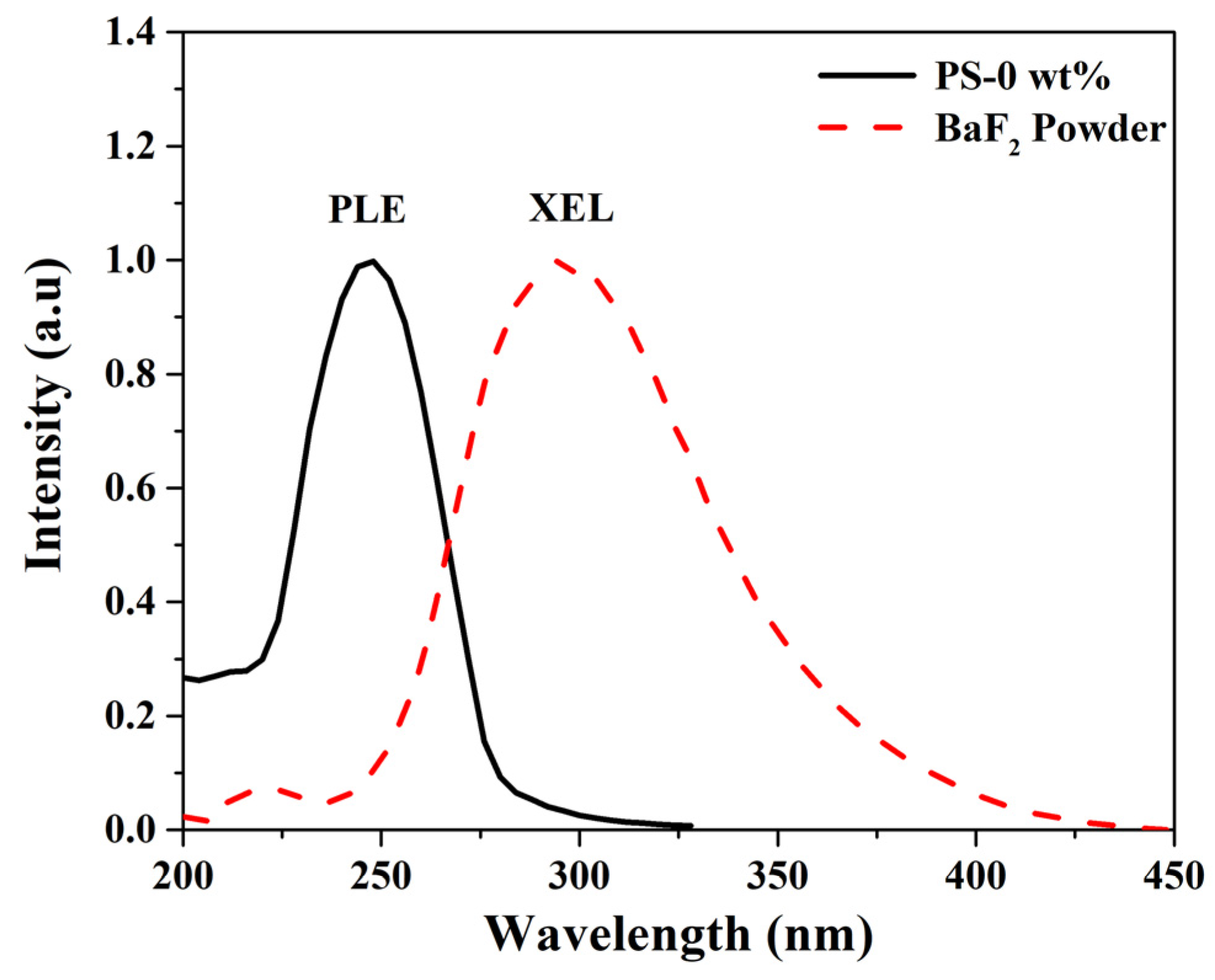
3.3. Mechanism of Luminescence Enhancement

To investigate the correlation of the stopping power to the BaF2 particle loading amounts and sample thickness, the pulse height spectra (PHS) under the gamma ray (59.5 KeV) radiation from an 241Am source were collected, using the Teflon reflection layer to resist the α particles. Figure 8 shows the PHS of different thicknesses and BaF2 amounts. The Compton edge gradually increased to a higher channel range with the increase of the BaF2 loading amounts. The trend remained unchanged until the addition was increased to 40 wt%, and then the Compton edge shifted to a lower channel when the loading amount was increased to 80 wt%. The possible reason for this is that the transmittance of the composite scintillator significantly decreased at 80 wt% loading amounts, as shown in Figure 5, and then the transmission of scintillation photons was reduced. The influence of transmittance, other than the loading amount, is the dominant factor for emission weakening. Figure 9 shows the gross count rate as a function with BaF2 loading amounts and thickness. The gross count rate of each sample was obtained by dividing the gross area by the collection time. The dramatically increased counts were collected at higher BaF2 loading amounts and thicknesses. It indicates that the stopping power of the composite scintillator increases, and more high-energy photons can be deposited in the plastic scintillator to excite the p-terphenyl to emit photons. This is the main reason for the enhanced luminescence of the composite scintillator after adding BaF2 particles.
In order to reveal the effect of thickness on the luminescence properties, the XEL intensity of samples with different thicknesses and BaF2 amounts was carried out, as shown in Figure 10. With the increase of the thickness and amounts, the luminous intensity gradually increased. The trend remained unchanged until the amount increased to 80 wt% and the thickness increased to 1 mm. When the thickness reached 2 mm, it showed a decreasing trend, and the linear relationship of the XEL luminescence intensity with the increasing BaF2 loading amount was changed as the thickness increased. When the thickness was greater than 1 mm, the 40 wt% loading amount had a higher luminescence intensity. This is mainly because the increase in the thickness and concentration will increase the photon yield, but at the same time it will also reduce the transparency of the sample. This will cause the photon generated by BaF2 under X-ray excitation to become difficult to transfer into the matrix, indicating the competition between the two aforementioned mechanisms. When the loading amount was 80 wt% and the thickness exceeded 1 mm, the opacity of the composite scintillator had a dominant influence on the reduction of the luminous intensity.
Figure 11 shows the XEL spectrum of BaF2 particles and the PLE spectrum of the pure plastic scintillator. The emission spectra of BaF2 particles partially overlapped with the excitation spectra of the pure plastic scintillator. On the other hand, since the emission of BaF2 overlaps with the absorption of p-terphenyl in PS, excitations on the BaF2 particles can transfer their energy to p-terphenyl in the PS matrix. Once excitons transfer their energy to p-terphenyl, they recombine to yield luminescence photons. It may be more beneficial for BaF2 particles to absorb and deposit energy and then transfer to p-terphenyl due to the fluorescence resonance energy transfer (FRET) overlap [11]. Although the emission of BaF2 overlaps with the absorption of terphenyl in polystyrene in Figure 12, such a small overlapping area contributes very little to the enhancement of the XEL intensity. This indicates that only a small portion of the photons emitted by BaF2 particles absorbing energy can excite p-terphenyl luminescence in the PS matrix. It is believed that the phenomenon of XEL enhancement is most likely related to the increased stopping power, as well as the FRET from BaF2 particles to the PS scintillator.

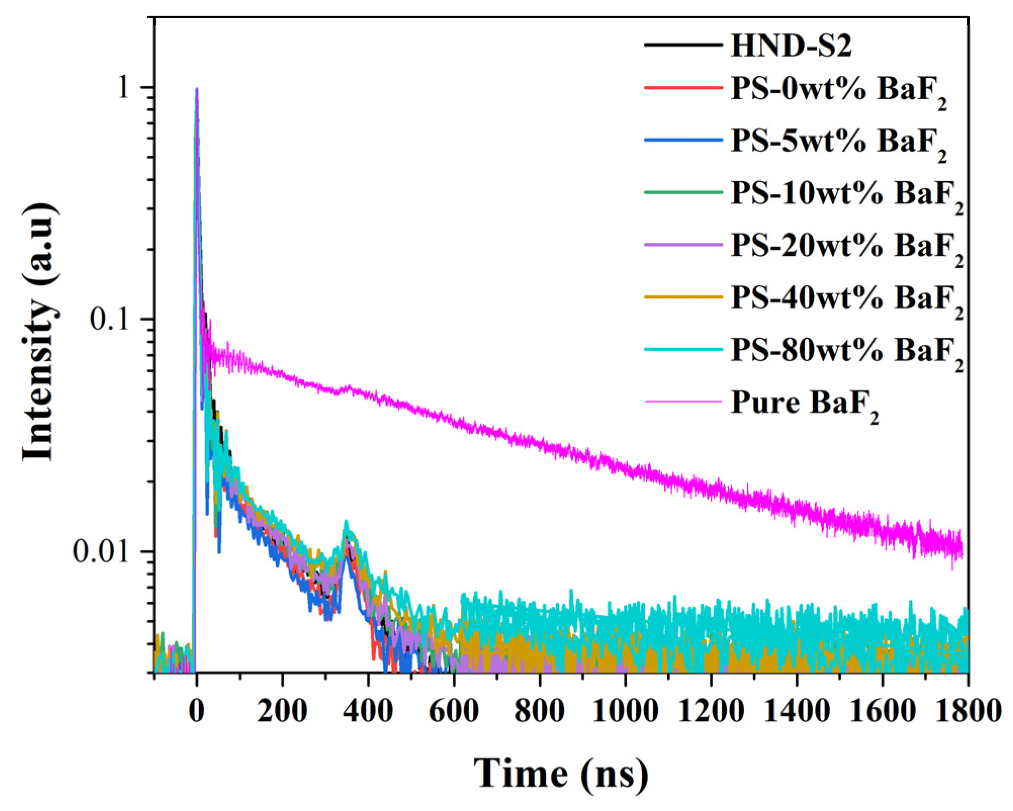
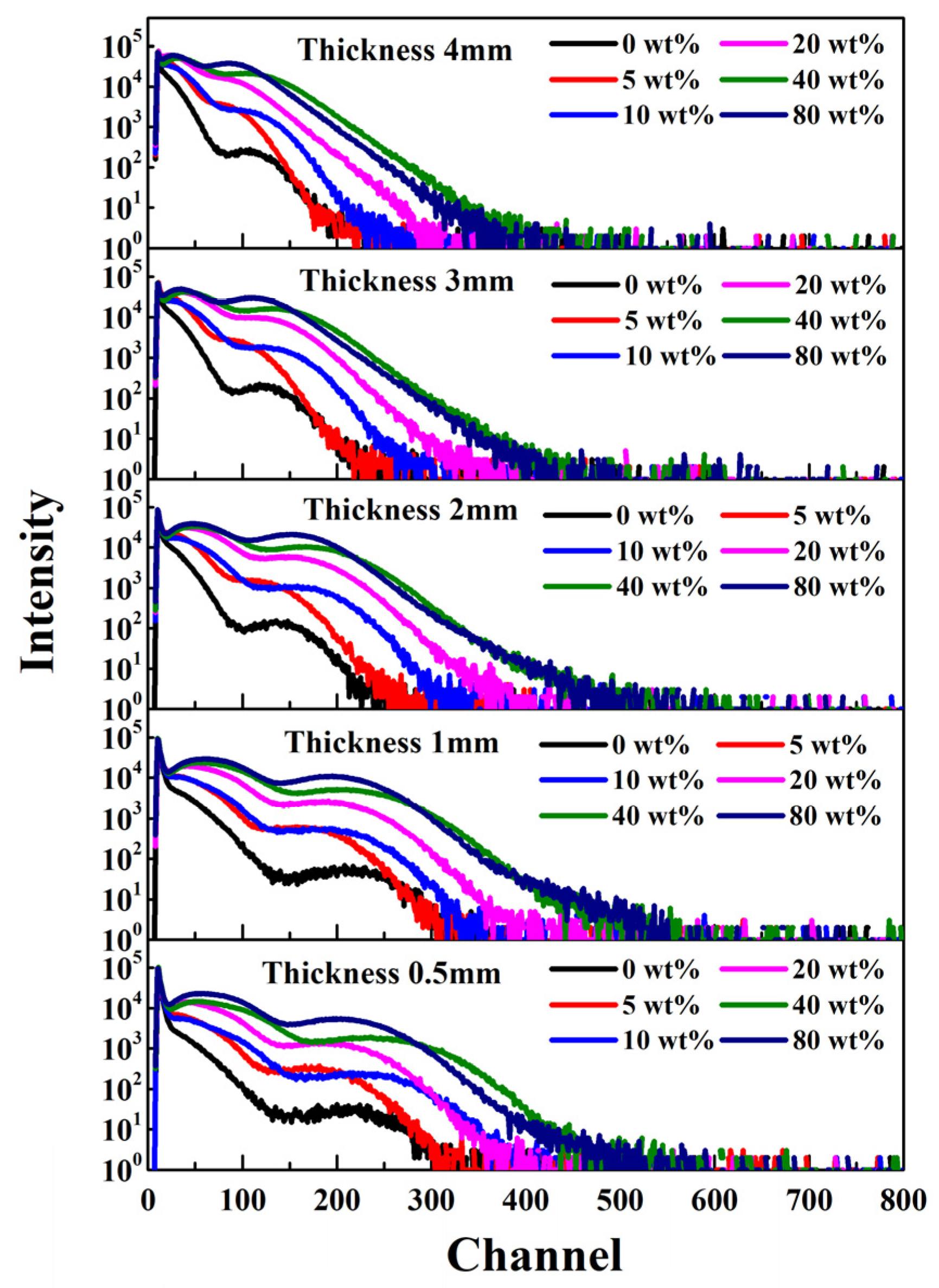
3.4. Scintillation Kinetics

The scintillation decay kinetics curves of composite scintillators loaded with different amounts of BaF2 particles are presented in Figure 13. Compared with pure BaF2, PS-BaF2 composite scintillators show obviously different decay kinetics. It is well known that bulk BaF2 crystal has a slow scintillation component peak at 310 nm, with a decay time of about 600 ns. This slow component limits its application in the field of high count rates [30]. However, no slow scintillation component of BaF2 at 310 nm was observed in the PS-BaF2. The composite scintillators remain the fast decay kinetics of the organic matrix. This result is similar to the experimental results of previous investigations [14,20,21,22,23]. The spectral and kinetic parameters of the composite scintillators are similar to those of the pure PS scintillator and are distinctly different from those of BaF2. This also shows that the prepared bulk PS-BaF2 composite scintillator combines the advantages of the fast decay of the organic matrix and the high stopping power of the inorganic BaF2 particles.

4. Conclusions

Bulk PS-BaF2 (0~80 wt%) composite scintillators were successfully prepared by combining in situ embedding of inorganic particles during the polymerization process with the mixed defoaming process. Inorganic BaF2 particles were uniformly dispersed in the organic matrix compared to the ultrasonic dispersion process through SEM morphological and EDS mapping analysis. The result of FTIR spectroscopy indicated that the loading of BaF2 particles had no effect on the polymerization structure of styrene. The PL and XEL properties of the composite scintillator were systematically investigated. The loading of BaF2 particles not only kept the emission peak of the organic matrix unchanged, but also the emission intensity of the 1 mm-thick composite scintillator with 80 wt% BaF2 particle loading was about 8 times that of the commercial HND-S2 plastic scintillator. However, when the thickness was greater than 1 mm, the composite scintillator with the 40 wt% BaF2 loading amount demonstrated a better performance. This is mainly because the increase in the thickness and concentration will increase the photon yield, but at the same time it will also reduce the transparency of the sample. The mechanism study showed that the XEL enhancement phenomenon is most likely related to the increased stopping power, as well as the FRET from BaF2 particles to the PS scintillator. Owing to the dramatically improved radioluminescence performance, and the retained fast decay kinetics feature of the PS scintillator, PS-BaF2 composite scintillators are promising for large-area radiation detection and imaging applications. This study is helpful for the development of reproducible, cost-effective, organic–inorganic composite scintillators for large-area radiation imaging applications.

Supplementary Materials

The following supporting information can be downloaded at: https://www.mdpi.com/article/10.3390/cryst13091334/s1, Figure S1: XRD pattern of BaF2 particles; Figure S2: SEM image of BaF2 particles; Figure S3: EDS analysis result of BaF2 particles; Figure S4: PS-BaF2 composite scintillators with 20 wt% BaF2 loading amounts prepared by using ultrasonic dispersion method; Table S1: EDS analysis results of BaF2 particles.

Author Contributions

Conceptualization, X.L. and J.C.; methodology, X.L.; validation, J.C. and Y.S.; investigation, Y.D. and X.Q.; data curation, X.L.; writing—original draft preparation, X.L.; writing—review and editing, M.D. and S.W.; visualization, Y.L.; supervision, J.C.; project administration, J.C.; funding acquisition, J.C. All authors have read and agreed to the published version of the manuscript.

Funding

This research was funded by the State Key Laboratory of Intense Pulsed Radiation Simulation and Effect, grant number SKLIPR1714, the Natural Science Foundation of China, grant number U1867211, and the Strategic Priority Research Program of the Chinese Academy of Sciences, grant number XDA25030600.

Data Availability Statement

The data underlying the results presented in this paper are not publicly available at this time but may be obtained from the authors upon reasonable request.

Conflicts of Interest

The authors declare no conflict of interest.

References

  1. Weber, M.J. Inorganic scintillators: Today and tomorrow. J. Lumin. 2002, 100, 35–45. [Google Scholar] [CrossRef]
  2. Chen, X.; Lu, H.; Gu, Q.; Zhang, N.; Wang, S.; Wu, S. Terbium-doped LiYbF4 nanomaterial-based scintillator responding to X-rays with high-resolution imaging applications. CrystEngComm 2022, 24, 2551–2557. [Google Scholar] [CrossRef]
  3. Novais, S.M.V.; Monteiro, T.J.; Andrade, A.B.; Gomes, M.A.; Dias, C.S.B.; Valerio, M.E.G.; Macedo, Z.S. Development of CdWO4-polystyrene scintillator composites for X-ray detection in imaging systems. Nucl. Instrum. Methods Phys. Res. Sect. A 2022, 1025, 166196. [Google Scholar] [CrossRef]
  4. Oliveira, J.; Martins, P.M.; Martins, P.; Correia, V.; Rocha, J.G.; Lanceros-Mendez, S. Gd2O3:Eu3+/PPO/POPOP/PS composites for digital imaging radiation detectors. Process. Appl. Phys. A 2015, 121, 581–587. [Google Scholar] [CrossRef]
  5. Sen, S.; Tyagi, M.; Sharma, K.; Sarkar, P.S.; Sarkar, S.; Basak, C.B.; Pitale, S.; Ghosh, M.; Gadkari, S.C. Organic−Inorganic composite Films based on Gd3Ga3Al2O12:Ce scintillator nanoparticles for X-ray Imaging applications. ACS Appl. Mater. Interfaces 2017, 9, 37310–37320. [Google Scholar] [CrossRef] [PubMed]
  6. Novais, S.M.V.; Monteiro, T.J.; Teixeira, V.C.; Gomes, M.A.; Valerio, M.E.G.; Macedo, Z.S.; Barbosa, L.B. Hydrothermal synthesis of CdWO4 for scintillator-polymer composite films development. J. Lumin. 2018, 199, 225–231. [Google Scholar] [CrossRef]
  7. Wang, B.; Yang, X.; Chen, S.; Lu, S.; Zhao, S.; Qian, Q.; Cai, W.; Wang, S.; Zang, Z. Flexible perovskite scintillators and detectors for X-ray detection. iScience 2022, 25, 105593. [Google Scholar] [CrossRef]
  8. Komendo, I.; Bondarev, A.; Fedorov, A.; Dosovitskiy, G.; Gurinovich, V.; Kazlou, D.; Kozhemyakin, V.; Mechinsky, V.; Mikhlin, A.; Retivov, V.; et al. New scintillator 6Li2CaSiO4:Eu2+ for neutron sensitive screens. Nucl. Instrum. Methods Phys. Res. Sect. A 2023, 1045, 167637. [Google Scholar] [CrossRef]
  9. Oliveira, J.; Correia, V.; Costa, P.; Francesko, A.; Rocha, G.; Lanceros-Mendez, S. Stretchable scintillator composites for indirect X-ray detectors. Compos. Part B-Eng. 2018, 133, 226–231. [Google Scholar] [CrossRef]
  10. Cai, W.; Wang, A.; Fu, L.; Hu, J.; Rao, T.; Wang, J.; Zhong, J.; Xiang, W. Preparation and luminescent properties of GdOF:Ce, Tb nanoparticles and their transparent PMMA nanocomposites. Opt. Mater. 2015, 43, 36–41. [Google Scholar] [CrossRef]
  11. Sahi, S.; Chen, W.; Jiang, K. Luminescence enhancement of PPO/PVT scintillators by CeF3 nanoparticles. J. Lumin. 2015, 159, 105–109. [Google Scholar] [CrossRef]
  12. Boyarintsev, A.Y.; Nepokupnaya, T.A.; Onufriyev, Y.D.; Tarasov, V.A. Composite Scintillator. In Proceedings of the Engineering of Scintillation Materials and Radiation Technologies, Minsk, Belarus, 26–30 January 2016. [Google Scholar]
  13. McKigney, E.A.; Del Sesto, R.E.; Jacobsohn, L.G.; Santi, P.A.; Muenchausen, R.E.; Ott, K.C.; McCleskey, T.M.; Bennett, B.L.; Smith, J.F.; Cooke, D.W. Nanocomposite scintillators for radiation detection and nuclear spectroscopy. Nucl. Instrum. Methods Phys. Res. Sect. A 2007, 579, 15–18. [Google Scholar] [CrossRef]
  14. Ouyang, X.; Liu, B.; Xiang, X.; Chen, L.; Xu, M.; Song, X.; Ruan, J.; Liu, L.; Liu, J.; Chen, C.; et al. CsI(Na) micron-scale particles-based composite material for fast pulsed X-ray detection. Nucl. Instrum. Methods Phys. Res. Sect. A 2020, 953, 163120. [Google Scholar] [CrossRef]
  15. Ziluei, H.; Azimirad, R.; Larijani, M.M.; Ziaie, F. Preparation and optimization of CdWO4-polymer nano-composite film as an alpha particle counter. Nucl. Instrum. Methods Phys. Res. Sect. A 2017, 852, 85–90. [Google Scholar] [CrossRef]
  16. Sun, Y.; Koshimizu, M.; Yahaba, N.; Nishikido, F.; Kishimoto, S.; Haruki, R.; Asai, K. High-energy X-ray detection by hafnium-doped organic-inorganic hybrid scintillators prepared by sol-gel method. Appl. Phys. Lett. 2014, 104, 174104. [Google Scholar] [CrossRef]
  17. Liu, C.; Hajagos, T.J.; Kishpaugh, D.; Jin, Y.; Hu, W.; Chen, Q.; Pei, Q. Facile single-precursor synthesis and surface modification of hafnium oxide nanoparticles for nanocomposite γ-ray scintillators. Adv. Funct. Mater. 2015, 25, 4607–4616. [Google Scholar] [CrossRef]
  18. Hamroun, M.S.E.; Bachari, K.; Guerbous, L.; Berrayah, A.; Mechernene, L. Structural and optical properties of LSO scintillator-polymer composite films. Optik 2019, 187, 111–116. [Google Scholar] [CrossRef]
  19. Vistovskyy, V.V.; Zhyshkovych, A.V.; Halyatkin, O.O.; Mitina, N.E.; Zaichenko, A.S.; Rodnyi, P.A.; Vasil’ev, A.N.; Gektin, A.V.; Voloshinovskii, A.S. The luminescence of BaF2 nanoparticles upon high-energy excitation. J. Appl. Phys. 2014, 116, 054308. [Google Scholar] [CrossRef]
  20. Demkiv, T.M.; Halyatkin, O.O.; Vistovskyy, V.V.; Gektin, A.V.; Voloshinovskii, A.S. X-ray excited luminescence of polystyrene-based scintillator loaded with LaPO4-Pr nanoparticles. J. Appl. Phys. 2016, 120, 144301. [Google Scholar] [CrossRef]
  21. Demkiv, T.M.; Vistovskyy, V.V.; Halyatkin, O.O.; Malyi, T.; Yakibchuk, P.M.; Gektin, A.V.; Voloshinovskii, A.S. Luminescence of polystyrene composites loaded with CeF3 nanoparticles. Nucl. Instrum. Methods Phys. Res. Sect. A 2018, 908, 309–312. [Google Scholar] [CrossRef]
  22. Demkiv, T.M.; Halyatkin, O.O.; Vistovskyy, V.V.; Hevyk, V.B.; Yakibehuk, P.M.; Gektin, A.V.; Voloshinovskii, A.S. X-ray excited luminescence of polystyrene composites loaded with SrF2 nanoparticles. Nucl. Instrum. Methods Phys. Res. Sect. A 2017, 847, 47–51. [Google Scholar] [CrossRef]
  23. Demkiv, T.M.; Halyatkin, O.O.; Vistovskyy, V.V.; Gektin, A.V.; Voloshinovskii, A.S. Luminescent and kinetic properties of the polystyrene composites based on BaF2 nanoparticles. Nucl. Instrum. Methods Phys. Res. Sect. A 2016, 810, 1–5. [Google Scholar] [CrossRef]
  24. Rajakrishna, K.; Dhanasekaran, A.; Yuvaraj, N.; Ajoy, K.C.; Venkatraman, B.; Jose, M.T. Thermal neutron sensitive inorganic compound loaded thin-film composite plastic scintillators. Appl. Radiat. Isot. 2022, 181, 110115. [Google Scholar] [CrossRef] [PubMed]
  25. Cai, W.; Chen, Q.; Cherepy, N.; Dooraghi, A.; Kishpaugh, D.; Chatziioannou, A.; Payne, S.; Xiang, W.; Pei, Q. Synthesis of bulk-size transparent gadolinium oxide-polymer nanocomposites for gamma ray spectroscopy. J. Mater. Chem. C 2013, 1, 1970–1976. [Google Scholar] [CrossRef] [PubMed]
  26. Gerasymov, I.; Nepokupnaya, T.; Boyarintsev, A.; Sidletskiy, O.; Kurtsev, D.; Voloshyna, O.; Trubaieva, O.; Boyarintseva, Y.; Sibilieva, T.; Shaposhnyk, A.; et al. GAGG:Ce composite scintillator for X-ray imaging. Opt. Mater. 2020, 109, 110305. [Google Scholar] [CrossRef]
  27. Feller, R.K.; Purdy, G.M.; Ortiz-Acosta, D.; Stange, S.; Li, A.; McKigney, E.A.; Esch, E.I.; Muenchausen, R.E.; Gilbertson, R.; Bacrania, M.; et al. Large-scale synthesis of CexLa1−xF3 nanocomposite scintillator materials. J. Mater. Chem. 2011, 21, 5716–5722. [Google Scholar] [CrossRef]
  28. Shevelev, V.S.; Ishchenko, A.V.; Vanetsev, A.S.; Nagirnyi, V.; Omelkov, S.I. Ultrafast hybrid nanocomposite scintillators: A review. J. Lumin. 2022, 242, 118534. [Google Scholar] [CrossRef]
  29. Athulya, P.A.; Chandrasekaran, N. Interactions of natural colloids with microplastics in aquatic environment and its impact on FTIR characterization of polyethylene and polystyrene microplastics. J. Mol. Liq. 2023, 369, 120950. [Google Scholar] [CrossRef]
  30. Chen, J.; Yang, F.; Zhang, L.; Zhu, R.-Y.; Du, Y.; Wang, S.; Sun, S.; Li, X. Slow scintillation suppression in yttrium doped BaF2 crystals. IEEE Trans. Nucl. Sci. 2018, 65, 2147–2151. [Google Scholar] [CrossRef]
Figure 1. Composite scintillators with 0~80 wt% BaF2 particle loading amounts and a thickness of 20 mm.
Crystals 13 01334 g001
Figure 2. Various PS-BaF2 composite scintillator samples, with a 0.5~4 mm thickness and 0~80 wt% loading amounts.
Crystals 13 01334 g002
Figure 3. (ad) SEM images and (fi) EDS mapping of PS-BaF2 composite scintillators with different BaF2 particle loading amounts (from top to bottom: PS-0 wt% BaF2, PS-5 wt% BaF2, PS-20 wt%, PS-80 wt% BaF2). (e,j) SEM image and EDS mapping of PS-BaF2 composite scintillators with 20 wt% loading amounts prepared using the ultrasonic dispersion method after pre-polymerization, for comparison.
Crystals 13 01334 g003
Figure 4. FTIR of composite scintillators with different loading amounts of BaF2 particles.
Crystals 13 01334 g004
Figure 5. Optical transmittance and EWLT of the composite scintillator with different loading amounts of BaF2 particles and a thickness of 1 mm.
Crystals 13 01334 g005
Figure 6. XEL of composite scintillators with different loading amounts of BaF2 particles.
Crystals 13 01334 g006
Figure 7. PL and PLE spectra of the pure PS scintillator and the PS-BaF2 composite scintillator with 20 wt% loading amount of BaF2 particles.
Crystals 13 01334 g007
Figure 8. PHS of composite scintillator under 241Am (59.5 KeV) excitation with different loading amounts and thicknesses.
Crystals 13 01334 g008
Figure 9. Gross count rate of composite scintillators as a function with BaF2 particle loading amounts and thicknesses under 241Am (59.5 KeV) excitation.
Crystals 13 01334 g009
Figure 10. XEL intensity of the composite scintillator as a function with the BaF2 loading amounts and sample thicknesses.
Crystals 13 01334 g010
Figure 11. XEL spectrum (red dashed line, BaF2 power) and PLE spectrum (black solid line, PS-0 wt% scintillator).
Crystals 13 01334 g011
Figure 12. XEL spectrum (red curve) of BaF2 powder and absorption spectrum (black curve) of the pure plastic scintillator.
Crystals 13 01334 g012
Figure 13. Scintillation decay kinetic curves of the composite scintillator with 0~80 wt% BaF2 particle loading.
Crystals 13 01334 g013
Disclaimer/Publisher’s Note: The statements, opinions and data contained in all publications are solely those of the individual author(s) and contributor(s) and not of MDPI and/or the editor(s). MDPI and/or the editor(s) disclaim responsibility for any injury to people or property resulting from any ideas, methods, instructions or products referred to in the content.

