Article

Characterization of GAGG Doped with Extremely Low Levels of Chromium and Exhibiting Exceptional Intensity of Emission in NIR Region

1
Faculty of Chemistry and Geosciences, Institute of Chemistry, Vilnius University, Naugarduko 24, LT-03225 Vilnius, Lithuania
2
Department of Chemical Engineering, Münster University of Applied Sciences, Stegerwaldstrasse 39, D-48565 Steinfurt, Germany
*
Author to whom correspondence should be addressed.
Crystals 2021, 11(6), 673; https://doi.org/10.3390/cryst11060673
Submission received: 27 May 2021 / Revised: 8 June 2021 / Accepted: 9 June 2021 / Published: 11 June 2021

Abstract

:
Cerium and chromium co-doped gadolinium aluminum gallium garnets were prepared using sol-gel technique. These compounds potentially can be applied for NIR-LED construction, horticulture and theranostics. Additionally, magnesium and calcium ions were also incorporated into the structure. X-ray diffraction data analysis confirmed the all-cubic symmetry with an Ia-3d space group, which is appropriate for garnet-type materials. From the characterization of the luminescence properties, it was confirmed that both chromium and cerium emissions could be incorporated. Cerium luminescence was detected under 450 nm excitation, while for chromium emission, 270 nm excitation was used. The emission of chromium ions was exceptionally intense, although it was determined that these compounds are doped only by parts per million of Cr3+ ions. Typically, the emission maxima of chromium ions are located around 650–750 nm in garnet systems. However, in this case, the emission maximum for chromium is measured to be around 790 nm, caused by re-absorption of Cr3+ ions. The main observation of this study is that the switchable emission wavelength in a compound of single phase was obtained, despite the fact that doping with Cr ions was performed in ppm level, causing an intense emission in NIR region.

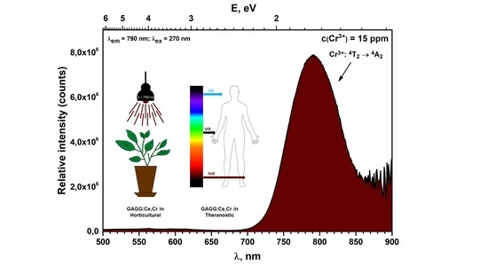
Graphical Abstract

1. Introduction

In recent years, more and more scientists have focused on the synthesis and development of functional inorganic materials. Two of the main considered groups of such compounds are the inorganic scintillators and phosphors. Inorganic scintillators are widely used in medicine and nuclear physics as the x-ray converter material for CT and others detectors [1,2,3,4], while inorganic phosphors are mainly used as material in light-emitting diodes (LEDs) [5,6,7].
Scintillator and phosphor materials usually consist of a host matrix and activator ions, which, in most cases, are lanthanide or transition metal ions [8]. While the host and activator strategy is the most common, there are other types of materials which possess intrinsic luminescence as well [9,10]. However, lanthanum, promethium, and lutetium are not suitable for such applications. One of the most popular groups of host materials are garnets, which are oxides crystallizing in a cubic structure with space group Ia3d, and which comprise three differently coordinated cation sites, namely dodecahedral, octahedral, and tetrahedral positions [11,12]. The host material used in this study was gadolinium aluminum gallium garnet (GAGG), with the formula Gd3Al2Ga3O12. GAGG:Ce garnets exhibited the brightest light yields of 46,000 ph/MeV among other oxide crystalline scintillators [13,14]. Despite the fact that there is a wide variety of dopant ions used in scintillator production, GAGG is mostly doped just by Ce3+ ions [15,16,17,18]. There has been a number of studies in recent years about various GAGG:Ce modifications and the resulting changes in luminescence properties. It has been shown that by changing the ratio between Al and Ga in the garnet structure, Ce emission band can be shifted due to the change of crystal field splitting. It is worthy to note that such changes in emission maxima are also present in other garnets doped with cerium ions, such as Lu3Al5O12:Ce (LuAG:Ce) or Y3Al5O12:Ce (YAG:Ce) [11,19,20,21]. Another type of modification for GAGG:Ce is co-doping with various metal cations. Studies have shown that co-doping of GAGG:Ce with mono/di/trivalent cations usually affects the luminescence, i.e., the absorption strength, quantum efficiency, quenching temperature, and decay time [17,22,23,24,25,26,27,28,29,30,31]. The change of these parameters depends not only on the dopant type, but also on its concentration. These changes are thought to be caused by the formation of various localized energy levels in the conduction band of the garnet, due to the crystal defects which are formed when Gd3+ ions are replaced with cations of different valence c(1+, 2+ or sometimes 4+) [11].
The impact of Cr3+ on Ce3+ decay time was observed in YAG:Ce,Cr. In this case, the decay time greatly increased with the introduction of high amount (500 ppm) of chromium ions [32]. It was proposed that Cr3+ co-doping results in the formation of electron trapping sites with an ideal trap depth for persistent luminescence at room temperature [28,32]. It was also proposed and proven that the same principles can be applied to the GAGG phosphor luminescence where co-doping of Cr3+ and Ce3+ results in persistent phosphor luminescence of excited Ce3+ sites and, in some cases, Cr3+ emission in the visible range [28,29,30,32,33]. In this study, a novel system consisting of GAGG matrix and four additional dopant elements (Ce3+, Cr3+, Mg2+ and Ca2+) was investigated. Since the chromium concentration is just in the ppm range, it exhibits not typical chromium emission but the maximum of emission band is shifted towards 790 nm. Such emission in the near infrared region (NIR) range might be useful for plant growth purposes [34]. Additionally, these compounds could be used in the fabrication of high power NIR-LED devices for night vision applications, and biosensors used to measure content of water, fat, sugar, protein of different products [35,36]. Therefore, in turn, these phosphors could be potentially applied in horticulture research experiments. Biological tissue transmits radiation in the range from 650 to 1300 nm (the first biological window), which allows deeper penetration, so such compounds could also be promising candidates in the field of theranostics [37,38].

2. Materials and Methods

Gadolinium aluminum gallium garnet powders were synthesized by the sol-gel method. Cerium concentration was kept at 0.05 mol% for all samples. All synthesized powder compounds are listed in Table 1.
For these compounds, Gd2O3, Ga2O3, Al(NO3)3·9H2O, (NH4)2Ce(NO3)6, Ca(NO3)2·4H2O and Mg(NO3)2·6H2O were used as precursors. Firstly, Gd2O3 and Ga2O3 were dissolved in an excess of concentrated nitric acid at 50 ℃. Then, the acid was evaporated and the remaining gel was washed with distilled water 2 or 3 times, followed by further evaporation of added water. An additional 200 mL of water was added after the washing, and Al(NO3)3·9H2O, (NH4)2Ce(NO3)6, Ca(NO3)2·9H2O and Mg(NO3)2·9H2O were dissolved. The solution was left under magnetic stirring for 2 h at 50–60 ℃. After that, citric acid was added to the solution with a ratio of 3:1 to metal ions, and was left to stir overnight. The solution was evaporated at the same temperature, and the obtained gels were dried at 140 ℃ for 24 h in the oven. The obtained powders were ground and annealed first at 1000 ℃ for 2 h in air, with 5 ℃/min heating rate. Secondly, the gained powders were heated at 1400 ℃ for 4 h, with 5 ℃/min heating rate.
X-ray diffraction (XRD) measurements of the powders were performed using the Rigaku MiniFlex II X-ray diffractometer (Rigaku Europe SE, Neu-Isenburg, Germany). Powders used for analysis were evenly dispersed on the glass sample holder using ethanol. Then diffraction patterns were recorded in the range of 2θ angles from 15° to 80° for all compounds. Cu Kα radiation (λ = 1.542 Å (Avarage of Cu Kα1 = 1.540 and Kα2 = 1.544)) was used for the analysis. The measurement parameters were set as follows: current was 15 mA, voltage −30 kV, X-ray detector movement step was 0.010° and dwell time was 5.0 s.
Measurements of emission and excitation: Edinburgh Instruments FLS980 spectrometer (Edinburgh Instruments, Livingston, UK) equipped with double excitation and emission monochromators and 450 W Xe arc lamp (Edinburgh Instruments, Livingston, UK) a cooled (−20 °C) single-photon counting photomultiplier (Hamamatsu R928(Edinburgh Instruments, Livingston, UK)) and mirror optics for powder samples were used for measuring the excitation and emission of the prepared samples. Obtained photoluminescence emission spectra were corrected using a correction file obtained from a tungsten incandescent lamp certified by National Physics Laboratory (NPL), UK. Excitation spectra were corrected by a reference detector [39]. The reflectance spectra from 250 to 600 nm were measured in an integrated (solid) sphere coated with barium sulphate. BaSO4 (99% Sigma-Aldrich, St. Louis, Missouri, United States) was used as a reference material, with excitation and emission gaps of 4 and 0.15 nm, respectively. Each measurement was performed 10 times. Measurements in the range of 120 to 400 nm were performed using a FLS920 fluorescence spectrometer (Edinburgh Instruments, Livingston, UK) with an R-UV excitation monochromator VM504 (Acton Research Corporation, Acton, Massachusetts, USA) and a deuterium lamp (Edinburgh Instruments, Livingston, UK) in a BAM:Eu (BaMgAl10O17:Eu2+)-coated integrated sphere. The measuring chamber containing the sample was continuously flushed with dry nitrogen gas to remove water and oxygen, since these molecules show vacuum UV absorption. The photoluminescence decay kinetics were studied for powders and thin films using the FLS980 spectrometer(Edinburgh Instruments, Livingston, UK). 450 nm lasers were used for these measurements [39].
Identification of quantification of Ca, Mg, and Cr in the synthesized species was performed by inductively coupled plasma optical emission spectrometry (ICP-OES) using Perkin-Elmer Optima 7000 DV spectrometer (Perkin-Elmer, Walthman, MA, USA). Sample decomposition procedure was carried out in concentrated nitric acid (HNO3, Rotipuran® Supra 69% (Roth, Karlsruhe, Germany)) using microwave reaction system Anton Paar Multiwave 3000 (Anton-Paar, Graz, Austria) equipped with XF100 rotor and PTFE liners (Anton-Paar, Graz, Austria). The following program was used for the dissolution of powders: during the first step, the microwave power was linearly increased to 800 W in 15 min and held at this point for the next 20 min. Once the vessels had been fully cooled and depressurized, the obtained clear solutions were quantitatively transferred into volumetric flasks and diluted up to 50 mL with deionized water. Calibration solutions were prepared by an appropriate dilution of the stock standard solutions (single-element ICP standards, 1000 mg/L, (Roth, Karlsruhe, Germany)).

3. Results and Discussion

3.1. X-ray Diffraction Analysis

To determine the phase purity of the powder garnet samples, XRD analysis was performed. Figure 1 shows the diffraction patterns of all synthesized garnets. From the displayed data, it can be concluded that all compounds, regardless of the concentrations of doping elements, possess cubic GAGG structure with Ia-3d space group ((PDF) #00-046-0448). No peaks corresponding to the constituent oxides were observed. However, we can observe a slight shift of the peaks from the PDF card data to smaller 2θ angles. This shift was due to additional doping of the compound with Mg2+, Ca2+, Ce3+, and/or Cr3+ ions, due to the ionic radii difference [40]. In summary, from the measured data, it can be concluded that garnets could be doped without the formation of additional impurity phases.

3.2. Luminescence Properties

For the practical application of luminescent materials, they have to emit a rather discreet wavelength light, due to the fact that light interacts differently based on its wavelength. Photons with NIR wavelengths are especially important, and they have deeper tissue penetration and can induce faster plant growth. Additionally, should the possibly arise to switch between NIR and other wavelengths in a single matrix, a new application area could potentially be available. In this case, the luminescence is induced by exciting one of the trivalent ions used as dopants (Ce3+, Cr3+). Upon recording the emission and excitation of the compounds, electronic transitions of both cerium and chromium ions can be observed. The absorption bands that are ascribed to cerium ions are due to interconfigurational [Xe]4f1–[Xe]5d1 transitions, while in the case of Cr3+ intraconfigurational [Ar]3d3–[A]3d3 transitions between the crystal-field components, 4A and 4T are observed. Due to the allowed nature of these electron transitions, broad emission bands are detected. During these processes, energy is either reemitted or just absorbed, which defines the optical properties of such garnets. Given the possibility to switch between cerium and chromium emission based on the excitation wavelength, as well as the large red shift of chromium emission toward 790 nm, such compounds could potentially be good candidates as multifunctional materials in horticultural and theranostic applications. However, further research for practical applications is still needed to evaluate such possibilities more in-depth.
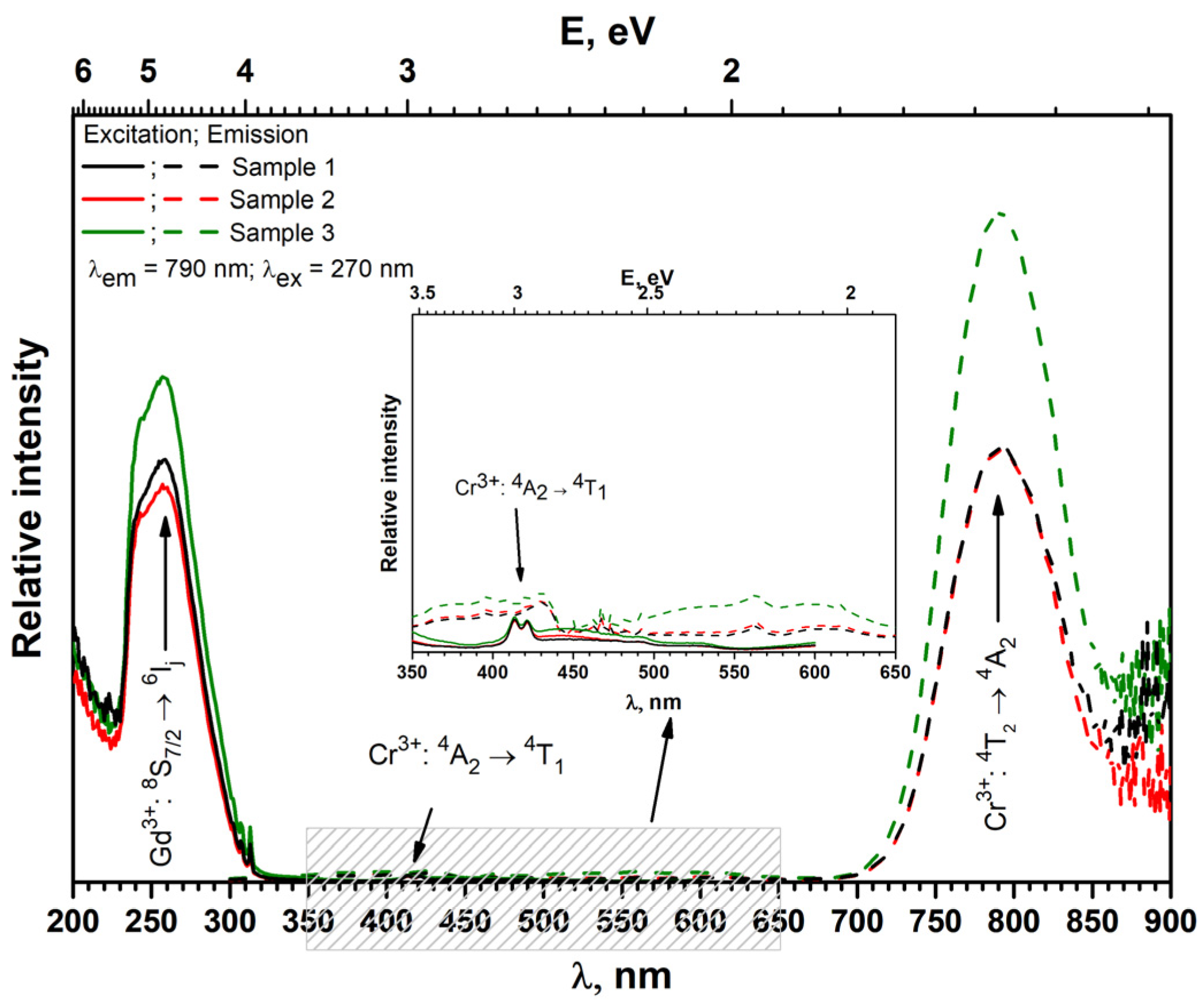
In order to estimate optical properties of the synthesized materials excitation, emission and reflectance spectra were recorded. The decay times were measured as well. Figure 2 and Figure 3 show the excitation and emission spectra of different GAGG compounds measured at room temperature. In Figure 2, the recorded chromium emission and excitation spectra are displayed. In excitation spectra, the most intense band is attributed to 8S7/26IJ transitions of Gd3+ ions with the maxima at 270 nm. At 415 and 575 nm, the evidence of excitation process of chromium ions resulting from the transitions between 4A1 to 4T1 and 4T2 orbitals was also observed. Meanwhile, the emission signal detected under 270 nm excitation is caused by the transition from 4T2 to 4A1 orbital. From the spectra given in Figure 2, it can be derived that the compound with the highest content of magnesium (Sample 3), i.e., 9 ppm, exhibits the most intense emission and excitation bands. The difference in the intensity of excitation and emission could be attributed to the different calcium content in the compounds. Since calcium has a 2+ charge and it is introduced instead of 3+ gadolinium ion, oxygen vacancy defects are most likely created, due to the aliovalent nature of substitution. It is commonly known that such defects reduce the luminescence intensity of most compounds [41,42]. In this case, the compound with the lowest Ca2+ amount (Sample 3) has the highest luminescence intensity, whereas Sample 1 and Sample 2 have similar amounts Ca2+ ions and show lower emission intensities. It should be noted that chromium is present in ppm fractions, nevertheless, its emission remains very intense. Commonly, chromium emission maximum in garnet matrix is around 650–750 nm, while in this case it was measured to be 790 nm. Such a shift to the red region was previously explained by the re-absorption of Cr3+ ions [43]. Studies by other researchers revealed that a high photoluminescence intensity of chromium ions emission was only recorded with 2000 times higher amounts (for example, 3 mol%) of Cr3+ in other compounds [44].
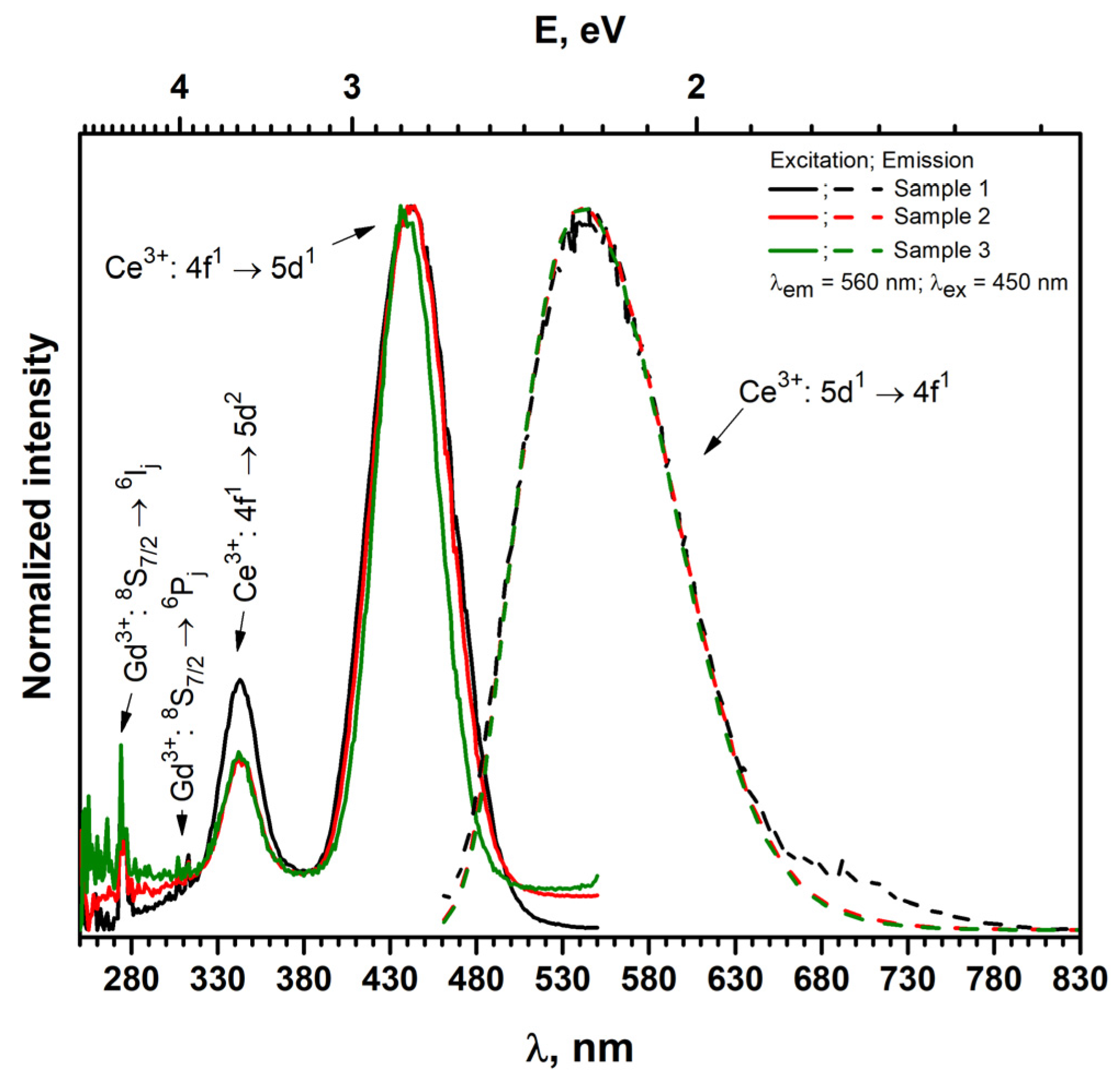
Since these garnets were doped with cerium as well, they also have the characteristic emission attributed to cerium ions. Normalized emission is attributed to cerium emission, and excitation spectra are shown in Figure 3. To measure cerium emission, the compounds were excited under 450 nm wavelength ([Xe]4f1 to [Xe]5d1 interconfigurational transitions of Ce3+), which cannot be attributed to chromium. The compounds appear to exhibit cerium-specific fluorescence at a wavelength of 540 nm, which arises from the 5d1 to 4f1 electronic transitions. Meanwhile, when investigating the excitation spectra of the compounds in addition to those cerium bands, the peaks contributing to gadolinium ions were observed in the 250–320 nm range [19,44].
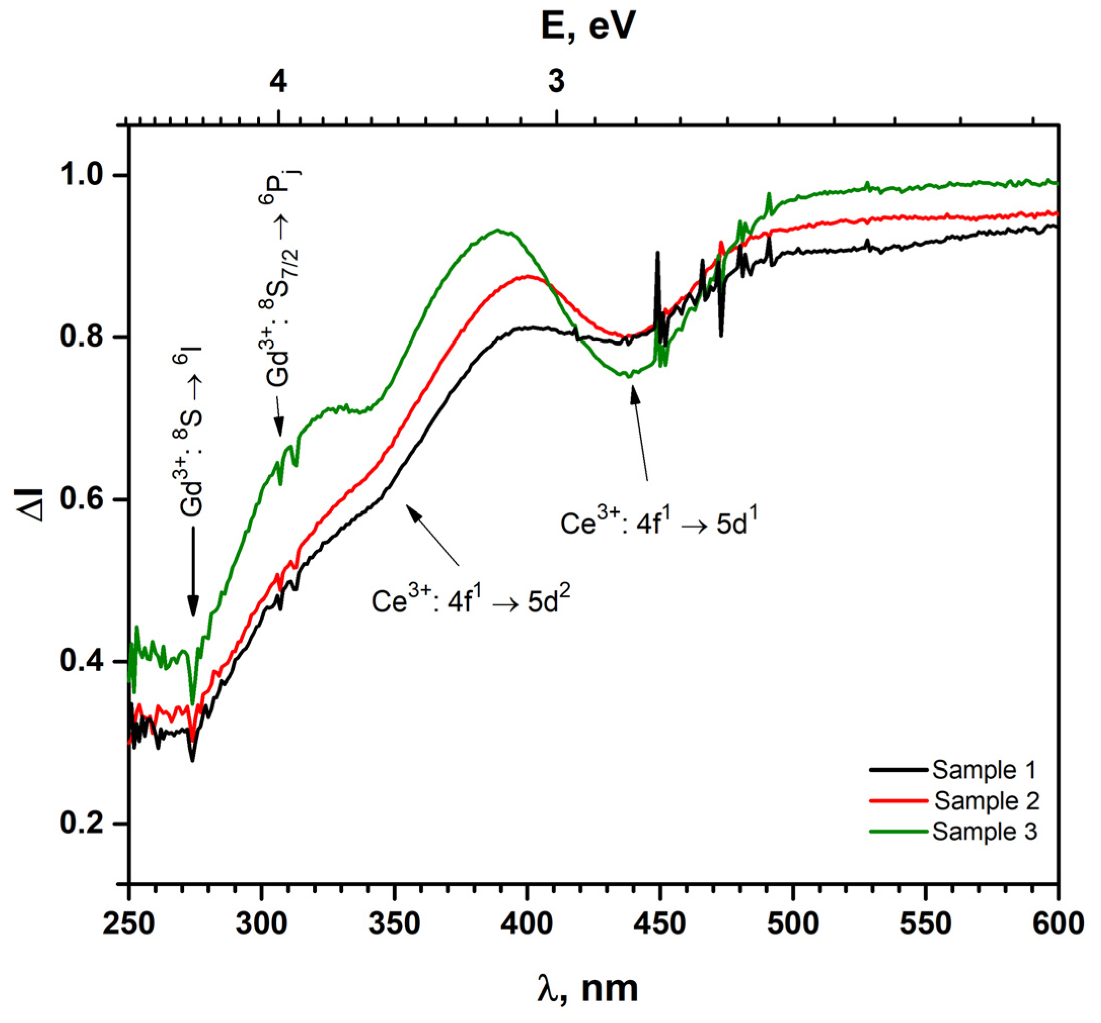
Figure 4 shows the reflectance spectra of the compounds. In the reflectance spectra of garnets doped with cerium, two absorption bands can be observed, one at about 350 nm, the another at 400–500 nm. These bands are assigned to the different crystal-field components of the interconfigurational Ce3+ [Xe]4f1 → [Xe]5d1 transitions. At approximately 275 nm, an absorption peak is observed in all spectra, which is assigned to the Gd3+ 8S7/26IJ interconfigurational transitions. The set of absorption peaks at about 312 nm is assigned to the Gd3+ 8S7/26PJ electron transitions [19,45]. It should be noted that the position of the Ce3+ absorption bands does not change upon changing the ions with which the garnet was doped, so it could be stated that doping with magnesium and calcium does not affect the absorption wavelength. However, it is obvious that garnets also have cerium. The absorption band at 360 and 440 nm is classified as a Ce3+ electronic transition. In addition, there is no bright absorption band at 600 nm, which is typically observed in Cr3+-containing compounds [44,46,47,48].
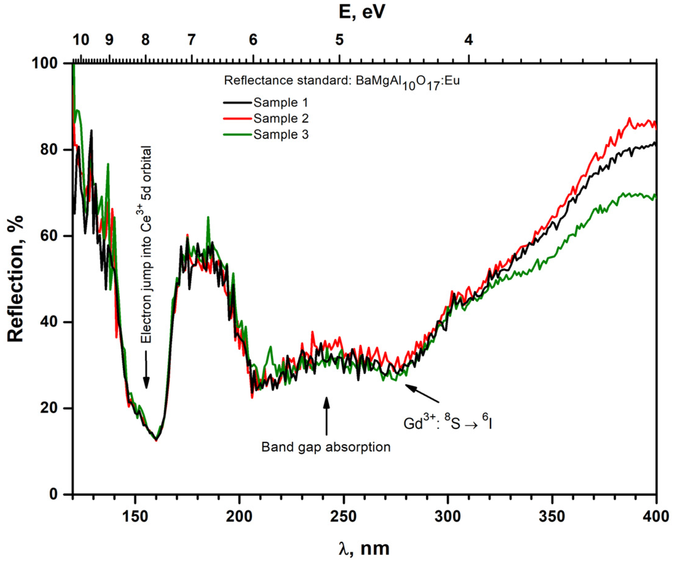
To better investigate the absorption of garnets in the UV region, the reflectance spectra of the synthesized gadolinium aluminum gallium garnets powders in the 130–400 nm range were recorded. The reflectance spectra are shown in Figure 5. It can be seen that in the 130–400 nm region, three clear absorption bands are visible: one in the 140–165 nm range, the second in the 190–240 nm range, and the last in 270–280 nm zone. The absorption in the 270–280 nm range can be attributed to Gd3+ 8S → 6I electron transitions [19]. The observed absorption in the 140–165 nm range can be interpreted as the electron jump into the Ce3+ 5d orbital [49,50]. Finally, the region of 200–300 nm can be explained as the band gap absorption of the garnet matrix [50,51].
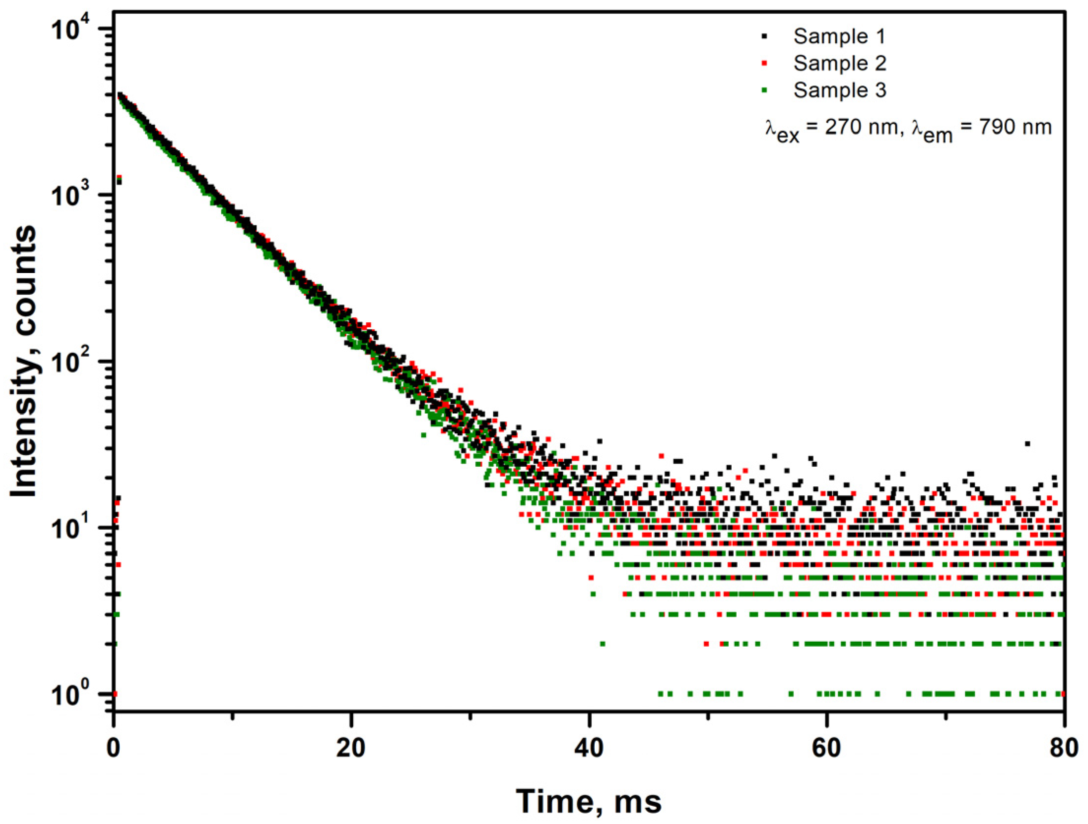
The photoluminescence decay curves of the chromium emission are shown in Figure 6. All calculated values of the decay times are listed in Table 2. As can be seen from Table 2, all compounds have almost the same decay times, which are between 6.0 and 5.9 ms. It can be seen that the samples have emission decay times showing the characteristics of Cr3+ ions, because characteristic decay times for Ce3+ ions are much shorter and reach about 50–60 ns [5,52]. Cr3+ ions electron transition are considered forbidden, which usually results in longer decay times in a rage of milliseconds [42,53]. However, for the Ce3+ ions, electron transitions are allowed, and the decay time is in the range of nanoseconds [5]. The obtained values are of chromium, since the 270 nm excitation wavelength was used. While there have been previous reports on the effect of calcium and magnesium doping in garnet matrix on decay times [17,54,55,56], in this case, no effect was observed, potentially due to the drastically smaller amounts to aforementioned ions.

4. Conclusions

Gadolinium aluminum gallium garnets doped with 0.05% cerium, chromium, magnesium, and calcium were synthesized by the sol-gel method whereby obtained garnet type samples are of single phase. Measurement of the excitation and emission spectra of these compounds revealed that extremely low levels of Cr3+ ions, i.e., only at a level of 15 ppm, caused intense emission in the NIR region. At the same time, but under different excitation wavelengths, a typical emission spectrum of Ce3+ was also observed, which proved that the single compound might emit in very broad range (from 470 to 850 nm). The most intense emission was located at a wavelength of 790 nm. The obtained compounds exhibit promising luminescence properties to illuminate plants and promote their growth. Since luminescence covered the first biological window and chromium ions exhibited a long specific decay time in the range of 6 ms, the synthesized compounds also demonstrate characteristics required for bio-imaging purposes.

Author Contributions

R.S. and G.I. conceived and planned the experiments and wrote the manuscript with support from other co-authors, G.L. and G.I. synthesized powders, R.S. supervised the project, A.Z. performed ICP measurements of powders, G.L. performed XRD measurements, D.E., G.L. and T.J. performed luminescence measurements, and T.J. analyzed luminescence properties. All authors provided critical feedback and helped shape the research, analysis and manuscript. All authors have read and agreed to the published version of the manuscript.

Funding

This research received no external funding.

Institutional Review Board Statement

Not applicable.

Informed Consent Statement

Not applicable.

Conflicts of Interest

The authors declare no conflict of interest.

References

  1. Paper, C.; Cherepy, N.; Livermore, L.; Seeley, Z.M.; Livermore, L.; Beck, P.; Livermore, L.; Hunter, S.L.; Livermore, L. High Energy Resolution Transparent Ceramic Garnet Scintillators. Int. Soc. Opt. Photonics 2014, 2. [Google Scholar] [CrossRef]
  2. Sakthong, O.; Chewpraditkul, W.; Wanarak, C.; Kamada, K. Nuclear Instruments and Methods in Physics Research A Scintillation Properties of Gd3Al2Ga3O12:Ce3þ Single Crystal Scintillators. Nucl. Inst. Methods Phys. Res. A 2014, 751, 1–5. [Google Scholar] [CrossRef] [Green Version]
  3. Belli, P.; Bernabei, R.; Cerulli, R.; Dai, C.J.; Danevich, F.A.; Incicchitti, A.; Kobychev, V.V.; Ponkratenko, O.A.; Prosperi, D.; Tretyak, V.I.; et al. Performances of a CeF3 Crystal Scintillator and Its Application to the Search for Rare Processes. Nucl. Instrum. Methods Phys. Res. Sect. A Accel. Spectrometers Detect. Assoc. Equip. 2003, 498, 352–361. [Google Scholar] [CrossRef]
  4. Van Eijk, C.W.E. Scintillator-Based Detectors; Elsevier B.V.: Amsterdam, The Netherlands, 2014; Volume 8. [Google Scholar] [CrossRef]
  5. Bachmann, V.; Ronda, C.; Meijerink, A. Temperature Quenching of Yellow Ce3+ Luminescence in YAG:Ce. Chem. Mater. 2009, 21, 2077–2084. [Google Scholar] [CrossRef]
  6. Khaidukov, N.; Zorenko, T.; Iskaliyeva, A.; Paprocki, K.; Batentschuk, M.; Osvet, A.; Van Deun, R.; Zhydaczevskii, Y.; Suchocki, A.; Zorenko, Y. Synthesis and Luminescent Properties of Prospective Ce3+ Doped Silicate Garnet Phosphors for White LED Converters. J. Lumin. 2017, 192, 328–336. [Google Scholar] [CrossRef]
  7. Ueda, J.; Dorenbos, P.; Bos, A.J.J.; Meijerink, A.; Tanabe, S. Insight into the Thermal Quenching Mechanism for Y3Al5O12:Ce3+ through Thermoluminescence Excitation Spectroscopy. J. Phys. Chem. C 2015, 119, 25003–25008. [Google Scholar] [CrossRef] [Green Version]
  8. Auffray, E.; Korjik, M.; Lucchini, M.T.; Nargelas, S.; Sidletskiy, O.; Tamulaitis, G.; Tratsiak, Y.; Vaitkevičius, A. Free Carrier Absorption in Self-Activated PbWO4 and Ce-Doped Y3(Al0.25Ga0.75)3O12 and Gd3Al2Ga3O12 Garnet Scintillators. Opt. Mater. 2016, 58, 461–465. [Google Scholar] [CrossRef] [Green Version]
  9. Seminko, V.; Maksimchuk, P.; Bespalova, I.; Masalov, A.; Viagin, O.; Okrushko, E.; Kononets, N.; Malyukin, Y. Defect and Intrinsic Luminescence of CeO2 Nanocrystals. Phys. Status Solidi 2017, 254, 1600488. [Google Scholar] [CrossRef]
  10. Zatsepin, D.A.; Boukhvalov, D.W.; Zatsepin, A.F.; Kuznetsova, Y.A.; Mashkovtsev, M.A.; Rychkov, V.N.; Shur, V.Y.; Esin, A.A.; Kurmaev, E.Z. Electronic Structure, Charge Transfer, and Intrinsic Luminescence of Gadolinium Oxide Nanoparticles: Experiment and Theory. Appl. Surf. Sci. 2018, 436, 697–707. [Google Scholar] [CrossRef] [Green Version]
  11. Kanai, T.; Satoh, M.; Miura, I. Characteristics of a Nonstoichiometric Gd3+δ(Al,Ga) 5-ΔO12:Ce Garnet Scintillator. J. Am. Ceram. Soc. 2008, 91, 456–462. [Google Scholar] [CrossRef]
  12. Geller, S. Magnetic Interactions and Distribution of Ions in the Garnets. J. Appl. Phys. 1960, 30. [Google Scholar] [CrossRef]
  13. Yanagida, T.; Kamada, K.; Fujimoto, Y.; Yagi, H.; Yanagitani, T. Comparative Study of Ceramic and Single Crystal Ce:GAGG Scintillator. Opt. Mater. 2013, 35, 2480–2485. [Google Scholar] [CrossRef]
  14. Fedorov, A.; Gurinovich, V.; Guzov, V.; Dosovitskiy, G.; Korzhik, M.; Kozhemyakin, V.; Lopatik, A.; Kozlov, D.; Mechinsky, V.; Retivov, V. Sensitivity of GAGG Based Scintillation Neutron Detector with SiPM Readout. Nucl. Eng. Technol. 2020, 52, 2306–2312. [Google Scholar] [CrossRef]
  15. Kitaura, M.; Sato, A.; Kamada, K.; Kurosawa, S.; Ohnishi, A.; Sasaki, M.; Hara, K. Photoluminescence Studies on Energy Transfer Processes In. Opt. Mater. 2015, 41, 45–48. [Google Scholar] [CrossRef]
  16. Kitaura, M.; Sato, A.; Kamada, K.; Ohnishi, A.; Sasaki, M. Phosphorescence of Ce-Doped Gd3Al2Ga3O12 Crystals Studied Using Luminescence Spectroscopy. J. Appl. Phys. 2014, 115, 10–18. [Google Scholar] [CrossRef]
  17. Lucchini, M.T.; Gundacker, S.; Lecoq, P.; Benaglia, A.; Nikl, M.; Kamada, K.; Yoshikawa, A.; Auffray, E. Timing Capabilities of Garnet Crystals for Detection of High Energy Charged Particles. Nucl. Instruments Methods Phys. Res. Sect. A Accel. Spectrometers Detect. Assoc. Equip. 2017, 852, 1–9. [Google Scholar] [CrossRef]
  18. Gundacker, S.; Turtos, R.M.; Auffray, E.; Lecoq, P. Precise Rise and Decay Time Measurements of Inorganic Scintillators by Means of X-Ray and 511 KeV Excitation. Nucl. Instrum. Methods Phys. Res. Sect. A Accel. Spectrometers Detect. Assoc. Equip. 2018, 891, 42–52. [Google Scholar] [CrossRef]
  19. Ogieg, J.M.; Katelnikovas, A.; Zych, A.; Ju, T.; Meijerink, A.; Ronda, C.R. Luminescence and Luminescence Quenching in Gd3 (Ga,Al)5O12 Scintillators Doped with Ce3+. J. Phys. Chem. A 2013, 117, 2479–2484. [Google Scholar] [CrossRef] [PubMed]
  20. Ueda, J.; Tanabe, S.; Nakanishi, T. Analysis of Ce3+ Luminescence Quenching in Solid Solutions between Y3Al5O12 and Y3Ga 5O12 by Temperature Dependence of Photoconductivity Measurement. J. Appl. Phys. 2011, 110, 053102. [Google Scholar] [CrossRef] [Green Version]
  21. Pan, Y.; Wu, M.; Su, Q. Tailored Photoluminescence of YAG:Ce Phosphor through Various Methods. J. Phys. Chem. Solids 2004, 65, 845–850. [Google Scholar] [CrossRef]
  22. Yoshino, M.; Kamada, K.; Kochurikhin, V.V.; Ivanov, M.; Nikl, M.; Okumura, S.; Yamamoto, S.; Yeom, J.Y.; Shoji, Y.; Kurosawa, S.; et al. Li+, Na+ and K+ Co-Doping Effects on Scintillation Properties of Ce:Gd3Ga3Al2O12 Single Crystals. J. Cryst. Growth 2018, 491, 1–5. [Google Scholar] [CrossRef]
  23. Kobayashi, T.; Yamamoto, S.; Yeom, J.; Kamada, K.; Yoshikawa, A. Development of High Resolution Phoswich Depth-of-Interaction Block Detectors Utilizing Mg Co-Doped New Scintillators. Nucl. Inst. Methods Phys. Res. A 2017. [Google Scholar] [CrossRef]
  24. Meng, F.; Koschan, M.; Wu, Y.; Melcher, C.L. Relationship between Ca2+ Concentration and the Properties of Codoped Gd3Ga3Al2O12:Ce Scintillators. Nucl. Instrum. Methods Phys. Res. Sect. A Accel. Spectrometers Detect. Assoc. Equip. 2015, 797, 138–143. [Google Scholar] [CrossRef]
  25. Kamada, K.; Shoji, Y.; Kochurikhin, V.V.; Nagura, A.; Okumura, S.; Yamamoto, S.; Yeom, J.Y.; Kurosawa, S.; Pejchal, J.; Yokota, Y.; et al. Single Crystal Growth of Ce:Gd3(Ga,Al)5O12 with Various Mg Concentration and Their Scintillation Properties. J. Cryst. Growth 2017, 468, 407–410. [Google Scholar] [CrossRef]
  26. Lucchini, M.T.; Babin, V.; Bohacek, P.; Gundacker, S.; Kamada, K.; Nikl, M.; Petrosyan, A.; Yoshikawa, A.; Auffray, E. Effect of Mg2+ Ions Co-Doping on Timing Performance and Radiation Tolerance of Cerium Doped Gd3Al2Ga3O12 Crystals. Nucl. Instrum. Methods Phys. Res. Sect. A Accel. Spectrometers Detect. Assoc. Equip. 2016, 816, 176–183. [Google Scholar] [CrossRef]
  27. Auffray, E.; Augulis, R.; Fedorov, A.; Dosovitskiy, G.; Grigorjeva, L.; Gulbinas, V.; Koschan, M.; Lucchini, M.; Melcher, C.; Nargelas, S.; et al. Excitation Transfer Engineering in Ce-Doped Oxide Crystalline Scintillators by Codoping with Alkali-Earth Ions. Phys. Status Solidi Appl. Mater. Sci. 2018, 215, 1–10. [Google Scholar] [CrossRef]
  28. Ueda, J.; Kuroishi, K.; Tanabe, S. Yellow Persistent Luminescence in Ce3+-Cr3+-Codoped Gadolinium Aluminum Gallium Garnet Transparent Ceramics after Blue-Light Excitation. Appl. Phys. Express 2014, 7. [Google Scholar] [CrossRef]
  29. Asami, K.; Ueda, J.; Tanabe, S. Trap Depth and Color Variation of Ce3+-Cr3+ Co-Doped Gd3(Al,Ga)5O12 Garnet Persistent Phosphors. Opt. Mater. 2016, 62, 171–175. [Google Scholar] [CrossRef]
  30. Gotoh, T.; Jeem, M.; Zhang, L.; Okinaka, N.; Watanabe, S. Synthesis of Yellow Persistent Phosphor Garnet by Mixed Fuel Solution Combustion Synthesis and Its Characteristic. J. Phys. Chem. Solids 2020, 142, 109436. [Google Scholar] [CrossRef]
  31. Tamulaitis, G.; Vaitkevičius, A.; Nargelas, S.; Augulis, R.; Gulbinas, V.; Bohacek, P.; Nikl, M.; Borisevich, A.; Fedorov, A.; Korjik, M.; et al. Subpicosecond Luminescence Rise Time in Magnesium Codoped GAGG:Ce Scintillator. Nucl. Instruments Methods Phys. Res. Sect. A Accel. Spectrometers Detect. Assoc. Equip. 2017, 870, 25–29. [Google Scholar] [CrossRef]
  32. Ueda, J.; Kuroishi, K.; Tanabe, S. Bright Persistent Ceramic Phosphors of Ce3+-Cr3+-Codoped Garnet Able to Store by Blue Light. Appl. Phys. Lett. 2014, 104, 5–9. [Google Scholar] [CrossRef]
  33. Blasse, G.; Grabmaier, B.C.; Ostertag, M. The Afterglow Mechanism of Chromium-Doped Gadolinium Gallium Garnet. J. Alloys Compd. 1993, 200, 17–18. [Google Scholar] [CrossRef]
  34. Yamada, A.; Tanigawa, T.; Suyama, T.; Matsuno, T.; Kunitake, T. Red:Far-Red Light Ratio and Far-Red Light Integral Promote or Retard Growth and Flowering in Eustoma Grandiflorum (Raf.) Shinn. Sci. Hortic. 2009, 120, 101–106. [Google Scholar] [CrossRef]
  35. Jia, Z.; Yuan, C.; Liu, Y.; Wang, X.J.; Sun, P.; Wang, L.; Jiang, H.; Jiang, J. Strategies to Approach High Performance in Cr3+-Doped Phosphors for High-Power NIR-LED Light Sources. Light Sci. Appl. 2020, 9, 2047–7538. [Google Scholar] [CrossRef]
  36. He, S.; Zhang, L.; Wu, H.; Wu, H.; Pan, G.; Hao, Z.; Zhang, X.; Zhang, L.; Zhang, H.; Zhang, J. Efficient Super Broadband NIR Ca2LuZr2Al3O12:Cr3+,Yb3+ Garnet Phosphor for Pc-LED Light Source toward NIR Spectroscopy Applications. Adv. Opt. Mater. 2020, 8, 1901684. [Google Scholar] [CrossRef]
  37. Ferrauto, G.; Carniato, F.; Di Gregorio, E.; Botta, M.; Tei, L. Photoacoustic Ratiometric Assessment of Mitoxantrone Release from Theranostic ICG-Conjugated Mesoporous Silica Nanoparticles. Nanoscale 2019, 11, 18031–18036. [Google Scholar] [CrossRef] [PubMed]
  38. Cao, R.; Shi, Z.; Quan, G.; Chen, T.; Guo, S.; Hu, Z.; Liu, P. Preparation and Luminescence Properties of Li2MgZrO4:Mn4+ Red Phosphor for Plant Growth. J. Lumin. 2017, 188, 577–581. [Google Scholar] [CrossRef]
  39. Inkrataite, G.; Zabiliute-Karaliune, A.; Aglinskaite, J.; Vitta, P.; Kristinaityte, K.; Marsalka, A.; Skaudzius, R. Study of YAG:Ce and Polymer Composite Properties for Application in LED Devices. Chempluschem 2020, 85, 1504–1510. [Google Scholar] [CrossRef]
  40. Chen, X.; Qin, H.; Zhang, Y.; Jiang, J.; Wu, Y.; Jiang, H. Effects of Ga Substitution for Al on the Fabrication and Optical Properties of Transparent Ce:GAGG-Based Ceramics. J. Eur. Ceram. Soc. 2017, 37, 4109–4114. [Google Scholar] [CrossRef]
  41. Panatarani, C.; Faizal, F.; Florena, F.F.; Jumhur, D.; Made Joni, I. The Effects of Divalent and Trivalent Dopants on the Luminescence Properties of ZnO Fine Particle with Oxygen Vacancies. Adv. Powder Technol. 2020, 31, 2605–2612. [Google Scholar] [CrossRef]
  42. Dickens, P.T.; Haven, D.T.; Friedrich, S.; Lynn, K.G. Scintillation Properties and Increased Vacancy Formation in Cerium and Calcium Co-Doped Yttrium Aluminum Garnet. J. Cryst. Growth 2019, 507, 16–22. [Google Scholar] [CrossRef]
  43. Mao, M.; Zhou, T.; Zeng, H.; Wang, L.; Huang, F.; Tang, X.; Xie, R.J. Broadband Near-Infrared (NIR) Emission Realized by the Crystal-Field Engineering of Y3-XCaxAl5-XSixO12:Cr3+ (x = 0-2.0) Garnet Phosphors. J. Mater. Chem. C 2020, 8, 1981–1988. [Google Scholar] [CrossRef]
  44. Butkute, S.; Gaigalas, E.; Beganskiene, A.; Ivanauskas, F.; Ramanauskas, R.; Kareiva, A. Sol-Gel Combustion Synthesis of High-Quality Chromium-Doped Mixed-Metal Garnets Y3Ga5O12 and Gd3Sc2Ga3O12. J. Alloys Compd. 2018, 739, 504–509. [Google Scholar] [CrossRef]
  45. Chen, L.; Chen, X.; Liu, F.; Chen, H.; Wang, H.; Zhao, E.; Jiang, Y.; Chan, T.S.; Wang, C.H.; Zhang, W.; et al. Charge Deformation and Orbital Hybridization: Intrinsic Mechanisms on Tunable Chromaticity of Y3Al5O12:Ce3+ Luminescence by Doping Gd3+ for Warm White LEDs. Sci. Rep. 2015, 5, 1–17. [Google Scholar] [CrossRef] [Green Version]
  46. Yanagida, T.; Fujimoto, Y.; Yamaji, A.; Kawaguchi, N.; Kamada, K.; Totsuka, D.; Fukuda, K.; Yamanoi, K.; Nishi, R.; Kurosawa, S.; et al. Study of the Correlation of Scintillation Decay and Emission Wavelength. Radiat. Meas. 2013, 55, 99–102. [Google Scholar] [CrossRef]
  47. Malysa, B.; Meijerink, A.; Jüstel, T. Temperature Dependent Luminescence Cr3+-Doped GdAl3(BO3)4 and YAl3(BO3)4. J. Lumin. 2016, 171, 246–253. [Google Scholar] [CrossRef]
  48. Zhou, X.; Luo, X.; Wu, B.; Jiang, S.; Li, L.; Luo, X.; Pang, Y. The Broad Emission at 785 Nm in YAG:Ce3+, Cr3+ Phosphor. Spectrochim. Acta Part A Mol. Biomol. Spectrosc. 2018, 190, 76–80. [Google Scholar] [CrossRef]
  49. Yanagida, T.; Fujimoto, Y.; Koshimizu, M.; Watanabe, K.; Sato, H.; Yagi, H.; Yanagitani, T. Positive Hysteresis of Ce-Doped GAGG Scintillator. In Optical Materials; Elsevier B.V.: Amsterdam, The Netherlands, 2014; Volume 36, pp. 2016–2019. [Google Scholar] [CrossRef] [Green Version]
  50. Liu, W.-R.; Lin, C.C.; Chiu, Y.-C.; Yeh, Y.-T.; Jang, S.-M.; Liu, R.-S.; Cheng, B.-M. Versatile Phosphors BaY2Si3O10:RE (RE = Ce3+, Tb3+, Eu3+) for Light-Emitting Diodes. Opt. Express 2009, 17, 18103. [Google Scholar] [CrossRef]
  51. Spassky, D.; Kozlova, N.; Zabelina, E.; Kasimova, V.; Krutyak, N.; Ukhanova, A.; Morozov, V.A.; Morozov, A.V.; Buzanov, O.; Chernenko, K.; et al. Influence of the Sc Cation Substituent on the Structural Properties and Energy Transfer Processes in GAGG:Ce Crystals. CrystEngComm 2020, 22, 2621–2631. [Google Scholar] [CrossRef]
  52. Katelnikovas, A.; Bettentrup, H.; Dutczak, D.; Kareiva, A.; Jüstel, T. On the Correlation between the Composition of Pr3 Doped Garnet Type Materials and Their Photoluminescence Properties. J. Lumin. 2011, 131, 2754–2761. [Google Scholar] [CrossRef]
  53. Zhou, D.; Tao, L.; Yu, Z.; Jiao, J.; Xu, W. Efficient Chromium Ion Passivated CsPbCl3:Mn Perovskite Quantum Dots for Photon Energy Conversion in Perovskite Solar Cells. J. Mater. Chem. C 2020, 8, 12323–12329. [Google Scholar] [CrossRef]
  54. Malysa, B.; Meijerink, A.; Jüstel, T. Temperature Dependent Cr3+ Photoluminescence in Garnets of the Type X3Sc2Ga3O12 (X = Lu, Y, Gd, La). J. Lumin. 2018, 202, 523–531. [Google Scholar] [CrossRef]
  55. Dantelle, G.; Boulon, G.; Guyot, Y.; Testemale, D.; Guzik, M.; Kurosawa, S.; Kamada, K.; Yoshikawa, A. Research on Efficient Fast Scintillators: Evidence and X-Ray Absorption Near Edge Spectroscopy Characterization of Ce4+ in Ce3+, Mg2+-Co-Doped Gd3Al2Ga3O12 Garnet Crystal. Phys. Status Solidi Basic Res. 2020, 257, 1900510. [Google Scholar] [CrossRef]
  56. Lucchini, M.T.; Buganov, O.; Auffray, E.; Bohacek, P.; Korjik, M.; Kozlov, D.; Nargelas, S.; Nikl, M.; Tikhomirov, S.; Tamulaitis, G.; et al. Measurement of Non-Equilibrium Carriers Dynamics in Ce-Doped YAG, LuAG and GAGG Crystals with and without Mg-Codoping. J. Lumin. 2018, 194, 1–7. [Google Scholar] [CrossRef]
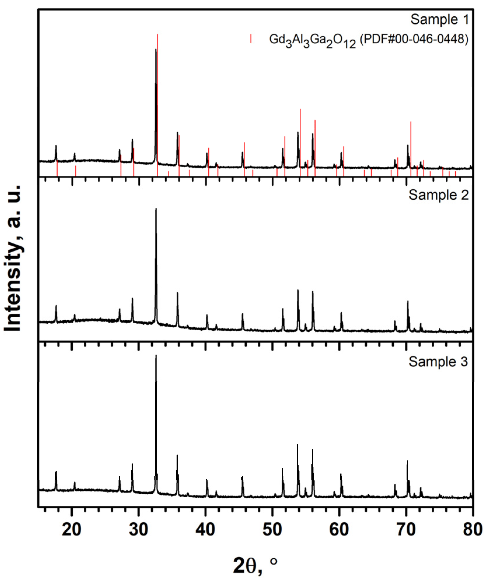
Figure 1. XRD patterns of the GAGG:Ce,Cr,Mg,Ca micropowder samples.
Crystals 11 00673 g001
Figure 2. Excitation and emission spectra of GAGG:Ce,Cr,Mg,Ca powder samples (λex = 270 nm). Inset represents zoomed in spectral region from 350 to 650 nm.
Crystals 11 00673 g002
Figure 3. Excitation and emission spectra of GAGG:Ce,Cr,Mg,Ca powder samples (λex = 450 nm).
Crystals 11 00673 g003
Figure 4. Diffuse reflection spectra of GAGG:Ce,Cr,Mg,Ca powder samples.
Crystals 11 00673 g004
Figure 5. Reflection spectra of GAGG:Ce,Cr,Mg,Ca powder samples in the UV region.
Crystals 11 00673 g005
Figure 6. Decay curves of the Cr3+ emission of the GAGG:Ce,Cr,Mg,Ca powder samples upon 270 nm excitation.
Crystals 11 00673 g006
Table 1. Chemical composition of synthesized powders determined by ICP-OES.
Name of SampleFormula of Sample
Sample 1GAGG: Ce 0.05%, Ca 100 ppm, Mg 7 ppm, Cr 15 ppm
Sample 2GAGG: Ce 0.05%, Ca 94 ppm, Mg 8 ppm, Cr 15 ppm
Sample 3GAGG: Ce 0.05%, Ca 57 ppm, Mg 9 ppm, Cr 15 ppm
Table 2. Synthesized powder samples.
SampleDecay Time (ms)
GAGG: Ce 0.05%, Ca 100 ppm, Mg 7 ppm, Cr 15 ppm (Sample 1)5.9
GAGG: Ce 0.05%, Ca 94 ppm, Mg 8 ppm, Cr 15 ppm (Sample 2)6
GAGG: Ce 0.05%, Ca 57 ppm, Mg 9 ppm, Cr 15 ppm (Sample 3)6
Publisher’s Note: MDPI stays neutral with regard to jurisdictional claims in published maps and institutional affiliations.