Abstract
Hydroxyapatite is one of the most studied biomaterials in the medical and dental field, because of its biocompatibility; it is the main constituent of the mineral part of teeth and bones. In dental science, hydroxyapatite nanoparticles (HAnps) or nano-hydroxyapatite (nano-HA) have been studied, over the last decade, in terms of oral implantology and bone reconstruction, as well in restorative and preventive dentistry. Hydroxyapatite nanoparticles have significant remineralizing effects on initial enamel lesions, and they have also been used as an additive material in order to improve existing and widely used dental materials, mainly in preventive fields, but also in restorative and regenerative fields. This paper investigates the role of HAnps in dentistry, including recent advances in the field of its use, as well as their advantages of using it as a component in other dental materials, whether experimental or commercially available. Based on the literature, HAnps have outstanding physical, chemical, mechanical and biological properties that make them suitable for multiple interventions, in different domains of dental science. Further well-designed randomized controlled trials should be conducted in order to confirm all the achievements revealed by the in vitro or in vivo studies published until now.
1. Introduction
Hydroxyapatite (Ca10(PO4)6·2(OH)) is the main mineral component of teeth and bones. Bone structure contains approximately 65 wt. % hydroxyapatite, a needle-shaped compound responsible for assuring the rigidity and strength, with a length of 60 nm and a width of 5–20 nm [1]. Additionally, enamel and dentine layers in the tooth’s structure are predominantly composed of hydroxyapatite (HA) crystals. The enamel layer consists of 96 wt.% inorganic matrix and organic constituents (such as proteins and lipids) and 4 wt.% water, whereas the dentin layer consist of 70 wt.% inorganic matrix, 20 wt.% organic matrix, and 10 wt.% water [2]. The average size of hydroxyapatite crystallites in enamel, calculated using X-ray diffraction (XRD), ranges between 48 and 78 nm [1]. The HA crystallites in enamel are hexagonal and linked to form 4 µm diameter rods, whereas in mature dentine, the crystallites are in the form of flattened plates, 60–70 nm in length, 3–4 nm in thickness and 20–30 nm in width [2].
With a stoichiometric calcium-to-phosphorus ratio of 1:67 (by weight), hydroxyapatite is the most stable, and the least soluble, form of calcium phosphate in nature [3]. Due to its excellent biocompatibility and osteogenic ability, this bioceramic has been extensively studied over the years for different biomedical applications, such as implant coating, bone scaffold, bone filler or drug delivery system [3,4,5].
In order to produce synthetic HA with a precise control of its structure, several methods have been described in the literature. Szcześ et al. [4] classified the methods of preparing HA into five categories: (1) dry methods (solid-state, mechanomechanical methods); (2) wet methods (chemical precipitation, hydrolysis, sol–gel, hydrothermal, emulsion or sonochemical); (3) high-temperature processes (combustion, pyrolysis method); (4) synthesis based on biogenic sources; (5) combination methods (such as hydrothermal–microemulsion, hydrothermal–mechanochemical, hydrothermal–hydrolysis, etc.) (Figure 1).
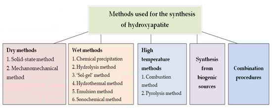
Figure 1.
Classification of hydroxyapatite synthesis procedures (Reprinted from Advances in Colloid and Interface Science, 249, Aleksandra Szcześ, Lucyna Hołysz, Emil Chibowski, Synthesis of hydroxyapatite for biomedical applications, 321–330, 2017, with permission from Elsevier).
Depending on the synthesis process, a variety of HA structures are available, such as nanorods, hierarchically nanostructures mesoporous microspheres, mesoporous rhombs, or hollow microspheres [4]. Different kinds of organic additives (aminoacids, surfactants) are used in the synthesis of hydroxyapatite, determining several types of structures, such as nanorods, nanowires, nanotubes, spindle-like particles, etc. Hydroxyapatite (HA) is currently used in clinical oral surgery, especially in bone tissue regeneration, such as for the treatment of periodontal bone defects, filling bone defects following cyst removal and apicoectomies, or, in the case of dental implant removal, to increase the width of atrophic alveolar ridges. Additionally, hydroxyapatite scaffolds are used in maxillofacial surgery to reconstruct parts of maxillary bones, or other parts of the facial skeleton, or even in pre-prosthetic surgery, in order to increase the width of the alveolar ridges [6,7].
Surface properties of nanosized HA are different from conventional micro-sized HA (Figure 2 and Figure 3); due to its unique quantum confining effects and the reactivity of the surface area, nanocrystalline hydroxyapatite display better bioactivity, biocompatibility and improved mechanical properties than microcrystalline and bulk HA [1,8,9,10,11,12].
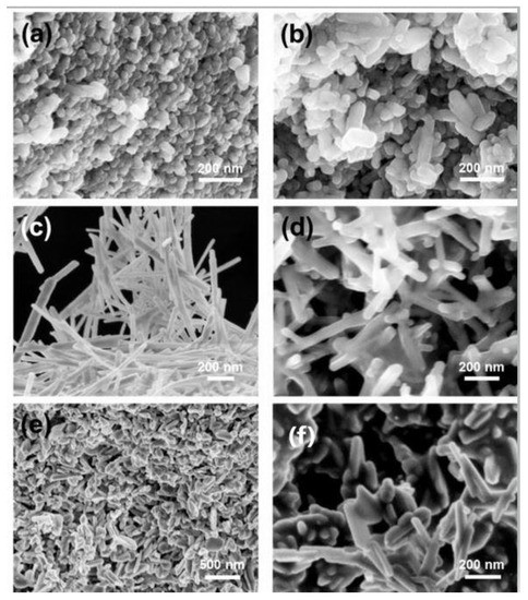
Figure 2.
SEM images of hydroxyapatite nanoparticles with different morphologies, obtained from the methods proposed by Sans et al. [5]: (a) spherical polymorphs; (b) rod shapes; (c) belts; (d) belts with smaller aspect ratios; (e) low magnification of needle-flakes; (f) high magnification of needle-flakes. Reprinted with permission from Jordi Sans, Vanesa Sanz, Jordi Puiggalí, Pau Turon and Carlos Alemán, Controlled Anisotropic Growth of Hydroxyapatite by Additive-Free Hydrothermal Synthesis, Cryst. Growth Des. 2021, 21, 2, 748–756. Copyright (2020) American Chemical Society. Accessed on 30 May 2021.
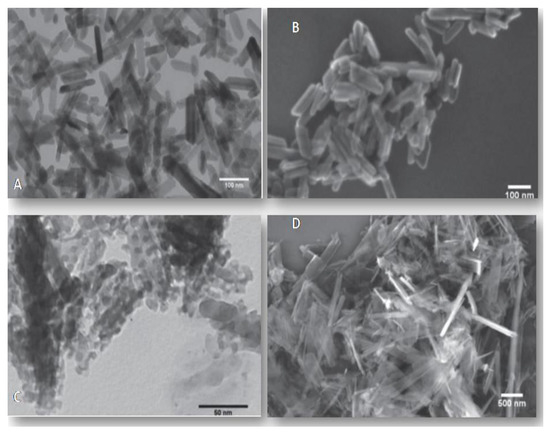
Figure 3.
Electron microscopy images (transmission electron microscopy—TEM, and scanning electron microscopy—SEM) of hydroxyapatite nanoparticles synthesized by hydrothermal processes by Arantes et al. [7]: (A) HA nanoparticles obtained at 100 °C, over 24 h, with the precursor concentrations of 0.03 mol/L−1 (TEM, scale bar value 100 nm); (B) HA nanoparticles obtained at 140 °C, over 48 h, with the precursor concentrations of 0.03 mol/L−1 (SEM, scale bar value 100 nm); (C) HA nanoparticles obtained at 100 °C, over 24 h, with the precursor concentrations of 0.1 mol/L−1 (TEM, scale bar value 50 nm) (D) HA nanoparticles obtained at 140 °C, over 48 h, with the precursor concentrations of 0.1 mol/L−1 (SEM, scale bar value 500 nm). Adapted from [7] under the terms of the Creative Commons CC BY license, accessed on 30 May 2021.
Hydroxyapatite nanoparticles (HAnps) have received great attention over the years in the medical field (for osteoporosis treatment), as well as in the oral surgery field (as a treatment in cases of alveolar bone destruction) [13,14,15,16]. HAnps can also prevent dental caries [16]; the particles can be introduced in toothpastes, where they will bond with both proteins and with fragments of plaque and bacteria, having the ability to act as a filler, repairing small defects on enamel surface [17]. Due to the high biocompatibility and bioactivity [17,18,19], as well as the antibacterial effect [20], hydroxyapatite nanoparticles have the ability to improve the properties of materials currently used in restorative dentistry [7,18].
Various types of hydroxyapatite reinforced materials (ion-doped HA, pure HA, or HA/polymer composites) have been investigated in experimental studies, clinical trials or systematic reviews. Although those materials are known to have non-immunogenic properties, are biocompatible and bioactive, and with good bone conductivity, there are also some disadvantages to be mentioned, such as brittleness and quick aggregation [21]. Nevertheless, due to the lack of toxic and allergenic properties, in dentistry, hydroxyapatite is used for coating dental implants, as a bone substitute, bone cement, or as a component of composite materials, dental cements and toothpastes [22].
In recent years, several reviews on hydroxyapatite or nano-hydroxyapatite have been published [20,23,24]. Material scientists and clinicians have studied hydroxyapatite-based materials for decades; therefore, reviews and literature updates are an excellent way for new researchers to gain a better understanding of the qualities of HAs. Thus, in this paper, we aimed to present the current state of knowledge and progress in the synthesis and the applicability of hydroxyapatite nanoparticles in restorative and conservative dentistry.
2. Characterization and Biocompatibility of Systems Based on Hydroxyapatite Nanoparticles
Hydroxyapatite nanoparticles (HAnps) vary in size from 20 to 80 nanometers (nm), with specific surface areas (SSAs) ranging from 15 to 50 m2/g. HAnps in the 100 nm range have also been observed, with SSAs ranging from 5 to 10 m2/g. Hydroxyapatite nanoparticles can be synthesized in ultra-pure and high-pure forms, either clear, coated, or in dispersed forms [25].
Natural hydroxyapatite presence in bone tissues is in the form of nanometer-size needle-like crystals of approximately 5–20nm width and 60nm length, with a poorly crystallized non-stoichiometric apatite phase containing CO32−, Na+, F−, and other ions in a collagen matrix. Compared to conventional hydroxyapatite bioceramics, nano-hydroxyapatite’s properties (such as surface grain size, pore size, or wettability) could control protein interactions, in the context of adsorption, configuration and bioactivity [26]. Therefore, HAnps could modulate subsequent enhanced osteoblast adhesion. Regarding this aspect, Webster et al. [27,28,29] proved that while these enhanced osteoblast functions refer to proliferation, alkaline phosphatase synthesis and calcium-containing mineral deposition, the size and topography and surface wettability of HAnps could promote increase in selective osteoblast adhesion (by increasing the adsorption of a protein—vitronectin—which mediates the osteoblast adhesion), and could also affect the conformations that enhance osteoblasts’ functions.
2.1. Synthesis of Hydroxyapatite Nanoparticles
Synthetic hydroxyapatite can be produced using different methods, including dry methods, wet chemical reactions, or mechanochemical reactions (Figure 1), and a few of these methods can generate nanosized hydroxyapatite [29,30,31,32]. During the synthesis of hydroxyapatite nanoparticles, the challenges refer to the formation of phase impurities (other than calcium phosphate salts), as well as the difficulties in controlling the size of the particles, the size distribution, morphology, crystallinity, and degree of particle agglomeration [29,33].
Among the synthetic procedures used for the production of hydroxyapatite nanoparticles, hydrolysis, sol–gel technique, mechanochemical or wet-chemical precipitation have previously been studied [33]. Hydroxyapatite nanoparticles (HAnps) obtained through hydrolysis (a low temperature procedure) have grain sizes between 20 and 50nm, with a diverse morphology, but a high phase purity of HAnps [33,34].With a crystalline size between 20 and 60nm, the ‘sol–gel’ method may also be used to obtain high phase purity of HAnps, with diverse morphology [33,35]. Mechanochemical procedures can be used to synthesize nanosized hydroxyapatite with an average particle size of 25 nm, but the result is non-stoichiometric HAnps with low phase purity, even though the process can be performed at room temperature and at a low cost [33,36].
Typical methods for producing HAnps require long processing times (e.g., 24 h) as well as extreme processing conditions (e.g., high pH, temperature, or ultra-sonication), which usually results in HAnps with properties that differ from biological apatite. The use of a simulated body fluid (SBF) with a salt composition similar to human blood plasma and an increased precursor concentration (i.e., Ca2+ and PO43− ions increased 3.5-fold) has been shown to be successful in overcoming these difficulties. The preparation of “bone-like” carbonated HAnps required 3 h, yielding HAnps crystals with a uniform rod shape and dimensions of about 27 nm × 7 nm. Due to the short time needed to prepare HAnps, this technique offers great potential for the production of HAnps coatings on metallic biomaterials, because productivity might be increased [37,38].
Although the precise control of particle size and particle size distribution is challenging in the nanoparticle technology field, Kuśnieruk et al. [1] proved that, by changing the parameters for hydrothermal synthesis, the diameter of hydroxyapatite nanoparticles (HAnps) could be controlled. The obtained nanoparticles with average particle diameters ranging from 8 to 39 nm were characterized by having a homogeneous morphology with a needle shape and a narrow particle size distribution, showing strong similarities with natural occurring apatite, found on dental or bone tissues. Moreover, the by-product of synthesizing HAnps by this method was only water. These results offer important information regarding the synthesis of HAnps with a controlled size (8 nm, 258m2/g), using room temperature as one of the parameters, and without any specialized equipment [1].
Over the last decade, non-synthetic sources of hydroxyapatite have also been studied. The majority of non-synthetic hydroxyapatite processing methods are variations on the above-mentioned synthetic methods. The main distinction is that some of the raw materials are derived from natural sources, such as plant sources, animal sources, biogenic sources and aquatic sources (e.g., eggshells, snail shells, plants, seashells, bovine or fish bones, etc.). The results of studies investigating the non-synthetic sources of HA revealed that they have the potential to be economically viable when compared with synthetic sources (in some cases, natural sources are waste products of other human activities). In addition, natural sources typically need calcination at a specific temperature, and nothing else, in order to produce HA with properties comparable to human bone apatite, with the right characteristics at an adequate Ca:P ratio [33].
Although the nanoprecipitation technique is the most frequently used method for HAnps synthesis, it is possible to obtain a high level of control over morphology and size by modifying experimental conditions [37,39,40] and by adding surfactants [41] (e.g., stearic acid, monosaccharides, and cetyltrimethylammonium bromide (CTAB) and chelating agents [42] (e.g., trisodium citrate and potassium sodium tartrate).
Synthetic templates, which serve as molecular cages capable of blocking reactions in the nanoscale region, represent a recent alternative method for preparing nanoparticles [43,44,45]. Using saccharide cages stabilized by heavy intermolecular interactions, this method has also been successfully used to prepare hydroxyapatite nanoparticles [46].
Several methods can be used for the synthesis of hydroxyapatite, although each method requires several processing parameters such as pH, temperature, a balanced molar ratio of calcium and phosphate precursors, etc., for producing stoichiometric HA particles. Although, a variety of methods can be used for preparing HA nanoparticles, only a few of them are satisfactory in terms of economic and performance, due to the diverse materials needed for the synthesis, complex and expensive procedures, severe aggregation and agglomeration, wide particle distribution, or various phase impurities that might occur in the crystal structure [24].
2.2. Characterization of Hydroxyapatite Particles
In order to characterize hydroxyapatite in the form of nanoparticles, rods, and discs, techniques such as spectroscopic and direct visualization have been used (Table 1); in this context, Fourier-transform infrared (FTIR) and Raman spectroscopies are often used to confirm the chemical composition of the HA. X-ray photoelectron spectroscopy (XPS) is a surface-sensitive quantitative spectroscopic technique which provides elemental composition at the parts per thousand range, as well as information regarding the empirical formula, chemical state, and electronic state of the elements within the material [47].

Table 1.
Classification, features, and limitations of the techniques used for HA characterization. Adapted from [47] under the terms of the Creative Commons CC BY license, accessed on 30 May 2021.
2.3. Synthesis of Hydroxyapatite Nanocomposites
Hydroxyapatite-based composites are essential in the field of bone tissue engineering. HA particles are commonly used for the fabrication of coatings, layers or thin films on the surface of prosthetic devices, in order to accelerate bone healing, shortly after implantation. In addition, HAs provide properties such as osteoinduction or antibiotic activity [48]. Different fabrication technique scan be used for hydroxyapatite coatings; electrochemical, electrophoretic or plasma spray deposition methods are frequently considered because of the flexibility in deposition parameters and the ability to fabricate smooth coatings with strong adhesion to the substrate [23].
The use of a scaffold is also an essential factor for hard tissue regeneration by providing the desired surface and space for cells to attach, proliferate, migrate, and differentiate to organize a normal bone tissue. In the field of dentistry and oral surgery, the preparation of biomaterials as an artificial scaffold, to act as a synthetic extracellular matrix, provides physical support to cells and the natural environment for cell proliferation and differentiation [48]. Biodegradable scaffolds represent a key point of the technology, in order to fulfill the requirements of optimum bone regeneration and repair. Hydroxyapatite, as a bioactive ceramic, can be used as a bone substitute, but also as a scaffold material in bone tissue engineering, because of its bioactivity, biocompatibility and osteoconductivity. However, due to HA’s low strength and toughness, various kinds of natural polymers, synthetic polymers (such as chitosan), protein (collagen), or other bioceramics have been used along with hydroxyapatite particles in order to develop different composite porous scaffolds [49].
Physical deposition techniques (such as thermal spraying techniques, electrophoretic deposition, or physical vapor deposition techniques) and wet chemical deposition procedures (such as chemical vapor deposition technique, biomimetic deposition techniques, sol–gel method, electrochemical deposition, micro-arc oxidation, electrospray deposition, drop or dropped-on-demand microdispensing) are the two main categories of methods used for HA particle coating production procedures. Nevertheless, the success or failure of the coating will depend on the prior preparation of the substrate, regardless of the method used. In order to remove dirt, oils, and other residues, surface cleaning is required, using either an ultrasonic bath of ethanol or acetone, followed by an acid treatment (etching) with/without sand or grit-blasting. Those procedures are required in order to facilitate the subsequent attachment and stability [50].
Due to their composition and crystalline structure, hydroxyapatite particles allow for several substitutions, such as: cationic substitutions—zinc-HAp; copper-substituted hydroxyapatite, silver-HAp, Mg-Hap, Sr-Hap) or anionic substitutions—silicon-substituted hydroxyapatite coatings—Si-HAp, fluoride-substituted hydroxyapatite coatings, carbonate-substituted hydroxyapatite coatings, or even co-substituted hydroxyapatite coatings. In vivo studies have revealed that substituted hydroxyapatite coatings can improve the performance of dental and orthopedic implants. In this context, Si-HAp coatings deposited on porous metallic implants have shown excellent bone regeneration capabilities, whereas other ionic substituents (Sr2+, Mg2+ or Zn2+) stimulate bone healing. Additionally, in vitro studies have revealed that substitutions with silver or copper have shown have antimicrobial properties against S.aureus and S. epidermidis [50]. Hydroxyapatite nanocomposites are multiphase materials and can be considered inside the bionanocomposite group. Those materials usually have improved structural and functional properties with respect to the constitutive phases. When compared to microscale hydroxyapatite composites, hydroxyapatite nanocomposites showed improved bioactivities (and mechanical properties) [51,52,53,54].
Hydroxyapatite nanocomposites can be produced by combining with other particles or coating on a matrix (polymer or metal) using a variety of methods. Using thermo-mechanical mixing, hydroxyapatite nanoparticles (as a filler) can be incorporated into the thermo-plastic polymer matrix (using a temperature above the melting point of the polymer matrix). Solvent casting methods refer to mixing hydroxyapatite nanoparticle dispersion with a polymer solution, followed by evaporation of the solvent for consolidation. By using in situ polymerization, hydroxyapatite/polymer nanocomposites can be obtained through the polymerization of monomers in which HAnps are dispersed. In situ precipitation refers to the precipitation of hydroxyapatite nanocrystals in the presence of a polymer matrix (usually a hydrogel), whereas co-precipitation involves the precipitation of HAnps in polymer solution which further induces the polymer precipitation. Several HA/ceramic nanocomposites can be obtained using sol–gel methods. By using powder metallurgy techniques, HA/metal composites (e.g., HA/titanium composites) can be prepared. This technique is a process of blending at least two kinds of powder, compressing them into a desired shape, and sintering the compressed material in a controlled atmosphere, in order to bond the powders. This technique can also be used to fabricate HA/ceramic composites [54].
Chitosan [poly-(β(1→4))-2-amino-2-deoxy-d-glucose] (CHI) is a natural, biocompatible, and biodegradable polymer, with antimicrobial and antioxidant properties, used in different biomedical fields, as well as in tissue engineering [55,56,57]. Carboxymethylated chitosan (CMC) is more soluble compared to chitosan and is capable of absorbing calcium ion, as well as enhancing bone regeneration [58]. HAnps were embedded in CHI and CMC matrices, using the co-precipitation method in water. Selected biopolymers function as capping ligands, controlling the nucleation and growth rate of HA particles during the precipitation process and forming a biocomposite network in the process. As a result of the higher surface area of the particles, CMC was able to generate HAnps with a smaller size and narrower size distribution, as well as higher cell viability of the bionanocomposite [37,59].
Technologies such as electrospinning enable the synthesis of micro/nanofibers by applying a strong electrical field, which allows the incorporation of drugs or nanoparticles into the fibers [60].
Due to the lack of adhesion between the inorganic and organic phases, the electrospinning of polymer solutions containing HAnps might determine a decrease in mechanical properties and a loss of structural stability of the final scaffold [61]. Therefore, coating HAnps with lipophilic compounds, such as oleic acid, represents a choice for improving interactions between the inorganic and organic components [62]. Additionally, the effective electrospinning of poly(lactic-co-glycolic acid) and HAnps grafted with polylactide (PLA) as a compatible agent, represent another option [63].
Considering the importance of preventing biofilm formation on the surfaces of implanted medical devices, any antimicrobial technique that can provide long-term biofilm protection is appealing and should be investigated. Antimicrobial scaffolds based on HAnps have the potential to be used in different medical domains. This aspect has revealed several research opportunities, although some caution should be exercised when silver ions are introduced into the scaffold, due to local cytotoxicity which may result from a high load or even a rapid and local release [37,64].
The incorporation of antibiotics has also been considered, but due to the growing resistance of microorganisms to antibiotics, other preventive strategies are being investigated. In the study published by Rauschmann et al., the antibiotic release properties and biocompatibility of a bioresorbable composite of calcium sulphate and HAnps were investigated. The nanocomposite revealed excellent resorption and biocompatibility properties, as well as an optimal release of gentamicin (94.7%) and vancomicin (96.3%). Within the first 24 h, and for three and four further days, released gentamicin and vancomycin concentrations exceeded 100-fold and 10-fold the minimal inhibitory concentrations (MICs) of susceptible Staphylococcus aureus [65].
Biocompatible nanosubstrates, such as HAnps, in combination with biomarkers, are promising candidates for studying disease etiology and progression through imaging technology [37]. Magnetic nanoparticles have substantial potential in medicine, for example, in non-invasive magnetic resonance imaging used in tumor detection [66]. Contrast agents use paramagnetic, super-paramagnetic, and ferromagnetic materials to alter the image contrasts between healthy and pathological tissues [67]. Due to their large surface area, the ability to target and deliver to a particular tumor, and control over the blood circulation half-life, nanosized contrast agents can be successfully used in medical imaging [68].
Gene therapy is under continuous development; therefore, non-viral transfecting agents based on HAnps have gained popularity over the last decade due to the lack of side effects. An in vitro study published by Cheang et al. in 2018 investigated the cytotoxicity and transfection efficiency of graphene oxide–hydroxyapatite nanocomposites (GO–Hap) when delivering thymidine kinase gene (HSV-TK) for the inhibition of human breast cancer growth. The nanocomposite particles were able to deliver efficiently pDNA without obvious cytotoxicity. In addition, GO–HAp/p-HRE/ERE-Sur-TK combined with ganciclovir (GCV) inhibited the proliferation and induced apoptosis in cancerous cells, and the cytotoxic effects were tolerable in normal breast cells [69].
Another study, published in 2019 by Fang et al., investigated the therapeutic effect on bone regeneration of hydroxyapatite microspheres (gelatin/nano-hydroxyapatite microsphere embedded with stromal cell-derived factor-1:GHM-S). An in vivo model of alveolar bone defects on rats was used to determine the effect of the biomimetic hydroxyapatite microspheres on bone formation, evaluated through micro-CT imaging and histological analysis. The results showed that biomimetic hydroxyapatite microspheres can improve alveolar bone regeneration [70].
By delivering drugs after crossing biological barriers and targeting diseases’ nuclei, regulated drug delivery systems are expected to open up new therapies. HAp has a variety of potential biomedical applications as a carrier different of therapeutic agents (such as drugs, genes, antigens, enzymes, and proteins) [71,72]. By masking the biomolecules, hydroxyapatite nanoparticles act as an excellent carrier which can deliver the drug just onto the target [37].
Although the results of in vivo or in vitro studies are promising, transferring new biomedical devices or materials from the lab to clinical applications is challenging. There are many fabrication methods that allow for manufacturing substituted nano-hydroxyapatite coatings or nanocomposites, which have evidenced satisfactory in vitro behavior. However, due to their size, nanoparticles can be inhaled or absorbed through teguments or digestive system. Their cytotoxicity will depend on their origin, but also on the dose and exposure time, aggregation, concentration, particle size and shape, surface area, and crystal structure, as well as any pre-exposure [73,74,75].
3. Applications of Systems Based on Hydroxyapatite Nanoparticles in Dental Science
The field of nanodentistry started to develop around the year 2000, as nanotechnology evolution affected the fields of diagnostics, materials, restorative dentistry and oral surgery. Nanodentistry aims to ensure comprehensive oral healthcare of the patient. As more advanced and accurate diagnostic methods become available, several oral diseases can be prevented or treated at early stages [76,77].
Among the nanomaterials available, nanocomposites, nanoimpression materials, and nanoceramics can be successfully used for the restoration of decayed, carious, missing and fractured teeth [76].
3.1. Hydroxyapatite Nanoparticles in Prophylactic and Regenerative Dentistry
With the help of nanotechnology, new strategies in prophylactic dentistry have been developed, related to the control and management of bacterial biofilms, and the remineralization of incipient carious lesions. Several dentifrices, mouth rinsing solutions, and remineralizing pastes (fluids) containing bioinspired nanosized apatites have been developed for the regulation of bio-adhesion and biofilm control in preventive dentistry [78,79,80].
Biomimetic hydroxyapatite nanoparticles (HAnps) facilitate remineralization of the enamel surface, providing excellent biological properties such as biocompatibility, lack of toxicity, and inflammatory and immunological responses [80]. In preventive dentistry, HAnps have been investigated in the context of different sub-domains, such as the remineralization of incipient carious lesions, dentinal hypersensitivity and dental erosion.
Regarding the remineralization of incipient carious lesions, different products (toothpastes, mouthwash products, chewing gums) are continuously being designed and investigated [81]. In an in vitro study published in 2011, Tschoppe et al. investigated the effect of different HAnps toothpastes on the remineralization of bovine enamel and dentine subsurface lesions, compared to amine fluoride toothpaste. Their results showed similar capacities to remineralize enamel and dentine subsurface lesions between HAnps toothpastes. In addition, fluoride toothpaste had the least remineralizing impact on all hard tissues, although an increase in lesion depths has also been observed [82].
A more recent study, published by Esteves-Oliveira et al. in 2017, investigated the caries-protective effect of different fluoride and non-fluoride toothpastes [83]. The hypothesis of their in vitro study was that no significant differences on the inhibition of demineralization would be observed between the anti-caries and anti-erosive toothpastes (based on fluoride), and the fluoride-free control (based on hydroxyapatite nanoparticles).The results showed that HAnps toothpaste did not significantly reduce enamel demineralization as compared to the control group, an aspect which was also observed by Souza et al. in a study published in 2015 [84].
The results of an in vitro study, performed on 30 extracted human premolars, by Sharma et al. (2017), proved that both HAnps and casein phosphopeptide–amorphous calcium (CPP–AC) phosphate have a remineralizing effect on early-stage caries. HAnps were, however, more effective in increasing the calcium and phosphorus content of enamel compared to CPP–AC, and this effect was more evident over a longer treatment period [85].
Utneja et al. (2018) investigated the use of HAnps as a fortifier for pits and fissure sealants (PFS). In their in vitro study, the remineralization potential was evaluated for a nano-HA filler sealant, a nano-HA filler with silica co-filler sealant, and a 10% nano-HA and nano-amorphous calcium phosphate (n-ACP) co-filler, using electron scanning microscopy (SEM). The authors investigated the degree of conversion (DOC), curing depth (CD), and microsphere bond strength (MBS) for all three sealants tested. The results revealed a remineralized region at the interface between sealant and enamel for all three hydroxyapatite-filled sealants. In addition, the sealant with 10% nano-HA along with 20% ACP had a significantly higher DOC, as well as an increased ion release compared to the control groups [86].
Jena et al. (2017) aimed to compare the tubule-occluding efficacy of four different desensitizing dentifrices using scanning electron microscopy (SEM). A total of 62 dentin blocks were obtained from extracted human molars and divided into five groups: two blocks were considered control groups (without any treatment), and four groups of 15 blocks each received a 2 min/day brush with toothpastes, for 14 days, and were stored in artificial saliva. Group 2 received Pepsodent Pro-sensitive relief and repair, Group 3 received Sensodyne repair and safety, Group 4 received Remin Pro, and Group 5 received a research dentifrice with 15% nano-HA crystals.SEM microscopy and a statistical study using the Kruskal–Wallis test were used to perform the assessment. The results revealed that the highest tubule occlusion percentage was for Groups 4 and 5, although an increased occlusion was observed among all study groups compared to the control blocks [87].
Vjayasankari et al. (2019) aimed to evaluate the remineralization potential of an experimental nano-HA paste on artificial carious lesions, by using scanning electron microscopy (SEM) with energy-dispersive X-ray (EDX). All four pastes tested (1% experimental nano-HA paste, 10% nano-HA experimental paste, 1% commercially available nano-HA paste and a caseinphosphopeptide–amorphous calcium phosphate (CPP–ACP)) showed significant changes in calcium and phosphorus weight percentages after remineralization. The 10% nano-HA experimental paste determined the highest mean value of calcium and phosphorus weight percentage. After remineralization, both 10% experimental nano-HA paste and CPP–ACP paste showed favorable enamel surface changes in SEM analysis [88].
Due to their remineralization properties, several researchers have investigated whether systems reinforced with HAnps could be successfully used in treating dentinal hypersensitivity (DH).
Wang et al. (2016) published a clinical trial that evaluated the effect of nano-HA pastes indicated for professional use (Desensibilize Nano-P) with or without experimental home-care application to Pro-Argin (a new technology) compared to a fluoride varnish (an already proven treatment) on DH relief after one and three months. The results obtained revealed that Desensibilize Nano (with or without home-care product association) had a comparable effectiveness in reducing dentinal hypersensitivity, compared to the fluoride varnish [89]. Their results confirmed the conclusions of a previous clinical study published by Gopinath et al. (2015) [90].
Another clinical study performed by Alhamed et al. (2020) on 90 carious lesions (incipient phase) assessed and compared the effectiveness of three different remineralizing agents (Tricalcium phosphate paste, Fluoride varnish, and Nano-hydroxyapatite gel) using a DIAGNOdent device. The authors concluded that all three remineralizing agents were effective in the treatment of initial carious lesion, while the most effective remineralizing agent was nano-hydroxyapatite [91].
An innovative strategy in the matter of reducing dentinal hypersensitive and remineralizing dentinal tubules is the use of more complex products, and combinations between hydroxyapatite nanoparticles and different compounds.
In an in vitro study, performed on 100 extracted teeth, Konagala et al. (2020) demonstrated the synergistic effect of arginine on the remineralizing potential of fluoride varnish and nano-hydroxyapatite. The results revealed an overall increase in the remineralization potential for the products combined with arginine, with a significant increase in fluoride gain for arginine+fluoride varnish [92].
3.2. Hydroxyapatite Nanoparticles in Aesthetic and Conservative Dentistry
In aesthetic dentistry, hypersensitivity may be an unwelcome side effect in cases of teeth-whitening procedures. Therefore, hydroxyapatite nanoparticle-reinforced products were also investigated in this context. In a randomized clinical trial, Vano et al. (2015) investigated the bleaching effect of 6% hydrogen peroxide (HP) with or without 2% nano-hydroxyapatite. The study included sixty subjects, and tooth color and tooth sensitivity were analyzed before and after treatment. Statistical analysis of the results revealed that both treatments showed significant improvements in tooth shade. Although the bleaching effectiveness of the tested products was comparable, the use of 6% HP with 2% nano-HA reduced the incidence of sensitivity during the bleaching treatment compared to a bleaching agent that did not contain n-HA [93].
An in vitro study was conducted by Kutuk et al. (2018) on 117 extracted teeth, assessing the color change, micro-hardness, and chemical composition of bleached enamel with different desensitizing agents. They concluded that using fluoride, casein phosphopeptide–amorphous calcium phosphate, potassium nitrate, or nano-hydroxyapatite as desensitizing agents after bleaching or combined with the bleaching agent did not inhibit the bleaching impact of the agent. Regardless of the desensitizing agent, the results showed that all treated enamel micro-hardness recovered after 14 days [94].
In dental science, the use of HAnps as a component that could enhance the properties of existing restorative dentistry materials has also been investigated. Bioactivity in dental resin composite restorations is one of the most significant achievements in modern biomaterial science and research. At the microscopic level, homogeneous dispersion of HAnps within the organic matrix of dental resin composite (and silanization of fillers) may enhance physical and mechanical properties [95,96,97].
An experimental study published in 2018 by Khalid et al. aimed to investigate (in terms of quantitative and qualitative analysis of monomer leaching) two experimental resin composites based on low concentrations of n-HA (30% silanized nano-HA;45% silanized nano-HA) as fillers with resin composites based on highly concentrated micro-fillers. The results revealed that 45% nano-HA based experimental composite showed minimal leaching of monomers compared to other composites [95].
Nobre et al. published an in situ pilot study in 2020, which investigated the effects of three hydroxyapatite-based solutions and their interactions with different dental material surfaces under oral conditions. Intraoral splints were implanted (with mounted samples from enamel and from three dental materials—titanium, ceramic and polymethyl-metacrylate—PMMA) into two volunteers. Additionally, three hydroxyapatite watery solutions (5%) were prepared with different shapes and sizes of HAnps (HAI, HA II, HA III). After 3 min of pellicle formation, a 10 mL rinse was performed for 30 s. A rinse with water served as the control. The particles were characterized using scanning electron microscopy (SEM) and transmission electron microscopy (TEM), whereas the pellicle–HA interactions were evaluated using SEM. The size ranges of the particles applied showed considerable variance in the SEM and TEM results. Under oral conditions, a heterogeneous hydroxyapatite coating was present on enamel, titanium, ceramic, and PMMA surfaces after 2 h. On enamel, titanium, and PMMA surfaces, bridge-like structures were visible between the HAnps and the pellicle. The authors concluded that under oral conditions, HAnps can bind to both enamel and artificial dental surfaces. The experiment revealed that the pellicle acts as a connection between the HAnps and the surface of the materials [98].
Jardim et al. investigated the synthesis of three dental composites with different concentrations of hydroxyapatite nanoparticles (HAnps) and evaluate their Ca2+ and PO43− release, enamel remineralization potential, and their physical–mechanical properties, compared to a non-HAnps composite. The results showed that HAnps incorporated in dental composites determined a long-term Ca2+ and PO43− release associated with a remineralizing potential of caries-like enamel lesions, mainly at potentially cariogenic pH levels (<5.5). The values of the flexural strength indicated that the experimental composites could be used in load-bearing areas (posterior teeth) [99].
Fixed orthodontic appliances promote plaque biofilm formation and increase the risk of full demineralization (also known as white spot accidents), which is the underlying cause of caries. Nanotechnology is also widely applied in fixed orthodontics; therefore, the addition of antimicrobial agents into orthodontic adhesives represents an optimal solution for the prevention of white spot formation.
In this context, Sodagar et al. (2016) investigated the antibacterial properties of a conventional orthodontic adhesive containing three different concentrations of silver (Ag)/hydroxyapatite nanoparticles. The results revealed that adding 5% Ag/HAnps to orthodontic adhesives reduces the growth of cariogenic bacteria, although the non-cariogenic effect against S. sanguinis was inferior compared to S. mutans and L. acidophilus, because it is capable of producing a bacterial growth inhibition zone [100].
A more recent in vitro experimental study, published by Gilani et al. (2020), assessed the shear bond strength of the enamel adhesive and bracket adhesive junctions, by adding silver and hydroxyapatite nanoparticles in different concentrations, in order to reduce the risk of white spot development. The Adhesive Remnant Index (ARI) score was used in order to investigate the debonding format and compare the study groups to the control group according to the amount of the adhesive remaining on the enamel surface after the debonding fracture test. The results showed that the 10% concentration silver-hydroxyapatite group has a similar fracture resistance to the control group and that it can be used as a suitable alternative for orthodontic bracket bonding, with the purpose of reducing the caries risk on the enamel surfaces [101].
Hasan investigated the effects of calcium hydroxyapatite nanoparticle incorporation on polymerization as well as the shear bond strength for an orthodontic adhesive (Heliosite®, IvoclarVivadent). The author concluded that 2% hydroxyapatite nanoparticle (HAnps) incorporation with a conventional adhesive system improved both the degree of conversion and the shear bond strength, whereas the incorporation of 4% HAnps proved to have both an inferior degree of conversion and shear bond strength for the orthodontic adhesive [102].
3.3. Hydroxyapatite Nanoparticles in Restorative Dentistry
In fixed prosthodontics, the correct cementation of indirect restorations to the tooth’s structure (such as all-metal, metal–ceramic, and all-ceramic crowns, as well as intracoronal restorations, laminate veneers, etc.) forms the basis of successful treatment. The primary function of a luting agent is to fill the void at the restoration–tooth interface and maintain the restoration in place to prevent its dislodgement during mastication [103].
Biocompatibility, adequate working time, flowability, compressive strength, minimal microleakage, low solubility in oral fluids, adhesiveness, aesthetics, low cost, and ease of removal in case of excess material are all essential requirements for an ideal luting agent, although no luting agent has been developed thus far capable of meeting all those requirements [103]. Glass–ionomer cements (GICs) offer good bonding with the enamel layer, and, to some extent, to dentin, while releasing fluoride [104]. Initially, GICs were used as restorative materials those, although these materials have further evolved into luting agents, liners, bases, fissure sealants, etc. [104].
A study published by Kheur et al. (2019) investigated the effect of incorporating HAnps in glass–ionomer luting agents, assessing the flexural strength and shear bond strength to the tooth, compared to conventional GIC, resin-modified GIC, and adhesive resin. The authors concluded that, although adhesive resin had the highest flexural strength and shear bond strengths, the experimental GIC modified with HAnps was revealed to have superior bonding between the carboxyl groups of the polyacid with calcium from natural tooth structures to synthetic hydroxyapatite [104].
Alatawi et al. (2018) performed a similar study, by incorporating different concentrations of hydroxyapatite nanoparticles into glass–ionomer cement, and the results showed that the addition of HAnps enhanced the fluoride ion release of the conventional glass–ionomer cement. In addition, the bacterial inhibition against S. Mutans was significant when a concentration of 8% nano-HA was used [105].
Noorani et al. evaluated the cytotoxic effects of nano-hydroxyapatite–silica incorporated into glass–ionomer cement (GIC) on human dental pulp stem cells (DPSCs), compared to a conventional GIC and a resin-modified GIC. Compared to the conventional GIC, the experimental nano-hydroxyapatite–silica–glass–ionomer cement had a comparable biocompatibility in terms of cytotoxicity, superior to the resin-modified GIC [106].
A more complex hybrid product was tested by Pagano et al. (2019). A glass–ionomer cement—nano-hydroxyapatite–mucosal defensive agent–antibiotic emulsion—was synthesized and evaluated in terms of its mechanical, thermal and biological properties. The null hypothesis of this study was that the new product would not change GIC’s mechanical or thermal properties, or the microbiological properties and their cytotoxicity levels. The results showed an improvement in mechanical properties, particularly in a wet environment such as the oral cavity and with the powder-formulated antibiotic. The addition of HAnps, an antibiotic agent, and mucosal defensive agent significantly reduced the cement’s overall cytotoxicity [107].
An intriguing strategy was developed by Juntaveeet al. (2018) by investigating the effects of nano-hydroxyapatite (NHA) gel and Clinpro (CP) on the remineralization potential of enamel and cementum at the cavosurface area of computer-aided design and computer-aided manufacturing ceramic restoration. In a clinical context, where irregularities at the cavosurface junction and microgaps at the tooth–restoration interfaces are always present and induce bacterial accumulation leading to tooth decay, their study indicated that both HAnps gel and CP are significantly capable of remineralizing in order to help in the recovery of demineralized enamel and cementum, compared to non-treated demineralized surfaces [15].
Our research reveals that hydroxyapatite (HA) and hydroxyapatite nanoparticles (HAnps) have received considerable attention over the last decade in oral and maxillo-facial surgery, as well as in the dentistry field (Figure 4). Although the majority of the studies published so far have confirmed the promising properties of hydroxyapatite nanoparticles, the results are divergent; although several experimental studies have revealed the potential of HAnps to repair enamel, others present no differences between the HAnps treatment and standard fluoride treatment regarding the remineralization effects on the enamel layer. The reason for this could be related to the variety of methodologies applied. Only few studies have reproduced real intraoral conditions, such as salivary flow, nutrition, or bacterial conditions.
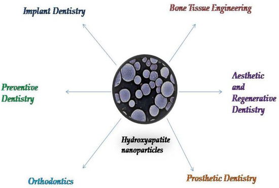
Figure 4.
The use of hydroxyapatite nanoparticles in different fields of dentistry.
4. Conclusions
Hydroxyapatite nanoparticles represent a revolutionary material with a wide use in dentistry. They provide a better source of free calcium (Ca), and, as a key element in the process of enamel remineralization, HAnps have the ability to protect against carious lesions, or even dental wear lesions. Hydroxyapatite nanoparticles have also been used as a reinforcing material to increase the quality of currently available and commonly used dental materials. This is the case with the experimental additions to conventional glass–ionomer cements, resin-based cements, or orthodontic adhesives, procedures that have resulted in major changes to these substances’ mechanical and chemical properties.
HAnps have also been investigated in cosmetic dentistry, as a bleaching adjuvant material, as well as a dentinal hypersensitivity and remineralization therapeutic factor, making them ideal for aesthetic and preventive dentistry.
Taking into consideration the increasing importance of hydroxyapatite nanoparticles in dentistry, it is very important to understand all the mechanisms of interaction of these particles with the intraoral environment. Despite the interesting results obtained thus far, there is a need for more clinical trials, with the inclusion of a large number of subjects, in order to better elucidate the biological effects of systems containing hydroxyapatite nanoparticles.
Author Contributions
Conceptualization, S.B. (Silvia Balhuc) and A.K.; methodology, M.N. and R.C.; software, M.N.; validation, S.B. (Smaranda Buduru), S.B. (Silvia Balhuc) and A.K.; resources, M.N and A.L.; writing—original draft preparation, A.L and Silvia Balhuc; writing—review and editing, A.K.; visualization, M.N.; supervision, R.C.; project administration, A.K. and S.B. (Smaranda Buduru). All authors have read and agreed to the published version of the manuscript.
Funding
This research received no external funding.
Institutional Review Board Statement
Not applicable.
Informed Consent Statement
Not applicable.
Data Availability Statement
Not applicable.
Conflicts of Interest
The authors declare no conflict of interest.
References
- Kuśnieruk, S.; Wojnarowicz, J.; Chodara, A.; Chudoba, T.; Gierlotka, S.; Lojkowski, W. Influence of hydrothermal synthesis parameters on the properties of hydroxyapatite nanoparticles. Beilstein. J. Nanotechnol. 2016, 7, 1586–1601. [Google Scholar] [CrossRef] [PubMed]
- De Dios Teruel, J.; Alcolea, A.; Hernández, A.; Ruiz, A.J.O. Comparison of chemical composition of enamel and dentine in human, bovine, porcine and ovine teeth. Arch. Oral. Biol. 2015, 60, 768–775. [Google Scholar] [CrossRef]
- Antoniac, I.; Miculescu, F.; Cotrut, C.; Ficai, A.; Rau, J.V.; Grosu, E.; Antoniac, A.; Tecu, C.; Cristescu, I. Controlling the Degradation Rate of Biodegradable Mg-Zn-Mn Alloys for Orthopedic Applications by Electrophoretic Deposition of Hydroxyapatite Coating. Materials 2020, 13, 263. [Google Scholar] [CrossRef] [PubMed]
- Szcześ, A.; Ho, L.; Chibowski, E. Synthesis of hydroxyapatite for biomedical applications. Adv. Colloid Interface Sci. 2017, 249, 321–330. [Google Scholar] [CrossRef] [PubMed]
- Sans, J.; Sanz, V.; Puiggalí, J.; Turon, P.; Alemán, C. Controlled Anisotropic Growth of Hydroxyapatite by Additive-Free Hydrothermal Synthesis. Cryst. Growth Des. 2020, 2, 748–756. [Google Scholar]
- Ghiasi, B.; Sefidbakht, Y.; Mozaffari-Jovin, S.; Gharehcheloo, B.; Mehrarya, M.; Khodadadi, A.; Rezaei, M.; Ranaei Siadat, S.O.; Uskoković, V. Hydroxyapatite as a biomaterial–A gift that keeps on giving. Drug Dev. Ind. Pharm. 2020, 46, 1035–1062. [Google Scholar] [CrossRef]
- Arantes, T.M.; Coimbra, L.M.M.; Cristovan, F.H.; Arantes, T.M.; Rosa, G.M.; Lião, L.M. Synthesis and optimization of colloidal hydroxyapatite nanoparticles by hydrothermal processes. J. Braz Chem. Soc. 2018, 29, 1894–1903. [Google Scholar] [CrossRef]
- Coelho, C.C.; Grenho, L.; Gomes, P.S.; Quadros, P.A.; Fernandes, M.H. Nano-hydroxyapatite in oral care cosmetics: Characterization and cytotoxicity assessment. Sci. Rep. 2019, 9, 1–10. [Google Scholar] [CrossRef]
- Sadat-Shojai, M.; Khorasani, M.-T.; Dinpanah-Khoshdargi, E.; Jamshidi, A. Synthesis methods for nanosized hydroxyapatite with diverse structures. Acta Biomater. 2013, 9, 7591–7621. [Google Scholar] [CrossRef]
- Molino, G.; Palmieri, M.C.; Montalbano, G.; Fiorilli, S.; Vitale-Brovarone, C. Biomimetic and mesoporous nano-hydroxyapatite for bone tissue application: A short review. Biomed. Mater. 2020, 27, 022001. [Google Scholar] [CrossRef]
- Dorozhkin, S.V. Nanosized and nanocrystalline calcium orthophosphates. Acta Biomater. 2010, 6, 715–734. [Google Scholar] [CrossRef]
- Varadarajan, N.; Balu, R.; Rana, D. Accelerated Sonochemical Synthesis of Calcium Deficient Hydroxyapatite Nanoparticles: Structural and Morphological Evolution. J. Biomater Tissue Eng. 2014, 4, 1–5. [Google Scholar] [CrossRef]
- Khajuria, D.K.; Vasireddi, R.; Trebbin, M.; Karasik, D.; Razdan, R. Novel therapeutic intervention for osteoporosis prepared with strontium hydroxyapatite and zoledronic acid: In vitro and pharmacodynamic evaluation. Mater. Sci. Eng. C Mater. Biol. Appl. 2017, 71, 698–708. [Google Scholar] [CrossRef] [PubMed]
- Sahana, H.; Khajuria, D.K.; Razdan, R.; Mahapatra, D.R.; Bhat, M.R.; Suresh, S.; Rao, R.R.; Mariappan, L. Improvement in bone properties by using risedronate adsorbed hydroxyapatite novel nanoparticle based formulation in a rat model of osteoporosis. J. Biomed. Nanotechnol. 2013, 9, 193–201. [Google Scholar] [CrossRef] [PubMed]
- Khajuria, D.K.; Zahra, S.F.; Razdan, R. Effect of locally administered novel biodegradable chitosan based risedronate/zinc-hydroxyapatite intra-pocket dental film on alveolar bone density in rat model of periodontitis. J. Biomater. Sci. Polym. Ed. 2018, 29, 74–91. [Google Scholar] [CrossRef] [PubMed]
- Juntavee, N.; Juntavee, A.; Plongniras, P. Remineralization potential of nano-hydroxyapatite on enamel and cementum surrounding margin of computer-aided design and computer-aided manufacturing ceramic restoration. Int. J. Nanomed. 2018, 13, 2755–2765. [Google Scholar] [CrossRef] [PubMed]
- Hannig, M.; Hannig, C. Nanotechnology and its role in caries therapy. Adv.Dent. Res. 2012, 24, 53–57. [Google Scholar] [CrossRef]
- Cai, Y.; Liu, Y.; Yan, W.; Hu, Q.; Tao, J.; Zhang, M. Role of Hydroxyapatite Nanoparticle Size in Bone Cell Proliferation Role of hydroxyapatite nanoparticle size in bone cell proliferation. J. Mater. Chem. 2007, 17, 3780–3787. [Google Scholar] [CrossRef]
- Goldberg, M.; Kulkarni, A.B.; Young, M.; Boskey, A. Dentin: Structure, composition and mineralization. Front. Biosci. 2011, 3, 711–735. [Google Scholar] [CrossRef]
- Huang, S.B.; Gao, S.S.; Yu, H.Y. Effect of nano-hydroxyapatite concentra- tion on remineralization of initial enamel lesion in vitro. Biomed. Mater. 2009, 4, 034104. [Google Scholar] [CrossRef]
- Shi, H.; Zhou, Z.; Li, W.; Fan, Y.; Li, Z.; Wei, J. Hydroxyapatite based materials for bone tissue engineering: A brief and comprehensive introduction. Crystals 2021, 11, 149. [Google Scholar] [CrossRef]
- Pajor, K.; Pajchel, L.; Kolmas, J. Hydroxyapatite and Fluorapatite in Conservative Dentistry and Oral Implantology—A Review. Materials 2019, 12, 2683. [Google Scholar] [CrossRef] [PubMed]
- Awasthi, S.; Pandey, S.K.; Arunan, E.; Srivastava, C. A review on hydroxyapatite coatings for the biomedical applications: Experimental and theoretical perspectives. J. Mater. Chem. B 2020, 9, 228–249. [Google Scholar] [CrossRef] [PubMed]
- Mohd Pu’ad, N.A.S.; Abdul Haq, R.H.; Mohd Noh, H.; Abdullah, H.Z.; Idris, M.I.; Lee, T.C. Synthesis method of hydroxyapatite: A review. Mater. Today Proc. 2019, 29, 233–239. [Google Scholar] [CrossRef]
- Ferraz, M.P.; Monteiro, F.J.; Manuel, C.M. Hydroxyapatite nanoparticles: A review of preparation methodologies. J. Appl. Biomater Biomech. 2004, 2, 74–80. [Google Scholar]
- Webster, T.J.; Siegel, R.W.; Bizios, R. Enhanced surface and mechanical properties of nano phase ceramics to achieve orthopaedic/dental implant efficacy. Key Eng. Mater. 2001, 192, 321–324. [Google Scholar]
- Webster, T.J.; Ergun, C.; Doremus, R.H.; Siegel, R.W.; Bizios, R. Enhanced functions of osteoblasts on nanophase ceramics. Biomaterials 2000, 21, 1803–1810. [Google Scholar] [CrossRef]
- Webster, T.J. Specific proteins mediate enhanced osteoblast adhesion on nanophase ceramics. J. Biomed. Mater. Res. 2000, 51, 475–483. [Google Scholar] [CrossRef]
- Pataquiva Mateus, A.; Monteiro, F.J.; Pía Ferraz, M. Nanoparticles of hydroxyapatite: Preparation, characterization and cellular approach-An Overview. Rev. Mutis 2013, 3, 43–56. [Google Scholar] [CrossRef]
- Fox, K.; Tran, P.A.; Tran, N. Recent advances in research applications of nanophase hydroxyapatite. Chemphyschem 2012, 13, 2495–2506. [Google Scholar] [CrossRef]
- Loo, S.C.; Moore, T.; Banik, B.; Alexis, F. Biomedical applications of hydroxyapatite nanoparticles. Curr. Pharm. Biotechnol. 2010, 11, 333–342. [Google Scholar] [CrossRef]
- Paz, A.; Guadarrama, D.; López, M.E.; González, J.; Brizuela, N.; Aragón, J. A comparative study of hydroxyapatite nanoparticles synthesized by different routes. Química Nova, 2012; 35, 1724–1727. [Google Scholar] [CrossRef]
- Agbeboh, N.I.; Oladele, I.O.; Daramola, O.O.; Adediran, A.A.; Olasukanmi, O.O.; Tanimola, M.O. Heliyon Environmentally sustainable processes for the synthesis of hydroxyapatite. Heliyon 2020, 6, e03765. [Google Scholar] [CrossRef]
- Shih, W.J.; Wang, M.C.; Hon, M.H. Morphology and crystallinity of the nanosizedhydroxyapatite synthesized by hydrolysis using cetyltrimethylammonium bromide(CTAB) as a surfactant. J. Cryst. Growth 2005, 275, 2339–2344. [Google Scholar] [CrossRef]
- Sanosh, K.P.; Chu, M.C.; Balakrishnan, A. Preparation and characterization of nano-hydroxyapatite powder using sol-gel technique. Bull. Mater. Sci. 2009, 32, 465–470. [Google Scholar] [CrossRef]
- Yeon, K.C.; Wang, J.; Ng, S.C. Mechanochemical synthesis of nanocrystalline hydroxyapatite from CaO and CaHPO4. Biomaterials 2001, 22, 2705–2712. [Google Scholar] [CrossRef]
- Turon, P.; del Valle, L.J.; Alemán, C.; Puiggalí, J. Biodegradable and biocompatible systems based on hydroxyapatite nanoparticles. Appl. Sci. 2017, 7, 60. [Google Scholar] [CrossRef]
- Leena, M.; Rana, D.; Webster, T.J.; Ramalingam, M. Accelerated synthesis of biomimetic nano hydroxyapatite using simulated body fluid. Mater. Chem. Phys. 2016, 180, 166–172. [Google Scholar] [CrossRef]
- Zhang, C.; Yang, J.; Quan, Z.; Yang, P.; Li, C.; Hou, Z.; Lin, J. Hydroxyapatite nano- and microcrystals withmultiform morphologies: Controllable synthesis and luminescence properties. Cryst. Growth Des. 2009, 9, 2725–2733. [Google Scholar] [CrossRef]
- Ren, F.; Leng, Y.; Ding, Y.; Wang, K. Hydrothermal growth of biomimetic carbonated apatite nanoparticles with tunable size, morphology and ultrastructure. Cryst. Eng. Commun. 2013, 15, 2137–2146. [Google Scholar] [CrossRef]
- Cao, M.; Wang, Y.; Guo, C.; Qi, Y.; Hu, C. Preparation of ultrahigh-aspect-ratio hydroxyapatite nanofibers in reverse micelles under hydrothermal conditions. Langmuir 2004, 20, 4784–4786. [Google Scholar] [CrossRef]
- Ma, M.G. Hierarchically nanostructured hydroxyapatite: Hydrothermal synthesis, morphology control, growth mechanism, and biological activity. Int. J. Nanomed. 2012, 7, 1781–1791. [Google Scholar] [CrossRef] [PubMed]
- McCaffrey, R.; Long, H.; Jin, Y.; Sanders, A.; Park, W.; Zhang, W. Template Synthesis of gold nanoparticles with an organic molecular cage. J. Am. Chem. Soc. 2014, 136, 1782–1785. [Google Scholar] [CrossRef] [PubMed]
- Li, J.L.; Liu, X.Y.; Wang, X.G.; Wang, R.Y. Controlling nanoparticle formation via sizable cages of supramolecular soft materials. Langmuir 2011, 27, 7820–7827. [Google Scholar] [CrossRef] [PubMed]
- Zhang, D.S.; Liu, X.Y.; Li, J.L.; Xu, H.Y.; Lin, H.; Chen, Y.Y. Design and fabrication of a new class of nano hybrid materials based on reactive polymeric molecular cages. Langmuir 2013, 29, 11498–11505. [Google Scholar] [CrossRef] [PubMed]
- Liu, Y.; Chi, W.; Zhao, D.; Liu, H.; Deng, Y. Molecular-cage method: An improvement of the precipitation method in synthesinzing nanoparticles. Ind. Eng. 2016, 55, 8403–8408. [Google Scholar] [CrossRef]
- Haider, A.; Haider, S.; Han, S.S.; Kang, I.K. Recent advances in the synthesis, functionalization and biomedical applications of hydroxyapatite: A review. RSC Adv. 2017, 7, 7442–7458. [Google Scholar] [CrossRef]
- Arcos, D.; Vallet-Regí, M. Substituted hydroxyapatite coatings of bone implants. J. Mater. Chem. B 2020, 8, 1781. [Google Scholar] [CrossRef]
- Ika, D.A. Bioceramics and Biocomposites-From Research to Clinical Practice; Antoniac, I., Ed.; John Wiley: Hoboken, NJ, USA, 2019; pp. 248–251. [Google Scholar]
- Surmenev, R.A.; Surmeneva, M.A.; Ivanova, A.A. Significance of Calcium Phosphate Coatings for the Enhancement of New Bone Osteogenesis–A Review. Acta Biomater. 2014, 10, 557–579. [Google Scholar] [CrossRef]
- Lewandrowski, K.U.; Bondre, S.P.; Wise, D.L.; Trantolo, D.J. Enhanced bioactivity of a poly(propylene fumarate) bone graft substituteby augmentation with nano-hydroxyapatite. Biomed. Mater. 2003, 13, 115–124. [Google Scholar]
- Ramay, H.R.; Li, Z.; Shum, E.; Zhang, M. Chitosan-Alginate Porous Scaffolds Reinforced by Hydroxyapatite Nano- and Micro-Particles: Structural, Mechanical, and Biological Properties. J. Biomed. Nanotechnol. 2005, 1, 151–160. [Google Scholar] [CrossRef]
- Li, J.; Dou, Y.; Yang, J.; Yin, Y.; Zhang, H.; Yao, F.; Wang, H.; Yao, K. Surface characterization and biocompatibilityof micro- and nano-hydroxyapatite/chitosan-gelatin network films. Mater. Sci. Eng. C 2009, 29, 1207–1215. [Google Scholar] [CrossRef]
- Okada, M.; Matsumoto, T. Fabrication methods of hydroxyapatite nanocomposites. Nano Biomed. 2016, 8, 15–26. [Google Scholar] [CrossRef]
- Muzzarelli, R.A.A.; Boudrant, J.; Meyer, D.; Manno, N.; Demarchis, M.; Paoletti, M.G. Current views on fungal chitin/chitosan, human chitinases, food preservation, glucans, pectins and inulin: A tribute to Henri Braconnot, precursor of the carbohydrate polymers science, on the chitin bicentennial. Carbohydr. Polym. 2012, 87, 995–1012. [Google Scholar] [CrossRef]
- Kaya, M.; Baran, T.; Asan-Ozusaglam, M.; Cakmak, Y.S.; Tozak, K.O.; Mol, A.; Mentes, A.; Sezen, G. Extraction and characterization of chitin and chitosan with antimicrobial and antioxidant activities from cosmopolitan Orthoptera species (Insecta). Biotechnol. Bioproc. Eng. 2015, 20, 168–179. [Google Scholar] [CrossRef]
- Kaya, M.; Cakmak, Y.S.; Baran, T.; Asan-Ozusaglam, M.; Mentes, A.; Tozak, K.O. New chitin, chitosan, and O-carboxymethyl chitosan sources from resting eggs of Daphnia longispina (Crustacea) with physicochemical characterization, and antimicrobial and antioxidant activities. Biotechnol. Bioproc. E 2014, 19, 58–69. [Google Scholar] [CrossRef]
- Liang, P.; Zhao, Y.; Shen, Q.; Wang, D.J.; Xu, D.F. The effect of carboxymethyl-chitosan on the precipitation of calcium carbonate. J. Cryst. Growth 2004, 261, 571–576. [Google Scholar] [CrossRef]
- Dumont, V.C.; Mansur, A.A.P.; Carvalho, S.M.; Medeiros Borsagli, F.G.L.; Pereira, M.M.; Mansur, H.S. Chitosan and carboxymethyl-chitosan capping ligands: Effects on the nucleation and growth of hydroxyapatite nanoparticles for producing biocomposite membranes. Mater. Sci. Eng. C 2016, 59, 265–277. [Google Scholar] [CrossRef] [PubMed]
- Lin, D.Y.; Johnson, M.A.; Vohden, R.A.; Chen, D.; Martin, D.C. Tailored nanofiber morphologies using modulated electrospinning for biomedical applications. Mater. Res. Soc. Symp. Proc. 2003, 736, D3.8.1–D3.8.6. [Google Scholar] [CrossRef]
- Yang, F.; Both, S.; Yang, X.; Walboomers, X.; Jansen, J. Development of an electrospun nano-apatite/PCL composite membrane for GTR/GBR application. Acta Biomater. 2009, 5, 3295–3304. [Google Scholar] [CrossRef]
- Yang, T.; Cui, X.; Kao, Y.; Wang, H.; Wen, J. Electrospinning PTMC/Gt/OA-HA composite fiber scaffolds and the biocompatibility with mandibular condylar chondrocytes. Colloids Surf. A 2016, 499, 123–130. [Google Scholar] [CrossRef]
- Song, X.; Ling, F.; Ma, L.; Yang, C.; Chen, X. Electrospun hydroxyapatite grafted poly(L-lactide)/poly(lactic-co-glycolic acid) nanofibers for guided bone regeneration membrane. Compos. Sci. Technol. 2013, 79, 8–14. [Google Scholar] [CrossRef]
- Lim, P.N.; Chang, L.; Thian, E.S. Development of nanosized silver-substituted apatite for biomedicalapplications: A review. Nanomed. NBM 2015, 11, 1331–1344. [Google Scholar] [CrossRef]
- Rauschmann, M.A.; Wichelhaus, T.A.; Stirnal, V.; Dingeldein, E.; Zichner, L.; Schnettler, R.; Alt, V. Nanocrystalline hydroxyapatite and calcium sulphate as biodegradable composite carrier material forlocal delivery of antibiotics in bone infections. Biomaterials 2005, 26, 2677–2684. [Google Scholar] [CrossRef]
- Hahn, M.A.; Singh, A.K.; Sharma, P.; Brown, S.C.; Moudgil, B.M. Nanoparticles as contrast agents for in vivobioimaging: Current status and future perspectives. Anal. Bioanal. Chem. 2011, 399, 3–27. [Google Scholar] [CrossRef] [PubMed]
- Pankhurst, Q.A.; Connolly, J.; Jones, S.; Dobson, J. Applications of magnetic nanoparticles in biomedicine. J. Phys. D Appl. Phys. 2003, 36, R167. [Google Scholar] [CrossRef]
- Laranjeira, M.S.; Moço, A.; Ferreira, J.; Coimbra, S.; Costa, E.; Santos-Silva, A.; Ferreira, P.J.; Monteiro, F.J. Different hydroxyapatite magnetic nanoparticles for medical imaging: Its effects on hemostatic, hemolyticactivity and cellular cytotoxicity. Colloid Surface B 2016, 146, 363–374. [Google Scholar] [CrossRef]
- Cheang, T.-Y.; Lei, Y.Y.; Zhang, Z.Q.; Zhou, H.Y.; Ye, R.Y.; Lin, Y.; Wang, S. Graphene oxide–hydroxyapatite nanocomposites effectively deliver HSV-TK suicide gene to inhibit human breast cancer growth. J. Biomater. Appl. 2018, 33, 216–226. [Google Scholar] [CrossRef] [PubMed]
- Fang, C.H.; Lin, Y.W.; Lin, F.H.; Sun, J.S.; Chao, Y.H.; Lin, H.Y.; Chang, Z.C. Biomimetic Synthesis of Nanocrystalline Hydroxyapatite Composites: Therapeutic Potential and Effects on Bone Regeneration. Int. J. Mol. Sci. 2019, 20, 6002. [Google Scholar] [CrossRef] [PubMed]
- Thomas, S.C.; Kumar Mishra, P.; Talegaonkar, S. Ceramic nanoparticles: Fabrication methods andapplications in drug delivery. Curr. Pharm. Des. 2015, 21, 6165–6188. [Google Scholar] [CrossRef]
- Thomas, S.C.; Sharma, H.; Rawat, P.; Verma, A.; Leekha, A.; Kumar, V.; Tyagi, A.; Gurjar, B.S.; Iqbal, Z.; Talegaonkar, S. Synergistic anticancer efficacy of bendamustine hydrochloride loaded bioactivehydroxyapatite nanoparticles: In-vitro, ex-vivo and in-vivo evaluation. Colloids Surf. B Biointerface 2016, 146, 852–860. [Google Scholar] [CrossRef]
- Bordea, R.I.; Candrea, S.; Alexescu, G.T.; Bran, S.; Băciuț, M.; Băciuț, G.; Lucaciu, O.; Dinu, C.M.; Todea, D.M. Nano-hydroxyapatite use in dentistry: A systematic review. Drug Metab. Rev. 2020, 52, 319–332. [Google Scholar] [CrossRef] [PubMed]
- Buzea, C.; Pacheco, I.I.; Robbie, K. Nanomaterials and nanoparticles: Sources and toxicity. Biointerphases 2007, 2, MR17–MR71. [Google Scholar] [CrossRef] [PubMed]
- Yazdani, J.; Ahmadian, E.; Sharifi, S.; Shahi, S.; Maleki Dizaj, S. A short view on nanohydroxyapatite as coating of dental implants. Biomed. Pharmacother. 2018, 105, 553–557. [Google Scholar] [CrossRef]
- Shashirekha, G.; Jena, A.; Mohapatra, S. Nanotechnology in Dentistry: Clinical Applications, Benefits, and Hazards. Compend. Cont. Educ. Dent. 2017, 38, e1–e4. [Google Scholar]
- Aeran, H.; Kumar, V.; Uniyal, S.; Tanwer, P. Nanodentistry: Is just a fiction or future. J. Oral Biol. Craniofac. Res. 2015, 5, 207–211. [Google Scholar] [CrossRef]
- Hannig, M.; Hannig, C. Nanomaterials in preventive dentistry. Nat. Nanotechnol. 2010, 5, 565–569. [Google Scholar] [CrossRef]
- Najibfard, K.; Ramalingam, K.; Chedjieu, I.; Amaechi, B.T. Remineralization of early caries by a nano-hydroxyapatite dentifrice. J.Clin. Dent. 2011, 22, 139–143. [Google Scholar]
- Koul, V.; Kharbanda, O.P. Applications of Nanomaterials in Dental Science: A Review. J. Nanosci. Nacotechnol. 2017, 17, 2235–2255. [Google Scholar] [CrossRef]
- Morgan, M.V.; Adams, G.G.; Bailey, D.L.; Tsao, C.E.; Fischman, S.L.; Reynolds, E.C. The anticariogenic effect of sugar-free gum containing CPP-ACP nanocomplexes on approximal caries determined using digital bitewing radiography. Caries Res. 2008, 42, 171–184. [Google Scholar] [CrossRef]
- Tschoppe, P.; Zandim, D.L.; Martus, P.; Kielbassa, A.M. Enamel and dentine remineralization by nano-hydroxyapatite toothpastes. J. Dent. 2011, 39, 430–437. [Google Scholar] [CrossRef]
- Esteves-Oliveira, M.; Santos, N.M.; Meyer-Lueckel, H.; Wierichs, R.J.; Rodrigues, J.A. Caries-preventive effect of anti-erosive and nano-hydroxyapatite-containing toothpastes in vitro. Clin. Oral Investig. 2017, 21, 291–300. [Google Scholar] [CrossRef] [PubMed]
- Souza, B.M.; Comar, L.P.; Vertuan, M.; Fernandes Neto, C.; Buzalaf, M.A.R.; Magalhaes, A.C. Effect of an experimental paste with hydroxyapatite nanoparticles and fluoride on dental demineralisation and remineralisation in situ. Caries Res. 2015, 49, 499–507. [Google Scholar] [CrossRef] [PubMed]
- Sharma, A.; Rao, A.; Shenoy, R.; Suprabha, B.S. Comparative evaluation of nano-hydroxyapatite and casein phospho- peptide-amorphous calcium phosphate on the remineral- ization potential of early enamel lesions: An in vitro study. J. Orofac. Sci. 2017, 9, 28. [Google Scholar] [CrossRef]
- Utneja, S.; Talwar, S.; Nawal, R.; Sapra, S.; Mittal, M.; Rajain, A.; Verma, M. Evaluation of remineralization potential and mechanical properties of pit and fissure sealants forti- fied with nano-hydroxyapatite and nano-amorphous cal- cium phosphate fillers: An in vitro study. J. Conserv. Dent. 2018, 21, 681. [Google Scholar] [CrossRef] [PubMed]
- Jena, A.; Kala, S.; Shashirekha, G. Comparing the effective- ness of four desensitizing toothpastes on dentinal tubule occlusion: A scanning electron microscope analysis. J. Conserv. Dent. 2017, 20, 269. [Google Scholar] [CrossRef]
- Vijayasankari, V.; Asokan, S.; GeethaPriya, P.R. Evaluation of remineralisation potential of experimental nano hydroxyapatite pastes using scanning electron microscope with energy dispersive X-ray analysis: An in-vitro trial. Eur.Arch. Paediatr. Dent. 2019, 20, 529–536. [Google Scholar] [CrossRef]
- Wang, L.; Magalhães, A.C.; Francisconi-Dos-Rios, L.F.; Calabria, M.P.; Araújo, D.; Buzalaf, M.; Lauris, J.; Pereira, J.C. Treatment of Dentin Hypersensitivity Using Nano-Hydroxyapatite Pastes: A Randomized Three-Month Clinical Trial. Oper. Dent. 2016, 41, E93–E101. [Google Scholar] [CrossRef]
- Gopinath, N.M.; John, J.; Nagappan, N.; Prabhu, S.; Kumar, E.S. Evaluation of Dentifrice Containing Nano-hydroxyapatite for Dentinal Hypersensitivity: A Randomized Controlled Trial. J. Int. Oral Health 2015, 7, 118–122. [Google Scholar]
- Lhamed, M.; Almalki, F.; Alselami, A.; Alotaibi, T.; Elkwatehy, W. Effect of different remineralizing agents on the initial carious lesions—A comparative study. Saudi Dent. J. 2020, 32, 390–395. [Google Scholar] [CrossRef]
- Konagala, R.K.; Mandava, J.; Anwarullah, A.; Uppalapati, L.V.; Karumuri, S.; Angadala, P.L. Synergistic Effect of Arginine on Remineralization Potential of Fluoride Varnish and Nanohydroxyapatite on Artificial Caries Lesions: An In VitroStudy. J. Contemp. Dent. Pract. 2020, 21, 1048–1110. [Google Scholar] [CrossRef]
- Vano, M.; Derchi, G.; Barone, A.; Genovesi, A.; Covani, U. Tooth bleaching with hydrogen peroxide and nano- hydroxyapatite: A 9-month follow-up randomized clinical trial. Int. J. Dent. Hygiene 2015, 13, 301–307. [Google Scholar] [CrossRef] [PubMed]
- Kutuk, Z.B.; Ergin, E.; Cakir, F.Y.; Gurgan, S. Effects of in- office bleaching agent combined with different desensitiz- ing agents on enamel. J. Appl. Oral. Sci. 2018, 27, e80. [Google Scholar] [CrossRef] [PubMed]
- Khalid, H.; Syed, M.R.; Rahbar, M.I.; Iqbal, H.; Ahmad, S.; Kaleem, M.; Matinlinna, J.P.; Khan, A.S. Effect of nano-bioceramics on monomer leaching and degree of conversion of resin-based composites. Dent. Mater. J. 2018, 37, 940–949. [Google Scholar] [CrossRef] [PubMed]
- Lung, C.Y.; Sarfraz, Z.; Habib, A.; Khan, A.S.; Matinlinna, J.P. Effect of silanization of hydroxyapatite fillers on physical and mechanical properties of a bis-GMA based resin composite. J. Mech. Behav. Biomed. Mater. 2016, 54, 283–294. [Google Scholar] [CrossRef]
- Lung, C.Y.K.; Matinlinna, J.P. Aspects of silane coupling agents and surface conditioning in dentistry: An overview. Dent. Mater. J. 2012, 28, 467–477. [Google Scholar] [CrossRef] [PubMed]
- Nobre, C.M.G.; Pütz, N.; Hannig, M. Adhesion of Hydroxyapatite Nanoparticles to Dental Materials under Oral Conditions. Scanning 2020, 2020, 6065739. [Google Scholar] [CrossRef]
- Jardim, R.N.; Rocha, A.A.; Rossi, A.M.; de Almeida Neves, A.; Portela, M.B.; Lopes, R.T.; Moreira da Silva, E. Fabrication and characterization of remineralizing dental composites containing hydroxyapatite nanoparticles. J. Mech. Behav. Biomed. Mater. 2020, 109, 103817–doi:10. [Google Scholar] [CrossRef]
- Sodagar, A.; Akhavan, A.; Hashemi, E.; Arab, S.; Pourhajibagher, M.; Sodagar, K.; Kharrazifard, M.J.; Bahador, A. Evaluation of the antibacterial activity of a conventional orthodontic composite containing silver/hydroxyapatite nanoparticles. Prog. Orthodont. 2016, 17, 40. [Google Scholar] [CrossRef]
- Gilani, M.A.H.; Ameli, N.; Ghorbani, R.; Akhavan, A.; Rabiei, A.; Zeinabadi, M.S.; Kameli, S. Effect of Adding Nano Silver-Hydroxyapatite to the Orthodontic Primer on Bracket-Enamel Shear Bond Strength. J. Evol. Med. Dent. Sci. 2020, 9, 3457–3462. [Google Scholar] [CrossRef]
- Hasan, L.A. Evaluation the properties of orthodontic adhesive incorporated with nano-hydroxyapatite particles. Saudi Dent. J. 2021. [Google Scholar] [CrossRef]
- Kheur, M.; Kantharia, N.; Iakha, T.; Kheur, S.; Husain, N.A.-H.; Özcan, M. Evaluation of mechanical and adhesion properties of glass ionomer cement incorporating nano-sized hydroxyapatite particles. Odontology 2020, 108, 66–73. [Google Scholar] [CrossRef] [PubMed]
- Sidhu, S.K.; Nicholson, J.W. A review of glass-ionomer cements for clinical dentistry. J. Funct. Biomater. 2016, 7, 16. [Google Scholar] [CrossRef] [PubMed]
- Alatawi, R.A.S.; Elsayed, N.H.; Mohamed, W.S. Influence of hydroxyapatite nanoparticles on the properties of glass ionomer cement. J. Mater. Res. Technol. 2019, 8, 344–349. [Google Scholar] [CrossRef]
- Noorani, T.Y.; Luddin, N.; Rahman, I.A.; Masudi, S.M. In Vitro Cytotoxicity Evaluation of Novel Nano-Hydroxyapatite-Silica Incorporated Glass Ionomer Cement. J. Clin. Diagn. Res. 2017, 11, ZC105–ZC109. [Google Scholar] [CrossRef] [PubMed]
- Pagano, S.; Chieruzzi, M.; Balloni, S.; Lombardo, G.; Torre, L.; Bodo, M.; Cianetti, S.; Marinucci, L. Biological, thermal and mechanical characterization of modified glass ionomer cements: The role of nanohydroxyapatite, ciprofloxacin and zinc l-carnosine. Mater. Sci. Eng. C Mater. Biol. Appl. 2019, 94, 76–85. [Google Scholar] [CrossRef]


Publisher’s Note: MDPI stays neutral with regard to jurisdictional claims in published maps and institutional affiliations. |
© 2021 by the authors. Licensee MDPI, Basel, Switzerland. This article is an open access article distributed under the terms and conditions of the Creative Commons Attribution (CC BY) license (https://creativecommons.org/licenses/by/4.0/).