Article

SnO2/Diatomite Composite Prepared by Solvothermal Reaction for Low-Cost Photocatalysts

1
College of Environmental Science and Engineering, Liaoning Technical University, Fuxin 123000, China
2
College of Materials Science and Engineering, Liaoning Technical University, Fuxin 123000, China
*
Author to whom correspondence should be addressed.
Catalysts 2019, 9(12), 1060; https://doi.org/10.3390/catal9121060
Submission received: 29 November 2019 / Revised: 7 December 2019 / Accepted: 10 December 2019 / Published: 13 December 2019
(This article belongs to the Section Environmental Catalysis)

Abstract

:
Abundant contaminants in wastewater have a negative effect on the natural environment and ecology. Developing highly efficient photocatalysts is a practical strategy to solve the pollution issue. In order to prevent the agglomeration of SnO2 nanoparticles and improve the photocatalytic efficiency, porous diatomite is adopted as a low-cost template to load monodispersed SnO2 nanoparticles by solvothermal reaction and sintering method. Through adjusting the mass of reactants, monodispersed SnO2 nanoparticles (~15 nm) generated on diatomite template achieved the maximum specific surface area of 23.53 m2·g−1. When served as a photocatalyst for degrading rhodamine B (Rh B) and methylene blue (MB), the composite presents an excellent photocatalytic activity close to pure SnO2, and achieves the fast degradation of Rh B and MB dye in 60 min. The degradation process is in well agreement with the first-order kinetic equation. The superior photocatalytic performance of SnO2/diatomite composite is attributed to the physical adsorption of dye molecules on the pores of diatomite, and the superior photocatalytic activity of monodispersed SnO2 nanoparticles. Due to the low-cost of diatomite and the easy preparation of SnO2 nanoparticles, the SnO2/diatomite composite has a promising application prospect, even better than pure SnO2 photocatalyst.

1. Introduction

A great amount of industrial wastewater seriously pollutes rivers and water sources, which brings great harm to animals, plants and human. Therefore, the controllable degradation of organic pollutants in wastewater is crucial to alleviate the damage to natural environment and ecology. Aiming at degrading the poisonous organic pollutants in wastewater, photocatalytic technology has been attracted much attention in recent years. The metal oxides semiconductors can be acted as high performance photocatalysts [1,2]. Under the irradiation of visible light, semiconductor materials can be excited to degrade organic pollutants into CO2, H2O and inorganic ions, and purify the wastewater. Among various semiconductor photocatalysts, SnO2 is an important wide band gap, n-type semiconductor material [3,4], which has abundant surface lattice oxygen defects. Moreover, SnO2 is a kind of surface resistance-controlled material with a strong adsorption capacity, high specific surface, internal activity, and fast reaction with pollutants, hence the SnO2 nanoparticles exhibit a superior photocatalytic performance [5]. In order to further improve the photocatalytic efficiency, various SnO2 nanostructures including nanoparticles, nanowires, spheres and nanorods, have been synthesized by adjusting the surfactants, reactants, reaction temperature and time [6,7]. However, as a photocatalyst, SnO2 aggregates seriously deteriorate the photocatalytic activity and photocatalytic efficiency. While it is hard to prevent the agglomeration of SnO2 nanoparticles during a synthesis process. Therefore, during the synthesis process, the selection of suitable templates is very crucial to control the uniform distribution of SnO2 nanoparticles. As we all know, SnO2 is often synthesized on graphene nanosheets to enhance the photocatalytic efficiency [8]. For example, Chen et al. [9] synthesized dense SnO2 layers on two sides of graphene sheets as high-efficient photocatalysts. Wei et al. [10] synthesized dandelion-like SnO2 on graphene sheets and presented an excellent photocatalytic performance when degrading Rhodamine B. However, the high cost of graphene or graphene oxide confines the wide application of graphene-metal oxides hybrid photocatalysts. As a cheap porous clay mineral, diatomite derives from a kind of single celled aquatic algae. It has some advantages, such as a low cost, large specific surface area, high porosity and chemical resistance. In the reported works about diatomite-based photocatalysts, diatomite is often acted as porous template to load TiO2 nanoparticles by sol-gel method [11] or hydrolysis-deposition method [12]. However, up to now, there are only two works about SnO2/diatomite composites. Zhong et al. [13] synthesized SnO2 nanowires on diatomite porous substrate. The composite was acted as gas sensing materials for detecting the response to SO2 gas. Zhen et al. [14] synthesized SnO2 on diatomaceous earth by hydrothermal method and investigated the potential application in the humidity sensing field. There are no reports about SnO2/diatomite photocatalysts.
About the synthesis of SnO2, hydrothermal or solvothermal method is often adopted to synthesize various SnO2 nanostructures. In our previous work, SnO2 nanosheets consisted of hierarchical SnO2 nanoparticles were synthesized on carbonized cotton cloth by solvothermal reaction and sintering method [15]. Considering the easy preparation of monodispersed SnO2 nanoparticles, the low cost of diatomite, and no reports about SnO2/diatomite photocatalysts, therefore, we synthesize monodispersed SnO2 nanoparticles on porous diatomite template by solvothermal reaction and sintering method. Firstly, SnS2 was synthesized on porous diatomite template by the solvothermal reaction of SnCl4∙5H2O and thioacetamide in isopropanol. Then, the SnS2/diatomite composite was sintered at 500 °C in air to produce SnO2/diatomite composite. Through changing the mass of precursors, four samples were prepared under the different mass ratios of SnO2 and diatomite as provided in Table 1. The samples were characterized by X-ray diffraction (XRD), scanning electron microscopy (SEM), transmission electron microscopy (TEM) and specific surface analysis (BET). In the photodegradation test, rhodamine B (Rh B) and methylene blue (MB) were adopted as simulated pollutants, and the relationship between SnO2 contents in composite and photocatalytic activity were discussed in detail. At last, the corresponding photodegradation mechanism of SnO2/diatomite composite was put forward.

2. Results and Discussion

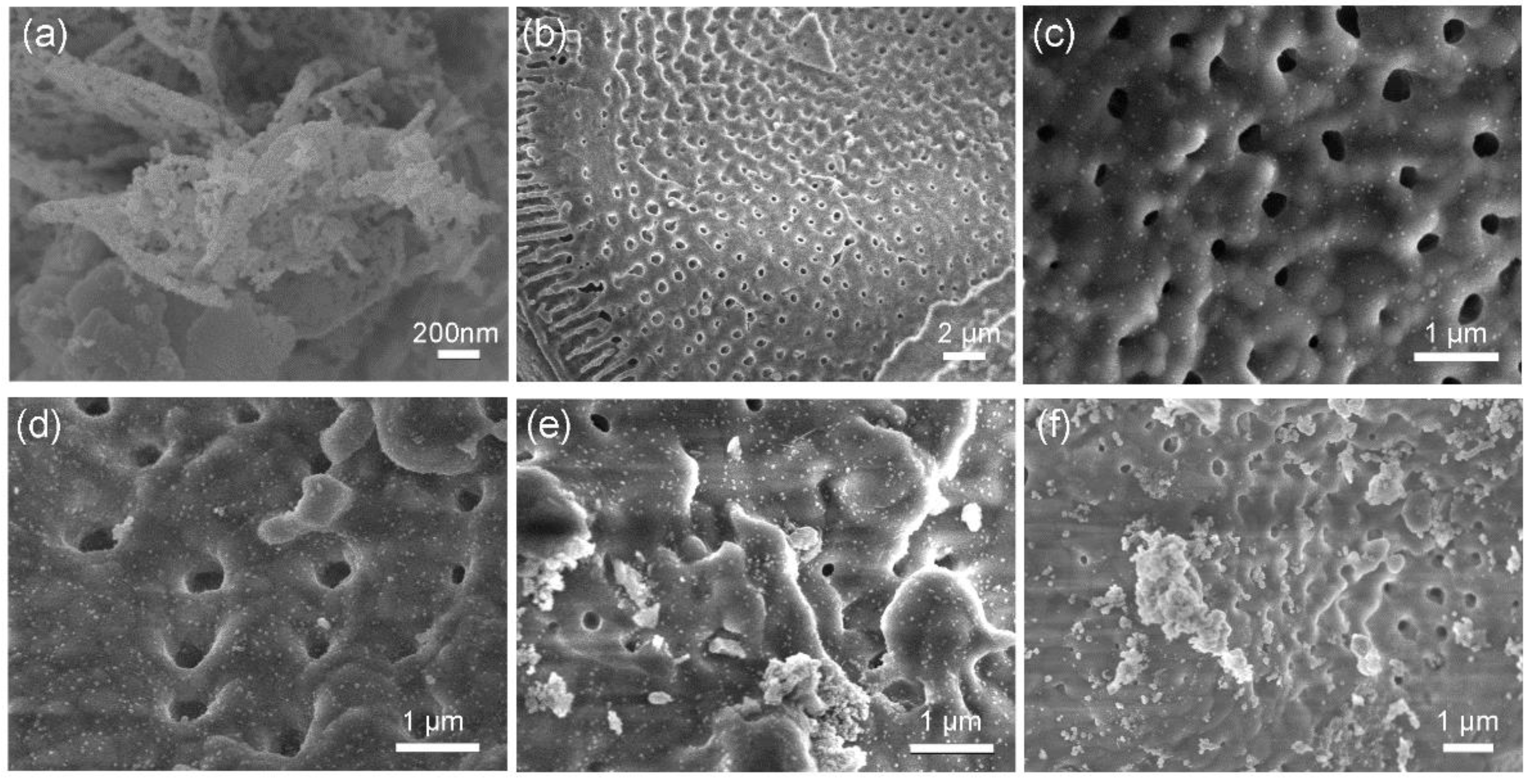
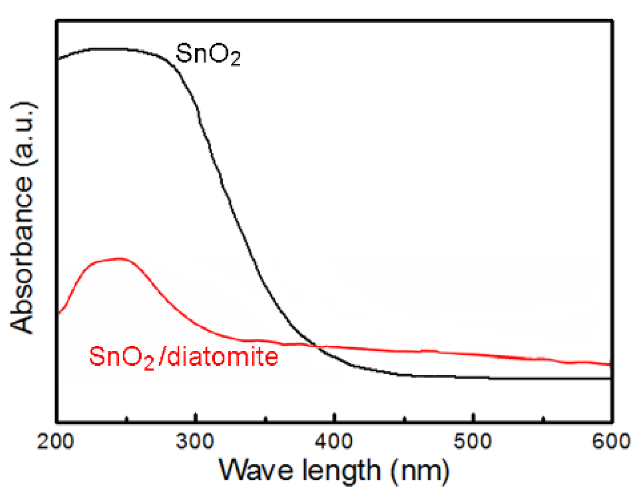
2.1. Composition and Structure of Materials

The XRD patterns of the pure SnO2, modified diatomite, and SnO2/diatomite composites are shown in Figure 1. The specific information of SnO2/diatomite composites is given in Section 3 and Table 1. From the spectrum of diatomite, it can be seen that there are obvious diffraction peaks at 21.98°, 28.44°, 31.46°, 36.08°, 42.66°, 44.84°, 47.06°, 48.61°, 57.08°, and 65.10°. These peaks are just corresponding to (101), (111), (102), (200), (211), (202), (113), (212), (301) and (312) crystal face of SiO2 [16], which reflect that the diatomite is a kind of porous SiO2. The characteristic peaks at 26.61°, 33.89°, 37.95°, 51.78°, 54.76°, 54.76°, 61.87°, 64.72°, 65.94°, 71.28° and 78.71° are assigned to the (110), (101), (200), (211), (220), (002), (310), (112), (301), (202), and (321) crystal face of pure SnO2, respectively [17]. From the spectra of different SnO2 loadings on diatomite, the peak intensity of SnO2 in SD4 sample is obviously higher than other samples, which may be attributed to the highest SnO2 fraction in the composite (Table 1). In addition, the composites also exhibit the feature peaks of diatomite at 21.98°, 28.44°, 31.46° and 36.08°, which further confirm that SnO2 particles are generated on the diatomite template successfully.
The microstructure of different materials are shown in Figure 2. In the absence of porous template, pure SnO2 (Figure 2a) exhibits a two-dimensional lamellar structure assembled by nanoparticles, with an average particle size of about 20 nm [18]. The possible reason for the formation of SnO2 aggregates is no template in the synthesis, resulting in a large number of nano-sized SnO2 particles assembled and connected together to form a lamellar structure. This aggregation phenomenon can be overcome by adding porous diatomite template during the synthesis process. Figure 2b shows the microstructure of modified diatomite. After the acidification treatment, the surface of the diatomite is smooth, in the form of a disc-shaped porous screen structure with a disc diameter of 20~40 μm and a pore diameter of 100~200 nm. Abundant porous structure ensures the diatomite with a large specific surface area, which can be served as an excellent template to prevent the aggregation of SnO2 nanoparticles. The microstructure of four SnO2/diatomite composites are presented in Figure 2c–f. It can be observed that the number of SnO2 nanoparticles on the surface of diatomite increase gradually with an increase of SnCl4∙5H2O precursors. When the resultant SnO2 content is less than 10%, corresponding to SD1 and SD2. There are ultrafine SnO2 nanoparticles dispersed on the surface of diatomite uniformly (Figure 2c), and the particle diameter of SnO2 is about 15 nm. At the same magnification of SEM, the SnO2 particles in the SD2 samples (Figure 2d) were denser than the SD1 sample. When the loading amount of SnO2 is more than 10%, the SnO2 nanoparticles are agglomerated on the surface of diatomite, especially for the SD4 sample in Figure 2f, the size of SnO2 agglomeration is more than 1 μm. The big aggregates undoubtedly reduce the specific surface area and deteriorate the photocatalytic activity of composite. The formation reason of big aggregates can be attributed to the self-aggregate of SnO2 nanoparticles in a high concentration of SnCl4∙5H2O precursor. Therefore, under the appropriate precursor concentration, porous diatomite template can effectively suppress the agglomeration of SnO2 nanoparticles. When the mass of SnCl4·5H2O and thioacetamide (TAA) are 212 and 128 mg separately, the resultant SnO2 content in composite is 7.6%, which achieves the monodispersed distribution of SnO2 nanoparticles (about 15 nm) on the surface of porous diatomite, and the particle size is even smaller than that of pure SnO2 (~20 nm) with no diatomite.
In addition, we also observed the microstructure of pure SnO2 and SnO2/diatomite-SD2 sample by TEM. Pure SnO2 is shown in Figure 3a. The SnO2 aggregates are composed of numerous SnO2 nanoparticles, and the particle diameter is less than 20 nm. However, from the SnO2/diatomite composite (SD2) in Figure 3b, there are much more SnO2 particles covering on the diatomite surface, and the magnified region in Figure 3c just shows the crystalline SnO2 nanoparticles dispersed on the matrix of amorphous diatomite. The lattice spacing of SnO2 is 0.334 nm, which is assigned to the (110) plane of tetragonal SnO2. Moreover, the particle size of SnO2 is less than 15 nm, which is in well agreement with the results obtained by SEM. In addition, the XPS general spectrum of pure SnO2 was obtained in Figure 3d, the wide survey scan exhibits the feature peaks of Sn and O elements. The high-resolution Sn spectrum was provided in Figure 3e, there are two strong peaks at 495.8 eV and 487.2 eV, corresponding to the spin orbits of Sn 3d3/2 and Sn 3d5/2 respectively. The binding energy gap is 8.6 eV, which is in accordance with the oxidation state of Sn4+. The result further confirmed the formation of SnO2, as reflected in the XRD result.
The specific surface area of diatomite and SnO2/diatomite composites were tested by N2 adsorption-desorption isotherm (BET) in Figure 4, and corresponding data were listed in Table 1. The specific surface area of modified diatomite was 13.72 m2·g−1. After loading with SnO2 nanoparticles, the SD2 sample exhibited the maximum specific surface area of 23.53 m2·g−1, which was attributed to the uniform distribution of SnO2 nanoparticles. On the contrary, the specific surface area of SD1 and SD3 samples was 17.15 and 14.95 m2·g−1 respectively, and the SD4 sample had the minimum specific surface area of 11.56 m2·g−1, even less than that of modified diatomite. The result further confirms that the much more SnO2 aggregates loading on the diatomite only increase the sample weight, which reduces the specific surface area of sample and brings a negative effect on the photocatalytic activity, which will be discussed later.

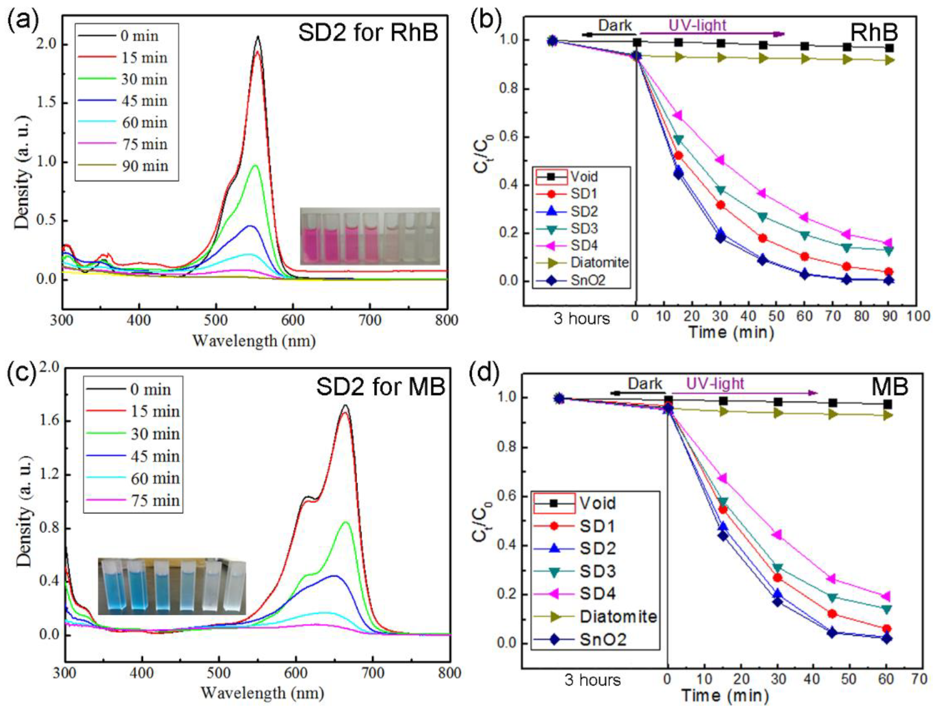
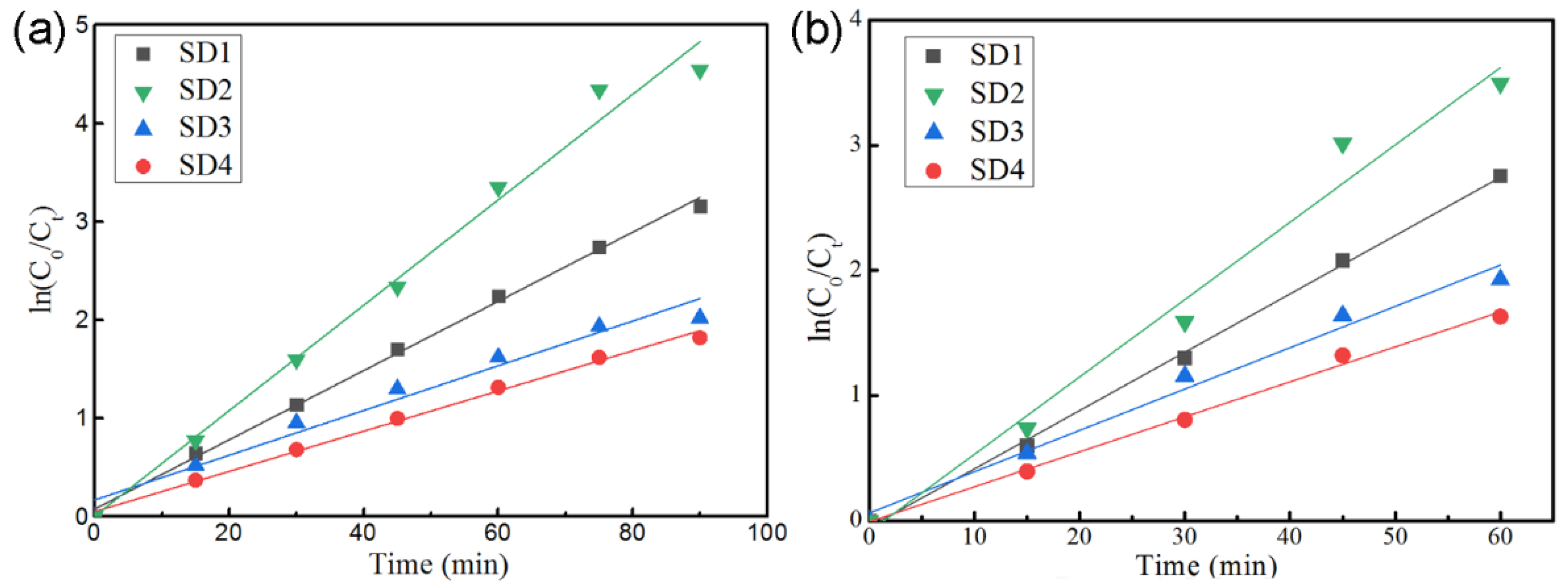
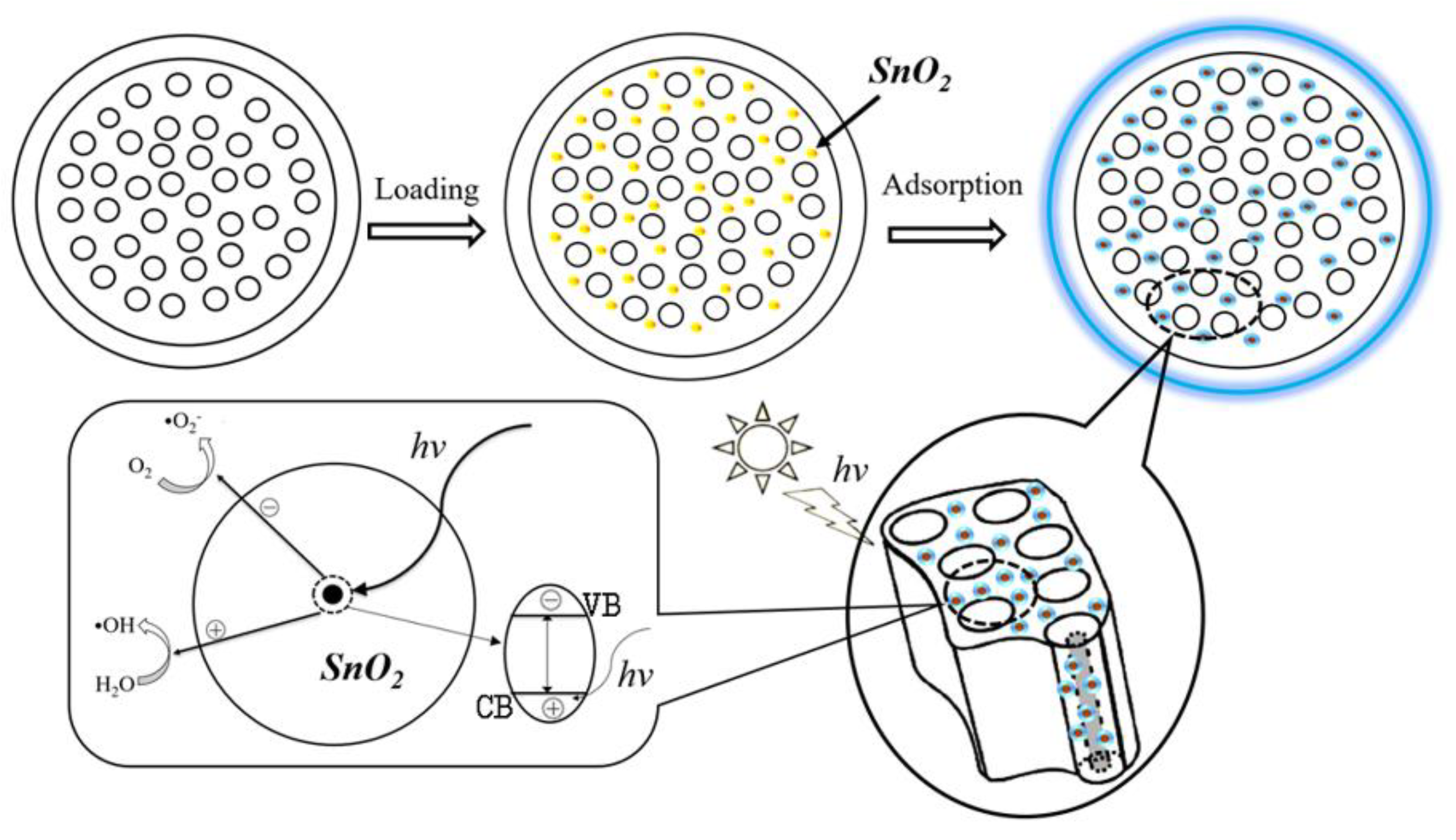
2.2. Photocatalytic Performance and Photodegradation Mechanism

The photodegradation performance of pure SnO2 and different SnO2/diatomite composites in Rh B solution is given in Figure 5. Figure 5a shows the change of adsorption peaks of SD2 sample in Rh B dye for different time. The peak at 546 nm just reflects the typical feature of Rh B. The decrease of peak intensity just shows the concentration change of dye. According to the relationship between the absorption peak intensity and the dye concentration, taking Ct/C0 as the ordinate and time as the horizontal ordinate, Figure 5b reflects the degradation efficiency curves of different photocatalysts. In the first 3 h, the degradation curves have no obvious change in dark condition, which is mainly due to the physical adsorption of dye on porous diatomite in the dark adsorption process. Under the irradiation of UV-light, pure dye (void) and diatomite have no obvious change, which indicates that the Rh B solution cannot be degraded by diatomite under light irradiation. While pure SnO2 and four SnO2/diatomite composites exhibit different photodegradation efficiencies. It can be observed that the photodegradation efficiency order is SnO2 > SD2 > SD1 > SD3 > SD4. Pure SnO2 has the best photocatalytic activity for the absence of inert diatomite template. However, when the irradiation time is more than 60 min, SD2 sample almost has the same photocatalytic activity with pure SnO2 and degrades Rh B dye by 96.8%. The main reason is attributed to the largest specific surface area, and the uniform distribution of SnO2 nanoparticles on the surface of diatomite. On the contrary, SD4 sample with the largest content of SnO2 exhibits the worst photodegradation activity, which is due to the smallest specific surface area and the SnO2 aggregates on diatomite. In addition, we also chose MB solution as a simulated pollutant and observed the degradation efficiency of pure SnO2 and different SnO2/diatomite composites. As shown in Figure 5c, the peak at 654 nm denotes the typical feature of MB, and the fading of peak intensity reflects the degradation of MB dye. Figure 5d shows the photocatalytic activity of different samples. Under the UV-light irradiation, the MB solution cannot be degraded by diatomite alone. The photocatalytic activity order of samples is SnO2 > SD2 > SD1 > SD3 > SD4. The photocatalytic activity for degrading MB solution is completely consistent with Rh B. Under the irradiation of 60 min, SD2 sample has the fastest degradation rate of 96.97%, which is close to that of pure SnO2. The result indicates that SD2 sample has a better photocatalytic activity in the degradation of MB dye.
We also simulate the photodegradation behavior of Rh B and MB dye solution according to the first-order reaction kinetic equation of ln (C0/Ct) = k · t [18], in which, k is the first-order reaction rate constant; C0 is the initial concentration of dye; Ct is the dye concentration under different irradiation time, and t is the irradiation time. Figure 6a,b shows the first-order kinetic fitting curves of photodegradation behavior of Rh B and MB dye, respectively. Corresponding k values and curve fitting degrees (R2) are listed in Table 2. The value of first-order reaction rate constant (k) denotes the slope of the fitting straight line, which reflects the photocatalytic efficiency or degradation rate. While the value of curve fitting degrees (R2) reflects the coincidence degree of first order reaction kinetics equation. When degrading Rh B dye, the constant k of four samples is 0.035, 0.054, 0.023 and 0.021 min−1, respectively. SD2 sample has the largest k value of 0.054 min−1, indicating the best photocatalytic activity. Moreover, the R2 value is 0.989, which confirms that the degradation process of Rh B is in good agreement with the first-order reaction kinetic equation. Meanwhile, when these samples are used to degrade MB dye, the constant k is 0.047, 0.061, 0.033 and 0.028 min−1 respectively. SD2 sample also exhibits the best photocatalytic activity with the largest k value of 0.061 min−1. The value of R2 is 0.981, which is in accordance with the first-order reaction kinetic equation. The k value in degradation of MB (0.061) is slightly higher than that of degrading Rh B (0.054), which reflects that the SD2 sample degrades the MB dye much easier.
To sum up, the photodegradation behavior of organic dyes mainly depends on the specific surface area and the dispersion state of SnO2 nanoparticles. Among these samples, SnO2/diatomite composite (SD2) has the maximum specific surface area of 23.53 m2·g−1, and numerous SnO2 nanoparticles are dispersed on the surface of porous diatomite template uniformly. Therefore, SD2 sample has the best photocatalytic activity, which is close to the pure SnO2. Under the irradiation of 60 min, the degradation rate of Rh B and MB dye is 96.8% and 96.97%, respectively.
In order to confirm the absorption feature of SnO2 and SnO2/diatomite composite, we observed their UV-vis absorption spectra in Figure 7. Compared to the absorption spectrum of pure SnO2, the SnO2/diatomite has a weak light absorption intensity and a narrow light absorption range. The reason can be attributed to the poor light absorption performance of diatomite. Therefore, the porous diatomite template weakens the light absorption of pure SnO2 in a certain extent. However, from the perspective of overcoming the aggregation of SnO2 nanoparticles and low-cost of diatomite, the composite of SnO2/diatomite has a practical application value.
The photocatalytic mechanism of SnO2/diatomite composite is put forward in Figure 8. The monodispersed SnO2 nanoparticles loading on porous diatomite is prepared by adjusting the mass of SnCl4∙5H2O precursor. When the SnO2/diatomite powders dispersed in the dye solution, the dye molecules physically adsorb to the disc-shaped surface and pores of the diatomite [19,20,21], and dye molecules contact with SnO2 nanoparticles closely. Under the irradiation of UV-light, the electrons in SnO2 nanoparticles are excited from the valence band to the conduction band, which generates photogenerated electrons and holes. The electrons and holes then diffuse to the surface of SnO2 catalyst. The redox reaction occurs between electrons/holes and oxygen in water adsorbed on the sample surface, producing some free radicals (•OH and •O2) with strong oxidation [22,23], just as the following reaction equations [8]:
SnO2/diatomite + hν → e + h+
e + O2 → •O2
•O2 + 2H+ + 2e → •OH + OH
h+ + H2O → •OH + H+
These free radicals oxidize the Rh B and MB molecules into CO2 and H2O, and degrade the organic dyes effectively without any secondary pollution. Among the existing diatomite-based composites, the TiO2/diatomite prepared by hydrolysis-deposition method had a Rh B decoloration rate of 100% within 30 min [24]. Granulated N-doped TiO2/diatomite exhibited a Rh B degradation rate of 84.8% within 3 h [16]. While pure SnO2 nanoparticles prepared by precipitation method achieved the degradation of MB (79%) under the irradiation time of 180 min [22]. Hierarchical SnO2 ultrathin nanosheets degraded 93% of MB within 150 min [25]. Iridium (Ir) nanoparticles decorated SnO2 nanorods decomposed more than 97% of MB under 60min irradiation [26]. In this work, the monodispersed SnO2 decorated diatomite composite achieved the fast degradation of Rh B (96.8%) and MB (96.97%) within 60 min. Through a comparison, the composite photocatalyst present an excellent photodegradation activity. Considering the low cost of diatomite (~3.4 USD per 1000 g), the high fraction of diatomite (above 90%), and easy preparation of SnO2 nanoparticles, the SnO2/diatomite composite has a good application prospect in the photodegradation of organic dyes.

3. Materials and Methods

3.1. Materials

SnCl4∙5H2O, thioacetamide (TAA), absolute ethanol and isopropanol were analytical purity, provided by Sinopharm Chemical Regeant Co. Ltd. (Shanghai, China). Diatomite (industrial grade) was purchased from Qingdao Shengtai Industry Co. Ltd. (Qingdao, China). Organic dye of Rhodamine B (Rh B, analytical purity) and methylene blue (MB, analytical purity) were provided by Nanjing Chemical Regent Co. Ltd. (Nanjing, China).

3.2. Preparation of Modified Diatomite and SnO2/Diatomite Composites

In order to obtain a perfect porous structure of diatomite, 10 g diatomite was added into 100 mL dilute hydrochloric acid (1 mol L−1). The suspension was put into a water bath at 80 °C and stirred for 2 h. Then, the solution was cooled to room temperature, and the acidified diatomite was filtered and rinsed to neutral by using abundant distilled water. After drying at 60 °C for 12 h, the acidified diatomite was sintered at 450 °C for 1 h to obtain modified diatomite. 1 g modified diatomite was added into 40 mL isopropanol, and the suspension was stirred continuously. Then, 106 mg SnCl4∙5H2O and 64 mg TAA were added into the suspension and stirred for 10 min. The suspension was transferred to a 100 mL PTFE-lined stainless-steel autoclave. The solvothermal reaction was carried out at 180 °C for 24 h. The precipitate was filtered and rinsed by ethanol and distilled water respectively to remove the impurities and ions, and SnS2/diatomite was obtained after drying at 60 °C for 12 h [15]. The SnS2/diatomite was sintered at 500 °C for 2 h in a muffle furnace to obtain SnO2/diatomite composite. Following this method, other three SnO2/diatomite composites were prepared by adjusting the mass of SnCl4∙5H2O and TAA precursor as 212 and 128 mg, 338 and 192 mg, 424 and 256 mg. According to the addition of precursors, four samples were denoted as SD1, SD2, SD3, and SD4, respectively. In addition, based on the weight difference between modified diatomite and resultant SnO2/diatomite, we calculated the fractions of SnO2 in composite as 4.9%, 7.6%, 11.7% and 16.5%, respectively. The specific recipe of sample is given in Table 1. In order to make a comparison, pure SnO2 powder was synthesized under the same procedures with no diatomite template.

3.3. Characterization

X-ray diffraction spectrum (XRD) was tested on X-Ray Spectrometer of X-7000 (SHIMADZU, Beijing, China) with a radiation source of Cu Kα. Specific surface area was obtained by using Brunauer-Emmett-Teller (BET) method in Micromeritics Instrument (ASAP 2020, Shanghai, China). Scanning electron microscopy (SEM) and transmission electron microscopy (TEM) was conducted on JSM-7500F (JEOL, Beijing, China) and JEM-2010 (JEOL, Beijing, China) separately. X-ray photoelectron spectroscopy (XPS, Thermo Scientific, Shanghai, China) was used with a K-Alpha+ photoelectron spectrometer. The photodegradation performance of samples were tested on ultraviolet-visible spectrophotometer (TU-1810, Beijing Persee Tongyong Instrument Co. Ltd., Beijing, China).

3.4. Photocatalytic Measurement

Rh B and MB served as simulated pollutants, and the concentration of two solutions were controlled as 10 mg L−1. The photocatalyst concentration in dye solution was fixed as 500 mg L−1. The experiment detail was given: 50 mg photocatalyst was added into 100 mL pollutant solution, and placed it in a dark chamber for 3 h to uniformly adsorb the dye molecules. Then, the suspension was transferred into a photochemistry reactor irradiated by a mercury lamp (500 W). Under the continuously stirring and irradiation of ultraviolet light, the dye solution (about 5 mL) was taken out from the photodegradation solution in a fixed time interval [8]. The supernatant was obtained by centrifuging the extracted dye solution and measured by using an ultraviolet-visible (UV-vis) spectrophotometer to check the change of dye featured peaks. The photodegradation rate of dye solution was obtained by the concentration change of dye under a fixed illumination time, which reflected the photocatalytic efficiency of photocatalysts. In this work, five photocatalysts were involved, including four SnO2/diatomite composites (SD1, SD2, SD3 and SD4) and pure SnO2.

4. Conclusions

In summary, SnO2/diatomite composite was synthesized by solvothermal reaction and sintering method. In the synthesis process, the weight of reactants was adjusted to effectively overcome the agglomeration of SnO2 nanoparticles. When the mass of SnCl4·5H2O and TAA was 212 and 128 mg, monodispersed SnO2 nanoparticles were uniformly generated on porous diatomite template with a particle size of about 15 nm (SD2). The SD2 composite has the maximum specific surface area of 23.53 m2·g−1. When served as a photocatalyst to degrade Rh B, and MB dye, it presented a superior degradation activity close to pure SnO2, and achieved a fast degradation of 96.8% Rh B and 96.97% MB in 60 min. The degradation process of Rh B and MB was in accordance with the first-order kinetic equation. The superior photocatalytic performance of SnO2/diatomite composite is attributed to the physical adsorption of dye molecules on the surface and pores of diatomite, and the highly efficient photocatalytic activity of monodispersed SnO2 nanoparticles. In addition, the SnO2/diatomite composite has the advantages of a low cost and easy preparation, and thus presents good application prospects in the field of photocatalysis.

Author Contributions

Conceptualization, H.J. and X.H.; methodology, H.J. and R.W.; Writing, H.J.; validation, D.W. and S.Y.

Funding

This research was funded by National Natural Science Foundation of China (51403094 and 51774175), and Program of Liaoning Education Department of China (No. LJ2017FBL002).

Conflicts of Interest

The authors declare no conflict of interest.

References

  1. Lin, L.; Huang, J.; Li, X.; Abass, M.A.; Zhang, S. Effective surface disorder engineering of metal oxide nanocrystals for improved photocatalysis. Appl. Catal. B Environ. 2017, 203, 615–624. [Google Scholar] [CrossRef]
  2. Xu, K.; Wu, J.; Tan, C.F.; Ho, G.W.; Wei, A.; Hong, M. Ag-CuO-ZnO metal-semiconductor multiconcentric nanotubes for achieving superior and perdurable photodegradation. Nanoscale 2017, 9, 1154–11583. [Google Scholar] [CrossRef] [PubMed] [Green Version]
  3. Manjula, P.; Boppella, R.; Manorama, S.V. A facile and green approach for the controlled synthesis of porous SnO2 nanospheres: Application as an efficient photocatalyst and an excellent gas sensing material. ACS Appl. Mater. Inter. 2012, 4, 6252–6260. [Google Scholar] [CrossRef] [PubMed]
  4. Sinha, A.K.; Pradhan, M.; Sarkar, S.; Pal, T. Large-scale solid-State synthesis of Sn-SnO2 nanoparticles from layered SnO by sunlight: A material for dye degradation in water by photocatalytic reaction. Environ. Sci. Technol. 2013, 47, 2339–2345. [Google Scholar] [CrossRef] [PubMed]
  5. Li, P.; Lan, Y.; Zhang, Q.; Zhao, Z.; Pullerits, T.; Zheng, K.; Zhou, Y. Iodinated SnO2 quantum dots: A facile and efficient approach to increase solar absorption for visible-light photocatalysis. J. Phys. Chem. C 2016, 120, 9253–9262. [Google Scholar] [CrossRef]
  6. Chen, X.; Chu, D.; Wang, L.; Hu, W.; Yang, H.; Sun, J.; Zhu, S.; Wang, G.; Tao, J.; Zhang, S. One-step synthesis of novel hierarchical flower-like SnO2 nanostructures with enhanced photocatalytic activity. J. Alloy. Compd. 2017, 729, 710–715. [Google Scholar] [CrossRef]
  7. Zhao, Q.; Gao, Y.; Bai, X.; Wu, C.; Xie, Y. Facile synthesis of SnO2 hollow nanospheres and applications in gas sensors and electrocatalysts. Eur. J. Inorg. Chem. 2006, 8, 1643–1648. [Google Scholar] [CrossRef]
  8. Hong, X.; Wang, R.; Li, S.; Fu, J.; Chen, L.; Wang, X. Hydrophilic macroporous SnO2/rGO composite prepared by melamine template for high efficient photocatalyst. J. Alloy. Compd. 2019. [Google Scholar] [CrossRef]
  9. Chen, Y.; Sun, F.; Huang, Z.; Chen, H.; Zhuang, Z.; Pan, Z.; Long, J.; Gu, F. Photochemical fabrication of SnO2 dense layers on reduced graphene oxide sheets for application in photocatalytic degradation of p-Nitrophenol. Appl. Catal. B Environ. 2017, 215, 8–17. [Google Scholar] [CrossRef]
  10. Wei, J.; Xue, S.L.; Xie, P.; Zou, R. Synthesis and photocatalytic properties of different SnO2 microspheres on graphene oxide sheets. Appl. Surf. Sci. 2016, 376, 172–179. [Google Scholar] [CrossRef]
  11. Wang, B.; Zhang, G.; Leng, X.; Sun, Z.; Zheng, S. Characterization and improved solar light activity of vanadium doped TiO2/diatomite hybrid catalysts. J. Hazard. Mater. 2015, 285, 212–220. [Google Scholar] [CrossRef] [PubMed] [Green Version]
  12. Zhang, J.; Wang, X.; Wang, J.; Wang, J.; Ji, Z. Effect of sulfate ions on the crystallization and photocatalytic activity of TiO2/diatomite composite photocatalyst. Chem. Phys. Lett. 2016, 643, 53–60. [Google Scholar] [CrossRef]
  13. Zhong, X.; Shen, Y.; Zhao, S.; Chen, X.; Han, C.; Wei, D.; Fang, P.; Meng, D. SO2 sensing properties of SnO2 nanowires grown on a novel diatomite-based porous substrate. Ceram. Int. 2019, 45, 2556–2565. [Google Scholar] [CrossRef]
  14. Zhen, Y.; Zhang, J.; Wang, W.; Li, Y.; Gao, X.; Xue, H.; Liu, X.; Jia, Z.; Xue, Q.; Zhang, J.; et al. Embedded SnO2/diatomaceous earth composites for fast humidity sensing and controlling properties. Sens. Actuat. B Chem. 2020, 303, 127137. [Google Scholar] [CrossRef]
  15. Hong, X.; Li, S.; Wang, R.; Fu, J. Hierarchical SnO2 nanoclusters wrapped functionalized carbonized cotton cloth for symmetrical supercapacitor. J. Alloy. Compd. 2019, 775, 15–21. [Google Scholar] [CrossRef]
  16. Chen, Y.; Liu, K. Preparation of granulated N-doped TiO2/diatomite composite and its applications of visible light degradation and disinfection. Powder Technol. 2016, 303, 176–191. [Google Scholar] [CrossRef]
  17. Jia, B.; Jia, W.; Qu, F.; Wu, X. General strategy for self assembly of mesoporous SnO2 nanospheres and their applications in water purification. RSC Adv. 2013, 3, 12140–12148. [Google Scholar] [CrossRef]
  18. Vishnuganth, M.A.; Remya, N.; Kumar, M.; Selvaraju, N. Photocatalytic degradation of carbofuran by TiO2-coated activated carbon: Model for kinetic, electrical energy per order and economic analysis. J. Environ. Manag. 2016, 181, 201–207. [Google Scholar] [CrossRef]
  19. Chen, Y.; Liu, K. Preparation and characterization of nitrogen-doped TiO2/diatomite integrated photocatalytic pellet for the adsorption-degradation of tetracycline hydrochloride using visible light. Chem. Eng. J. 2016, 302, 682–696. [Google Scholar] [CrossRef]
  20. Zhang, G.; Sun, Z.; Duan, Y.; Ma, R.; Zheng, S. Synthesis of nano-TiO2/diatomite composite and its photocatalytic degradation of gaseous formaldehyde. Appl. Surf. Sci. 2017, 412, 105–112. [Google Scholar] [CrossRef]
  21. Chen, Y.; Wu, Q.; Zhou, C.; Jin, Q. Enhanced photocatalytic activity of La and N co-doped TiO2/diatomite composite. Powder Technol. 2017, 322, 296–300. [Google Scholar] [CrossRef]
  22. Kim, S.P.; Choi, M.Y.; Choi, H.C. Photocatalytic activity of SnO2 nanoparticles in methylene blue degradation. Mater. Res. Bull. 2016, 74, 85–89. [Google Scholar] [CrossRef]
  23. Chen, H.; Pu, X.; Gu, M.; Zhu, L.; Chen, L. Tailored synthesis of SnO2@graphene nanocomposites with enhanced photocatalytic response. Ceram. Int. 2016, 42, 17717–17722. [Google Scholar] [CrossRef]
  24. Chen, H.; Pu, X.; Gu, M.; Zhu, L.; Chen, L. The influence of carriers on the structure and photocatalytic activity of TiO2/diatomite composite photocatalysts. Adv. Powder Technol. 2015, 26, 595–601. [Google Scholar]
  25. Huang, R.; Huang, S.; Chen, D.; Zhang, Q.; Le, T.; Wang, Q.; Hu, Z.; Chen, Z.; Jiang, Y.; Zhao, B. Insight into efficient pollutant degradation from paramorphic SnO2 hierarchical superstructures. J. Alloy. Compd. 2019, 776, 287–296. [Google Scholar] [CrossRef]
  26. Dhanalakshmi, M.; Saravanakumar, K.; Prabavathi, S.L.; Abinaya, M.; Muthuraj, V. Fabrication of novel surface plasmon resonance induced visible light driven iridium decorated SnO2 nanorods for degradation of organic contaminants. J. Alloy. Compd. 2018, 763, 512–524. [Google Scholar] [CrossRef]
Figure 1. XRD spectra of pure SnO2, modified diatomite and SnO2/diatomite composites.
Catalysts 09 01060 g001
Figure 2. The microstructure of (a) pure SnO2, (b) modified diatomite, (c) SD1, (d) SD2, (e) SD3 and (f) SD4 by SEM.
Catalysts 09 01060 g002
Figure 3. The microstructure of (a) pure SnO2 and (b,c) SnO2/diatomite-SD2 by TEM, and the XPS spectrum of pure SnO2 (d,e).
Catalysts 09 01060 g003
Figure 4. N2 adsorption-desorption isotherm of diatomite and different SnO2/diatomite composites.
Catalysts 09 01060 g004
Figure 5. (a) UV-visible adsorption spectra of SD2 sample in Rh B solution under different irradiation time; (b) Photodegradation curves of different samples in Rh B solution; (c) UV-visible adsorption spectra of SD2 sample in MB solution under different irradiation time; (d) Photodegradation curves of different samples in MB solution.
Catalysts 09 01060 g005
Figure 6. The first-order reaction kinetic fitting curves of (a) Rh B solution and (b) MB solution.
Catalysts 09 01060 g006
Figure 7. UV-vis absorption spectra of SnO2 and SnO2/diatomite.
Catalysts 09 01060 g007
Figure 8. Photocatalytic mechanism of SnO2/diatomite composite in degradation of organic dyes.
Catalysts 09 01060 g008
Table 1. The specific surface area of different SnO2/diatomite composites derived from BET method.
SampleDiatomiteSD1SD2SD3SD4
SnCl4∙5H2O/thioacetamide0/0106/64 mg212/128 mg338/192 mg424/256 mg
SnO2 fraction04.9%7.6%11.7%16.5%
Specific surface area/(m2·g−1) 13.7217.1523.5314.9511.56
Table 2. Corresponding first-order reaction kinetic fitting parameters of samples in Rh B and MB.
Sample/DyeSD1/RhBSD2/RhBSD3/RhBSD4/RhBSD1/MBSD2/MBSD3/MBSD4/MB
k/min−10.0350.0540.0230.0210.0470.0610.0330.028
R20.9970.9890.9710.9960.9980.9810.9850.996

