Article

Enhanced Visible Light Photodegradation of Microplastic Fragments with Plasmonic Platinum/Zinc Oxide Nanorod Photocatalysts

1
Department of Sustainable Development, Environmental Science and Engineering (SEED), School of Architecture and Built Environment, KTH Royal Institute of Technology, 100 44 Stockholm, Sweden
2
Department of Civil and Environmental Engineering (CEE), Islamic University of Technology, Gazipur 1704, Dhaka, Bangladesh
3
Functional Materials, Department of Applied Physics, School of Engineering Sciences, KTH Royal Institute of Technology, 164 40 Kista, Stockholm, Sweden
*
Author to whom correspondence should be addressed.
Catalysts 2019, 9(10), 819; https://doi.org/10.3390/catal9100819
Submission received: 14 September 2019 / Revised: 23 September 2019 / Accepted: 26 September 2019 / Published: 28 September 2019
(This article belongs to the Special Issue Photocatalysis: Activity of Nanomaterials)

Abstract

:
Microplastics are persistent anthropogenic pollutants which have become a global concern owing to their widespread existence and unfamiliar threats to the environment and living organisms. This study demonstrates the degradation of fragmented microplastics particularly low-density polyethylene (LDPE) film in water, through visible light-induced plasmonic photocatalysts comprising of platinum nanoparticles deposited on zinc oxide (ZnO) nanorods (ZnO-Pt). The ZnO-Pt nanocomposite photocatalysts were observed to have better degradation kinetics for a model organic dye (methylene blue) compared to bare ZnO nanorods, attributed to the plasmonic effects leading to better interfacial exciton separation and improved hydroxyl radical activity along with a 78% increase in visible light absorption. These demonstrations of the plasmonically enhanced photocatalyst enabled it to effectively degrade microplastic fragments as confirmed following the changes in carbonyl and vinyl indices in infrared absorption. In addition, visual proof of physical surface damage of the LDPE film establishes the efficacy of using plasmonically enhanced nanocomposite photocatalytic materials to tackle the microplastic menace using just sunlight for a clean and green approach towards mitigation of microplastics in the ecosystem.

Graphical Abstract

1. Introduction

Plastic based commodities are among the most widely used items in the modern world. The term ‘microplastic (MP)’ refers to any fragmented plastic material with sizes less than 5 mm or lower. They can be broadly categorized into primary and secondary microplastics that are mostly used in personal care products, cosmetics, medicines, textiles or disintegrate from larger plastic products [1,2]. The actual amount of microplastics in the ecosystem is beyond quantification, due to its infinitesimal size, light-weight, persistent characteristics, and widespread occurrence in the environmental system. It has been estimated that between 0.48–1.27 million/annum of plastic debris enters the ocean from different sources [3], with the major sources being: Effluents discharged from wastewater treatment plants (WWTPs), surface runoff from contaminated landscape or disposal lands, and beach littering [4,5].
Removal of microplastics from water or wastewater is difficult, due to the intrinsic physical and chemical characteristics. Few studies suggest advanced treatment technologies like membrane bioreactor, bacterial oxidation, retrofilteration that could be suitable for removing microplastics from aqueous media, but can be time-consuming and expensive often with high energy requirements [6,7]. Other available options are removal by physical or mechanical means or degradation by thermal, chemical or catalytic processes [8,9]. Certain type of chemicals, such as persulfate salts, have been widely applied to produce reactive oxygen species (ROS) and hydroxyl radical for advanced oxidation. Previous studies showed that transition metals (e.g., Fe, Co, Mn) and their oxides are excellent activators for persulfate salts in producing sulfate radicals during catalytic process for organic/inorganic contaminants removal [10]. Recently, carbon nanomaterials emerged as metal-free alternatives and were applied for activation of persulfate salts in catalytic degradation of microplastics [11]. On the other hand, photo-mediated catalytic degradation, one of the advanced oxidation processes (AOPs), has been shown to be an energy-efficient process to degrade microplastics in water wherein the typically nanostructured photocatalyst play an important role for expediting the degradation process by generating reactive species like hydroxyl (•OH) and superoxide ( O 2 ) radicals in the presence of light with appropriate energy. These species directly initiate the degradation process, which leads to polymeric chain breaking, branching, crosslinking and complete mineralization [12,13,14].
Nanoscale semiconductors, such as TiO2, ZnO and ZnS, have been extensively studied in photocatalysis applications from water splitting to pollutant degradation. TiO2 nanomaterials have been reported on their capability to photocatalytically degrade microplastics, due to their high photocatalytic activity [15,16]. In comparison to other photocatalysts, nanostructured zinc oxide (ZnO), a semiconductor with a band gap of 3.37 eV, has been used in water and wastewater treatments, due to its high redox potential, higher electron mobility at room temperature and better catalytic performance [17,18]. Hexagonal ZnO nanorods have a higher surface area and stability in comparison to spherical nanoparticles, leading to a more efficient photo-oxidation process [14]. For further improvement of photocatalytic activities of ZnO, metals, nonmetals, and carbon materials can be deposited on ZnO to modify their optical characteristics and to improve electron-hole pair separation, which is essential for enhancing the photocatalytic process [19]. Studies have shown that by depositing platinum, a plasmonic metal, the overall photocatalytic efficiency can be improved, due to reduced electron-hole pair recombination and enhanced visible light absorption. Both these aspects are a consequence of surface plasmon resonance phenomenon, wherein a particular wavelength of electromagnetic spectra incident on metal nanostructures of platinum, gives rise to the resonance of interfacial conduction electrons, leading to enhanced absorption of light in such nanocomposite [18,19,20,21,22,23].
In this study, the photocatalytic activity of ZnO nanorods was modified by depositing platinum nanoparticles on the nanorod surface, and the photocatalytic performance of the nanocomposite was studied. Multiple characterization tools were applied to follow the effective degradation of low-density polyethylene (LDPE) films, which are the most widely found microplastic pollutant in water and wastewater systems [2,6].

2. Results and Discussion

2.1. Morphology and Crystal Structure of Designed Photocatalysts

The average diameter and length of synthesized ZnO nanorods and ZnO nanorods-supported platinum nanoparticles (ZnO-Pt) were estimated to be 55 µm and 960 µm (same for both cases) respectively, in SEM images, as shown in Figure 1. The micrographs clearly indicate that there is a considerable increase in ZnO nanorod surface roughness and morphology after deposition of platinum nanoparticles. The weight percentage of Zn, O and Pt in ZnO-Pt samples were found to be 70.6%, 23.9%, 5.4% respectively (see Figure S2 in Supplementary Materials).
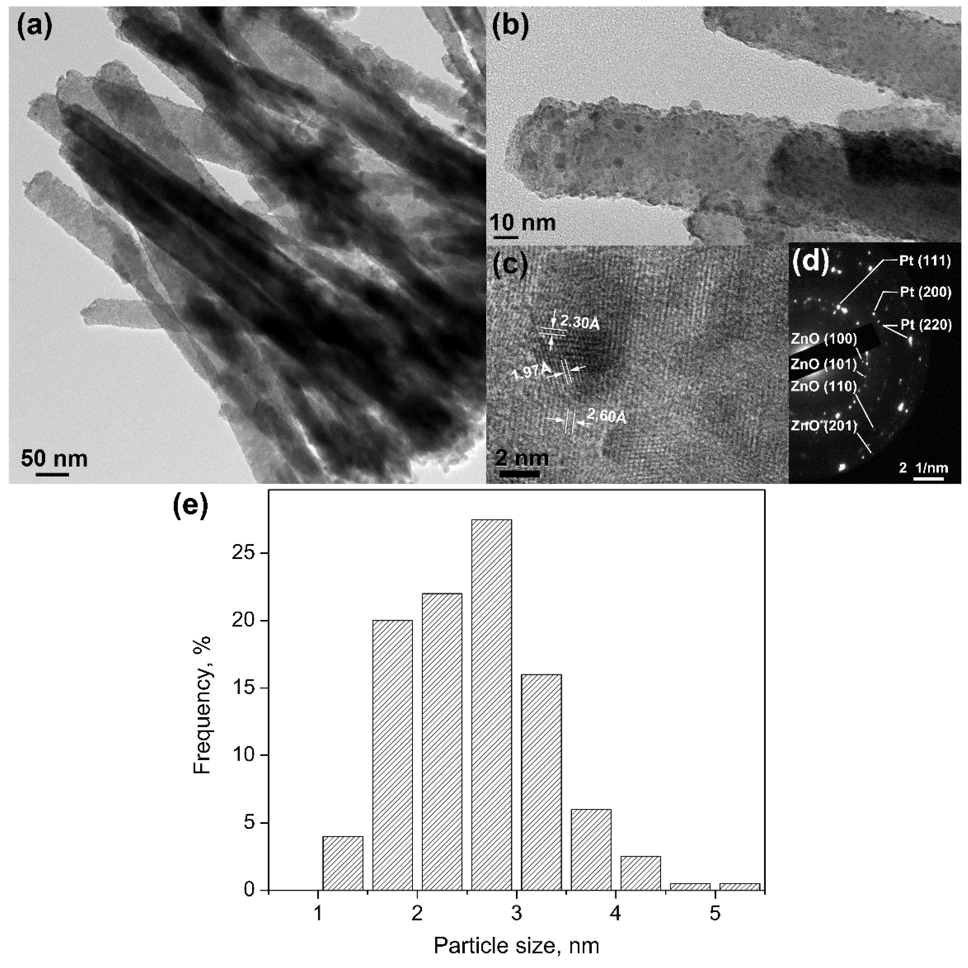
High resolution transmission electron microscope (HR-TEM) was also used to obtain a more detailed insight on morphology and crystal structure of deposited Pt nanoparticles on the ZnO-Pt nanorods. Figure 2 indicates that the platinum nanoparticles were distributed homogenously on the ZnO nanorod surface as confirmed from their lattice spacing’s of 2.30 Å and 1.97 Å (see Figure 2c), that respectively correspond to (111) and (200) planes of a face centered cubic crystal structure of platinum. The lattice spacing of ZnO nanorod as background in Figure 2c is about 2.60 Å, which corresponds to (002) plane of hexagonal wurtzite zinc oxide crystals. Figure 2d shows the selected-area electron diffraction (SAED) pattern of ZnO-Pt nanorods. The diffraction spots on rings can be indexed from (100), (101), (110) and (201) crystal planes of ZnO, as well as from (111), (200) and (220) planes of platinum, labelled in Figure 2d. Thereafter, the hexagonal wurtzite crystal structure of ZnO and face-centered cubic structure of platinum are, thus, confirmed [24]. The average size of platinum nanoparticles is about 2.6 nm with a standard deviation (σ) of 0.8 nm (n = 200). The size distribution of platinum nanoparticles is plotted in Figure 2e.

2.2. Modification of Optical Properties of ZnO/Pt Nanocomposite

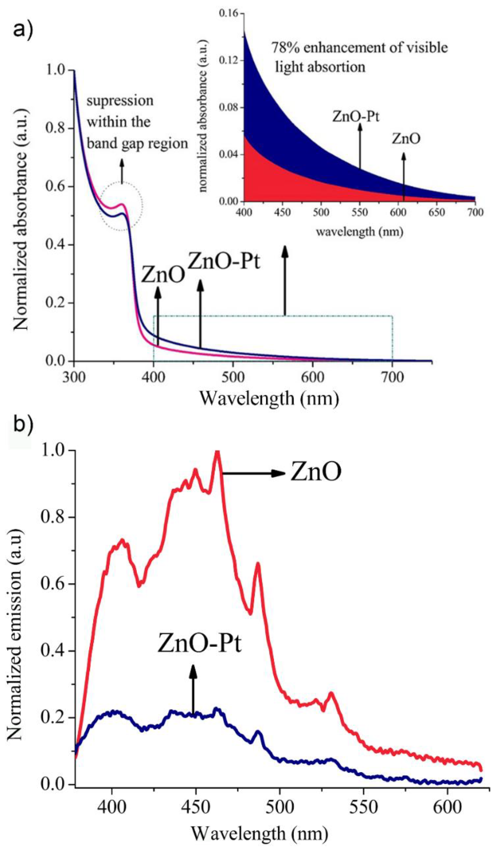
UV-VIS absorbance spectra of unmodified ZnO nanorods and ZnO nanorods loaded with platinum nanoparticles were recorded over 300 to 750 nm. As can be observed from Figure 3, due to their inherent semiconductor properties, ZnO nanorods showed strong absorption in the UV region, but a weaker absorption in the visible region. However, upon deposition of platinum nanoparticles on the ZnO nanorods surface, a 78% enhancement in the absorption in the visible region could be observed, which is attributed to the surface plasmon resonance effect of platinum nanoparticles at 385 mm [20].
Simultaneously, there was a reduction in the ZnO band-to-band excitation at 362 nm which could be a result of the increased defect density within the ZnO nanorod structure, due to UV-light exposure prior to platinum deposition (also indicated by the extended absorption tail in the visible region) and reduced light intensity incident on the surface of ZnO nanorods. Thus, both the phenomena contribute to the improvement in optical absorption in the visible region. Compared to as-grown ZnO nanorod samples, PL spectrum of ZnO-Pt (Figure 3b) displays a considerable reduction in the radiative recombination of the photo-generated excitons. This is in accordance with the band positions and Fermi levels on either side of the ZnO-Pt interface, where the work function of platinum (−5.65 eV w.r.t. vacuum) and electron affinity of ZnO (−4.5 eV w.r.t. vacuum), along with the higher Fermi level of n-type ZnO gives rise to a Schottky junction with a barrier height of approximately φB = 1.15 eV. Therefore, an electric field will be built between ZnO and Pt; and the direction of this electric field is from ZnO to Pt. Due to the relative position of conduction band of ZnO and Fermi level of platinum, the platinum nanoparticle coating will act as a sink for the photo-excited electrons in the interface with ZnO, reducing the viability of radiative and non-radiative recombination processes for the photo mediated exciton pairs in ZnO. This, in turn, will lead to a reduction in the PL intensity as observed from Figure 3b. However, this phenomenon leads to a higher degree of radical species to be formed in the presence of water, due to charge transfer reactions between water molecules and the individual excitons (electrons and holes), which can aid the photocatalytic degradation process [18,23,25,26,27].

2.3. Photocatalytic Activity Test for ZnO and ZnO-Pt

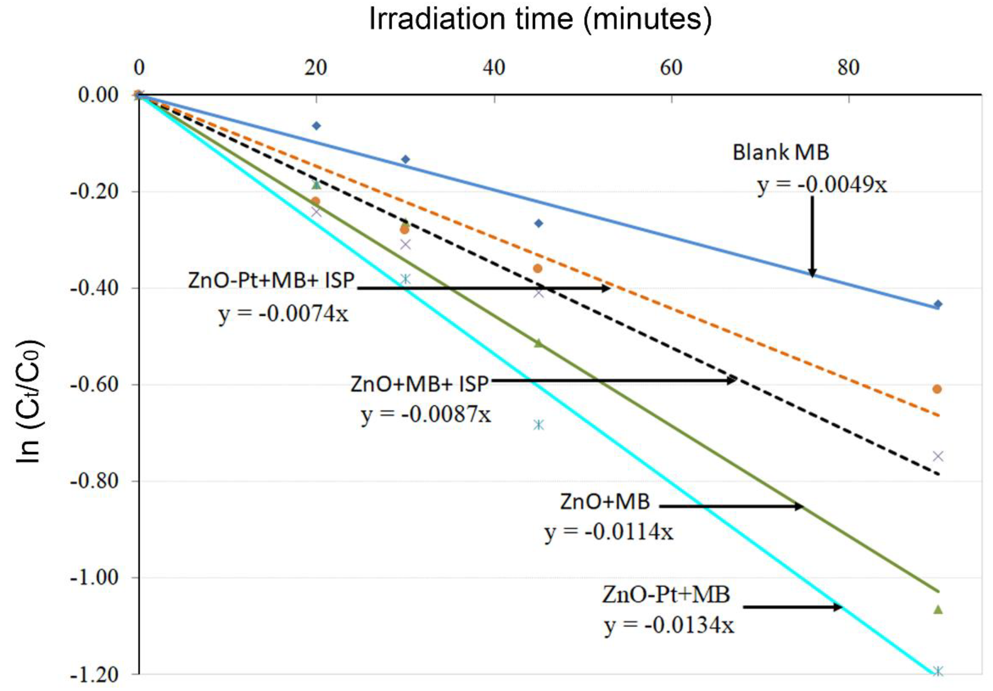
Experiments were carried out using methylene blue (MB) dye as a model organic contaminant to test the photocatalytic propensity of the ZnO and ZnO-Pt catalysts. It is well documented that MB can undergo photolysis under visible light irradiation (without a catalyst), a phenomenon also observed in our experiments, as shown in Figure 4 [28,29]. With the inclusion of a ZnO nanorod catalyst in the process, a substantial enhancement in the photodegradation of methylene blue could be observed. This is expected as the photo generated electrons and holes (excitons) on the ZnO nanorods will react with the surrounding water to form radical species (•OH, O 2 ) which can degrade MB [18,30]. The degradation phenomenon was further enhanced by almost 18% with the addition of platinum nanoparticles on the ZnO nanorods. This additional enhancement is activated by the improved charge separation, mediated by the favorable energy band alignment at the ZnO-Pt interface as explained previously, along with additional electrons pumped into the ZnO nanorods from the platinum nanoparticles, due to the induced surface plasmon resonance [19,27].
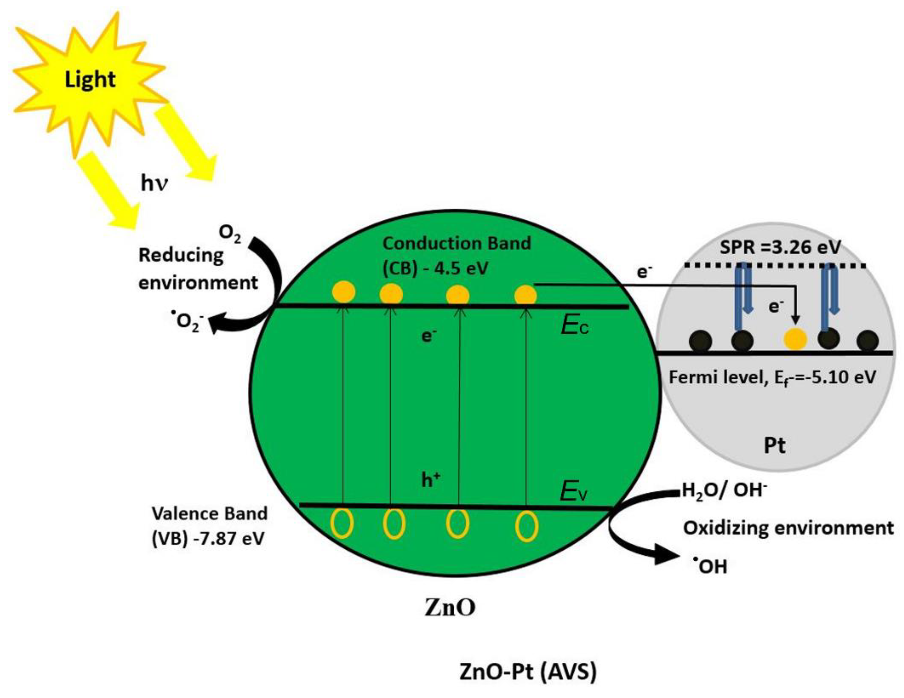
Further proof of the effect of a plasmonic nanoparticle coating was provided by experiments specifically carried out to identify the primary radical species (•OH, O 2 ) taking part in the degradation process (Figure 4). Photocatalytic oxidation of methylene blue was carried out under visible light in the presence of isopropanol (ISP), which is a known hole scavenger. As observed from the reduction in degradation kinetics and efficiency in the presence of isopropanol for both ZnO and ZnO-Pt photo-catalysts (Figure 4), it is clear that hydroxyl radicals are the primary species mediating the oxidation of MB. However, it is interesting to note that the reduction in MB oxidation is much more pronounced in the presence of ZnO-Pt catalysts, suggesting that the plasmonic nanoparticle coating leads to increased hydroxyl radical generation (compared to unmodified ZnO nanorod samples), which in turn is an additional proof that the platinum coating improves the exciton separation efficiency. The above-mentioned process is pictorially depicted in Figure 5 to illustrate the band alignment at the ZnO-Pt interface scheme, where the platinum nanoparticles act as electron sinks to arrest/reduce the exciton recombination process to aid in the photocatalytic reactions [20,31,32].

2.4. Photocatalytic Degradation of Low Density Polyethylene Films

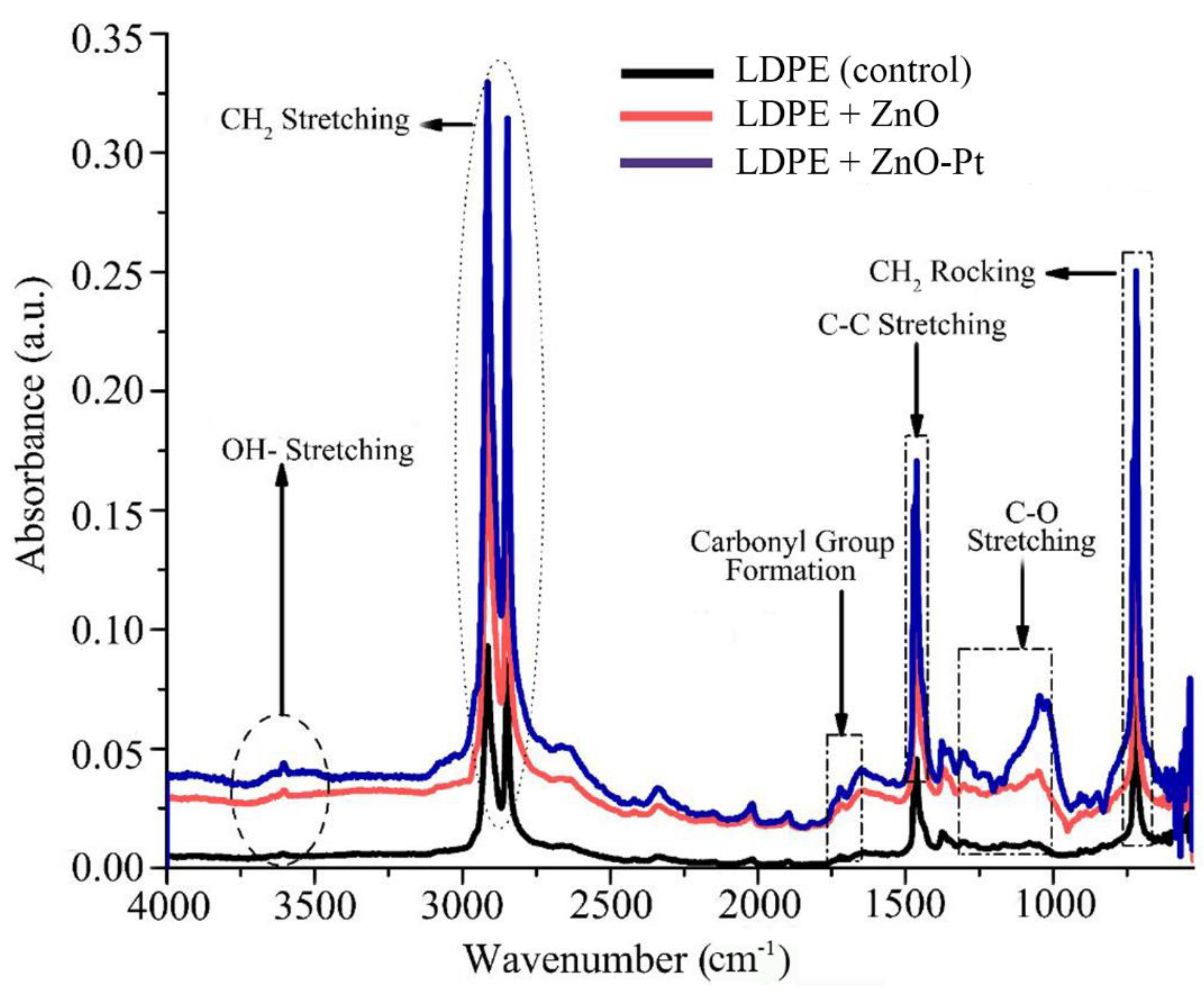
Photocatalysts were subsequently tested for their ability to break down the most prolific microplastic pollutant in water, low density polyethylene (LDPE). As observed from the FTIR spectra of LDPE fragments, there is a considerable change in its chemical structure before and after visible light photocatalysis with ZnO and ZnO-Pt catalysts (Figure 6). Compared to un-irradiated LDPE fragments, several functional groups can be identified having peaks at 710 cm−1, and 719 cm−1 (medium type rocking deformation of -CH2), 2847 cm−1, 2915 cm−1 (symmetric and asymmetric strong stretching of -CH2), 1462 cm−1, 1472 cm−1 (stretching of -C=C- bond), and 1377 cm−1 (weak symmetric deformation of -CH3 group), which remain unchanged after the photocatalytic process [33,34]. However, several oxygenated groups like carbonyl, hydroperoxide, peroxides and unsaturated groups within the band from 1700–1760 cm−1, 3600–3610 cm−1, 1100–1300 cm−1, and 880–920 cm−1 respectively, appear during the photodegradation process which provide evidence of the chemical transformation of LDPE as suggested previously in References [14,35,36,37].
Furthermore, carbonyl index (CI) and vinyl index (VI) of photocatalyzed LDPE were calculated to compare the performance of ZnO and ZnO-Pt photocatalysts as these are used for indirect quantification of the degree of degradation [13]. Table 1 presents the carbonyl index and Vinyl index of LDPE films, showing an increment after irradiation. It is clear that the photodegradation efficiency is improved in the presence of ZnO nanorods with platinum nanoparticles. On average, the carbonyl index and vinyl index increased by 13% and 15% respectively, suggesting that the degradation efficiency of ZnO-Pt plasmonic photocatalysts is substantially improved compared to the unmodified ZnO nanorods.

2.5. Microstructure Analysis of Non-Irradiated and Irradiated Low Density Polyethylene Films

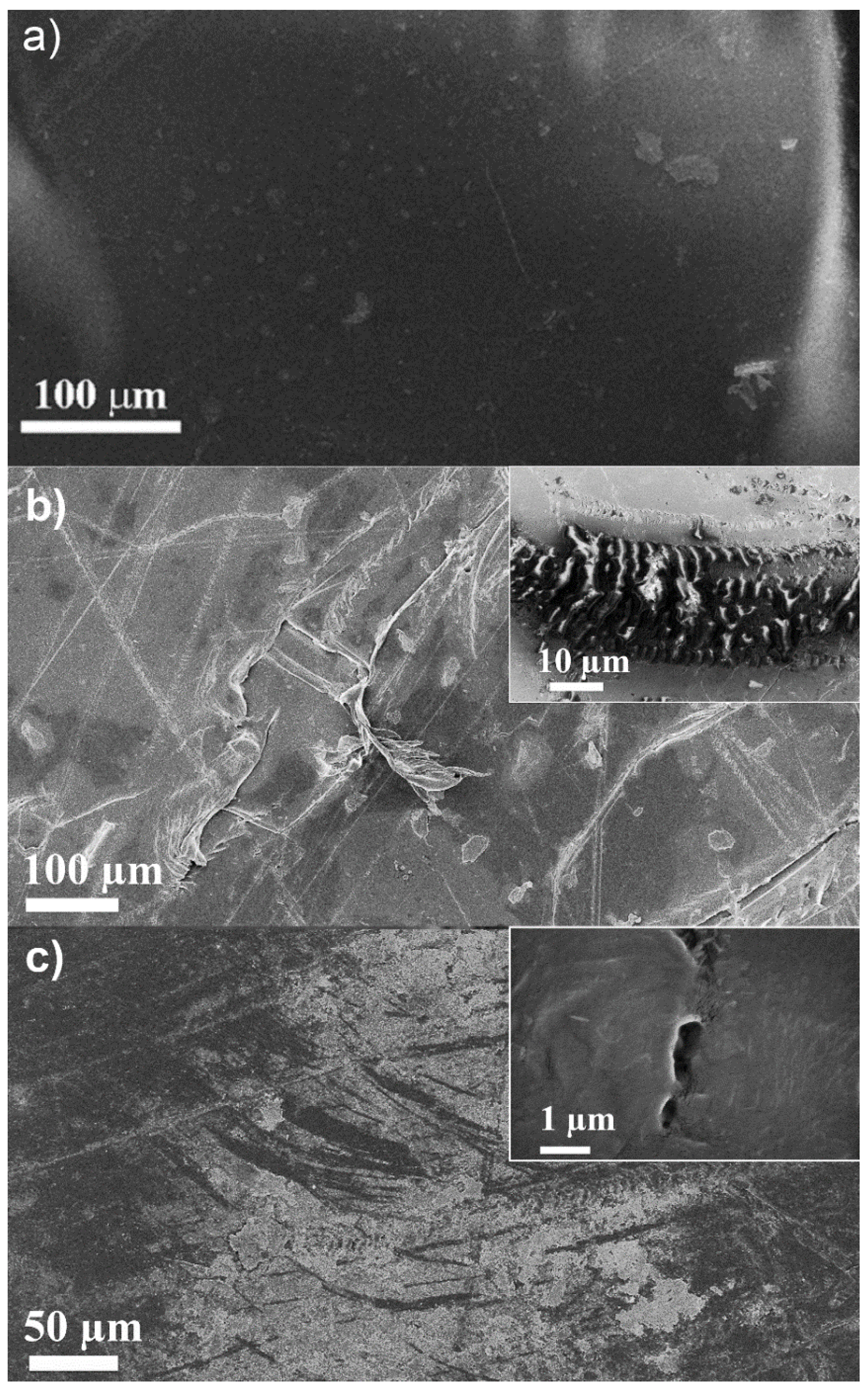
Additional visual data was obtained from SEM images (Figure 7a–c), where the morphological changes of the photo-catalytically degraded LDPE films show substantial changes in nanometer scale. The surface of the non-irradiated LDPE was found to be smooth except for a few manufacturing defects and spots [12]. Several wrinkles, cracks and cavities of varying sizes were detected all over the photodegraded films as a result of newly formed oxygenated groups and volatile organic compounds [38]. Deeper cavities and wrinkles were observed on LDPE film when photo-irradiated with ZnO-Pt catalyst in comparison with ZnO upon photodegradation for 175 hours.

3. Materials and Methods

3.1. Reagents

Reagents of zinc acetate dihydrate (Zn(CH3COO)2·2H2O), hexamethylenetetramine (C6H12N4), zinc nitrate hexahydrate (Zn(NO3)2·6H2O), and potassium hexachloroplatinate (K2PtCl6) were purchased from Sigma-Aldrich Chemie GmbH (Taufkirchen, Munich, Germany) and used without further purification. LDPE film in commercial use, with a thickness of 50 microns, was used to study the photocatalytic degradation studies.

3.2. Synthesis of Nanostructured Catalysts

ZnO nanorods were grown by a hydrothermal process which involved seeding of substrates with ZnO nanoparticles followed by 5 hours’ growth process in growth equimolar solution containing 10 mM of hexamethylenetetramine and zinc nitrate hexahydrate at 90 °C. Following the growth of nanorod coatings, the samples were washed extensively with deionized water and then annealed in ambient conditions at 350 °C for 1 hour prior to further processing [39].

3.3. Deposition of Platinum Nanoparticles on ZnO Nanorods

Pt-deposited ZnO nanorods (ZnO-Pt) were prepared using UV-C photoreduction method in which generation of surface defects on the ZnO nanorods was carried out by exposing the nanorods to a 100 W vapor mercury UV-C lamp for 30 minutes at a distance of 14 cm followed by photo mediated crystal growth from 1mM potassium hexachloroplatinate solution for 10 minutes. After deposition, the substrate was annealed in ambient air at 450 °C for 1 hour [22,40].

3.4. Photocatalysis Test Setup

The performance of designed catalysts was evaluated by following a process defined by the International Organization for Standardization (ISO 10678:2010) which include the degradation of 10 μM methylene blue (MB) dye solution (3 mL) for 90 minutes under light irradiation using ZnO and ZnO-Pt substrates, having dimensions of 2.5 cm × 0.75 cm, as the photocatalysts. Photocatalysis of low-density polyethylene film of size (1 cm × 1 cm) was carried out for 175 h in a Petri dish containing synthesized catalysts and deionized water. A 50 W dichroic halogen lamp in ambient air was used for visible light illumination (≈60–70 klux) from a distance of 10 cm for all photocatalytic degradation studies reported here, as shown in Figure S1 in Supplementary Materials.

3.5. Characterization Techniques

Surface topography and morphology of both catalysts and low-density polyethylene were examined by scanning electron microscopy (SEM) (GEMINI® Ultra 55, ZEISS, Oberkochen, Germany ). Energy dispersive X-ray spectroscopy (EDX) was utilized for the identification and determination of the abundance of elements in the designed photocatalysts. Platinum nanoparticle-coated ZnO nanorods were removed from the substrate by scratching with a sharp blade and drop-casted on copper grids to observe the morphology and crystal structure using high resolution transmission electron microscopy (HR-TEM, JEOL JEM-2100F, Tokyo, Japan).
The optical absorbance of designed catalysts and the degradation kinetics of MB dye were determined using UV-VIS spectrophotometer (LAMBDA-750, PerkinElmer, Waltham, MA, USA). Photoluminescence (PL) spectrophotometer (model LS 55, PerkinElmer, Waltham, MA, USA) was used for the assessment of intrinsic and UV-induced defect density for ZnO and ZnO-Pt samples. Emission spectra were obtained using an excitation wavelength of 320 nm [41].
Fourier transformation infra-red (FTIR) spectra of low-density polyethylene film was obtained using FTIR spectrometer (Nicolet™ iS10, Thermo Fisher Scientific, Waltham, MA, USA) to observe the variations of functional groups of polymer during photocatalytic degradation. Changes in the carbonyl index (CI) determined by the ratio of the area under the absorbance of 1710 cm−1 to the area under the reference peak at 1380 cm−1 and the Vinyl index (VI) which is the ratio of area under the absorbance of 909 cm−1 to the area under the reference peak at 1380 cm−1 were monitored [13,14].

4. Conclusions

Enhancement of visible light absorption, due to plasmon absorption in the platinum nanoparticles, along with the diffusion of photogenerated electrons into platinum from the ZnO nanorod interfaces reduces electron-hole recombination. This leads to more than 15% enhancement of photocatalytic performance with visible light irradiation, due to the combined effects enumerated above of the plasmonic catalyst. The photocatalytic activity for the degradation of organic matters (using methylene blue dye as a contaminant) was found to occur primarily, due to the contribution of superoxides and hydroxyl radicals that form during the photocatalysis. The plasmonic photocatalyst (ZnO-Pt) was found to degrade microplastic pollutants like residual LDPE films in water effectively. Formation of oxygenated groups like hydro-peroxides, peroxides, and unsaturated groups resulted in wrinkles, cracks and holes ensure the photocatalytic degradation of LDPE films. Modified ZnO-Pt catalysts showed ca. 13% higher potential for oxidation of LDPE film compared to the as-grown ZnO nanorods. Thus, modification of ZnO with plasmonic metal could be a viable way for expediting the oxidation of microplastic pollutants in water using sunlight.

Supplementary Materials

The following are available online at https://www.mdpi.com/2073-4344/9/10/819/s1, Figure S1: Experimental setup for photocatalytic degradation of LDPE film, Figure S2: X-ray dispersive spectroscopy (EDX) result showing the relative elemental composition of ZnO-Pt catalyst.

Author Contributions

Conceptualization, J.D.; Formal analysis, T.S.T., F.Y. and K.L.K.; Funding acquisition, J.D.; Investigation, T.S.T. and F.Y.; Methodology, T.S.T. and J.D.; Supervision, J.D.; Writing—original draft, T.S.T., F.Y. and K.L.K.; Writing—review and editing, J.D.

Funding

This research was funded by the European Commission through the EU research and innovation funding programme Horizon 2020, grant number 774586 (CLAIM-Cleaning Litter by developing and Applying Innovative Methods in european seas).

Acknowledgments

The publication of this article was funded by KTH Royal Institute of Technology.

Conflicts of Interest

The authors declare no conflict of interest.

References

  1. Barnes, D.K.A.; Galgani, F.; Thompson, R.C.; Barlaz, M. Accumulation and fragmentation of plastic debris in global environments. Philos. Trans. R. Soc. London Ser. B 2009, 364, 1985–1998. [Google Scholar] [CrossRef] [PubMed] [Green Version]
  2. Bergmann, M.; Gutow, L.; Klages, M. Marine Anthropogenic Litter; Springer International Publishing: Cham, Switzerland, 2015. [Google Scholar]
  3. Wu, W.-M.; Yang, J.; Criddle, C.S. Microplastics pollution and reduction strategies. Front. Environ. Sci. Eng. 2016, 11, 6. [Google Scholar] [CrossRef]
  4. Talvitie, J.; Heinonen, M.; Pääkkönen, J.P.; Vahtera, E.; Mikola, A.; Setälä, O.; Vahala, R. Do wastewater treatment plants act as a potential point source of microplastics? Preliminary study in the coastal Gulf of Finland, Baltic Sea. Water Sci. Technol. 2015, 72, 1495–1504. [Google Scholar] [CrossRef] [PubMed]
  5. Auta, H.S.; Emenike, C.U.; Fauziah, S.H. Distribution and importance of microplastics in the marine environment: A review of the sources, fate, effects, and potential solutions. Environ. Int. 2017, 102, 165–176. [Google Scholar] [CrossRef] [PubMed]
  6. Talvitie, J.; Mikola, A.; Koistinen, A.; Setälä, O. Solutions to microplastic pollution—Removal of microplastics from wastewater effluent with advanced wastewater treatment technologies. Water Res. 2017, 123, 401–407. [Google Scholar] [CrossRef] [PubMed]
  7. Westphalen, H.; Abdelrasoul, A. Challenges and treatment of microplastics in water. In Water Challenges of an Urbanizing World; Glavan, M., Ed.; IntechOpen: London, UK, 2017. [Google Scholar]
  8. Singh, B.; Sharma, N. Mechanistic implications of plastic degradation. Polym. Degrad. Stab. 2008, 93, 561–584. [Google Scholar] [CrossRef]
  9. Klein, S.; Dimzon, I.K.; Eubeler, J.; Knepper, T.P. Analysis, occurrence, and degradation of microplastics in the aqueous environment. In Freshwater Microplastics. The Handbook of Environmental Chemistry; Wagner, M., Lambert, S., Eds.; Springer: Cham, Switzerland, 2018; Volume 58, pp. 51–57. [Google Scholar]
  10. Zhou, D.; Chen, L.; Li, J.; Wu, F. Transition metal catalyzed sulfite auto-oxidation systems for oxidative decontamination in waters: A state-of-the-art minireview. Chem. Eng. J. 2018, 346, 726–738. [Google Scholar] [CrossRef]
  11. Kang, J.; Zhou, L.; Duan, X.; Sun, H.; Ao, Z.; Wang, S. Degradation of cosmetic microplastics via functionalized carbon nanosprings. Matter 2019, 1, 745–758. [Google Scholar] [CrossRef]
  12. Yousif, E.; Haddad, R. Photodegradation and photostabilization of polymers, especially polystyrene: Review. SpringerPlus 2013, 2, 398. [Google Scholar] [CrossRef] [PubMed]
  13. Ali, S.S.; Qazi, I.A.; Arshad, M.; Khan, Z.; Voice, T.C.; Mehmood, C.T. Photocatalytic degradation of low density polyethylene (LDPE) films using titania nanotubes. Environ. Nanotechnol. Monit. Manag. 2016, 5, 44–53. [Google Scholar] [CrossRef] [Green Version]
  14. Tofa, T.S.; Kunjali, K.L.; Paul, S.; Dutta, J. Visible light photocatalytic degradation of microplastic residues with zinc oxide nanorods. Environ. Chem. Lett. 2019. [Google Scholar] [CrossRef]
  15. Ariza-Tarazona, M.C.; Villarreal-Chiu, J.F.; Barbieri, V.; Siligardi, C.; Cedillo-González, E.I. New strategy for microplastic degradation: Green photocatalysis using a protein-based porous N-TiO2 semiconductor. Ceram. Int. 2019, 45, 9618–9624. [Google Scholar] [CrossRef]
  16. Li, S.; Xu, S.; He, L.; Xu, F.; Wang, Y.; Zhang, L. Photocatalytic degradation of polyethylene plastic with polypyrrole/TiO2 nanocomposite as photocatalyst. Polym. Plast. Technol. Eng. 2010, 49, 400–406. [Google Scholar] [CrossRef]
  17. Laxman, K.; Al Rashdi, M.; Al Sabahi, J.; Al Abri, M.; Dutta, J. Supported versus colloidal zinc oxide for advanced oxidation processes. Appl. Surf. Sci. 2017, 411, 285–290. [Google Scholar] [CrossRef]
  18. Qi, K.; Cheng, B.; Yu, J.; Ho, W. Review on the improvement of the photocatalytic and antibacterial activities of ZnO. J. Alloys Compd. 2017, 727, 792–820. [Google Scholar] [CrossRef]
  19. Bora, T.; Dutta, J. Plasmonic photocatalyst design: Metal-semiconductor junction affecting photocatalytic efficiency. J. Nanosci. Nanotechnol. 2019, 19, 383–388. [Google Scholar] [CrossRef] [PubMed]
  20. Lin, J.M.; Lin, H.Y.; Cheng, C.L.; Chen, Y.F. Giant enhancement of bandgap emission of ZnO nanorods by platinum nanoparticles. Nanotechnology 2006, 17, 4391–4394. [Google Scholar] [CrossRef] [Green Version]
  21. Wang, P.; Huang, B.; Dai, Y.; Whangbo, M.H. Plasmonic photocatalysts: Harvesting visible light with noble metal nanoparticles. Phys. Chem. Chem. Phys. 2012, 14, 9813–9825. [Google Scholar] [CrossRef] [PubMed]
  22. Al-Alawi, R.; Laxman, K.; Dastgir, S.; Dutta, J. Role of bonding mechanisms during transfer hydrogenation reaction on heterogeneous catalysts of platinum nanoparticles supported on zinc oxide nanorods. Appl. Surf. Sci. 2016, 377, 200–206. [Google Scholar] [CrossRef]
  23. Bora, T.; Zoepfl, D.; Dutta, J. Importance of plasmonic heating on visible light driven photocatalysis of gold nanoparticle decorated zinc oxide nanorods. Sci. Rep. 2016, 6, 26913. [Google Scholar] [CrossRef] [PubMed]
  24. Chichagov, A.; Belonozhko, A.; Lopatin, A.; Dokina, T.; Samokhvalova, O.; Ushakovskaya, T.; Shilova, Z. Information computing system from structural data on minerals (Mincryst). Kristallografiya 1990, 35, 610–616. [Google Scholar]
  25. Sun, F.; Qiao, X.; Tan, F.; Wang, W.; Qiu, X. One-step microwave synthesis of Ag/ZnO nanocomposites with enhanced photocatalytic performance. J. Mater. Sci. Mater. Electron. 2012, 47, 7262–7268. [Google Scholar] [CrossRef]
  26. Yang, T.; Wang, M.; Duan, C.; Hu, X.; Huang, L.; Peng, J.; Huang, F.; Gong, X. Inverted polymer solar cells with 8.4% efficiency by conjugated polyelectrolyte. Energy Environ. Sci. 2012, 5, 8208–8214. [Google Scholar] [CrossRef]
  27. Zhang, N.; Tang, W.; Wang, P.; Zhang, X.; Zhao, Z. In situ enhancement of NBE emission of Au–ZnO composite nanowires by SPR. Cryst. Eng. Comm. 2013, 3301–3304. [Google Scholar] [CrossRef]
  28. Anandan, S.; Vinu, A.; Sheeja Lovely, K.L.P.; Gokulakrishnan, N.; Srinivasu, P.; Mori, T.; Murugesan, V.; Sivamurugan, V.; Ariga, K. Photocatalytic activity of La-doped ZnO for the degradation of monocrotophos in aqueous suspension. J. Mol. Catal. A Chem. 2007, 266, 149–157. [Google Scholar] [CrossRef]
  29. Yu, C.; Yang, K.; Xie, Y.; Fan, Q.; Yu, J.C.; Shu, Q.; Wang, C. Novel hollow Pt-ZnO nanocomposite microspheres with hierarchical structure and enhanced photocatalytic activity and stability. Nanoscale 2013, 5, 2142. [Google Scholar] [CrossRef] [PubMed]
  30. Baruah, S.; Mahmood, M.A.; Myint, M.T.Z.; Bora, T.; Dutta, J. Enhanced visible light photocatalysis through fast crystallization of zinc oxide nanorods. Beilstein J. Nanotechnol. 2010, 1, 14–20. [Google Scholar] [CrossRef] [PubMed] [Green Version]
  31. Flyunt, R.; Knolle, W.; Kahnt, A.; Halbig, C.E.; Lotnyk, A.; Häupl, T.; Prager, A.; Eigler, S.; Abel, B. High quality reduced graphene oxide flakes by fast kinetically controlled and clean indirect UV-induced radical reduction. Nanoscale 2016, 8, 7572–7579. [Google Scholar] [CrossRef] [PubMed] [Green Version]
  32. Vinoth, R.; Babu, S.G.; Ramachandran, R.; Neppolian, B. Bismuth oxyiodide incorporated reduced graphene oxide nanocomposite material as an efficient photocatalyst for visible light assisted degradation of organic pollutants. Appl. Surf. Sci. 2017, 418, 163–170. [Google Scholar] [CrossRef]
  33. Gulmine, J.V.; Janissek, P.R.; Heise, H.M.; Akcelrud, L. Polyethylene characterization by FTIR. Polym. Test. 2002, 21, 557–563. [Google Scholar] [CrossRef]
  34. Mouallif, I.; Latrach, A.; Benali, C.M.H.; Barbe, N. FTIR study of HDPE structural changes, moisture absorption and mechanical properties variation when exposed to sulphuric acid aging in various temperatures. In 20ÈME Congrès Français de Mécanique, AFM; Maison de la Mécanique: Besançon, France, 2011; pp. 2011–25044. [Google Scholar]
  35. Luongo, J.P. Infrared study of oxygenated groups formed in polyethylene during oxidation. J. Polym. Sci. 1960, 42, 139–150. [Google Scholar] [CrossRef]
  36. Qin, H.; Zhao, C.; Zhang, S.; Chen, G.; Yang, M. Photo-oxidative degradation of polyethylene/montmorillonite nanocomposite. Polym. Degrad. Stab. 2003, 81, 497–500. [Google Scholar] [CrossRef]
  37. Gardette, M.; Perthue, A.; Gardette, J.L.; Janecska, T.; Földes, E.; Pukánszky, B.; Therias, S. Photo- and thermal-oxidation of polyethylene: Comparison of mechanisms and influence of unsaturation content. Polym. Degrad. Stab. 2013, 98, 2383–2390. [Google Scholar] [CrossRef] [Green Version]
  38. Shang, J.; Chai, M.; Zhu, Y. Photocatalytic degradation of polystyrene plastic under fluorescent light. Environ. Sci. Technol. 2003, 37, 4494–4499. [Google Scholar] [CrossRef]
  39. Baruah, S.; Dutta, J. Hydrothermal growth of ZnO nanostructures. Sci. Technol. Adv. Mater. 2009, 10, 013001. [Google Scholar] [CrossRef] [PubMed]
  40. Myint, M.T.Z.; Kumar, N.S.; Hornyak, G.L.; Dutta, J. Hydrophobic/hydrophilic switching on zinc oxide micro-textured surface. Appl. Surf. Sci. 2013, 264, 344–348. [Google Scholar] [CrossRef]
  41. Baral, A.; Khanuja, M.; Islam, S.S.; Sharma, R.; Mehta, B.R. Identification and origin of visible transitions in one dimensional (1D) ZnO nanostructures: Excitation wavelength and morphology dependence study. J. Lumin. 2017, 183, 383–390. [Google Scholar] [CrossRef]
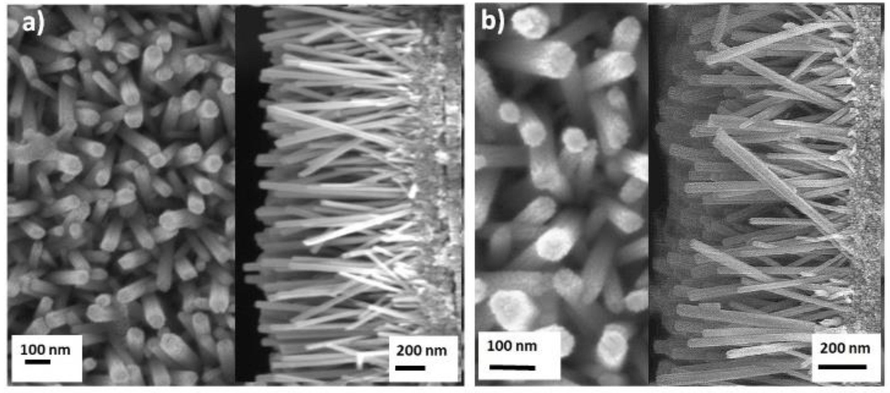
Figure 1. SEM images of (a) ZnO nanorods and (b) platinum nanoparticles-deposited of ZnO nanorods (ZnO-Pt). Top and cross-sectional views show the diameter and length of nanorods, respectively.
Catalysts 09 00819 g001
Figure 2. TEM images of platinum nanoparticles deposited on ZnO nanorods: (a,b) Uniformly deposited platinum nanoparticles on ZnO nanorods; (c) High resolution image showing the lattice spacing of platinum nanoparticles and ZnO nanorods, (d) SAED pattern of ZnO-Pt; and (e) particle size distribution of platinum nanoparticles grown on the surface of ZnO nanorods.
Catalysts 09 00819 g002
Figure 3. (a) UV-VIS adsorption spectra of ZnO nanorods and platinum nanoparticle-deposited ZnO nanorods. (b) Emission spectra of ZnO nanorods and ZnO-Pt with 320 nm excitation. Reduction in the PL intensity for ZnO-Pt indicates lower electron-hole recombination rate than that for ZnO nanorods.
Catalysts 09 00819 g003
Figure 4. Photocatalytic activities of ZnO and ZnO-Pt catalysts and the effect of •OH scavengers on the degradation kinetics. (MB, methylene blue; ISP, isopropanol).
Catalysts 09 00819 g004
Figure 5. The suggested mechanism of enhanced photocatalytic activities with ZnO-Pt (energy levels: Absolute vacuum scale (AVS)).
Catalysts 09 00819 g005
Figure 6. FTIR spectra of low-density polyethylene films before and after irradiation along with ZnO and ZnO-Pt catalysts upon 175 hours exposure under visible light.
Catalysts 09 00819 g006
Figure 7. SEM images of low-density polyethylene film after 175 hours of visible light exposure (a) controlled LDPE, (b) ZnO, (c) ZnO-Pt reveal the configuration of cracks, spots and wrinkles; plasmonic catalysts showed better catalytic activities than ZnO alone.
Catalysts 09 00819 g007
Table 1. Variations in the carbonyl index and vinyl index of photocatalyzed low-density polyethylene films.
CatalystCarbonyl index (CI)Vinyl index (VI)
LDPE (control)0.710.51
LDPE + ZnO 1.381.12
LDPE + ZnO-Pt1.491.35