Article

Improving the Indigo Carmine Decolorization Ability of a Bacillus amyloliquefaciens Laccase by Site-Directed Mutagenesis

College of Life Sciences, Northeast Forestry University, Harbin 150040, China
*
Author to whom correspondence should be addressed.
Catalysts 2017, 7(9), 275; https://doi.org/10.3390/catal7090275
Submission received: 14 August 2017 / Revised: 29 August 2017 / Accepted: 12 September 2017 / Published: 15 September 2017
(This article belongs to the Special Issue Biocatalysis and Biotransformations)

Abstract

:
Indigo carmine is a typical recalcitrant dye which is widely used in textile dyeing processes. Laccases are versatile oxidases showing strong ability to eliminate hazardous dyes from wastewater. However, most laccases require the participation of mediators for efficient decolorization of indigo carmine. Here we describe the improvement of the decolorization ability of a bacterial laccase through site-directed mutagenesis. A D501G variant of Bacillus amyloliquefaciens laccase was constructed and overexpressed in Escherichia coli. The laccase activity in the culture supernatant achieved 3374 U·L−1 for the mutant. Compared with the wild-type enzyme, the D501G exhibited better stability and catalytic efficiency. It could decolorize more than 92% of indigo carmine without additional mediators in 5 h at pH 9.0, which was 3.5 times higher than the wild-type laccase. Isatin sulfonic acid was confirmed to be the main product of indigo carmine degradation by UV-vis and LC-MS analyses.

Graphical Abstract

1. Introduction

Textile industries discharge great amounts of effluents to the environment, and about 15–20% of dyes are lost in the effluents during the dyeing process [1,2]. Since synthetic dyes are designed to be chemically and photolytically stable, they are highly persistent and their removal from wastewater is rather difficult. The discharge of untreated dye effluents in water bodies may cause severe environmental and health hazards due to the toxicity of most synthetic dyes [1].
China is the largest dyestuff production country in the world. The national total output of dye in 2015 was 9.2 × 10⁵ tons, accounting for about 2/3 of the global output [3]. The export volume of indigo dye in China increased 12.5% annually during the 12th Five-Year Plan period (2011–2015), which was the highest among all dyes [3]. Indigo carmine is among the most widely used textile dyeing agents for the dyeing of polyester fibers and denim. It is a typical recalcitrant dye and is toxic to human health [4,5]. Various techniques have been employed to remove this dye from wastewater, such as photocatalytic degradation, electrochemical oxidation, and ozonation [4,5,6]. However, these methods are usually quite expensive and consume high amounts of chemicals and/or energy [7]. Biological processes, due to their eco-friendly and cost-competitive benefits, are considered to be an alternative to the traditional physicochemical methods in dye effluents treatment [1,7].
Currently, laccase-catalyzed decolorization has received much attention due to its potential ability to degrade of a wide range of dyes [2,8]. Laccases (EC 1.10.3.2) are enzymes that belong to the broader family of multicopper oxidases. They are able to catalyze the oxidation of various aromatic compounds with the reduction of molecular oxygen to water [9]. The first commercial laccase was launched in 1996 by Novozyme, and was used for denim bleaching with the help of a redox mediator. The product exhibited good performance in oxidizing the indigo dye on denim garments into uncolored compounds [9]. To date, most commercial laccases are of fungal origin, and they are usually active in moderate temperatures and an acidic pH range [9]. Since wastewater from denim processing is characterized by high pH and high temperature [10], the application of fungal laccases for the treatment of textile effluent is limited by their instability under these drastic conditions.
Compared with fungal laccases, bacterial laccases are highly active and much more stable under alkaline and high temperature conditions [9]. In our previous work, we heterologously expressed and characterized a novel laccase from Bacillus amyloliquefaciens LC02. The recombinant enzyme showed a half-life of inactivation about 3.5 h at 70 °C, and retained about 70% of its initial activity after 10 days of incubation at pH 9.0 [11]. In addition, it could efficiently decolorize indigo carmine with acetosyringone as a mediator. Nevertheless, the extent of decolorization in the absence of a mediator was rather low. The application of the laccase-mediator system suffers from drawbacks of high cost and the possible toxicity of most mediators, which hamper its application at the industrial scale [12]. Therefore, this work aims to increase the decolorization of indigo carmine by laccase without using mediators. The aspartic acid at position 501 in B. amyloliquefaciens laccase is close to the T1 Cu site (Figure S1), and adjacent to methionine 502, which is the axial ligand of the T1 copper ion [13]. It has been suggested that Asp501 in B. subtilis laccase lies at the surface of the water channel by which the dioxygen molecule approaches the trinuclear copper center. It also is involved in the formation of hydrogen bonds with the solvent molecules in the channel [14]. Multiple sequence alignments showed that Asp501 has only been found in laccases from Bacillus sp., whereas a glycine residue at the corresponding position existed in other bacterial and fungal laccases (Figure S2). It has been reported that the substitution of Asp500 (equivalent to Asp501 in B. amyloliquefaciens and B. subtilis laccase) by glycine could increase the catalytic efficiency and functional expression of Bacillus sp. HR03 laccase [15]. Thus, the site-directed mutagenesis method was employed to improve the catalytic efficiency of B. amyloliquefaciens laccase (Scheme 1). The mutant enzyme was overexpressed in Escherichia coli to achieve a high-yield production. The degradation products of indigo carmine were also characterized using UV-vis and LC-MS analyses.

2. Results and Discussion

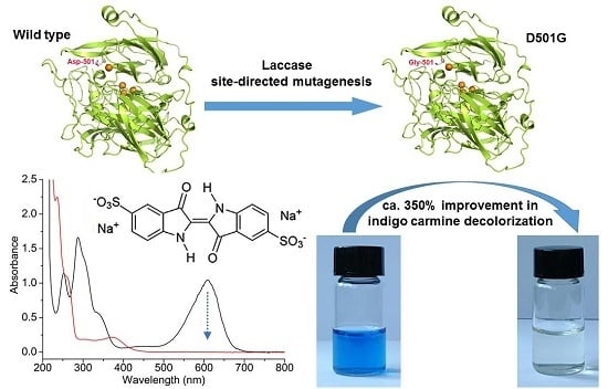
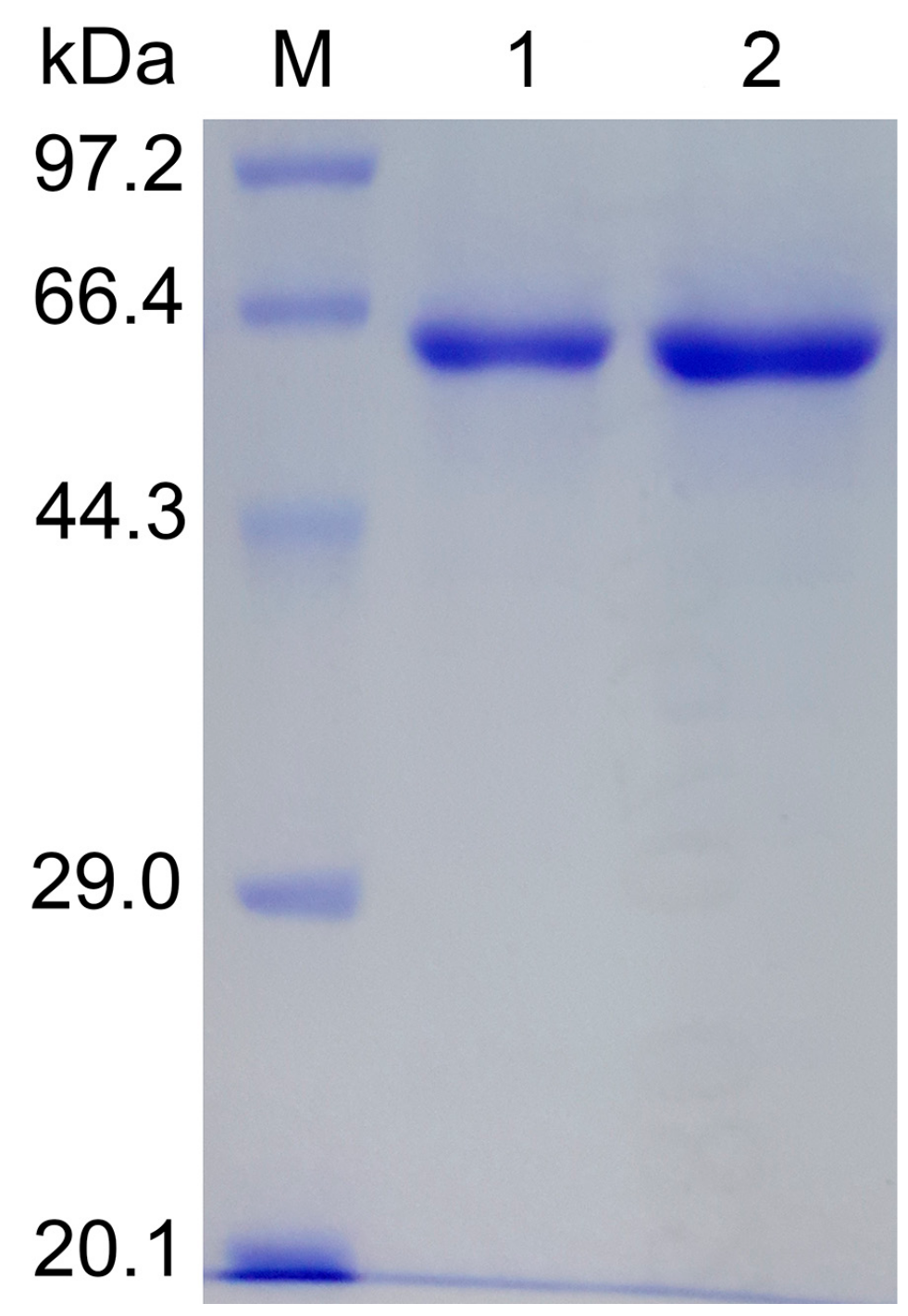
2.1. Site-Directed Mutagenesis and Expression in E. coli

Both the wild-type and D501G laccase could be extracellularly expressed in E. coli. The volumetric activity with 2,2′-azino-bis(3-ethylbenzothiazoline-6-sulfonate) (ABTS) as a substrate reached 4319 U·L−1 for the wild-type enzyme and 3374 U·L−1 for D501G. The expression level is significantly higher than that of the wild-type laccase expressed in Pichia pastoris (380 U·L−1) [11]. Moreover, the induction time was reduced from seven days to one day when E. coli was used as the host for recombinant protein production. In most cases, the expression of bacterial laccases in E. coli resulted in the formation of inclusion bodies, which were difficult to refold and purify and resulted in low yields [13,16,17,18]. Furthermore, the recombinant laccases were usually located intracellularly, and cell disruption was required to extract the target proteins. Extracellular expression of recombinant proteins, which has the advantages of decreasing protein aggregation and simplifying the purification process, affords an opportunity to overcome these issues [19]. Unfortunately, the secretory expression of bacterial laccases in P. pastoris were unsatisfactory due to the low yield and long cultivation time [11,20,21]. Recently, the B. subtilis laccase was successfully expressed in secretory form in E. coli by altering the induction conditions, and high laccase activity (2401 U·L−1) was achieved after the optimization of induction parameters [22]. In the present study, we also detected high activity in the culture medium for both the wild-type and D501G laccase using the two phase induction strategy. However, the D501G variant exhibited a lower volumetric activity than the wild-type laccase. Previous reports indicated that replacing aspartic acid with glycine at the same position (D500G) resulted in the enhancement of the soluble expression of B. licheniformis and Bacillus sp. HR03 laccases in E. coli [15,23]. Therefore, the lower laccase content in the culture supernatant of D501G (41 and 71 mg·L−1 for D501G and the wild-type, respectively) might be due to the increase of the soluble expression in the E. coli cells.

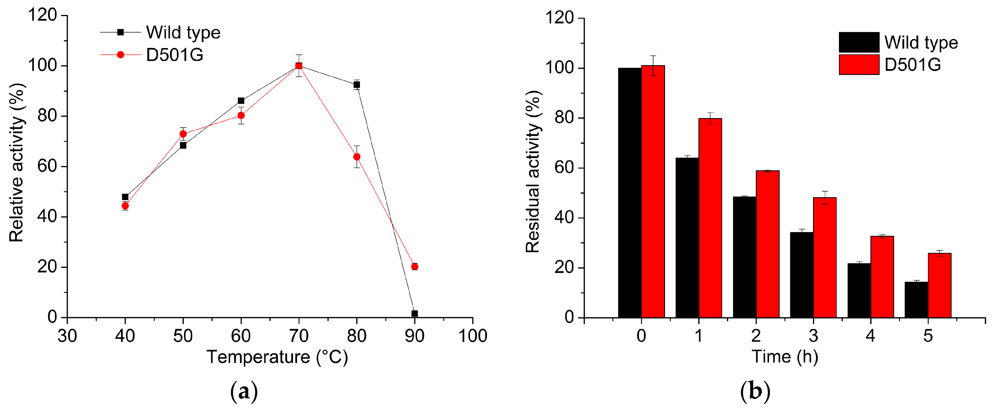
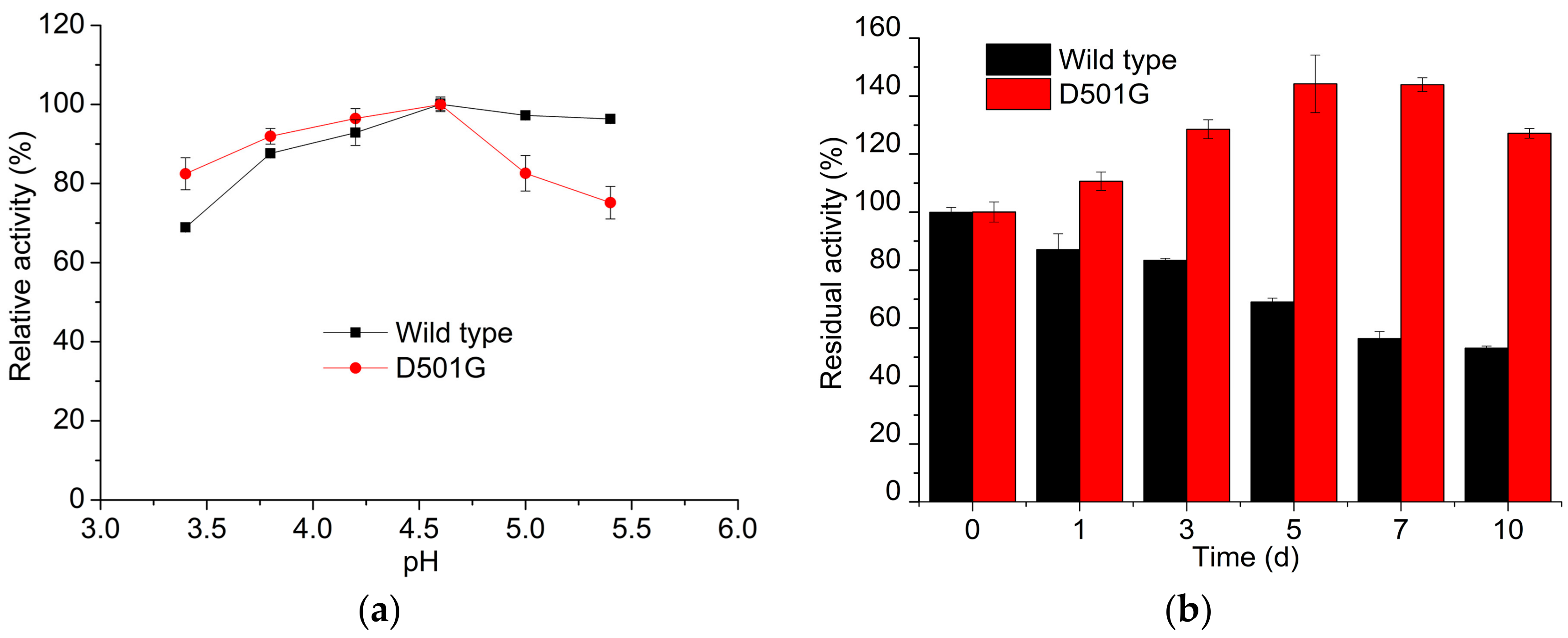
2.2. Enzyme Characterization

SDS-PAGE analysis showed a band with a molecular weight of about 65 kDa for the purified mutant and wild-type laccases (Figure 1). Further, a similar pH profile was observed for the mutant and the wild-type. The optimal pH for ABTS oxidation was 4.6 (Figure 2a), with a slight shift toward a more alkaline range compared with that of the P. pastoris-expressed laccase [11]. The D501G variant exhibited a higher stability at pH 9.0 (Figure 2b). It was fully active after 10 days of incubation at pH 9.0, while the wild-type enzyme retained 53% of the initial activity.
Although the D501G mutant had the same optimal temperature (70 °C) as the wild-type, it demonstrated a higher thermostability (Figure 3). The half-lives of inactivation at 70 °C were about 2 h and 3 h for the wild-type and mutant laccase, respectively (Figure 3b). As shown in Table 1, there was a 35% decrease in Km and a 2.3-fold increase in kcat, which resulted in about a 3.7-fold higher catalytic efficiency. Similar results have been found for the D500G mutant of Bacillus sp. HR03 laccase [15], while a slight decrease in catalytic efficiency was measured for the D500G variant of B. licheniformis laccase [23].

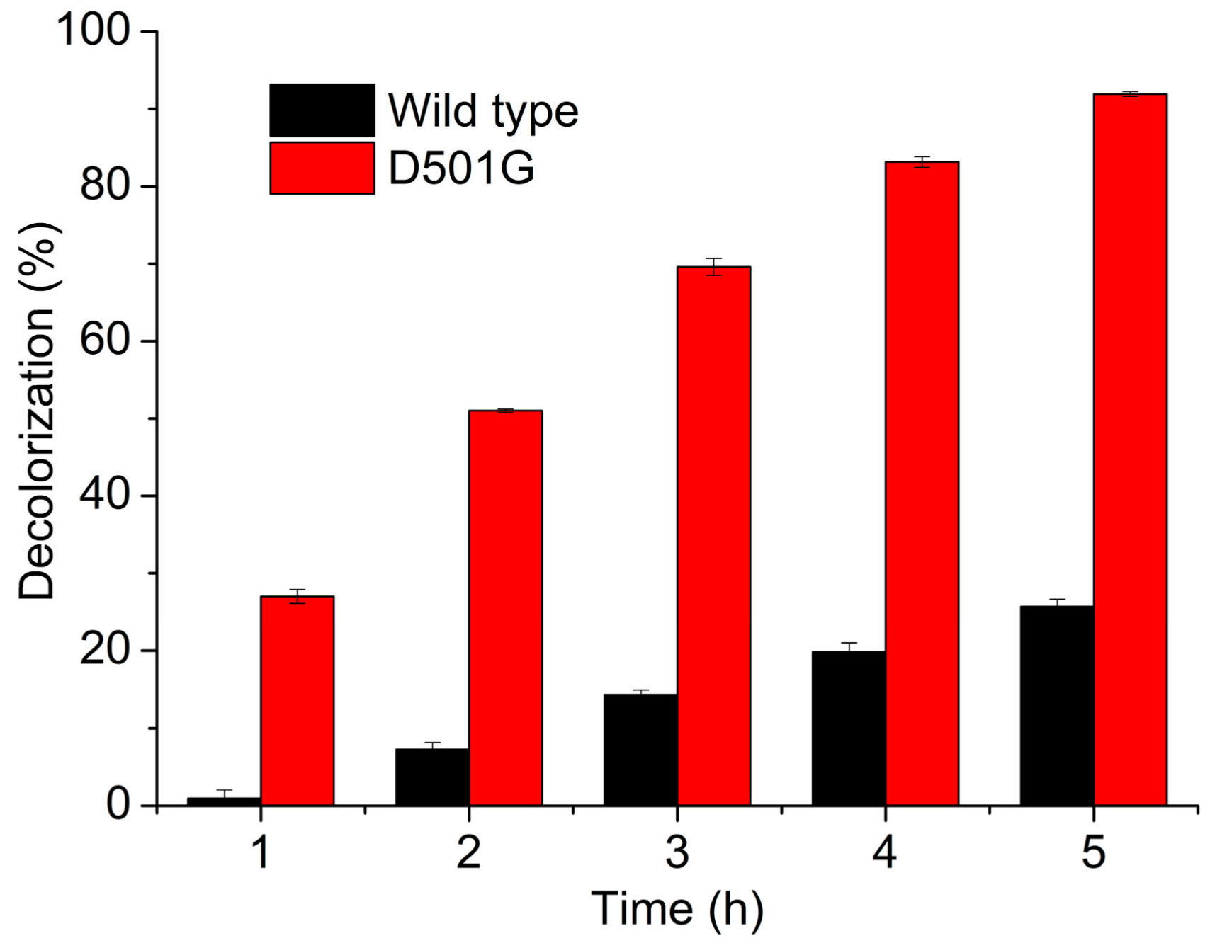
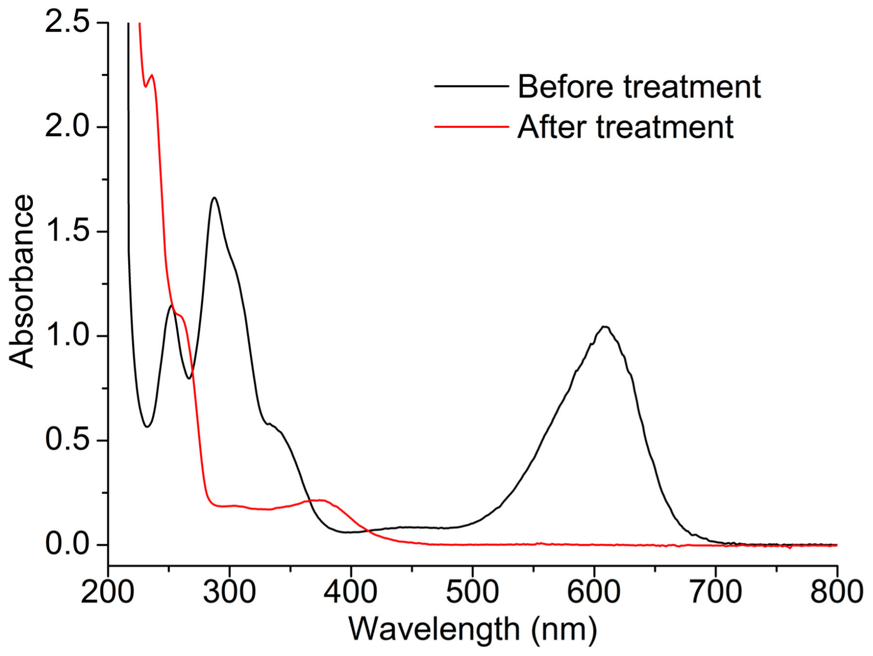
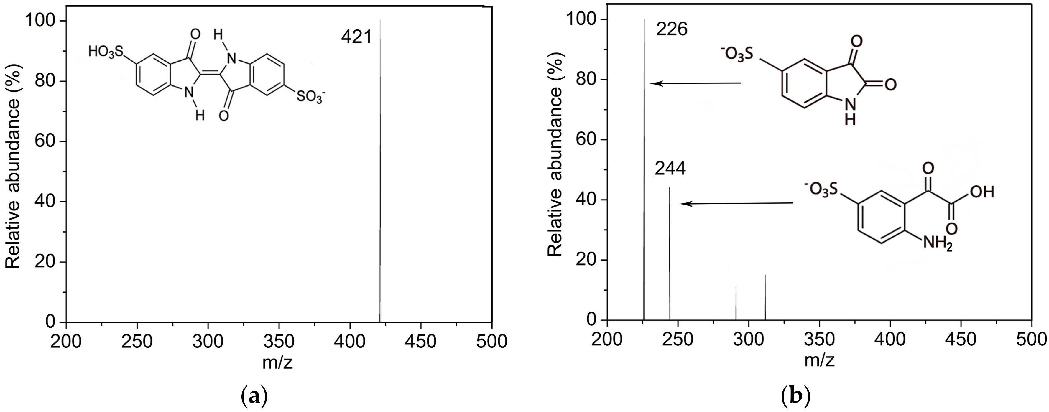
2.3. Indigo Carmine Decolorization

Dye decolorization tests revealed a remarkable enhancement of indigo carmine degradation by laccase after mutagenesis. No obvious decolorization occurred in the first 1 h when the wild-type laccase was used without an additional mediator, and only 26% of indigo carmine was decolorized after 5 h (Figure 4). In contrast, the D501G mutant could decolorize more than 92% of indigo carmine in 5 h at pH 9.0, which was about 3.5 times higher than that of the wild-type enzyme. Protein structure analysis indicated that the replacement of Asp501 caused a decrease in the number of hydrogen bonds formed at position 501 and a larger space between Gly501 and Met 503 (Figure S3), which may increase the conformational flexibility of this region. Conformational flexibility in the active site is important for substrate binding and for enzyme catalysis, and there is usually a positive correlation between conformational flexibility and enzyme activity [24,25,26]. As mentioned above, the Asp501 is adjacent to the axial ligand (Met502) of the T1 copper ion and lies at the surface of the water channel. Met502 has been proven to play an important role in affecting the redox potential of the T1 site, where the substrate was oxidized by the copper ion [27]. Additionally, the less steric hindrance at position 501 could increase the accessibility of solvent molecules in the water channel, and consequently influence the redox potential of the copper center [15]. Therefore, the increase in the flexibility near the T1 site may impact the redox potential of the laccase and lead to a higher catalytic efficiency for indigo carmine decolorization. Generally, mediators are needed for efficient decolorization of indigo carmine by fungal or bacterial laccases. For example, laccases from Trametes hirsuta Bm2 alone were not able to oxidize indigo carmine, even under high enzymatic activity (18,000 U·L−1) [28]. Indigo carmine was poorly decolorized by Streptomyces coelicolor laccase in the absence of mediators, while more than 85% of the dye was decolorized by laccase with syringaldehyde as a mediator [29]. Although the laccases-mediator system demonstrates high efficiency in dye decolorization, it also increases the application cost as most mediators are expensive, and they cannot be co-immobilized when an immobilized enzyme is used. Moreover, the toxicity of some mediators limits their practical application. Hence, the development of laccase-catalyzed oxidation without mediators is much more desirable for wastewater treatment.
The cleavage of the chromophore is a crucial step in dye degradation, which makes the dye fragments more susceptible to the subsequent biodegradation [7]. The chromophore center of indigo carmine consists of a C=C double bond substituted by two NH donor groups and two CO acceptor groups [30]. As shown in Figure 5, the characteristic peaks of indigo carmine are situated at 610 nm, 287 nm, and 250 nm in the UV-visible absorption spectrum. The peak at 610 nm, which accounts for the blue color of indigo carmine, disappeared after the laccase treatment. Meanwhile, the peak at 287 nm also vanished and the absorption band at 250 nm increased and blue-shifted to 235 nm. The results suggested the cleavage of the chromophoric structure of the dye and the generation of a new compound. The spectrum of the laccase-treated solution is very similar to that of isatin sulfonic acid (ISA) [31], which can be formed from indigo carmine by breaking down the double bond in the H-chromophore [31,32].
To identify the decolorization products, the solutions before and after laccase treatment were analyzed by LC-MS. The main peak at m/z 421, which corresponds to a single charged anion of indigo carmine [33], was present in the mass spectrum of the initial dye solution (Figure 6a). After 5 h of treatment by laccase, this anion was no longer detected, indicating that the dye was completely biotransformed. Two most abundant peaks at m/z 226 and 244 were observed in the mass spectrum (Figure 6b). The m/z peak at 226 is a characteristic peak of ISA, which can be further converted to the anion of m/z 244 by the addition of H2O. Both products of m/z 226 and 244 can be identified during indigo carmine degradation by other methods [32,33,34]. These results are consistent with that of the UV-visible spectra, which revealed the characteristic peak of ISA at 235 nm.

3. Materials and Methods

3.1. Materials

2,2′-Azino-bis(3-ethylbenzothiazoline-6-sulfonate) (ABTS), syringaldazine, and indigo carmine were products of Sigma-Aldrich (St. Louis, MO, USA). Fast Mutagenesis System was supplied by TransGen (Beijing, China). Gel Extraction Kit and Plasmid Mini Kit I were purchased from Omega Bio-Tek (Norcross, GA, USA). E. coli BL21(DE3) competent cells and Bradford Protein Assay Kit were obtained from Tiangen (Beijing, China). Expression vector pET-20b(+) was a product of Novagen (Billerica, MA, USA). Recombinant plasmid pPICZαA/lac harboring the wild-type laccase gene from B. amyloliquefaciens LC02 was constructed previously and preserved in our laboratory [11]. Restriction enzymes and T4 DNA ligase were purchased from NEB (Ipswich, MA, USA). HisTrap HP affinity columns were from GE Healthcare (Piscataway, NJ, USA). Other chemicals were of analytical reagent grade.

3.2. Site-Directed Mutagenesis

The wild-type laccase gene was amplified from the plasmid pPICZαA/lac with forward primer 5′-GCGTCCATATGGCACTGGAAAAATTTGCAGATG-3′ and reverse primer 5′-CCGCTCGAGCTGCTTATCCGTGACGTCCATC-3′ [11]. Recognition sites of Nde I and Xho I are indicated by underline. The amplified product was purified and digested with Nde I and Xho I. Then, it was cloned into pET-20b(+) vector, and the recombinant plasmid pET-20b(+)/lac was used as a template for site-directed mutagenesis. A mutant D501G was constructed using Fast Mutagenesis System according to the supplier’s protocol. For creation of the variant, the following primers were used: D501G-F 5′-GAGCACGAAGATTACGGTATGATGAGACCGAT-3′ and D501G-R 5′-ACCGTAATCTTCGTGCTCTAAAATATGACAGTGC-3′. The sequences of the inserted genes were verified by sequencing. Plasmids containing the wild-type and the mutant laccase gene were then transformed into E. coli BL21(DE3) for protein expression.
Multiple sequence alignment was performed with Clustal Omega (http://www.ebi.ac.uk/Tools/msa/clustalo/). All protein sequences were searched from the NCBI (National Center for Biotechnology Information, https://www.ncbi.nlm.nih.gov). Homology modeling was performed using the SWISS-MODEL Server (http://swissmodel.expasy.org/). CotA protein from B. subtilis (Protein Data Bank (PDB) ID: 1GSK) that showed 76.76% identity to B. amyloliquefaciens laccase was used as a template. The protein structures were analyzed with PyMol viewer.

3.3. Protein Expression and Purification

E. coli BL21(DE3) cells harboring the wild-type laccase and its variant were grown overnight in 5 mL LB (Luria-Bertani) medium supplemented with 100 mg·mL−1 of ampicillin (Solarbio, Beijing, China) at 30 °C, 180 rpm. The overnight culture (100 μL) was inoculated into 10 mL fresh LB/ampicillin medium and incubated at 30 °C with shaking (180 rpm). Protein expression was induced at an OD600 of 0.8 by adding 0.1 mM isopropyl-β-d-thiogalactoside (IPTG) (Solarbio, Beijing, China) and 2 mM CuSO4 (Sinopharm, Shanghai, China) (final concentration). After incubating at 25 °C, 120 rpm for 4 h, the shaker was turned off to achieve microaerobic conditions. Incubation was continued at 30 °C for a further 20 h, and cells were harvested by centrifugation at 8000 rpm for 10 min. The supernatant was collected and concentrated with poly(ethylene glycol) (PEG) 20,000 (Solarbio, Beijing, China). The obtained sample was loaded on a HisTrap HP affinity column, which was pre-equilibrated with 20 mM Tris-HCl (pH 7.4) containing 500 mM NaCl and 40 mM imidazole. The target protein was eluted with 200 mM imidazole in the same buffer. The pooled active fractions were concentrated and desalted by ultrafiltration (cutoff 30 kDa). Then the purified enzymes were stored at −20 °C for further use.

3.4. Enzyme Assay and Characterization

Laccase activity was measured at 30 °C using ABTS and syringaldazine as substrates [13]. One unit of enzyme activity was defined as the amount of enzyme required to oxidize 1 μmol of substrate per minute.
Kinetic parameters for the purified laccase were determined at 30 °C using different concentrations of ABTS (50–1000 μM). Protein concentration was determined using the Bradford Protein Assay Kit with bovine serum albumin as the standard. All assays were carried out in triplicate.
The optimal pH and temperature as well as pH and thermal stability of the wild-type and mutant enzyme were determined as previously described [11].

3.5. Indigo Carmine Decolorization

Decolorization was conducted with 100 U·L−1 of laccase and 25 mg·L−1 indigo carmine in 50 mM Tris-HCl buffer (pH 9.0). Reactions were initiated by the addition of laccase, and incubated at 40 °C under mild shaking conditions. Control samples were run in parallel without laccase. The residual dye concentration was determined using a UV-visible spectrophotometer (BioSpectrometer, Eppendorf, Humburg, Germany). All reactions were performed in triplicate.
To analyze the degradation products, laccases were removed from the reaction mixture using Amicon Ultra-15 centrifugal filters (10 kDa) (Millipore, Billerica, MA, USA). HPLC (Surveyor, Thermo Finnigan, Waltham, MA, USA) analysis was performed on a Symmetry C18 column (2.1 × 150 mm), using a mixture of 100% acetonitrile and 0.1% formic acid (7:3, v/v−1) as the mobile phase. The flow rate of the mobile phase was 0.3 mL·min−1. ESI-MS (Electrospray Ionization mass spectrometry) analysis was performed with a mass spectrometer (LCQ Deca XP MAX, Thermo Finnigan, Waltham, MA, USA) operating in the negative ion mode. Typical ESI conditions were as follows: sheath gas flow rate 70 bar; auxiliary gas flow rate 15 bar; spray voltage 4.5 kV; capillary voltage 15 V; and capillary temperature 350 °C.

4. Conclusions

A D501G variant of B. amyloliquefaciens laccase was constructed by site-directed mutagenesis. Both the wild-type and D501G laccases could be extracellularly overexpressed in E. coli. The D501G exhibited higher stability and catalytic efficiency than the wild-type laccase. In addition, indigo carmine decolorization by laccase was improved 3.5 times after mutagenesis. In the absence of mediators, more than 92% of indigo carmine was decolorized by D501G in 5 h at pH 9.0. Isatin sulfonic acid was confirmed to be the main product of indigo carmine degradation.

Supplementary Materials

The following are available online at www.mdpi.com/2073-4344/7/9/275/s1, Figure S1: Homology model of the wild-type laccase from B. amyloliquefaciens; Figure S2: Protein sequence alignment of fungal and bacterial laccases; Figure S3: Partial structure of the wild-type laccase (a) and D501G variant (b).

Acknowledgments

This work was supported by the National Natural Science Foundation of China (31200394) and the Fundamental Research Funds for the Central Universities (2572017CA22, 2572015EA02).

Author Contributions

L.L. and F.F. designed the study; J.W. and L.L. carried out the experiments; J.W., L.L. and F.F. analyzed the data; J.W. and L.L. wrote the paper.

Conflicts of Interest

The authors declare no conflict of interest.

References

  1. Imran, M.; Crowley, D.E.; Khalid, A.; Hussain, S.; Mumtaz, M.W.; Arshad, M. Microbial biotechnology for decolorization of textile wastewaters. Rev. Environ. Sci. Biotechnol. 2015, 14, 73–92. [Google Scholar] [CrossRef]
  2. Bilal, M.; Asgher, M.; Parra-Saldivar, R.; Hu, H.; Wang, W.; Zhang, X.; Iqbal, H.M.N. Immobilized ligninolytic enzymes: An innovative and environmental responsive technology to tackle dye-based industrial pollutants-A review. Sci. Total Environ. 2017, 576, 646–659. [Google Scholar] [CrossRef] [PubMed]
  3. China Dyestuff Industry Association Secretariat. The development situation and trends of China’s dyestuff industry in “Twelfth Five-Year”. Fine Spec. Chem. 2016, 24, 5–12. [Google Scholar]
  4. Hernández-Gordillo, A.; Rodríguez-González, V.; Oros-Ruiz, S.; Gómez, R. Photodegradation of indigo carmine dye by CdS nanostructures under blue-light irradiation emitted by LEDs. Catal. Today 2016, 266, 27–35. [Google Scholar] [CrossRef]
  5. Palma-Goyes, R.E.; Silva-Agredo, J.; González, I.; Torres-Palma, R.A. Comparative degradation of indigo carmine by electrochemical oxidation and advanced oxidation processes. Electrochim. Acta 2014, 140, 427–433. [Google Scholar] [CrossRef]
  6. Morali, E.K.; Uzal, N.; Yetis, U. Ozonation pre and post-treatment of denim textile mill effluents: Effect of cleaner production measures. J. Clean. Prod. 2016, 137, 1–9. [Google Scholar] [CrossRef]
  7. Holkar, C.R.; Jadhav, A.J.; Pinjari, D.V.; Mahamuni, N.M.; Pandit, A.B. A critical review on textile wastewater treatments: Possible approaches. J. Environ. Manag. 2016, 182, 351–366. [Google Scholar] [CrossRef] [PubMed]
  8. Pezzella, C.; Guarino, L.; Piscitelli, A. How to enjoy laccases. Cell. Mol. Life Sci. 2015, 72, 923–940. [Google Scholar] [CrossRef] [PubMed]
  9. Martins, L.O.; Durão, P.; Brissos, V.; Lindley, P.F. Laccases of prokaryotic origin: Enzymes at the interface of protein science and protein technology. Cell. Mol. Life Sci. 2015, 72, 911–922. [Google Scholar] [CrossRef] [PubMed]
  10. Ticha, M.B.; Meksi, N.; Drira, N.; Kechida, M.; Mhenni, M.F. A promising route to dye cotton by indigo with an ecological exhaustion process: A dyeing process optimization based on a response surface methodology. Ind. Crops Prod. 2013, 46, 350–358. [Google Scholar] [CrossRef]
  11. Chen, B.; Xu, W.Q.; Pan, X.R.; Lu, L. A novel non-blue laccase from Bacillus amyloliquefaciens: Secretory expression and characterization. Int. J. Biol. Macromol. 2015, 76, 39–44. [Google Scholar] [CrossRef] [PubMed]
  12. Singh, G.; Kaur, K.; Puri, S.; Sharma, P. Critical factors affecting laccase-mediated biobleaching of pulp in paper industry. Appl. Microbiol. Biotechnol. 2015, 99, 155–164. [Google Scholar] [CrossRef] [PubMed]
  13. Martins, L.O.; Soares, C.M.; Pereira, M.M.; Teixeira, M.; Costa, T.; Jones, G.H.; Henriques, A.O. Molecular and biochemical characterization of a highly stable bacterial laccase that occurs as a structural component of the Bacillus subtilis endospore coat. J. Biol. Chem. 2002, 277, 18849–18859. [Google Scholar] [CrossRef] [PubMed]
  14. Enguita, F.J.; Marçal, D.; Martins, L.O.; Grenha, R.; Henriques, A.O.; Lindley, P.F.; Carrondo, M.A. Substrate and dioxygen binding to the endospore coat laccase from Bacillus subtilis. J. Biol. Chem. 2004, 279, 23472–23476. [Google Scholar] [CrossRef] [PubMed]
  15. Nasoohi, N.; Khajeh, K.; Mohammadian, M.; Ranjbar, B. Enhancement of catalysis and functional expression of a bacterial laccase by single amino acid replacement. Int. J. Biol. Macromol. 2013, 60, 56–61. [Google Scholar] [CrossRef] [PubMed]
  16. Mollania, N.; Khajeh, K.; Ranjbar, B.; Rashno, F.; Akbari, N.; Fathi-Roudsari, M. An efficient in vitro refolding of recombinant bacterial laccase in Escherichia coli. Enzyme Microb. Technol. 2013, 52, 325–330. [Google Scholar] [CrossRef] [PubMed]
  17. Kim, H.W.; Lee, S.Y.; Park, H.; Jeon, S.J. Expression, refolding, and characterization of a small laccase from Thermus thermophilus HJ6. Protein Expr. Purif. 2015, 114, 37–43. [Google Scholar] [CrossRef] [PubMed]
  18. Kumar, S.; Jain, K.K.; Rani, S.; Bhardwaj, K.N.; Goel, M.; Kuhad, R.C. In Vitro refolding and characterization of recombinant laccase (CotA) from Bacillus pumilus MK001 and its potential for phenolics degradation. Mol. Biotechnol. 2016, 58, 789–800. [Google Scholar] [CrossRef] [PubMed]
  19. Natarajan, A.; Haitjema, C.H.; Lee, R.; Boock, J.T.; DeLisa, M.P. An engineered survival-selection assay for extracellular protein expression uncovers hypersecretory phenotypes in Escherichia coli. ACS Synth. Biol. 2017, 6, 875–883. [Google Scholar] [CrossRef] [PubMed]
  20. Lu, L.; Wang, T.N.; Xu, T.F.; Wang, J.Y.; Wang, C.L.; Zhao, M. Cloning and expression of thermo-alkali-stable laccase of Bacillus licheniformis in Pichia pastoris and its characterization. Bioresour. Technol. 2013, 134, 81–86. [Google Scholar] [CrossRef] [PubMed]
  21. Wang, T.N.; Lu, L.; Wang, J.Y.; Xu, T.F.; Li, J.; Zhao, M. Enhanced expression of an industry applicable CotA laccase from Bacillus subtilis in Pichia pastoris by non-repressing carbon sources together with pH adjustment: Recombinant enzyme characterization and dye decolorization. Process Biochem. 2015, 50, 97–103. [Google Scholar] [CrossRef]
  22. Wang, T.N.; Zhao, M. A simple strategy for extracellular production of CotA laccase in Escherichia coli and decolorization of simulated textile effluent by recombinant laccase. Appl. Microbiol. Biotechnol. 2017, 101, 685–696. [Google Scholar] [CrossRef] [PubMed]
  23. Koschorreck, K.; Schmid, R.D.; Urlacher, V.B. Improving the functional expression of a Bacillus licheniformis laccase by random and site-directed mutagenesis. BMC Biotechnol. 2009, 9, 12. [Google Scholar] [CrossRef] [PubMed]
  24. Chen, C.H.; Namanja, A.T.; Chen, Y. Conformational flexibility and changes underlying activation of the SUMO-specific protease SENP1 by remote substrate binding. Nat. Commun. 2014, 5, 4968. [Google Scholar] [CrossRef] [PubMed]
  25. Kazuyo, F.; Hong, S.Y.; Yeon, Y.J.; Joo, J.C.; Yoo, Y.J. Enhancing the activity of Bacillus circulans xylanase by modulating the flexibility of the hinge region. J. Ind. Microbiol. Biotechnol. 2014, 41, 1181–1190. [Google Scholar] [CrossRef] [PubMed]
  26. Stoisser, T.; Brunsteiner, M.; Wilson, D.K.; Nidetzky, B. Conformational flexibility related to enzyme activity: Evidence for a dynamic active-site gatekeeper function of Tyr215 in Aerococcus viridans lactate oxidase. Sci. Rep. 2016, 6, 27892. [Google Scholar] [CrossRef] [PubMed]
  27. Durão, P.; Bento, I.; Fernandes, A.T.; Melo, E.P.; Lindley, P.F.; Martins, L.O. Perturbations of the T1 copper site in the CotA laccase from Bacillus subtilis: Structural, biochemical, enzymatic and stability studies. J. Biol. Inorg. Chem. 2006, 11, 514–526. [Google Scholar] [CrossRef] [PubMed]
  28. Zapata-Castillo, P.; Villalonga-Santana, L.; Islas-Flores, I.; Rivera-Muñoz, G.; Ancona-Escalante, W.; Solís-Pereira, S. Synergistic action of laccases from Trametes hirsuta Bm2 improves decolourization of indigo carmine. Lett. Appl. Microbiol. 2015, 61, 252–258. [Google Scholar] [CrossRef] [PubMed]
  29. Dubé, E.; Shareck, F.; Hurtubise, Y.; Daneault, C.; Beauregard, M. Homologous cloning, expression, and characterisation of a laccase from Streptomyces coelicolor and enzymatic decolourisation of an indigo dye. Appl. Microbiol. Biotechnol. 2008, 79, 597–603. [Google Scholar] [CrossRef] [PubMed]
  30. Vautier, M.; Guillard, C.; Herrmann, J.M. Photocatalytic degradation of dyes in water: Case study of indigo and of indigo carmine. J. Catal. 2001, 201, 46–59. [Google Scholar] [CrossRef]
  31. Kandelbauer, A.; Kessler, W.; Kessler, R.W. Online UV-visible spectroscopy and multivariate curve resolution as powerful tool for model-free investigation of laccase-catalysed oxidation. Anal. Bioanal. Chem. 2008, 390, 1303–1315. [Google Scholar] [CrossRef] [PubMed]
  32. De Andrade, F.V.; de Lima, G.M.; Augusti, R.; Coelho, M.G.; Ardisson, J.D.; Romero, O.B. A versatile approach to treat aqueous residues of textile industry: The photocatalytic degradation of indigo carmine dye employing the autoclaved cellular concrete/Fe2O3 system. Chem. Eng. J. 2012, 180, 25–31. [Google Scholar] [CrossRef]
  33. Chacón-Patiño, M.L.; Blanco-Tirado, C.; Hinestroza, J.P.; Combariza, M.Y. Biocomposite of nanostructured MnO2 and Fique fibers for efficient dye degradation. Green Chem. 2013, 15, 2920–2928. [Google Scholar] [CrossRef]
  34. Dalmázio, I.; de Urzedo, A.P.F.M.; Alves, T.M.A.; Catharino, R.R.; Eberlin, M.N.; Nascentes, C.C.; Augusti, R. Electrospray ionization mass spectrometry monitoring of indigo carmine degradation by advanced oxidative processes. J. Mass Spectrom. 2007, 42, 1273–1278. [Google Scholar] [CrossRef] [PubMed]
Scheme 1. Site-directed mutagenesis of laccase from B. amyloliquefaciens for improving indigo carmine decolorization.
Catalysts 07 00275 sch001
Figure 1. SDS-PAGE analysis of the purified D501G mutant and wild-type laccase. Lane M: protein marker; Lane 1: the D501G mutant; Lane 2: the wild-type laccase.
Catalysts 07 00275 g001
Figure 2. Effect of pH on activity (a) and stability (b) of the wild-type and D501G. (a) Laccase activity was measured with ABTS as a substrate; (b) the stability was tested at pH 9.0. Error bars represent the standard deviation of three replicates.
Catalysts 07 00275 g002
Figure 3. Effect of temperature on activity (a) and stability (b) of the wild-type and D501G. (a) Laccase activity was measured with ABTS as a substrate; (b) the stability was tested at 70 °C. Error bars represent the standard deviation of three replicates.
Catalysts 07 00275 g003
Figure 4. Decolorization of indigo carmine by the wild-type and D501G mutant at pH 9.0. Error bars represent the standard deviation of three replicates.
Catalysts 07 00275 g004
Figure 5. UV-vis absorption spectra of indigo carmine before and after treatment by D501G.
Catalysts 07 00275 g005
Figure 6. Mass spectra of indigo carmine before (a) and after (b) treatment by D501G.
Catalysts 07 00275 g006
Table 1. Catalytic parameters of the purified wild-type and mutant laccases.
EnzymeKm (μM)kcat (s−1)kcat/Km (s−1·μM−1)Specific Activity (U·mg−1)
Wild-type436.8 ± 48.250.1 ± 4.40.1160.5± 0.7
D501G282.6 ± 20.9117.2 ± 4.60.4181.4± 1.9

