Review

The Frc–Oxc Pathway in Microbial Oxalate Metabolism and Its Therapeutic Potential for Calcium Oxalate Kidney Stones


1
College of Marine Science and Engineering, Nanjing Normal University, Nanjing 210023, China
2
Jiangsu Key Laboratory for Microbes and Genomics, College of Life Science, Nanjing Normal University, Nanjing 210023, China
*
Author to whom correspondence should be addressed.
Catalysts 2026, 16(7), 648; https://doi.org/10.3390/catal16070648
Submission received: 6 May 2026 / Revised: 3 July 2026 / Accepted: 7 July 2026 / Published: 16 July 2026
(This article belongs to the Section Catalysis for Pharmaceuticals)

Abstract

Gut microbial oxalate degradation has emerged as a potential strategy for reducing hyperoxaluria and calcium oxalate kidney stone risk. Among microbial oxalate-degrading systems, the OxlT–Frc–Oxc pathway is one of the best-characterized routes because it couples oxalate catabolism to membrane transport and bacterial energy conservation. Frc activates oxalate by transferring CoA from formyl-CoA to oxalate, whereas Oxc, a thiamine-diphosphate-dependent enzyme, decarboxylates oxalyl-CoA to formyl-CoA and CO2, allowing pathway cycling. Published sequence-mining studies have identified more than 1300 putative OXC homologs distributed across seven major clusters, indicating that oxalate-degradation capacity is broader than Oxalobacter formigenes alone. However, clinical translation remains inconsistent. Recent human colonization and microbiome studies show that O. formigenes administration can reduce urinary oxalate in some individuals, but response depends on colonization stability, baseline oxalate-degrading gene abundance, diet, antibiotic exposure, and intestinal niche conditions. This review synthesizes biochemical, structural, functional, ecological, and therapeutic evidence for the Frc–Oxc pathway and discusses why gene carriage alone does not guarantee in vivo oxalate degradation. Finally, the Oxalate Niche Index (ONI) is considered as a conceptual framework for integrating genetic capacity, pathway activity, niche permissiveness, ecological persistence, and inhibitory pressure in future microbiome-based strategies for hyperoxaluria and calcium oxalate kidney stone disease.

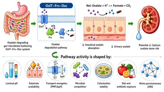
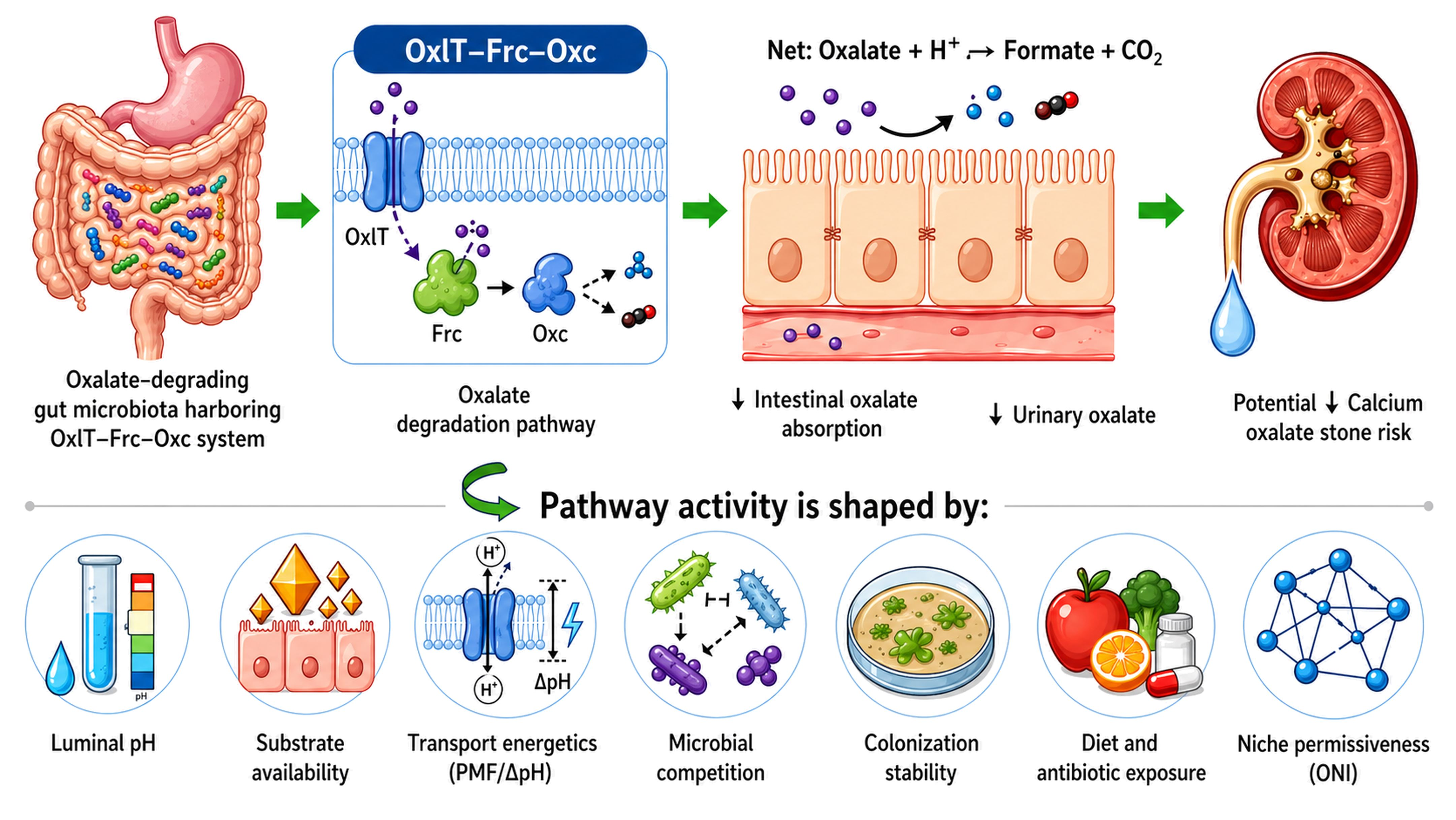
Graphical Abstract

1. Introduction

Oxalate accumulation contributes to hyperoxaluria, calcium oxalate nephrolithiasis, and kidney injury, and because mammals lack an oxalate-catabolic pathway, gut microbes provide the primary sink limiting oxalate absorption [1,2,3]. The best-characterized microbial route is the formyl-CoA transferase–oxalyl-CoA decarboxylase (Frc–Oxc) system, in which Frc/Oxc couple oxalate metabolism to OxlT-mediated oxalate/formate exchange and proton-motive-force generation, motivating microbiome-based strategies to lower systemic oxalate burden [4,5,6]. These mechanistic insights have driven efforts to restore oxalate degradation through probiotics and recolonization strategies, with the assumption that the mere presence of oxalate-degrading microbes will reduce systemic oxalate levels and protect human health.
However, clinical translation has been inconsistent. Probiotic and synbiotic interventions have failed to deliver consistent reductions in urinary oxalate or stone recurrence, revealing a critical barrier in effective therapeutic application [7,8]. Oxalobacter formigenes (a common intestinal oxalate-degrading bacterium) colonization varies, is often transient, and is antibiotic-sensitive [9,10], while supplementation effects are heterogeneous and often strongest in low-baseline individuals [11,12]. Furthermore, metagenomic studies demonstrate that frc/oxc carriage does not consistently predict in vivo oxalate degradation, revealing a persistent gap between genetic potential and measurable function [13,14]. Collectively, these findings have fueled an ongoing debate: Is O. formigenes depletion a causal factor in hyperoxaluria, or just a sign of a non-permissive gut niche formed by diet, bile acids, inflammation, and community competition [15,16]?
To address these challenges, the field has changed from taxon-centric to ecological thinking. It has come to understand that the efficacy of a probiotic depends on the recipient gut environment and metabolic niche rather than the specific strain employed [11,12,17]. In this situation, the proposed Oxalate Niche Index (ONI, discussed further in the future perspectives in Section 6.5) may serve as a conceptual framework for integrating niche permissiveness with pathway-related capacity. Rather than treating oxalate degradation as a static property of bacterial presence alone, this perspective highlights that meaningful Frc–Oxc activity depends on favorable transport energetics, regulatory activation, and ecological persistence within the gut. This review focuses scientifically on the biochemical, structural, functional, ecological, and therapeutic relevance of the Frc–Oxc pathway in microbial oxalate metabolism, with particular emphasis on hyperoxaluria and calcium oxalate kidney stone disease rather than all forms of human calculi or urolithiasis. In addition, the proposed Oxalate Niche Index (ONI) is discussed as a conceptual framework for explaining variability in oxalate-targeted microbiome therapy.

Clinical Background: Oxalate Homeostasis and Calcium Oxalate Stone Formation

Oxalate homeostasis in humans is controlled by the balance among endogenous oxalate production, dietary oxalate intake, intestinal absorption, microbial degradation, and renal excretion [3,18]. Because mammals do not possess an enzymatic pathway for oxalate degradation, oxalate acts largely as a metabolic end product that must either be eliminated by the kidneys or degraded by intestinal microorganisms before absorption [19,20]. This makes the gut–kidney axis clinically important in hyperoxaluria and calcium oxalate kidney stone disease.
Human oxalate originates from both endogenous and exogenous sources. Endogenous oxalate is generated mainly through hepatic metabolism of glyoxylate, glycolate, glycine, hydroxyproline, and ascorbate-derived intermediates [3,21]. In primary hyperoxaluria, inherited defects in glyoxylate metabolism cause excessive endogenous oxalate production, resulting in marked hyperoxaluria, calcium oxalate deposition, nephrolithiasis, nephrocalcinosis, and progressive kidney injury [22,23]. Exogenous oxalate is derived from the diet, especially oxalate-rich foods such as spinach, rhubarb, nuts, cocoa, tea, and some leafy vegetables [18,24,25]. However, the clinical effect of dietary oxalate depends not only on oxalate intake, but also on intestinal solubility, calcium and magnesium availability, gut transit, fat absorption, microbiota composition, and intestinal absorptive status [26,27,28].
In the gastrointestinal tract, soluble oxalate can be absorbed across the intestinal epithelium and later excreted in urine [21]. Dietary calcium and magnesium can bind oxalate in the gut lumen, forming poorly soluble complexes and thereby reducing intestinal oxalate absorption [18,26]. In contrast, low dietary calcium intake, fat malabsorption, bile acid disturbances, intestinal inflammation, bariatric surgery, or disruption of oxalate-degrading microbiota can increase the soluble oxalate pool and promote enteric hyperoxaluria [23,29,30]. Under these conditions, more oxalate becomes available for intestinal absorption, increasing urinary oxalate excretion and calcium oxalate supersaturation [3,31].
Renal excretion is the principal route for removing absorbed oxalate from the body. Because urinary oxalate concentration strongly influences calcium oxalate supersaturation, even moderate increases in urinary oxalate can increase stone risk [3,21,24]. Calcium oxalate stone formation is a multistep process involving urinary supersaturation, crystal nucleation, crystal growth, aggregation, and retention within the kidney [32]. Additional urinary and host factors, including low urine volume, hypercalciuria, hypocitraturia, reduced magnesium, altered urinary macromolecules, Randall’s plaque formation, epithelial injury, and inflammation, can further influence crystal retention, stone growth, and recurrence risk, supporting the need for long-term multimodal prevention strategies [32,33].
The gut microbiota may modify this clinical process by degrading luminal oxalate before absorption [19,20]. Oxalate-degrading bacteria, particularly those carrying the OxlT–Frc–Oxc system, can convert oxalate into formate and CO2, thereby reducing the intestinal oxalate pool and potentially lowering urinary oxalate burden [19,20]. However, microbial protection is not determined only by the presence of oxalate-degrading taxa or genes. Effective oxalate degradation requires pathway expression, substrate availability, suitable intestinal conditions, active transport, and colonization stability [34,35]. This clinical background provides the rationale for focusing on the Frc–Oxc pathway as a therapeutic target, while also explaining why probiotic outcomes may vary among patients with different dietary, microbial, metabolic, and disease contexts [17,36].

2. Biochemical Pathway of Oxalate Degradation Involving Frc and Oxc

2.1. Overview of the Oxalate Catabolic Pathway (OCP)

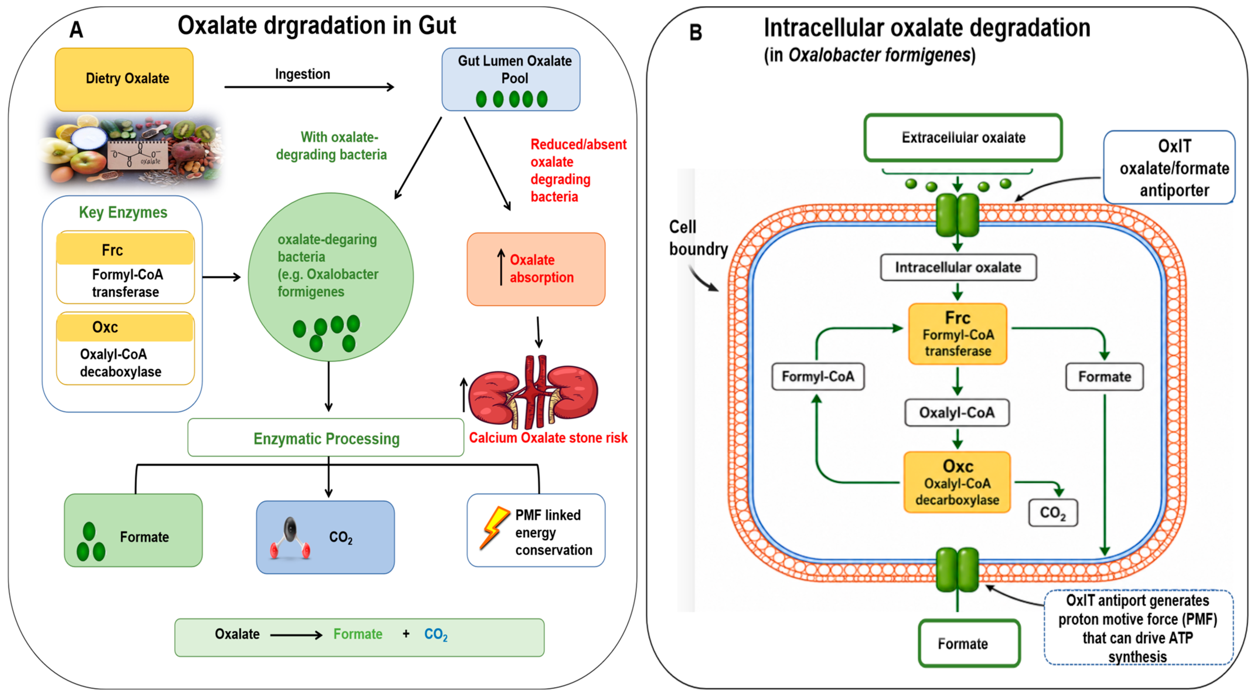
One of the major microbiological recycling processes is bacterial oxalate degradation, in which oxalate is converted to formate and CO2, thereby completing the consecutive reactions of frc and oxc [37]. This two-step anaerobic route is a crucial metabolic trait of certain symbionts of the gut, including O. formigenes (Figure 1A,B) [37]. By constantly ingesting luminal oxalate, these microbes play a role in intestinal and general oxalate homeostasis maintenance [38]. Frc–Oxc pathway has diverse ecological purposes outside the gut [39,40].
The frcoxc genes can be arranged in an operon, facilitating their coordinated expression during oxalate degradation [41,42]. Their transcription is regulated by environmental conditions like pH and availability of oxalate [4]. The oxalate comes into the cell via the OxlT antiporter, and then Frc converts it to oxalyl-CoA, and Oxc to formyl-CoA and CO2 [43,44]. Proteomic analyses indicate that oxalotrophic bacteria highly upregulate Frc and Oxc in reaction to oxalate; e.g., one of the Azospirillum species is able to degrade whole calcium oxalate in 72 h with significant enhancement in both enzymes [44]. Together, these findings suggest that coordinated expression of OxlT, Frc, and Oxc is essential for efficient oxalate degradation under conditions where oxalate is available (Table 1).
One of the key characteristics of this pathway is that it is tightly linked to membrane energetics. OxlT is an electrogenic oxalate 2-/formate-antiporter, and this charge gradient can be used to produce proton-motive-force, which can be used to generate ATP [5]. Consequently, to achieve efficient oxalate degradation in vivo, not only are the frc and oxc genes required, but so are the efficiency of transport, formate efflux, and a favorable pH gradient across the cell membrane [45]. This energetic dependence may help explain why some bacteria carrying the pathway fail to show meaningful oxalate-degrading activity in the intestinal environment.

2.2. Mechanism of Frc: Activation of Oxalate to Oxalyl-CoA

A formyl-CoA transferase, Frc, is the enzyme that starts the core pathway of oxalate degradation by transferring CoA to activated oxalate to generate oxalyl-CoA [46]. This is a critical step because it activates oxalate for decarboxylation and is a suggested rate-limiting step of the pathway [44]. Structural studies reveal that Frc is highly conserved and that residues in the active site play a critical role in CoA transfer and transition-state stabilization [47]. A thiamine pyrophosphate-dependent enzyme, Oxc, catalyzes the oxalyl-CoA decarboxylation process to formyl-CoA and CO2. This reaction restores the CoA donor needed to proceed with Frc activity, and promotes cyclic pathway activity [48]. Oxc requires cofactors (thiamine pyrophosphate and Mg2+) for activity and typically works under slightly acidic environments, which are found in certain gut microenvironments [48]. Metagenomic surveys suggest that oxc homologs are commonly found in oxalate-rich environments [44], whereas probiotic strains show significant oxalate reduction mediated by Oxc, which is controlled by pH and growth conditions [36]. Together, Frc and Oxc form a tightly linked metabolic unit that enables efficient oxalate utilization under anaerobic conditions in the gut.
From a therapeutic perspective, Frc and Oxc are crucial not only due to their enzymatic roles but also because they are coupled with the OxlT-dependent transport and PMF production (Figure 1) [40]. OxlT-based oxalate/formate exchange also plays a role in the production of proton-motive-force, connecting oxalate degradation to bacterial energy conservation [5,44]. In other words, the activity of the Frc–Oxc pathway in vivo relies on the presence of a functional system and not just the abundance of enzymes [40]. This is especially relevant when considering probiotic applications, because strains that contain frc and oxc may still be useless if the intestinal environment of the body does not support transport energetics or pathway induction.

2.3. Comparison with Alternative Oxalate Degrading Pathways (ODPs)

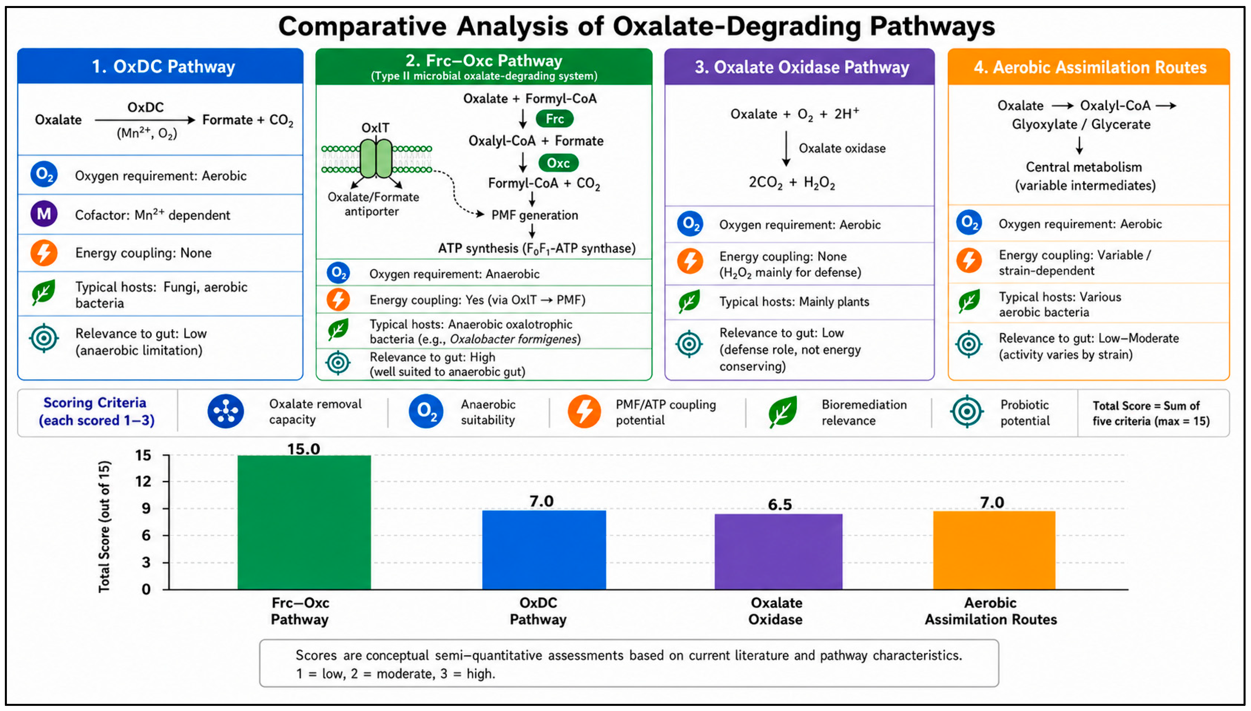
ODPs are a broad category covering all biological mechanisms that break down oxalate [10]. The OCP is a subset of ODPs that precisely involves only CoA-dependent routes [49]. The Frc–Oxc pathway differs from alternative oxalate degradation schemes in apparatus, oxygen requirement and energetic yield [39,40]. Some fungal and aerobic bacterial OxDCs can directly convert oxalate to formate and CO2 but involve oxygen and manganese, making them inappropriate for anaerobic gut environments (Figure 2(1)) [50]. Plant oxalate oxidase generates H2O2, which functions primarily in defense rather than energy uptake (Figure 2(3)) [51]. Type V oxdC pathways in bacteria also decarboxylate oxalate, although they do not provide the energy-coupled transport advantage provided by OxlT [40]. Aerobic integration pathways not only use glyoxylate and hydroxypyruvate as intermediates to enter central metabolism but also show inconsistent activity due to strain-specific polymorphisms (Figure 2(4)) [49]. The Frc–Oxc pathway is very effective at degrading insoluble oxalates, and it allows biomineralization, which has been used to detoxify metal oxalates (e.g., lead oxalate to phosgenite) (Figure 2(2)) [52]. Its strict anaerobic efficiency also makes it a capable therapeutic target for probiotic development [12]. Collectively, these biochemical steps highlight that Frc–Oxc oxalotrophy is inseparable from OxlT-mediated oxalate/formate exchange and PMF generation.

3. Structural and Functional Characteristics of Frc and Oxc

3.1. Crystal Structures and Active Sites of Frc

Structural analyses show that Frc is a homodimer, and each monomer cooperates in a shared CoA-binding site, which is crucial for the formation of oxalyl-CoA [46,47]. Residues (Q17, W48, D169 and G259-G260) are highly conserved among different groups of organisms and participate in transition state stabilization and prevent unproductive hydrolysis [47,53,54]. The dimeric architecture forms a narrow access channel and a flexible substrate entrance that protect intermediates [47]. Mutant studies show that changes in residues like G259 may inhibit dimer formation and the enzyme’s activity [53]. Environmental adaptation in extremophiles, such as modified surface properties, may help to increase enzyme stability under high salt conditions [55]. These features make Frc a promising candidate for engineering strains with enhanced oxalate-degrading capacity [47]. Structural studies indicate that Frc should be viewed not only as a simple CoA-transfer enzyme but also as a conserved catalytic platform that controls the entry of oxalate into the energy-linked degradation cycle. The crystal structure of O. formigenes formyl-CoA transferase revealed an interlocked homodimeric architecture, with the CoA-binding site positioned within a protected structural environment that supports efficient substrate handling [47]. Kinetic and mechanistic studies further confirmed that Frc catalyzes CoA transfer from formyl-CoA to oxalate, producing oxalyl-CoA as the activated intermediate required for downstream Oxc activity [46]. Therefore, disruption of dimer stability, CoA binding, or active-site organization could reduce pathway flux even when the frc gene is present.

3.2. TPP-Dependent Catalysis of Oxc

Oxc is a thiamine pyrophosphate (TPP)-dependent enzyme with quaternary structures ranging from dimers in eukaryotes to tetramers in bacteria, and stabilized under anaerobic conditions [56]. The active site lies within a cleft, with TPP bound to pyrimidine-binding (PYR) and pyrophosphate-binding (PP) domains with Mg2+ and highly conserved residues (E56, Y120, E121 and R555) [48,57]. The enzyme catalyzes non-oxidative decarboxylation by forming a covalent bond between TPP and oxalyl-CoA. Adenosine diphosphate (ADP) positively allosterically activates the reaction by binding to regulatory pocket residues, thereby significantly increasing the substrate turnover rate [57]. The enzyme’s optimal pH of 5.5–6.0 is adapted to low pH environments of the gut and soils [56]. Engineered Oxc variants have been explored for synthetic one-carbon metabolism and carboligase applications [15]. TPP-binding residues are conserved among microorganisms to maintain enzyme activity [56]. The Oxc reaction provides the biochemical driving force that maintains the cyclic Frc–Oxc pathway. Oxc is a thiamine diphosphate-dependent decarboxylase that converts oxalyl-CoA to formyl-CoA and CO2, thereby regenerating the formyl-CoA donor required for the Frc reaction [57]. Structural analysis of Oxc from O. formigenes showed that cofactor binding, Mg2+ coordination, and activation by ADP are important for catalytic function [57]. Thus, Oxc activity is influenced not only by the presence of oxc but also by cofactor availability, protein assembly, pH, and metabolic state.

3.3. Gene Organization and Operon Architecture

In many oxalotrophic bacteria, frc and oxc and sometimes oxlT genes are organized as a small operon, allowing for co-regulation and rapid response to oxalate presence [56,58]. Genomic analyses reveal widespread presence of the operon in Proteobacteria, Actinobacteria and Firmicutes, suggesting horizontal gene transfer in various environments [12,59]. The frc and oxc genes are separated by a short intergenic sequence, which is mostly less than 100 bp and enables a bicistronic transcript. Operon promoters are pH-sensitive with increased activity, facilitating transcription in the acidic conditions of oxalate-enriched habitats [60,61]. Research on co-expression suggests that the structure of an operon may improve its degradation capacity and make it a helpful approach for engineered microbial systems [15]. The organization of oxlT, frc, and oxc is functionally important because oxalate uptake, CoA transfer, decarboxylation, and formate export must operate as a coordinated module. OxlT mediates oxalate/formate exchange, while Frc and Oxc convert imported oxalate into formate and CO2, allowing the pathway to function as an integrated transport–metabolism system [40,42]. When these genes are physically linked or co-regulated, bacteria can more efficiently activate the complete oxalate-degradation pathway in response to oxalate availability. In contrast, fragmented organization or weak regulatory coupling may result in incomplete pathway activity despite the presence of individual pathway genes [14,39].

3.4. Regulation of Enzyme Expression

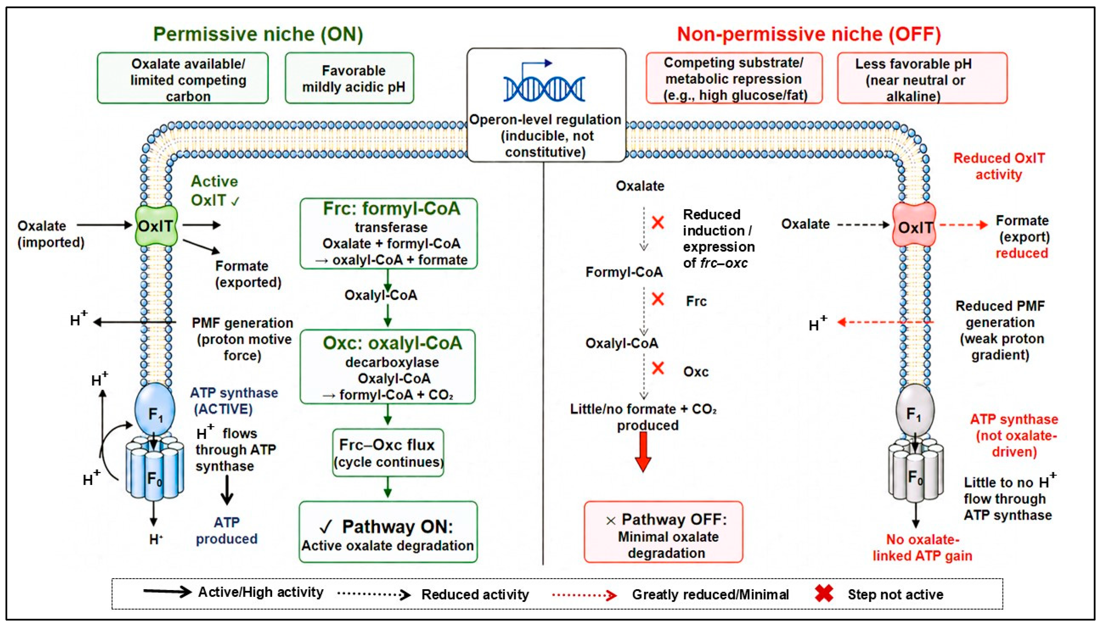
The frc and oxc genes are generally induced by oxalate and regulated by pH, with optimal expression at low pH levels [12,60]. Oxalate likely acts as a co-inducer, binding to regulatory proteins or sensor-kinase complexes regulating transcription of the operon [42,62,63]. Carbon availability also affects expression, with glucose being able to inhibit the operon in some strains [36,44]. Environmental isolates demonstrate time-dependent induction, with the highest transcript abundance occurring after prolonged exposure to oxalate [42,45,64]. The proteomics information shows that Frc and Oxc are more abundant under oxalate exposure, while high pH suppresses the operon and prevents deleterious side effects [42,44]. This ON/OFF regulatory strategy reflects adaptation to a wide range of environmental habitats [10,65]. A common oversight of probiotic studies is that frc–oxc is not always on; it is a highly regulated stress-response/energy-salvage operon. For many facultative degraders, the operon is turned on only under low-carbon, mildly acidic and oxalate-rich environments. High-carbon diets (glucose or fat) can invoke catabolite repression and turn off gene transcription, allowing these bacteria to ferment more favored substrates [66]. Therefore, the same probiotic strain may degrade oxalate robustly in vitro but not in the human host with a diet rich in other carbon. This regulatory logic has direct clinical relevance, predicting that probiotic efficacy is highly niche-dependent and remains mechanistically incomplete without concurrent management of substrate, diet, and local pH [25]. In O. formigenes, oxalate metabolism is linked to proton-motive-force generation, and external pH influences the electrical and chemical components of the proton gradient during oxalate metabolism [67,68]. In lactobacilli, oxc and frc homologs can be transcriptionally and functionally associated with oxalate degradation, but activity varies with strain and culture conditions [42]. Consequently, a bacterium may show strong oxalate degradation under controlled in vitro conditions but weaker activity in vivo, where oxalate availability, pH, bile acids, antibiotics, and microbial competition vary among individuals [36]. The inducible, catabolite-gated nature of frc–oxc operon (summarized in Figure 3) provides a mechanistic explanation for why oxalate-degradation genes frequently fail to translate into in vivo activity.

3.5. Structure-Function Analysis of Mutants and Recombinant Enzymes

Catalytic insights into Frc and Oxc have been revealed by mutant and recombinant approaches [47,48]. oxc and frc knockout mutants lose the ability to degrade oxalate [42]. Coenzyme-binding site mutations in Oxc (e.g., E121Q, S568A, Y497A) impact the binding or rates of TPP turnover, although directed evolution has improved the catalysis for artificial purposes [48,69]. Studies of fungus-growing termite nests have linked oxalate-degrading bacterial communities with environmental oxalate–carbonate transformation [70]. Together, these functional studies highlight the adaptability and potential uses for Frc–Oxc [36]. The structural basis for Frc/Oxc performance and inducible operon regulation shows that oxalate degradation is permissive and easily shut down by carbon sufficiency and non-permissive pH. These regulatory constraints refine the ONI model and directly imply that responder status depends on niche permissiveness as much as on gene carriage. Mutant, recombinant, and structure–function studies collectively show that the Frc–Oxc pathway is modular but not automatically functional. OxlT structure–function studies demonstrated that membrane topology and transporter residues are important for oxalate/formate exchange [5,71], while Frc and Oxc structural studies showed that active-site integrity and cofactor-dependent catalysis are essential for pathway flux [47,57]. Functional studies in lactobacilli further show that the presence of frc and oxc homologs must be supported by measurable oxalate-degrading activity [42]. Therefore, future strain screening should combine genomic detection with enzyme activity assays, oxalate-degradation measurements, transport-function assessment, and colonization-performance studies.

4. Evolution and Distribution of frc and oxc Genes

4.1. Phylogenetic Distribution and Functional Diversity

The frc and oxc genes are distributed across diverse bacterial lineages, indicating that microbial oxalate metabolism is not restricted to O. formigenes [13,72]. Among these organisms, O. formigenes remains the best-characterized intestinal oxalotroph and uses the OxlT–Frc–Oxc system for anaerobic oxalate transport, CoA transfer, decarboxylation, formate production, and transport-linked energy conservation [73]. Comparative OXC analyses further show that oxalyl-CoA decarboxylase homologs occur across several bacterial groups, including Proteobacteria, Actinobacteria, Firmicutes/lactobacilli-associated taxa, Bacteroidetes-related taxa, and other environmental or host-associated bacteria [72]. This broad distribution suggests that oxalate-degrading capacity may contribute to different ecological functions, including oxalate detoxification, intestinal oxalate turnover, carbon cycling, pH modulation, and environmental oxalate transformation [13,72,73]. In host-associated systems, oxalate-degrading bacteria have been reported in human and animal gut microbiomes, where their abundance and activity may influence luminal oxalate availability and urinary oxalate burden [11,74,75].
However, oxalate-degrading potential should not be inferred from taxonomy alone. Some lactobacilli, bifidobacteria, enterococci, and other gut-associated bacteria show strain-dependent oxalate-degrading activity, but functional validation is required because gene presence does not necessarily prove active oxalate degradation in vivo [75,76]. Because the former broad genus Lactobacillus was taxonomically reorganized in 2020, this review uses the lowercase, non-italicized term “lactobacilli” when referring broadly to organisms historically grouped within Lactobacillaceae [77]. Where species identity is clear, updated genus names should be used; where older studies used pre-2020 nomenclature, the original name is retained only to remain consistent with the cited literature. Therefore, candidate probiotic strains should be evaluated using genomic screening together with frc/oxc expression, enzyme activity, oxalate-degradation assays, transport function, and colonization performance.

4.2. Identification of Oxc Homolog Clusters

Comparative analyses of oxalyl-CoA decarboxylase (OXC/oxc) homologs show that this enzyme family is taxonomically diverse and is not restricted to O. formigenes. Jiang et al. classified 1396 putative OXC sequences into seven major clusters based on sequence similarity, taxonomic distribution, gene-context organization, and human microbiome body-site abundance [72]. This cluster framework provides a useful baseline for mapping OXC diversity within human microbiome datasets, although it should be interpreted primarily as a bioinformatic classification rather than direct evidence that every recovered homolog is functionally active in vivo. Cluster 1 includes the experimentally characterized OXC from O. formigenes, an obligate anaerobic gut bacterium that relies strongly on oxalate metabolism for growth and transport-linked energy conservation [72,73]. Cluster 2 contains the E. coli OXC homolog and related Gammaproteobacteria-associated sequences; in E. coli, YfdU/OXC and YfdW/Frc are required for oxalate-induced acid tolerance, indicating that some OXC homologs may contribute to acid-resistance physiology as well as oxalate metabolism [62]. Clusters 3 and 5 are mainly associated with Actinobacteria, including Mycobacterium- and Bifidobacterium-related representatives, whereas Cluster 4 contains Bacilli-associated homologs, including lactobacilli-associated OXC sequences [72]. Functional studies support the biological relevance of several non-Oxalobacter OXC-containing bacteria. In lactobacilli, frc and oxc homologs have been transcriptionally and functionally associated with oxalate degradation, although activity is strain-dependent [42,78]. In bifidobacteria, OXC/oxc has been molecularly characterized in Bifidobacterium lactis, and oxalate-degrading activity has been reported in Bifidobacterium animalis subsp. lactis, including under acidic conditions [79,80]. These findings demonstrate that OXC homologs occur across probiotic-associated bacterial groups, but functional activity should be experimentally validated rather than inferred only from broad taxonomic assignments. Table 2 summarizes the seven OXC clusters according to representative taxa, dominant taxonomic associations, reported microbiome distribution, and evolutionary interpretation. Overall, OXC cluster assignment should be combined with frc/oxc co-occurrence, gene-expression evidence, enzyme assays, oxalate-degradation measurements, and ecological context when evaluating candidate probiotic strains for kidney stone prevention.

4.3. Evolutionary Conservation and Selection Pressures

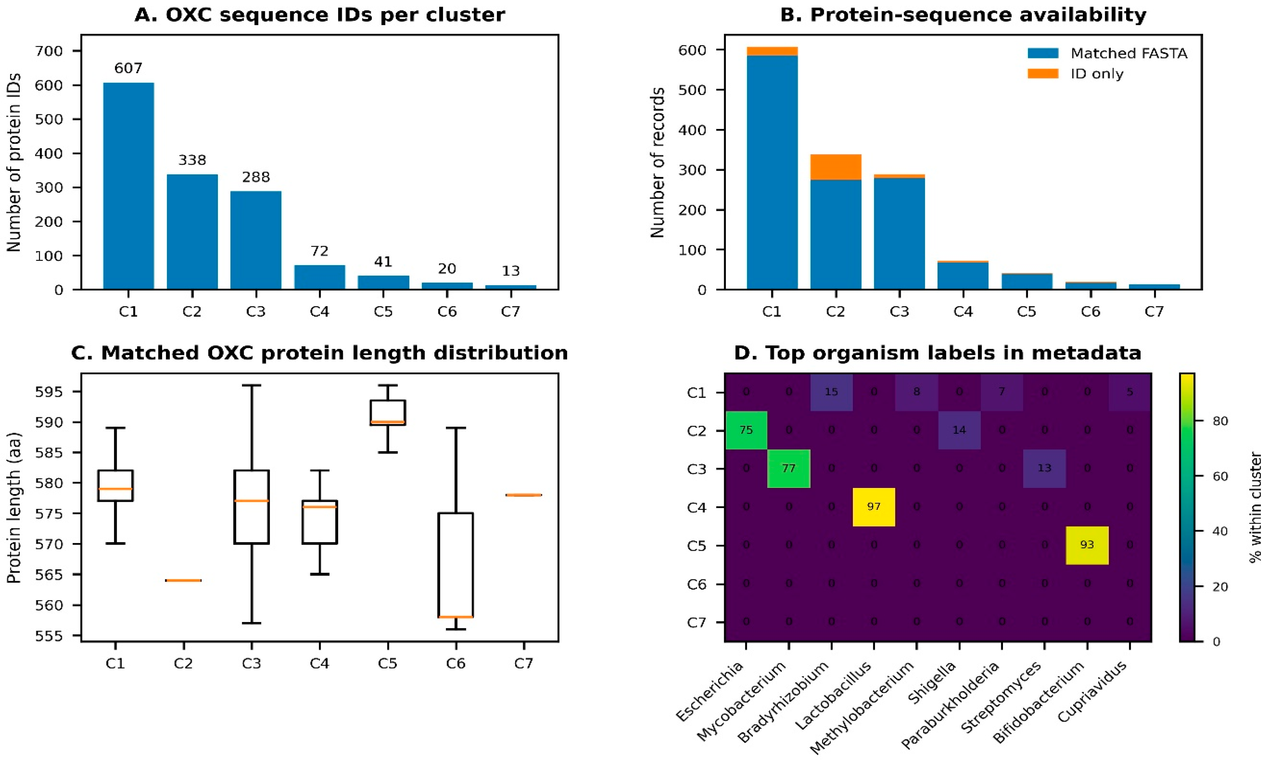
Evolutionary analyses indicate that frc and oxc are generally conserved across oxalate-degrading microbial lineages, particularly in regions associated with catalysis, substrate binding, and cofactor-dependent function [72]. This conservation suggests that oxalate metabolism provides a selective advantage in microbial niches where oxalate is available as a carbon or energy-related substrate [13,73]. The strong purifying selection on key residues in specialized OxB reflects the Frc–Oxc pathway’s essential role for growth and survival under oxalate-rich environments. Figure 4 further illustrates the broad distribution and taxonomic structuring of OXC homologs across several bacterial clusters based on reanalysis of the Jiang et al. supplementary dataset [72]. The conservation of these genes also has practical significance. It indicates that the enzymatic core of the Frc–Oxc pathway is stable across different taxa [73], which may help in identifying candidate probiotic strains or predicting oxalate-degrading potential from genomic data. At the same time, conserved sequence alone should not be interpreted as proof of an effective in vivo function because pathway activity remains dependent on regulation, transport coupling, and ecological context.

4.4. Horizontal Gene Transfer Events

Phylogenetically diverse bacteria that contain frc and oxc suggest that horizontal gene transfer (HGT) may have contributed to the spread of oxalate-degrading capacity [41,72]. The inter-lineage transfer of highly conserved, operon-like gene arrangements may have enabled distinct taxa across contrasting ecological niches to rapidly acquire specialized oxalate-associated metabolic functions [64]. This evolutionary mechanism explains the widespread presence of homologous pathway components in organisms possessing vastly different metabolic strategies and lifestyles [74,81]. From a translational perspective, the occurrence of these historical HGT events indicates that functional oxalate-degrading traits are distributed across a diverse range of gut-associated taxa rather than being restricted to a single specialized organism [82,83]. In summary, horizontal gene transfer may have shaped the genetic diversity and ecological distribution of the oxalate-degrading genes within the human microbiome [13,15]. These evolutionary insights support the paradigm that host oxalate homeostasis is mediated by a multi-taxonomic spectrum of organisms, suggesting that clinical strategies targeting hyperoxaluria must adopt a community-level, ecosystem-based approach to successfully restore metabolic function.

4.5. Prevalence in Human Microbiomes

Large-scale metagenomic studies show that frc and oxc are widely distributed in the human gut microbiome, indicating that oxalate degradation is a common functional trait [10,80,84]. A study of 660 healthy subjects found that oxc was detected in approximately 84% of individuals, while frc was detected in approximately 88%. These genes were predominantly associated with Cluster 1 O. formigenes [10]. In contrast, complete oxalotrophy is a relatively rare metabolic trait, largely confined to the gut, with limited occurrence in oral and respiratory microbiomes [13,41,85]. Dysbiosis in kidney stone patients is associated with reduced oxc prevalence, potentially contributing to elevated urinary oxalate excretion and increased stone risk [85,86]. The prevalence varies with age and diet, with higher abundance observed in adults compared to neonates, and in populations consuming oxalate-rich diets [2,12]. In inflammatory bowel disease (IBD), reduction of oxalate degraders further exacerbates hyperoxaluria [10]. These patterns highlight the pathway’s clinical relevance and support interventions aimed at restoring the oxalotrophic taxonomic group in dysbiotic communities [85,87]. Overall, the evolutionary distribution of frc and oxc supports the central importance of the Frc–Oxc pathway in microbial oxalate metabolism. At the same time, phylogenetic breadth and genomic conservation do not eliminate the need to consider pathway regulation and ecological context when evaluating therapeutic potential in human calculi.

5. Environmental Factors Influencing Oxalate Degradation

5.1. Impact of pH and Carbon Sources

Environmental pH is an important regulator of microbial oxalate degradation, but its effect differs between specialist and facultative oxalate degraders. In the obligate oxalotroph O. formigenes, optimal oxalate degradation and growth are generally reported under mildly acidic to near-neutral conditions, approximately pH 6.0–7.0, whereas stronger acidity, such as pH 4.5, can substantially reduce transcriptional activity and degradation efficiency [13,88,89]. In contrast, some lactic acid bacteria (LAB) and Bifidobacterium strains may use frc–oxc-associated oxalate degradation as part of an acid-stress adaptation response, where subinhibitory acidic conditions can stimulate operon expression and improve oxalate-degrading activity [88,90]. Therefore, the pH requirement for oxalate degradation should be interpreted as strain- and niche-dependent rather than universal across all oxalate-degrading bacteria.
Carbon availability further modulates oxalate-degrading activity through distinct ecological mechanisms. In facultative oxalate degraders, easily fermentable carbohydrates such as glucose and sucrose may enhance biomass formation and indirectly support enzyme production [36,78]. Under carbon-limited conditions, some generalist degraders may increase reliance on alternative substrates and induce oxc transcription to utilize oxalate [47,56,91]. However, excess preferred carbon sources may reduce oxalate-pathway activity through metabolic regulation or carbon catabolite repression mechanisms [64,92]. Prebiotics may also influence oxalate degradation by supporting bacterial growth, modifying gut niche conditions, or improving substrate availability; for example, inulin-containing formulations have been reported to enhance degradation performance in selected kinetic or optimization assays [4,9,45]. However, these effects should be considered formulation- and strain-specific rather than generalizable to all oxalate-degrading bacteria.
In the gut, these factors have practical therapeutic implications. Local pH gradients, substrate availability, and competing nutrients can determine whether the Frc–Oxc pathway is active or functionally silent [28,93,94]. Thus, probiotic efficacy depends not only on bacterial administration but also on whether the intestinal environment supports pathway induction and transport-linked oxalate metabolism. Table 3 summarizes the ideal conditions for representative taxa.

5.2. Role of Prebiotics and Nutritional Supplements

Prebiotics significantly enhance intestinal oxalate degradation by providing fermentable substrates that drive the proliferation and metabolic activity of oxalate-consuming bacterial cohorts [36]. Non-digestible oligosaccharides, such as inulin and fructooligosaccharides (FOS), have been reported to increase the oxalate-degrading activity of certain probiotic strains by approximately 38%, while altered gut microbiota is associated with renal stone risk [76,98]. This metabolic enhancement is driven primarily by biomass expansion, improved mucosal colonization, and the production of short-chain fatty acids that lower local pH, which indirectly facilitates the upregulation of frc–oxc operon expression [42]. Notably, in vivo models of hyperoxaluria have demonstrated that prebiotic administration successfully reduces urinary oxalate excretion [99].
Synbiotic strategies leverage these dynamics by co-administering synergistic probiotic strains alongside specific prebiotic substrates, thereby maximizing bacterial survival, metabolic transit, and functional consistency within the competitive gastrointestinal tract [100,101]. Beyond luminal degradation, certain systemic nutritional adjuncts can target endogenous pathways. For instance, vitamin B6 serves as an essential cofactor for hepatic alanine-glyoxylate aminotransferase (AGT); its supplementation prevents the metabolic shunting of glyoxylate toward endogenous oxalate production [4,12,102]. Although these findings are encouraging, the main value of prebiotics in this context is that they may help create a more supportive environment for pathway activity rather than serving as stand-alone therapies [74,103]. This makes them especially relevant for improving the consistency of microbiome-based interventions targeting hyperoxaluria and calcium oxalate stone risk.

5.3. Effects of Antibiotics and Microbial Disruptions

Antibiotic administration can impair intestinal oxalate degradation by disrupting oxalate-degrading microbial communities, particularly O. formigenes. Population-based studies have reported an association between oral antibiotic exposure and increased odds of nephrolithiasis, with stronger associations after recent exposure and in younger individuals [20,104,105]. This relationship is biologically plausible because antibiotics can reduce colonization by oxalotrophic bacteria and disturb microbiome functions involved in oxalate handling. Broad-spectrum antibiotic classes, including fluoroquinolones, cephalosporins, sulfonamides, nitrofurantoin, and broad-spectrum penicillins, have been associated with increased kidney stone risk in epidemiological analyses [106,107,108]. Microbial disruption may be especially relevant in inflammatory bowel disease (IBD) and other dysbiotic gut conditions. Multi-omics studies indicate that oxalate-degrading gene expression, including frc and oxc transcripts, can be reduced in IBD-associated microbiomes, even when oxalate-degradation genes are still detectable [10,43].
Repeated or prolonged exposure to antimicrobial agents can persistently alter gut microbiome structure. This selective pressure may reduce the community-level capacity to degrade metabolic compounds while favoring resistant microbial populations that may lack beneficial oxalate-degrading functions [86,109]. Human strains of O. formigenes exhibit sensitivity to several widely prescribed outpatient antibiotic classes, including macrolides, fluoroquinolones, clindamycin, tetracyclines, aminoglycosides, and metronidazole [105]. This susceptibility profile provides a mechanistic explanation for why antibiotic exposure can compromise colonization stability and decrease functional oxalate degradation, even when oxalate-degrading genes remain detectable by metagenomic analysis. Ultimately, these dynamics underscore the importance of antibiotic stewardship and highlight the need for targeted adjuvant strategies designed to conserve, restore, or protect native oxalotrophic populations during clinical interventions [12,74,82].

5.4. Influence of Host Diet and Oxalate Load

Host diet is an important driver shaping the composition and metabolic activity of the oxalate-degrading gut microbiota [2]. Diets rich in oxalate may increase the abundance or activity of frc/oxc-positive taxa and enhance luminal oxalate-degrading capacity, whereas prolonged restriction of dietary oxalate may reduce substrate availability for specialized oxalotrophs [15,73]. At the molecular level, elevated oxalate availability can induce oxc expression in O. formigenes, while dietary fibers and prebiotic substrates may support the growth of selected facultative oxalate-degrading bacteria, including some LAB [11,36,76]. Animal studies further suggest that oxalate-rich diets, including spinach-supplemented regimens, can modify intestinal oxalate-degrading activity, whereas high-fat-diet-associated dysbiosis may suppress native oxalotrophic populations and promote intestinal oxalate absorption [2,89,110]. Human dietary and clinical studies also indicate that diet-dependent changes in gut microbial function and oxalate availability can influence urinary oxalate excretion and nephrolithiasis risk, supporting the therapeutic potential of targeted nutritional modulation [12,14,61,111].
These dietary dynamics are important because urinary oxalate excretion depends strongly on gastrointestinal solubility and absorption kinetics, not on oxalate intake alone. Co-ingested dietary calcium can bind free oxalate in the intestinal lumen to form poorly soluble calcium oxalate complexes, thereby reducing the bioavailable pool of soluble oxalate [18,23]. Conversely, low dietary calcium intake, high soluble oxalate intake, fat malabsorption, and altered intestinal transit can increase soluble oxalate absorption and subsequent urinary oxalate excretion [18,112]. Therefore, diet can influence Frc–Oxc-based therapy in two opposite ways: sufficient oxalate may be needed to induce microbial oxalate-degradation pathways, but excessive soluble oxalate absorption can increase urinary stone risk if microbial degradation or mineral binding is insufficient.

5.5. In Vitro and In Vivo Modulation Strategies

Experimental studies show that oxalate degradation can be improved in vitro through optimization of pH, substrate conditions and prebiotic supplementation [28,86]. These in vitro systems are useful for identifying favorable growth and degradation conditions, but their results should be interpreted as strain- and assay-specific rather than directly transferable to the human gut [95,96]. Multi-strain probiotic and synbiotic approaches have also been tested in oxalate-degradation contexts, where responses appear to depend on strain compatibility, formulation design, and environmental conditions [12,76,86]. In vivo strategies aim to move beyond culture optimization by restoring or enhancing oxalate-degrading function within the host intestinal ecosystem. Probiotic and synbiotic administration, fecal microbiota transplantation, and engineered live biotherapeutic products have all been investigated as approaches to increase host oxalate-degrading capacity [13,113,114]. However, successful in vivo activity requires more than the presence of an administered organism; the intestinal niche must also support colonization, pathway induction, and sustained oxalate metabolism [11,74].
Local pH, carbon co-substrates, oxalate availability, and prebiotic support are key culture-related axes of “oxalate-niche permissiveness”. Host bile acid profiles and antibiotic history further modify this niche by altering gut microbiome structure and oxalotrophic stability [96,97]. Together, these factors may determine whether the frc–oxc pathway is induced, weakly active, or functionally inactive, thereby influencing whether administered strains can colonize, persist, and produce measurable oxalate-lowering effects [74]. Recent in vivo validation studies support this niche-based interpretation. In a high-oxalate diet model, fecal microbiota transplantation (FMT) reduced urinary oxalate excretion and renal calcium oxalate crystal deposition while restoring gut microbial and metabolomic profiles [114]. Similarly, induced O. formigenes colonization in healthy adults reduced urinary oxalate excretion, although persistence and response varied among individuals [74]. Baseline abundance of oxalate-degrading bacteria and frc/oxc genes has also been linked to variation in response to O. formigenes administration [11]. These findings support a therapeutic framework in which effective oxalate reduction depends on colonization stability, dietary compatibility, and a permissive host intestinal environment, rather than the mere presence of an oxalate-degrading organism.

6. Probiotic and Microbiome-Based Therapeutic Applications

6.1. Oxalate-Degrading Bacteria of Therapeutic Interest

Oxalate-degrading biotherapies aim to reduce the soluble intestinal oxalate pool before absorption and thereby lower urinary oxalate burden [25,113]. Among these organisms, O. formigenes remains the most specialized gut oxalotroph; it uses oxalate as a major metabolic substrate and relies on the coordinated OxlT–Frc–Oxc system for oxalate transport, CoA transfer, decarboxylation, formate production, and transport-linked energy conservation [73,74]. This metabolic specialization makes O. formigenes an important candidate for oxalate-targeted biotherapy, although its clinical performance depends on colonization stability, substrate availability, antibiotic exposure, and host intestinal ecology [74,105]. Therefore, delivery strategies for O. formigenes should prioritize preservation of viability, protection from oxygen and gastrointestinal stress, and support for intestinal engraftment [104,115]. Alternative candidate probiotics include strains of lactobacilli, bifidobacteria, and enterococci, which show distinct, strain-dependent oxalate-degrading phenotypes [76].
However, functional oxalate degradation cannot be inferred from genus-level identity alone. The term “lactobacilli” is used here when referring broadly to organisms historically grouped within Lactobacillaceae, following the major 2020 taxonomic reorganization of the former broad genus Lactobacillus [77]. These facultative degraders remain clinically attractive because many strains have established probiotic use, acid-stress tolerance, and compatibility with commercial probiotic manufacturing pipelines [25,76]. Multi-strain consortia may offer advantages over single-strain interventions by combining complementary oxalate-degrading and niche-supporting functions [76]. However, consortium efficacy remains formulation- and host-dependent and should be validated using functional oxalate-degradation assays rather than inferred from strain lists alone [76]. Beyond wild-type isolates, genetically engineered live biotherapeutic products represent an additional therapeutic avenue [113]. Engineered platforms may be designed to enhance gastrointestinal oxalate consumption and improve predictable oxalate-lowering activity, but they require rigorous evaluation of safety, stability, containment, and long-term clinical efficacy before use in calcium oxalate nephrolithiasis prevention [113].

6.2. Preclinical Evidence: In Vitro, Animal, and Engineered-Strain Studies

Preclinical investigations provide foundational evidence for oxalate-targeted microbial therapeutics. In vitro optimization models demonstrate that microbial oxalate degradation depends on environmental pH, substrate availability, nutrient composition, and prebiotic supplementation, although these responses remain strain- and assay-specific [76,96,113]. These systems are useful for identifying favorable metabolic conditions, but static in vitro cultures cannot fully reproduce the complex physiology of the host gastrointestinal tract, including regional pH gradients, bile acid exposure, microbial competition, and host-specific colonization barriers [73,97].
To bridge this translational gap, animal models and engineered live biotherapeutic products provide functional evidence for whether microbial oxalate degradation can alter host oxalate handling. Synthetic biology has emerged as a promising strategy to improve the predictability and robustness of oxalate-degrading activity. A prominent example is SYNB8802, an engineered Escherichia coli Nissle 1917 strain designed to consume oxalate in the gastrointestinal tract [113,116]. In preclinical studies, oral SYNB8802 significantly reduced urinary oxalate excretion in healthy mice and non-human primates, and in silico modeling predicted potential urinary oxalate lowering in humans with enteric hyperoxaluria [113,117]. Similarly, Whitaker et al. developed NB1000S, an engineered porphyran-utilizing Phocaeicola vulgatus strain carrying an oxalate-degradation pathway and a conditional attenuation system for controlled gut colonization. In a dietary enteric-hyperoxaluria rat model, the engineered strain reduced urinary oxalate by 47% compared with a control strain lacking the oxalate-degradation pathway, while the human phase 1/2a study showed controlled colonization but also highlighted genetic stability and long-term treatment-control challenges [118]. These engineered platforms may help overcome some limitations of native obligate oxalotrophs, including strict colonization requirements and variable persistence. However, translating preclinical success into human therapy requires rigorous evaluation of long-term biosafety, dose–response behavior, ecological stability within the native microbiome, biocontainment strategies, and sustained oxalate-lowering efficacy in vivo [113,119].

6.3. Human Clinical Evidence and Variable Outcomes

Human clinical evidence for oxalate-targeted microbial interventions remains mixed and is characterized by notable inter-individual variability. Early studies using mixed lactic acid bacterial preparations, such as Oxadrop®, reported reductions in urinary oxalate excretion in small cohorts of patients with enteric hyperoxaluria [7,30,120]. However, a later randomized, double-blind, placebo-controlled trial of Oxadrop® in calcium stone formers with idiopathic hyperoxaluria did not show a significant reduction in urinary oxalate compared with placebo [121]. These findings indicate that conventional microbial supplementation alone is insufficient to guarantee reproducible oxalate-lowering efficacy. Clinical trials using native O. formigenes have also produced variable outcomes. In primary hyperoxaluria, orally administered O. formigenes was generally well tolerated, but larger randomized trials did not consistently show significant urinary oxalate reduction compared with placebo. This limitation is biologically plausible because primary hyperoxaluria is mainly driven by hepatic oxalate overproduction, which may exceed the capacity of localized intestinal microbial degradation [111,122,123].
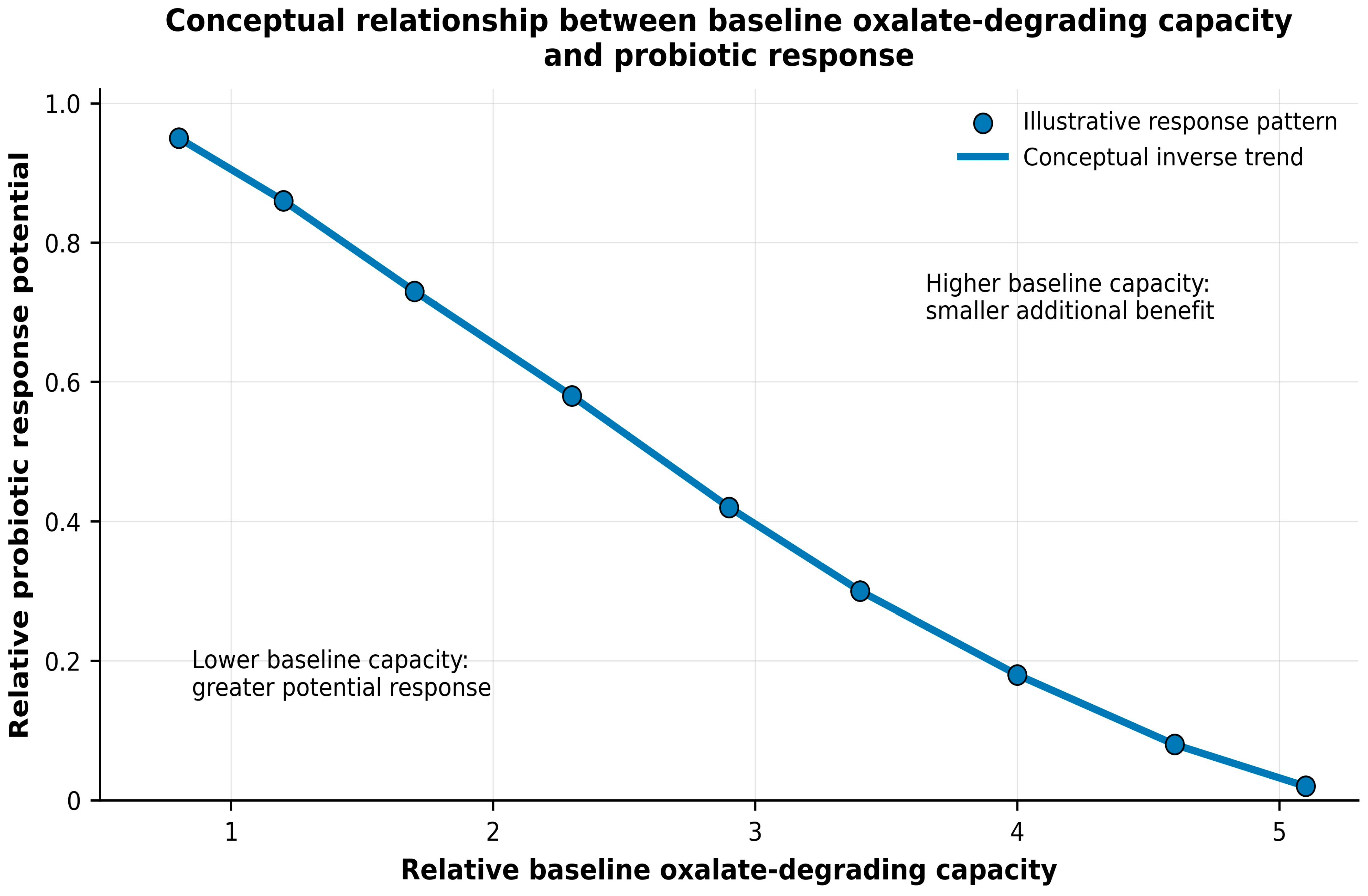
In contrast, gut-directed microbial therapies may be more mechanistically relevant in enteric or idiopathic hyperoxaluria, where intestinal oxalate absorption and luminal oxalate bioavailability contribute directly to urinary oxalate burden [23,120]. In idiopathic calcium oxalate nephrolithiasis, therapeutic response to microbiome-based degradation is likely influenced by diet, baseline hyperoxaluria severity, recent antibiotic exposure, and the functional structure of the existing gut microbiome [8,121]. Recent human colonization and multi-omics studies refine this interpretation. Fargue et al. showed that exogenously administered O. formigenes can colonize the gastrointestinal tract of healthy adults and reduce urinary oxalate excretion under controlled conditions [74]. Suryavanshi et al. further reported that baseline abundance of oxalate-degrading bacteria and frc/oxc genes is associated with stool oxalate, urinary oxalate, and variation in response to O. formigenes administration [11]. These findings support the conceptual framework presented in Figure 5: individuals with lower baseline oxalate-degrading capacity may have greater response potential, whereas individuals with higher baseline capacity may gain less additional benefit from microbial supplementation [11,74].

6.4. Why Clinical Translation Often Fails

Several factors may explain why oxalate-degrading probiotics do not consistently produce clinical benefit. First, colonization is a major limitation. O. formigenes is an oxygen-sensitive anaerobe with demanding growth requirements, and administered cells must survive gastric passage, bile exposure, oxygen stress, intestinal transit, and competition with resident microbiota [73,74]. Even when short-term fecal recovery occurs, long-term persistence may remain weak or variable [74]. Second, recipient microbiome structure can limit probiotic benefit. If the gut already contains active oxalate-degrading organisms, an introduced strain may provide little additional functional gain; conversely, individuals with lower baseline oxalate-degrading capacity may have greater response potential. This helps explain why baseline microbial function and community context may predict response better than probiotic dose alone [11,74].
Third, antibiotic exposure can disrupt oxalate-degrading communities. Human strains of O. formigenes are sensitive to several commonly used antibiotics, including macrolides, fluoroquinolones, tetracyclines, clindamycin, aminoglycosides, and metronidazole [105]. Oral antibiotic exposure has also been associated with increased odds of nephrolithiasis, supporting the need to consider antibiotic history when interpreting probiotic failure or designing clinical trials [106]. Fourth, dietary and host factors influence efficacy. Oxalate availability may support pathway induction, whereas dietary calcium can reduce soluble oxalate absorption by forming poorly soluble calcium oxalate complexes [18,23]. Fat malabsorption, bile acid disturbances, intestinal inflammation, and altered gut transit can increase soluble oxalate absorption and may create a less permissive niche for administered bacteria [23,97]. These factors indicate that probiotic therapy is unlikely to work consistently unless it is integrated with diet, intestinal physiology, and microbiome context.
Importantly, these clinical limitations should be interpreted according to hyperoxaluria subtype. In enteric or diet-associated hyperoxaluria, intestinal oxalate absorption and luminal oxalate availability are major contributors to urinary oxalate burden; therefore, gut-directed microbial degradation may have direct mechanistic relevance [23,120]. In contrast, primary hyperoxaluria is driven primarily by endogenous hepatic oxalate overproduction caused by inherited defects in glyoxylate metabolism [124,125]. Therefore, gut-restricted probiotic or microbiome-based approaches are unlikely to function as stand-alone treatments for primary hyperoxaluria and should be regarded as supportive or adjunctive strategies that complement established disease-specific management.

6.5. Strategies to Improve Microbiome-Based Therapy

Several strategies may improve the translational success of Frc–Oxc-targeted therapy. First, patient stratification should be incorporated into trial design. Patients should be grouped according to hyperoxaluria subtype, baseline urinary oxalate, dietary oxalate/calcium intake, recent antibiotic exposure, gut oxalate-degrading gene abundance, and fecal oxalate-degradation activity [6,11,17,73]. This approach would help identify patients most likely to benefit from microbial therapy instead of treating all calcium oxalate stone formers as a single homogeneous group. Second, synbiotic and niche-supporting approaches may improve engraftment and pathway activity. Combining oxalate-degrading bacteria with prebiotics, dietary modulation, or controlled oxalate/calcium balance may help create a gut environment that supports pathway induction, microbial persistence, and functional oxalate degradation [18,76,95,96]. This approach is more realistic than administering bacteria without modifying the ecological conditions required for their activity.
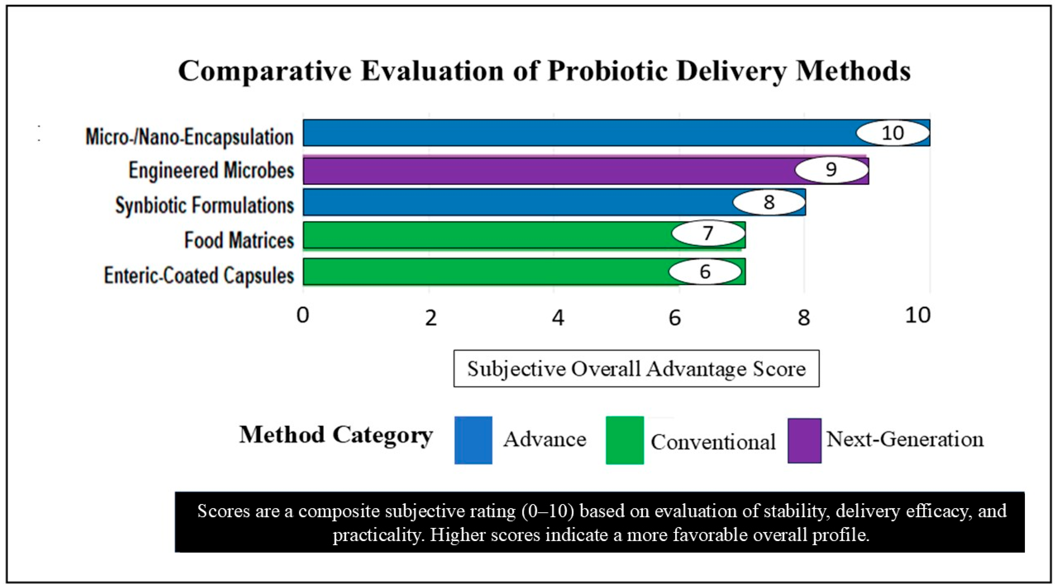
Third, improved delivery systems are needed. Protective formulations, anaerobic stabilization, and stress-tolerant delivery approaches may improve survival through the upper gastrointestinal tract and enhance delivery to intestinal sites where oxalate degradation is most relevant. For obligate anaerobes such as O. formigenes, formulation stability and viability during storage and transit remain especially important [73,104,115,126]. A qualitative comparison of major probiotic delivery strategies for oxalate-degrading microbial therapy is summarized in Figure 6, highlighting the relative advantages of conventional capsules, enteric-coated formulations, microencapsulation, anaerobic stabilization, defined consortia, and engineered live biotherapeutic products. Fourth, defined microbial consortia and engineered live biotherapeutics may offer advantages over single-strain probiotics, because microbiome engineering can expand genetically tractable gut microbes and enable rational design of strains or consortia with improved stability, targeted metabolic activity, and controlled therapeutic functions [119,127]. Defined consortia could combine oxalate degradation with ecological support functions, whereas engineered strains may provide more predictable oxalate-degrading activity. However, these approaches require careful safety evaluation, containment strategies, and long-term monitoring before clinical use [13,76,113]. Overall, the therapeutic potential of the Frc–Oxc pathway should be viewed as microbiome-based functional therapy rather than simple probiotic replacement. Successful clinical translation will likely require matching the right microbial function to the right patient, supported by diet, niche permissiveness, delivery technology, and longitudinal monitoring of urinary oxalate and stone-recurrence outcomes [11,23,113,128].

6.6. Safety and Efficacy in Special Populations

Oxalate-degrading probiotics have generally shown acceptable tolerability in the studied patient groups, although efficacy varies with host physiology, disease context, and baseline microbiome composition [11,13,121]. In idiopathic stone formers, probiotic interventions have generally shown acceptable tolerability, although urinary oxalate responses have been inconsistent across studies [25,121,129]. These outcomes may also be influenced by the baseline abundance of endogenous oxalate-degrading taxa [11]. However, patients with primary hyperoxaluria type 1 (PH1) generally have variable responses despite good tolerance due to difficulties in establishing optimal colonization of O. formigenes [123]. Greater therapeutic relevance may be expected in enteric hyperoxaluria, particularly in post-bariatric surgery settings, because intestinal oxalate absorption is a major contributor to urinary oxalate burden. In these patients, gut-directed microbial products may reduce luminal oxalate availability, although reported oxalate-lowering effects remain variable and should be interpreted cautiously across different study designs [7,30,120]. Evidence in patients with chronic kidney disease remains limited [130], while evidence in children, older adults, and patients with IBD is insufficient to draw population-specific conclusions. In summary, although oxalate-degrading probiotics are generally safe, their efficacy varies and is highly dependent on individual physiological and microbial characteristics [116,121,122].

7. Future Perspectives

The next generation of microbiome therapy for oxalate management should focus on function, not strain. The question should not be whether a probiotic strain has frc and oxc but whether the host’s gut is an appropriate environment for Frc–Oxc activity. This strategy suggests that approaches are needed to promote the intestinal environment consistent with pathway activation, persistence and oxalate degradation. This could involve diet modification, prebiotics, and removal of factors that downregulate oxalate-degrading activity. A further avenue is the design of new live biotherapeutics such as engineered strains or specific microbial communities, with enhanced oxalate-degrading activity, survival and persistence in the gut, and resistance to environmental stress.
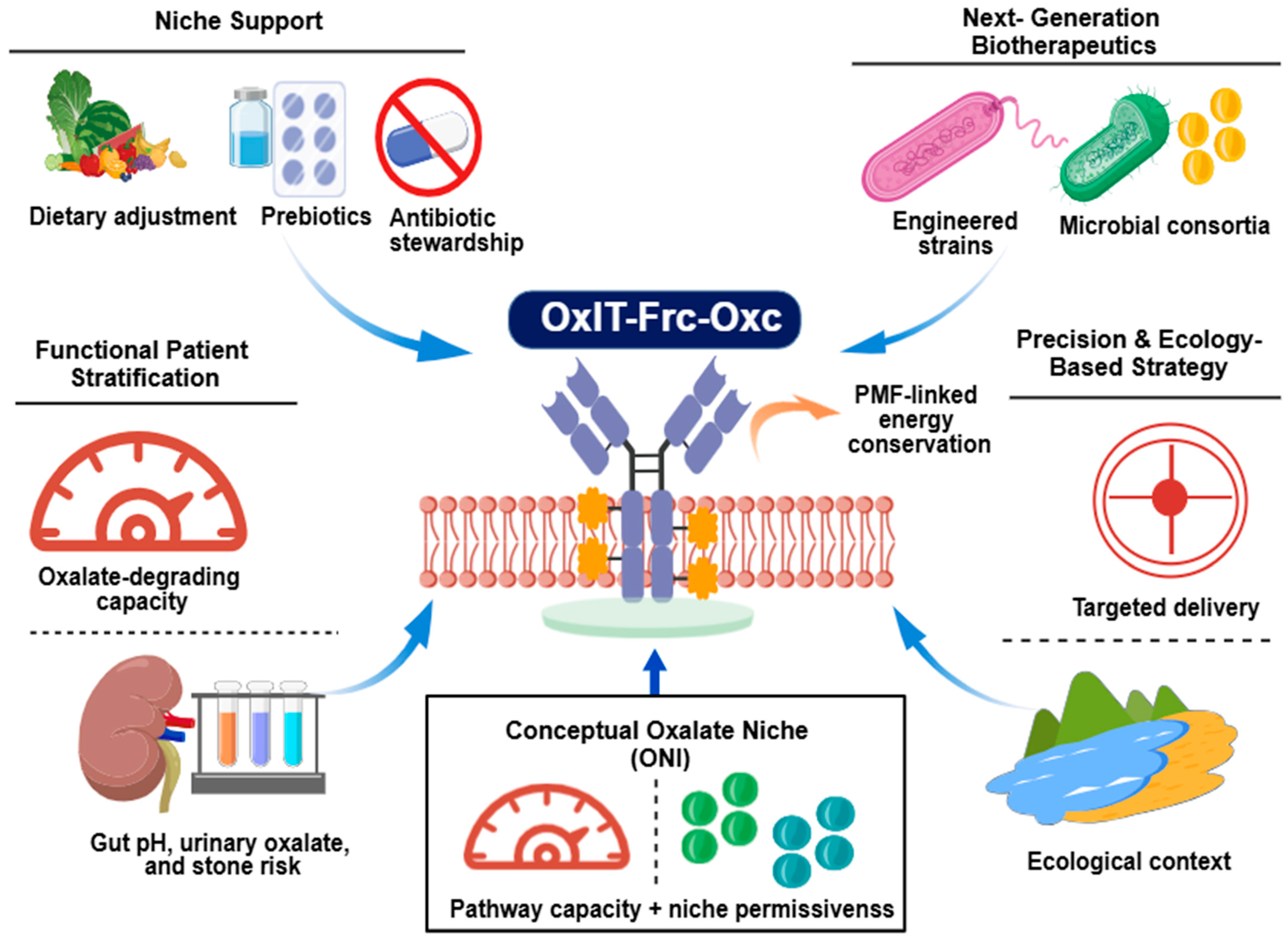
Future studies should also emphasize functional patient stratification. Rather than relying only on taxonomic abundance or gene carriage, patients may be classified according to baseline oxalate-degrading capacity, ecological context, and disease subtype. In this setting, the proposed ONI may serve as a useful conceptual framework for integrating niche permissiveness with pathway-related capacity. Conceptually, ONI can be calculated as
ONI = 100 × (G × A × N × P × (1 − I))
where G represents the genetic capacity for oxalate degradation, reflected by the abundance or presence of key pathway genes such as frc, oxc, and oxlT. A represents pathway activity, which may be estimated through Frc/Oxc expression, enzyme activity, or experimentally measured oxalate-degradation capacity. N represents niche permissiveness, including intestinal pH, oxalate availability, anaerobic conditions, and transport energetics that support OxlT-mediated oxalate/formate exchange. P represents ecological persistence, including colonization stability, microbial survival, and compatibility with the resident gut community. I represent inhibitory pressure, including antibiotic exposure, bile acids, inflammation, unfavorable diet, and microbial competition. Each component is conceptually normalized from 0 to 1, producing a final ONI score ranging from 0 to 100. A higher ONI would indicate a gut environment more favorable for functional oxalate degradation, whereas a lower ONI would suggest that genetic potential may not translate into measurable activity. Although ONI is not yet clinically validated, it provides a hypothesis-generating framework for future studies using metagenomic, metatranscriptomic, enzyme-activity, fecal oxalate-degradation, urinary oxalate, dietary-control, and longitudinal stone-recurrence datasets. Overall, the most promising direction is a precise, ecology-informed strategy that combines microbial function, host environment, and targeted delivery to improve clinical consistency in hyperoxaluria and calcium oxalate kidney stone disease. Figure 7 provides a visual illustration of the future perspective section.

8. Conclusions

Frc–Oxc is an important route of oxalate degradation by microbes and a significant interaction between the gut microbiota and oxalate homeostasis. This pathway allows certain bacteria to break down oxalate in anaerobic conditions via the Frc/Oxc system and thus can possibly decrease intestinal absorption and the formation of calcium oxalate stones. However, gene carriage or abundance alone does not determine the stone-preventive potential of this pathway. Experimental and clinical evidence indicates that in vivo oxalate degradation is influenced by transport energetics, pH, microbial competition, colonization stability, diet, and antibiotic exposure. In this way, these multifactorial ecological and physiological constraints are seen in the variable efficacy of probiotic and microbiome-based therapies across studies and patient groups. To conclude, the Frc–Oxc pathway could be a therapeutic option in managing hyperoxaluria and oxalate stone disease, but this will need a strategy that includes the use of ecology and microbial delivery to activate and sustain the pathway. In this aspect, ONI should be regarded as a conceptual and hypothesis-generating framework rather than a clinically validated index. After future validation using metagenomic, metatranscriptomic, enzyme-activity, fecal oxalate-degradation, urinary oxalate, dietary-control, and longitudinal stone-recurrence datasets, ONI may help explain variability in treatment responses and guide the development of more predictable microbiome-based therapies for hyperoxaluria and calcium oxalate kidney stone disease.

Author Contributions

M.T. designed, drafted, and finalized the manuscript. M.L. and S.N. contributed to literature collection. W.N. revised the manuscript. B.L. conceived the review, supervised the work, and revised the manuscript for intellectual content. All authors have read and agreed to the published version of the manuscript.

Funding

This work was partly supported by the Science and Technology Support Plan of Guizhou Province (Grant Number 2023-145).

Data Availability Statement

No new primary datasets were generated in this review. The secondary analysis presented in Figure 4 used publicly available supplementary data reported by Jiang et al. [72]. All data discussed are derived from publicly available published studies cited in the manuscript.

Acknowledgments

The authors thank the university department for institutional support during this work. Data-based figures were prepared by the authors in the cloud-based Python 3.12.13 environment using Google Colaboratory, https://colab.research.google.com (accessed on 23 June 2026), and Matplotlib 3.10.0. Conceptual illustrations were prepared by the authors using BioGDP [131] and BioRender (https://BioRender.com) and edited in Microsoft PowerPoint, followed by thorough author review, correction, and finalization for scientific accuracy.

Conflicts of Interest

The authors declare no conflicts of interest.

Abbreviations

AbbreviationFull FormAbbreviationFull Form
FrcFormyl-CoA transferaseOxcOxalyl-CoA decarboxylase
OxlTOxalate/formate antiporterPMFProton motive force
ONIOxalate Niche IndexLABLactic acid bacteria
OxDCOxalate DecarboxylasePHPrimary hyperoxaluria
CKDChronic kidney diseaseFOSFructooligosaccharides
TPPThiamine pyrophosphateATPAdenosine triphosphate
ADP Adenosine diphosphateoxdCOxalate decarboxylase
ODPOxalate-Degrading PathwayOxBOxalate-degrading bacteria
NAD+Nicotinamide adenine dinucleotide (oxidized form)NADHNicotinamide adenine dinucleotide (reduced form)
PPInorganic pyrophosphateOCPOxalate Carbonate Pathway
CoACoenzyme AHMM Hidden Markov Model
MFSMajor Facilitator SuperfamilyOFAOxalate:Formate Antiporter

References

  1. Grocholski, C.; Dubourg, L.; Guebre-Egziabher, F.; Bourdain, C.A.; Abid, N.; Bacchetta, J.; Chambrier, C.; Lemoine, S. Oxalate: De la physiologie à la pathologie. Nephrol. Ther. 2023, 19, 201–214. [Google Scholar] [CrossRef] [Scilit] [PubMed]
  2. Zayed, A.; Adly, G.M.; Farag, M.A. Management strategies for the anti-nutrient oxalic acid in foods: A comprehensive overview of its dietary sources, roles, metabolism, and processing. Food Bioprocess. Technol. 2025, 18, 4280–4300. [Google Scholar] [CrossRef] [Scilit]
  3. Ermer, T.; Nazzal, L.; Tio, M.C.; Waikar, S.; Aronson, P.S.; Knauf, F. Oxalate homeostasis. Nat. Rev. Nephrol. 2023, 19, 123–138. [Google Scholar] [PubMed]
  4. Al-Kabe, S.H.; Niamah, A.K. Current trends and technological advancements in the use of oxalate-degrading bacteria as starters in fermented foods—A review. Life 2024, 14, 1338. [Google Scholar] [CrossRef] [Scilit] [PubMed]
  5. Jaunet-Lahary, T.; Shimamura, T.; Hayashi, M.; Nomura, N.; Hirasawa, K.; Shimizu, T.; Yamashita, M.; Tsutsumi, N.; Suehiro, Y.; Kojima, K. Structure and mechanism of oxalate transporter OxlT in an oxalate-degrading bacterium in the gut microbiota. Nat. Commun. 2023, 14, 1730. [Google Scholar] [CrossRef] [Scilit] [PubMed]
  6. Zaidan, N.; Wang, C.; Chen, Z.; Lieske, J.C.; Milliner, D.; Seide, B.; Ho, M.; Li, H.; Ruggles, K.V.; Modersitzki, F.; et al. Multiomics assessment of the gut microbiome in rare hyperoxaluric conditions. Kidney Int. Rep. 2024, 9, 1836–1848. [Google Scholar] [CrossRef] [Scilit] [PubMed]
  7. Bhardwaj, M.; Singhal, A.; Bhardwaj, G.; Dukic, I. Probiotic and synbiotic interventions targeting oxalate-degrading gut bacteria for the prevention of kidney stones: A systematic review. Cureus 2025, 17, e98728. [Google Scholar] [CrossRef] [Scilit] [PubMed]
  8. Lieske, J.C. Probiotics for prevention of urinary stones. Ann. Transl. Med. 2017, 5, 29. [Google Scholar] [CrossRef] [Scilit] [PubMed]
  9. Pei, X.; Liu, M.; Yu, S. How is the human microbiome linked to kidney stones? Front. Cell. Infect. Microbiol. 2025, 15, 1602413. [Google Scholar] [CrossRef] [Scilit] [PubMed]
  10. Liu, M.; Devlin, J.C.; Hu, J.; Volkova, A.; Battaglia, T.W.; Ho, M.; Asplin, J.R.; Byrd, A.; Loke, P.; Li, H.; et al. Microbial genetic and transcriptional contributions to oxalate degradation by the gut microbiota in health and disease. eLife 2021, 10, e63642. [Google Scholar] [CrossRef] [Scilit] [PubMed]
  11. Suryavanshi, M.; Franklin, A.; Fargue, S.; Assimos, D.G.; Knight, J.; Miller, A.W. Baseline abundance of oxalate-degrading bacteria determines response to Oxalobacter formigenes probiotic therapy. Gut Microbes 2025, 17, 2562337. [Google Scholar] [CrossRef] [Scilit] [PubMed]
  12. Yang, J.; Li, D.; Li, T.; Jia, B. Probiotics in the prevention and treatment of calcium oxalate kidney stones: Mechanisms and therapeutic potential. Front. Microbiol. 2025, 16, 1663138. [Google Scholar] [CrossRef] [Scilit] [PubMed]
  13. Mukherjee, S.D.; Batagello, C.; Adler, A.; Agudelo, J.; Zampini, A.; Suryavanshi, M.; Nguyen, A.; Orr, T.; Dearing, D.; Monga, M.; et al. Complex system modeling reveals oxalate homeostasis is driven by diverse oxalate-degrading bacteria. eLife 2025, 14, RP104121. [Google Scholar] [CrossRef] [Scilit] [PubMed]
  14. Youssef, H.I.A. Detection of oxalyl-CoA decarboxylase (oxc) and formyl-CoA transferase (frc) genes in novel probiotic isolates capable of oxalate degradation in vitro. Folia Microbiol. 2024, 69, 423–432. [Google Scholar] [CrossRef] [Scilit]
  15. Wan, W.; Wu, W.; Amier, Y.; Li, X.; Yang, J.; Huang, Y.; Xun, Y.; Yu, X. Engineered microorganisms: A new direction in kidney stone prevention and treatment. Synth. Syst. Biotechnol. 2024, 9, 294–303. [Google Scholar] [CrossRef] [Scilit] [PubMed]
  16. Chen, T.; Qian, B.; Zou, J.; Luo, P.; Zou, J.; Li, W.; Chen, Q.; Zheng, L. Oxalate as a potent promoter of kidney stone formation. Front. Med. 2023, 10, 1159616. [Google Scholar] [CrossRef] [Scilit]
  17. Suryavanshi, M.; Mukherjee, S.D.; Miller, A.W. Predicting probiotic success: Lessons from Oxalobacter and oxalate metabolism. npj Biofilms Microbiomes 2026, 12, 30. [Google Scholar] [CrossRef] [Scilit] [PubMed]
  18. Mitchell, T.; Kumar, P.; Reddy, T.; Wood, K.D.; Knight, J.; Assimos, D.G.; Holmes, R.P. Dietary oxalate and kidney stone formation. Am. J. Physiol. Ren. Physiol. 2019, 316, F409–F413. [Google Scholar] [CrossRef] [Scilit]
  19. Hatch, M. Gut microbiota and oxalate homeostasis. Ann. Transl. Med. 2017, 5, 36. [Google Scholar] [CrossRef] [Scilit] [PubMed]
  20. Abratt, V.R.; Reid, S.J. Oxalate-degrading bacteria of the human gut as probiotics in the management of kidney stone disease. Adv. Appl. Microbiol. 2010, 72, 63–87. [Google Scholar] [CrossRef] [Scilit] [PubMed]
  21. Crivelli, J.J.; Mitchell, T.; Knight, J.; Wood, K.D.; Assimos, D.G.; Holmes, R.P.; Fargue, S. Contribution of dietary oxalate and oxalate precursors to urinary oxalate excretion. Nutrients 2021, 13, 62. [Google Scholar]
  22. Demoulin, N.; Aydin, S.; Gillion, V.; Morelle, J.; Jadoul, M. Pathophysiology and management of hyperoxaluria and oxalate nephropathy: A review. Am. J. Kidney Dis. 2022, 79, 717–727. [Google Scholar] [CrossRef] [Scilit] [PubMed]
  23. Witting, C.; Langman, C.B.; Assimos, D.; Baum, M.A.; Kausz, A.; Milliner, D.; Tasian, G.; Worcester, E.; Allain, M.; West, M. Pathophysiology and treatment of enteric hyperoxaluria. Clin. J. Am. Soc. Nephrol. 2021, 16, 487–495. [Google Scholar] [PubMed]
  24. Bargagli, M.; Tio, M.C.; Waikar, S.S.; Ferraro, P.M. Dietary oxalate intake and kidney outcomes. Nutrients 2020, 12, 2673. [Google Scholar] [CrossRef] [Scilit] [PubMed]
  25. Lieske, J.C.; Tremaine, W.J.; De Simone, C.; O’Connor, H.M.; Li, X.; Bergstralh, E.J.; Goldfarb, D.S. Diet, but not oral probiotics, effectively reduces urinary oxalate excretion and calcium oxalate supersaturation. Kidney Int. 2010, 78, 1178–1185. [Google Scholar] [CrossRef] [Scilit] [PubMed]
  26. Liebman, M.; Al-Wahsh, I.A. Probiotics and other key determinants of dietary oxalate absorption. Adv. Nutr. 2011, 2, 254–260. [Google Scholar] [CrossRef] [Scilit] [PubMed]
  27. Stepanova, N. Oxalate homeostasis in non-stone-forming chronic kidney disease: A review of key findings and perspectives. Biomedicines 2023, 11, 1654. [Google Scholar] [CrossRef] [Scilit] [PubMed]
  28. Worcester, E.M.; Coe, F.L. Clinical practice. Calcium kidney stones. N. Engl. J. Med. 2010, 363, 954–963. [Google Scholar] [CrossRef] [Scilit] [PubMed]
  29. Kumar, R.; Lieske, J.C.; Collazo-Clavell, M.L.; Sarr, M.G.; Olson, E.R.; Vrtiska, T.J.; Bergstralh, E.J.; Li, X. Fat malabsorption and increased intestinal oxalate absorption are common after Roux-en-Y gastric bypass surgery. Surgery 2011, 149, 654–661. [Google Scholar] [CrossRef] [Scilit] [PubMed]
  30. Siener, R.; Ernsten, C.; Welchowski, T.; Hesse, A. Metabolic profile of calcium oxalate stone patients with enteric hyperoxaluria and impact of dietary intervention. Nutrients 2024, 16, 2688. [Google Scholar] [CrossRef] [Scilit] [PubMed]
  31. Nazzal, L.; Puri, S.; Goldfarb, D.S. Enteric hyperoxaluria: An important cause of end-stage kidney disease. Nephrol. Dial. Transplant. 2016, 31, 375–382. [Google Scholar] [PubMed]
  32. Khan, S.R.; Pearle, M.S.; Robertson, W.G.; Gambaro, G.; Canales, B.K.; Doizi, S.; Traxer, O.; Tiselius, H.-G. Kidney stones. Nat. Rev. Dis. Prim. 2016, 2, 16008. [Google Scholar] [CrossRef] [Scilit] [PubMed]
  33. Zisman, A.L. Effectiveness of treatment modalities on kidney stone recurrence. Clin. J. Am. Soc. Nephrol. 2017, 12, 1699–1708. [Google Scholar] [CrossRef] [Scilit] [PubMed]
  34. Mehta, M.; Goldfarb, D.S.; Nazzal, L. The role of the microbiome in kidney stone formation. Int. J. Surg. 2016, 36, 607–612. [Google Scholar] [CrossRef] [Scilit] [PubMed]
  35. Pang, S.; Zhang, Z.; Ma, Q.; Liu, Y.; Wang, S.; Wang, J.; Bi, Y. The gut–kidney microbiome–oxalate axis in calcium oxalate nephrolithiasis: Mechanisms and microbiome-based interventions. Front. Cell. Infect. Microbiol. 2026, 16, 1804800. [Google Scholar] [CrossRef] [Scilit] [PubMed]
  36. Karamad, D.; Khosravi-Darani, K.; Khaneghah, A.M.; Miller, A.W. Probiotic oxalate-degrading bacteria: New insight of environmental variables and expression of the oxc and frc genes on oxalate degradation activity. Foods 2022, 11, 2876. [Google Scholar] [CrossRef] [Scilit] [PubMed]
  37. Grąz, M. Role of oxalic acid in fungal and bacterial metabolism and its biotechnological potential. World J. Microbiol. Biotechnol. 2024, 40, 178. [Google Scholar] [CrossRef] [Scilit] [PubMed]
  38. Jafari, G.A.; Fotouhi Ardakani, R.; Akhavan Sepahi, M.; Nowroozi, J.; Soltanpour, M.S. Development of an innovative method by optimizing qPCR technique for isolating and determining Oxalobacter formigenes microbial load in the stool of patients with urolithiasis. Iran. J. Kidney Dis. 2021, 15, 190–198. [Google Scholar] [PubMed]
  39. Hervé, V.; Junier, T.; Bindschedler, S.; Verrecchia, E.; Junier, P. Diversity and ecology of oxalotrophic bacteria. World J. Microbiol. Biotechnol. 2016, 32, 28. [Google Scholar] [CrossRef] [Scilit] [PubMed]
  40. Sonke, A.; Trembath-Reichert, E. Expanding the taxonomic and environmental extent of an underexplored carbon metabolism—Oxalotrophy. Front. Microbiol. 2023, 14, 1161937. [Google Scholar] [CrossRef] [Scilit] [PubMed]
  41. Junier, T.; Palmieri, F.; Ubags, N.D.; Trompette, A.; Koutsokera, A.; Junier, P.; Pagni, M.; Neuenschwander, S. Prevalence of oxalotrophy in the human microbiome. BMC Genom. 2025, 26, 954. [Google Scholar] [CrossRef] [Scilit]
  42. Azcarate-Peril, M.A.; Bruno-Bárcena, J.M.; Hassan, H.M.; Klaenhammer, T.R. Transcriptional and functional analysis of oxalyl-coenzyme A (CoA) decarboxylase and formyl-CoA transferase genes from Lactobacillus acidophilus. Appl. Environ. Microbiol. 2006, 72, 1891–1899. [Google Scholar] [CrossRef] [Scilit] [PubMed]
  43. Prasanna, A.N.; Parween, S.; Froehlich, K.; Schmidt, T.; Shekhawat, K.; Prabhu, D.D.; Saad, M.M.; Hirt, H. Genomic and functional divergence of oxalate metabolism pathways in bacteria from contrasting ecosystems. Microb. Genom. 2026, 12, 001587. [Google Scholar] [CrossRef] [Scilit] [PubMed]
  44. Xia, D.; Nie, W.; Li, X.; Finlay, R.D.; Lian, B. Secondary products and molecular mechanism of calcium oxalate degradation by the strain Azospirillum sp. OX-1. Sci. Rep. 2024, 14, 23506. [Google Scholar] [CrossRef] [Scilit] [PubMed]
  45. Lv, Y.; Liu, Y.; Lin, Y.; Zheng, H.; Yan, J.; Zhang, Y.; Miao, W.; Wu, W.; Lin, C. Functional diversification of oxalate decarboxylases in terms of enzymatic activity, morphosporogenesis, stress regulation and virulence in Colletotrichum siamense. Front. Microbiol. 2025, 16, 1547950. [Google Scholar] [CrossRef] [Scilit] [PubMed]
  46. Jonsson, S.; Ricagno, S.; Lindqvist, Y.; Richards, N.G. Kinetic and mechanistic characterization of the formyl-CoA transferase from Oxalobacter formigenes. J. Biol. Chem. 2004, 279, 36003–36012. [Google Scholar] [CrossRef] [Scilit] [PubMed]
  47. Ricagno, S.; Jonsson, S.; Richards, N.; Lindqvist, Y. Formyl-CoA transferase encloses the CoA binding site at the interface of an interlocked dimer. EMBO J. 2003, 22, 3210–3219. [Google Scholar] [CrossRef] [Scilit] [PubMed]
  48. Berthold, C.L.; Toyota, C.G.; Moussatche, P.; Wood, M.D.; Leeper, F.; Richards, N.G.; Lindqvist, Y. Crystallographic snapshots of oxalyl-CoA decarboxylase give insights into catalysis by nonoxidative ThDP-dependent decarboxylases. Structure 2007, 15, 853–861. [Google Scholar] [CrossRef] [Scilit] [PubMed]
  49. Sun, Q.; Li, J.; Finlay, R.D.; Lian, B. Oxalotrophic bacterial assemblages in the ectomycorrhizosphere of forest trees and their effects on oxalate degradation and carbon fixation potential. Chem. Geol. 2019, 514, 54–64. [Google Scholar] [CrossRef] [Scilit]
  50. Zan, X.; Yan, Y.; Chen, G.; Sun, L.; Wang, L.; Wen, Y.; Xu, Y.; Zhang, Z.; Li, X.; Yang, Y.; et al. Recent advances of oxalate decarboxylase: Biochemical characteristics, catalysis mechanisms, and gene expression and regulation. J. Agric. Food Chem. 2024, 72, 10163–10178. [Google Scholar] [CrossRef] [Scilit] [PubMed]
  51. Gómez-Espinoza, O.; Rojas-Villalta, D.; Zúñiga-Pereira, A.M.; Chacón-Díaz, C.; Bravo, L.A.; Reyes-Díaz, M. Plant oxalate oxidases: Key enzymes in redox and stress regulation. J. Exp. Bot. 2025, 76, 4896–4909. [Google Scholar] [CrossRef] [Scilit] [PubMed]
  52. Syed, S.; Buddolla, V.; Lian, B. Lead oxalates in some Chinese leafy vegetable cultivation: Their biomineralization and remediation by oxalate degrading Streptomyces sp. 3 Biotech 2022, 12, 284. [Google Scholar] [CrossRef] [Scilit]
  53. Berthold, C.L.; Toyota, C.G.; Richards, N.G.; Lindqvist, Y. Reinvestigation of the catalytic mechanism of formyl-CoA transferase, a class III CoA-transferase. J. Biol. Chem. 2008, 283, 6519–6529. [Google Scholar] [CrossRef] [Scilit] [PubMed]
  54. Toyota, C.G.; Berthold, C.L.; Gruez, A.; Jónsson, S.; Lindqvist, Y.; Cambillau, C.; Richards, N.G. Differential substrate specificity and kinetic behavior of Escherichia coli YfdW and Oxalobacter formigenes formyl coenzyme A transferase. J. Bacteriol. 2008, 190, 2556–2564. [Google Scholar] [CrossRef] [Scilit] [PubMed]
  55. Shima, S.; Thauer, R.K.; Ermler, U. Hyperthermophilic and salt-dependent formyltransferase from Methanopyrus kandleri. Biochem. Soc. Trans. 2004, 32, 269–272. [Google Scholar] [CrossRef] [Scilit] [PubMed]
  56. Cheng, N.; Paris, V.; Rao, X.; Wang, X.; Nakata, P.A. A conserved oxalyl-coenzyme A decarboxylase in oxalate catabolism. Plant Signal. Behav. 2022, 17, 2062555. [Google Scholar] [CrossRef] [Scilit]
  57. Berthold, C.L.; Moussatche, P.; Richards, N.G.; Lindqvist, Y. Structural basis for activation of the thiamin diphosphate-dependent enzyme oxalyl-CoA decarboxylase by adenosine diphosphate. J. Biol. Chem. 2005, 280, 41645–41654. [Google Scholar] [CrossRef] [Scilit] [PubMed]
  58. Nattermann, M.; Burgener, S.; Pfister, P.; Chou, A.; Schulz, L.; Lee, S.H.; Paczia, N.; Zarzycki, J.; Gonzalez, R.; Erb, T.J. Engineering a highly efficient carboligase for synthetic one-carbon metabolism. ACS Catal. 2021, 11, 5396–5404. [Google Scholar] [CrossRef] [Scilit] [PubMed]
  59. Dhanya Raj, C.T.; Kandaswamy, S.; Suryavanshi, M.V.; Ramasamy, K.P.; Rajasabapathy, R.; Arthur James, R. Genomic and metabolic properties of Staphylococcus gallinarum FCW1 MCC4687 isolated from naturally fermented coconut water towards GRAS assessment. Gene 2023, 867, 147356. [Google Scholar] [CrossRef] [Scilit] [PubMed]
  60. Xu, M.; Qin, Y.; Xia, Y.; Wang, G.; Xiong, Z.; Song, X.; Ai, L. Screening of oxalate-degrading probiotics and preventive effect of Lactiplantibacillus plantarum AR1089 on kidney stones. Food Funct. 2024, 15, 10163–10178. [Google Scholar] [CrossRef] [Scilit] [PubMed]
  61. Zhang, M.; Fan, L.; Li, J. Prediction of calcium oxalate kidney stones: A comprehensive analysis of clinical and gut microbiome characteristics. Medicine 2025, 104, e43103. [Google Scholar] [CrossRef] [Scilit] [PubMed]
  62. Fontenot, E.M.; Ezelle, K.E.; Gabreski, L.N.; Giglio, E.R.; McAfee, J.M.; Mills, A.C.; Qureshi, M.N.; Salmon, K.M.; Toyota, C.G. YfdW and YfdU are required for oxalate-induced acid tolerance in Escherichia coli K-12. J. Bacteriol. 2013, 195, 1446–1455. [Google Scholar] [CrossRef] [Scilit] [PubMed]
  63. Masuda, N.; Church, G.M. Regulatory network of acid resistance genes in Escherichia coli. Mol. Microbiol. 2003, 48, 699–712. [Google Scholar] [CrossRef] [Scilit] [PubMed]
  64. Mehra, Y.; Rajesh, N.G.; Viswanathan, P. Analysis and characterization of Lactobacillus paragasseri and Lacticaseibacillus paracasei: Two probiotic bacteria that can degrade intestinal oxalate in hyperoxaluric rats. Probiotics Antimicrob. Proteins 2022, 14, 854–872. [Google Scholar] [CrossRef] [Scilit] [PubMed]
  65. Masoleh, A.M.; Khoshnood, S.; Haddadi, M.H.; Rostamzad, A.; Narki, M.; Hoshmandfar, R.; Sadeghifard, N. Potential role of Lactobacillus and Bifidobacterium for preventing kidney stones. Clin. Lab. 2023, 69, 889–895. [Google Scholar] [CrossRef] [Scilit]
  66. Görke, B.; Stülke, J. Carbon catabolite repression in bacteria: Many ways to make the most out of nutrients. Nat. Rev. Microbiol. 2008, 6, 613–624. [Google Scholar] [CrossRef] [Scilit] [PubMed]
  67. Kuhner, C.H.; Hartman, P.A.; Allison, M.J. Generation of a proton motive force by the anaerobic oxalate-degrading bacterium Oxalobacter formigenes. Appl. Environ. Microbiol. 1996, 62, 2494–2500. [Google Scholar] [CrossRef] [Scilit] [PubMed]
  68. Stewart, C.S.; Duncan, S.H.; Cave, D.R. Oxalobacter formigenes and its role in oxalate metabolism in the human gut. FEMS Microbiol. Lett. 2004, 230, 1–7. [Google Scholar] [CrossRef] [Scilit] [PubMed]
  69. Burgener, S.; Cortina, N.S.; Erb, T.J. Oxalyl-CoA decarboxylase enables nucleophilic one-carbon extension of aldehydes to chiral α-hydroxy acids. Angew. Chem. Int. Ed. 2020, 59, 5526–5530. [Google Scholar] [CrossRef] [Scilit]
  70. Sun, Q.; Li, J.; Syed, S.; Li, X.; Yuan, H.; Lian, B. Roles of oxalate-degrading bacteria in fungus-growing termite nests. Biodivers. Data J. 2024, 12, e130041. [Google Scholar] [CrossRef] [Scilit] [PubMed]
  71. Ye, L.; Jia, Z.; Jung, T.; Maloney, P.C. Topology of OxlT, the oxalate transporter of Oxalobacter formigenes, determined by site-directed fluorescence labeling. J. Bacteriol. 2001, 183, 2490–2496. [Google Scholar] [CrossRef] [Scilit]
  72. Jiang, T.; Chen, W.; Cao, L.; He, Y.; Zhou, H.; Mao, H. Abundance, functional, and evolutionary analysis of oxalyl-coenzyme A decarboxylase in human microbiota. Front. Microbiol. 2020, 11, 672. [Google Scholar] [CrossRef] [Scilit] [PubMed]
  73. Daniel, S.L.; Moradi, L.; Paiste, H.; Wood, K.D.; Assimos, D.G.; Holmes, R.P.; Nazzal, L.; Hatch, M.; Knight, J. Forty years of Oxalobacter formigenes, a gutsy oxalate-degrading specialist. Appl. Environ. Microbiol. 2021, 87, e0054421. [Google Scholar] [CrossRef] [Scilit] [PubMed]
  74. Fargue, S.; Suryavanshi, M.; Wood, K.D.; Crivelli, J.J.; Oster, R.A.; Assimos, D.G.; Miller, A.; Knight, J. Inducing Oxalobacter formigenes colonization reduces urinary oxalate in healthy adults. Kidney Int. Rep. 2025, 10, 1518–1528. [Google Scholar] [CrossRef] [Scilit] [PubMed]
  75. Tavasoli, S.; Alebouyeh, M.; Naji, M.; Shakiba majd, G.; Shabani Nashtaei, M.; Broumandnia, N.; Basiri, A. Association of intestinal oxalate-degrading bacteria with recurrent calcium kidney stone formation and hyperoxaluria: A case–control study. BJU Int. 2020, 125, 133–143. [Google Scholar] [PubMed]
  76. Önal Darilmaz, D.; Sönmez, S.; Beyatlı, Y. The effects of inulin as a prebiotic supplement and the synbiotic interactions of probiotics to improve oxalate degrading activity. Int. J. Food Sci. Technol. 2018, 54, 121–131. [Google Scholar] [CrossRef] [Scilit]
  77. Zheng, J.; Wittouck, S.; Salvetti, E.; Franz, C.M.; Harris, H.M.; Mattarelli, P.; O’toole, P.W.; Pot, B.; Vandamme, P.; Walter, J. A taxonomic note on the genus Lactobacillus: Description of 23 novel genera, emended description of the genus Lactobacillus Beijerinck 1901, and union of Lactobacillaceae and Leuconostocaceae. Int. J. Syst. Evol. Microbiol. 2020, 70, 2782–2858. [Google Scholar] [CrossRef] [Scilit] [PubMed]
  78. Turroni, S.; Vitali, B.; Bendazzoli, C.; Candela, M.; Gotti, R.; Federici, F.; Pirovano, F.; Brigidi, P. Oxalate consumption by lactobacilli: Evaluation of oxalyl-CoA decarboxylase and formyl-CoA transferase activity in Lactobacillus acidophilus. J. Appl. Microbiol. 2007, 103, 1600–1609. [Google Scholar] [CrossRef] [Scilit] [PubMed]
  79. Federici, F.; Vitali, B.; Gotti, R.; Pasca, M.R.; Gobbi, S.; Peck, A.B.; Brigidi, P. Characterization and heterologous expression of the oxalyl coenzyme A decarboxylase gene from Bifidobacterium lactis. Appl. Environ. Microbiol. 2004, 70, 5066–5073. [Google Scholar] [CrossRef] [Scilit] [PubMed]
  80. Turroni, S.; Bendazzoli, C.; Dipalo, S.C.; Candela, M.; Vitali, B.; Gotti, R.; Brigidi, P. Oxalate-degrading activity in Bifidobacterium animalis subsp. lactis: Impact of acidic conditions on the transcriptional levels of the oxalyl coenzyme A (CoA) decarboxylase and formyl-CoA transferase genes. Appl. Environ. Microbiol. 2010, 76, 5609–5620. [Google Scholar] [CrossRef] [Scilit] [PubMed]
  81. Al, K.F.; Joris, B.R.; Daisley, B.A.; Chmiel, J.A.; Bjazevic, J.; Reid, G.; Gloor, G.B.; Denstedt, J.D.; Razvi, H.; Burton, J.P. Multi-site microbiota alteration is a hallmark of kidney stone formation. Microbiome 2023, 11, 263. [Google Scholar] [CrossRef] [Scilit] [PubMed]
  82. Du, S.; Sun, K.; Xiao, B.; Liu, Z. Bioinformatic analysis of oxalate-degrading enzymes in probiotics: A systematic genome-scale and structural survey. Microorganisms 2025, 13, 2553. [Google Scholar] [CrossRef] [Scilit] [PubMed]
  83. Miller, A.W.; Kohl, K.D.; Dearing, M.D. The gastrointestinal tract of the white-throated woodrat (Neotoma albigula) harbors distinct consortia of oxalate-degrading bacteria. Appl. Environ. Microbiol. 2014, 80, 1595–1601. [Google Scholar] [CrossRef] [Scilit] [PubMed]
  84. Stern, J.M.; Moazami, S.; Qiu, Y.; Kurland, I.; Chen, Z.; Agalliu, I.; Burk, R.; Davies, K.P. Evidence for a distinct gut microbiome in kidney stone formers compared to non-stone formers. Urolithiasis 2016, 44, 399–407. [Google Scholar] [CrossRef] [Scilit] [PubMed]
  85. Lo, C.Y.Z.; Khor, Q.H.; Abdullatif, V.A.; Delgado, C.; Lu, Y.; Katz, J.; Sur, R.L. Systematic review of pharmacological, complementary and alternative therapies for the prevention of calcium oxalate stones. Asian J. Urol. 2025, 12, 169–188. [Google Scholar] [CrossRef] [Scilit] [PubMed]
  86. Wigner, P.; Bijak, M.; Saluk-Bijak, J. Probiotics in the prevention of the calcium oxalate urolithiasis. Cells 2022, 11, 284. [Google Scholar] [CrossRef] [Scilit] [PubMed]
  87. Liu, Y.; Wang, S.; Wang, L.; Lu, H.; Zhang, T.; Zeng, W. Characterization of genomic, physiological, and probiotic features of Lactiplantibacillus plantarum JS21 strain isolated from traditional fermented Jiangshui. Foods 2024, 13, 1082. [Google Scholar] [CrossRef] [Scilit] [PubMed]
  88. Ran, J.; Tang, Y.; Mao, W.; Meng, X.; Jiao, L.; Li, Y.; Zhao, R.; Zhou, H. Optimization of the fermentation process and antioxidant activity of mixed lactic acid bacteria for honeysuckle beverage. Front. Microbiol. 2024, 15, 1364448. [Google Scholar] [CrossRef] [Scilit] [PubMed]
  89. Rahman, M.M.; Abdullah, R.B.; Wan Khadijah, W.E. A review of oxalate poisoning in domestic animals: Tolerance and performance aspects. J. Anim. Physiol. Anim. Nutr. 2013, 97, 605–614. [Google Scholar]
  90. Popova-Krumova, P.; Danova, S.; Atanasova, N.; Yankov, D. Lactic acid production by Lactiplantibacillus plantarum AC 11S —Kinetics and modeling. Microorganisms 2024, 12, 739. [Google Scholar] [CrossRef] [Scilit] [PubMed]
  91. Cowan, D.A.; Babenko, D.; Bird, R.; Botha, A.; Breecker, D.O.; Clarke, C.E.; Francis, M.L.; Gallagher, T.; Lebre, P.H.; Nel, T. Oxalate and oxalotrophy: An environmental perspective. Sustain. Microbiol. 2024, 1, qvad004. [Google Scholar] [CrossRef] [Scilit]
  92. Siener, R.; Ernsten, C.; Speller, J.; Scheurlen, C.; Sauerbruch, T.; Hesse, A. Intestinal oxalate absorption, enteric hyperoxaluria, and risk of urinary stone formation in patients with Crohn’s disease. Nutrients 2024, 16, 264. [Google Scholar] [CrossRef] [Scilit] [PubMed]
  93. Sadaf, H.; Raza, S.I.; Hassan, S.W. Role of gut microbiota against calcium oxalate. Microb. Pathog. 2017, 109, 287–291. [Google Scholar] [CrossRef] [Scilit] [PubMed]
  94. Shah, A.; Leslie, S.; Ramakrishnan, S. Hyperoxaluria. In StatPearls [Internet]; StatPearls Publishing: Treasure Island, FL, USA, 2025. [Google Scholar]
  95. Karamad, D.; Khosravi-Darani, K.; Hosseini, H.; Tavasoli, S.; Miller, A.W. Assessment of the process variables for degradation of oxalate by Lactobacillus acidophilus ATCC 4356 using simulated rumen fluid media and tea. Appl. Food Biotechnol. 2020, 7, 195–204. [Google Scholar]
  96. Karamad, D.; Khosravi-Darani, K.; Hosseini, H.; Tavasoli, S.; Miller, A.W. Evaluation of Oxalobacter formigenes DSM 4420 biodegradation activity for high oxalate media content: An in vitro model. Biocatal. Agric. Biotechnol. 2019, 22, 101378. [Google Scholar] [CrossRef] [Scilit] [PubMed]
  97. Liu, L.; Ma, Y.; Jian, Z.; Liao, B.; Li, Y.; Lin, L.; Wang, M.; Chen, J.; Wei, J.; Yang, M. Gut microbiota-bile acid crosstalk contributes to calcium oxalate nephropathy through Hsp90α-mediated ferroptosis. Cell Rep. 2025, 44, 115936. [Google Scholar] [CrossRef] [Scilit] [PubMed]
  98. Kim, H.N.; Kim, J.H.; Chang, Y.; Yang, D.; Joo, K.J.; Cho, Y.S.; Park, H.J.; Kim, H.L.; Ryu, S. Gut microbiota and the prevalence and incidence of renal stones. Sci. Rep. 2022, 12, 3732. [Google Scholar] [CrossRef] [Scilit] [PubMed]
  99. Tian, J.; Peng, J.; Ge, W.H.; Zhang, J. Therapeutic effect of inulin on enteric hyperoxaluria in rats. Zhongguo Yi Xue Ke Xue Yuan Xue Bao 2014, 36, 313–316. [Google Scholar] [CrossRef] [Scilit] [PubMed]
  100. Li, X.M.; Amier, Y.; Wan, W.L.; Wang, Y.Q.; Lyu, G.R.Y.; Lu, J.L.; Rao, J.Y.; Yang, Y.Y.; Xun, Y.; Yu, X. A novel synbiotic formulation prevents calcium oxalate stones by restoring gut microbiota homeostasis. BMC Microbiol. 2025, 25, 673. [Google Scholar] [CrossRef] [Scilit] [PubMed]
  101. Stepanova, N.; Akulenko, I.; Serhiichuk, T.; Dovbynchuk, T.; Savchenko, S.; Tolstanova, G. Synbiotic supplementation and oxalate homeostasis in rats: Focus on microbiota oxalate-degrading activity. Urolithiasis 2022, 50, 249–258. [Google Scholar] [CrossRef] [Scilit] [PubMed]
  102. Singh, P.; Chebib, F.T.; Cogal, A.G.; Gavrilov, D.K.; Harris, P.C.; Lieske, J.C. Pyridoxine responsiveness in a type 1 primary hyperoxaluria patient with a rare (atypical) AGXT gene mutation. Kidney Int. Rep. 2020, 5, 955–958. [Google Scholar] [CrossRef] [Scilit] [PubMed]
  103. Abbas, M.N.; Zain-ul-abideen; Zia, M.M. Use of probiotics and prebiotics for the controlling of different types of kidney stones. Indo Am. J. Pharm. Sci. 2020, 7, 1762–1765. [Google Scholar]
  104. Hiremath, S.; Viswanathan, P. Oxalobacter formigenes: A new hope as a live biotherapeutic agent in the management of calcium oxalate renal stones. Anaerobe 2022, 75, 102572. [Google Scholar] [CrossRef] [Scilit] [PubMed]
  105. Lange, J.N.; Wood, K.D.; Wong, H.; Otto, R.; Mufarrij, P.W.; Knight, J.; Akpinar, H.; Holmes, R.P.; Assimos, D.G. Sensitivity of human strains of Oxalobacter formigenes to commonly prescribed antibiotics. Urology 2012, 79, 1286–1289. [Google Scholar] [CrossRef] [Scilit] [PubMed]
  106. Tasian, G.E.; Jemielita, T.; Goldfarb, D.S.; Copelovitch, L.; Gerber, J.S.; Wu, Q.; Denburg, M.R. Oral antibiotic exposure and kidney stone disease. J. Am. Soc. Nephrol. 2018, 29, 1731–1740. [Google Scholar] [CrossRef] [Scilit] [PubMed]
  107. Nazzal, L.; Francois, F.; Henderson, N.; Liu, M.; Li, H.; Koh, H.; Wang, C.; Gao, Z.; Perez, G.P.; Asplin, J.R. Effect of antibiotic treatment on Oxalobacter formigenes colonization of the gut microbiome and urinary oxalate excretion. Sci. Rep. 2021, 11, 16428. [Google Scholar] [CrossRef] [Scilit] [PubMed]
  108. Ferraro, P.M.; Curhan, G.C.; Gambaro, G.; Taylor, E.N. Antibiotic use and risk of incident kidney stones in female nurses. Am. J. Kidney Dis. 2019, 74, 736–741. [Google Scholar] [CrossRef] [Scilit] [PubMed]
  109. Dethlefsen, L.; Relman, D.A. Incomplete recovery and individualized responses of the human distal gut microbiota to repeated antibiotic perturbation. Proc. Natl. Acad. Sci. USA 2011, 108, 4554–4561. [Google Scholar] [PubMed]
  110. Afkari, R.; Feizabadi, M.M.; Ansari-Moghadam, A.; Safari, T.; Bokaeian, M. Simultaneous use of oxalate-degrading bacteria and herbal extract to reduce the urinary oxalate in a rat model: A new strategy. Int. Braz. J. Urol. 2019, 45, 1249–1259. [Google Scholar] [CrossRef] [Scilit] [PubMed]
  111. Milliner, D.; Hoppe, B.; Groothoff, J. A randomised phase II/III study to evaluate the efficacy and safety of orally administered Oxalobacter formigenes to treat primary hyperoxaluria. Urolithiasis 2018, 46, 313–323. [Google Scholar] [PubMed]
  112. Desenclos, J.; Forté, V.; Clément, C.; Daudon, M.; Letavernier, E. Pathophysiology and management of enteric hyperoxaluria. Clin. Res. Hepatol. Gastroenterol. 2024, 48, 102322. [Google Scholar] [CrossRef] [Scilit] [PubMed]
  113. Lubkowicz, D.; Horvath, N.G.; James, M.J.; Cantarella, P.; Renaud, L.; Bergeron, C.G.; Shmueli, R.B.; Anderson, C.; Gao, J.R.; Kurtz, C.B.; et al. An engineered bacterial therapeutic lowers urinary oxalate in preclinical models and in silico simulations of enteric hyperoxaluria. Mol. Syst. Biol. 2022, 18, e10539. [Google Scholar] [CrossRef] [Scilit] [PubMed]
  114. An, L.; Li, S.; Chang, Z.; Lei, M.; He, Z.; Xu, P.; Zhang, S.; Jiang, Z.; Iqbal, M.S.; Sun, X. Gut microbiota modulation via fecal microbiota transplantation mitigates hyperoxaluria and calcium oxalate crystal depositions induced by high oxalate diet. Gut Microbes 2025, 17, 2457490. [Google Scholar] [CrossRef] [Scilit] [PubMed]
  115. Ellis, M.L.; Dowell, A.E.; Li, X.; Knight, J. Probiotic properties of Oxalobacter formigenes: An in vitro examination. Arch. Microbiol. 2016, 198, 1019–1026. [Google Scholar] [CrossRef] [Scilit] [PubMed]
  116. Puurunen, M.; Denney, W.; Searle, S.; Scalera, S.; Marsh, A.; Lubkowicz, D.; Kurtz, C.; Brennan, A.; Riese, R. PD21-10 Safety and tolerability of an oxalate-consuming synthetic biotic medicine: SYNB8802 in healthy volunteers with induced dietary hyperoxaluria. J. Urol. 2021, 206, e378–e379. [Google Scholar] [CrossRef] [Scilit]
  117. Rutter, J.W.; Dekker, L.; Owen, K.A.; Barnes, C.P. Microbiome engineering: Engineered live biotherapeutic products for treating human disease. Front. Bioeng. Biotechnol. 2022, 10, 1000873. [Google Scholar] [CrossRef] [Scilit] [PubMed]
  118. Whitaker, W.R.; Russ, Z.N.; Stanley Shepherd, E.; Popov, L.M.; Louie, A.; Lam, K.; Zong, D.M.; Gill, C.C.; Gehrig, J.L.; Rishi, H.S. Controlled colonization of the human gut with a genetically engineered microbial therapeutic. Science 2025, 389, 303–308. [Google Scholar] [CrossRef] [Scilit] [PubMed]
  119. Bai, X.; Huang, Z.; Duraj-Thatte, A.M.; Ebert, M.P.; Zhang, F.; Burgermeister, E.; Liu, X.; Scott, B.M.; Li, G.; Zuo, T. Engineering the gut microbiome. Nat. Rev. Bioeng. 2023, 1, 665–679. [Google Scholar] [CrossRef] [Scilit]
  120. Lieske, J.C.; Goldfarb, D.S.; De Simone, C.; Regnier, C. Use of a probiotic to decrease enteric hyperoxaluria. Kidney Int. 2005, 68, 1244–1249. [Google Scholar] [CrossRef] [Scilit] [PubMed]
  121. Goldfarb, D.S.; Modersitzki, F.; Asplin, J.R. A randomized, controlled trial of lactic acid bacteria for idiopathic hyperoxaluria. Clin. J. Am. Soc. Nephrol. 2007, 2, 745–749. [Google Scholar] [CrossRef] [Scilit] [PubMed]
  122. Hoppe, B.; Groothoff, J.W.; Hulton, S.-A.; Cochat, P.; Niaudet, P.; Kemper, M.J.; Deschênes, G.; Unwin, R.; Milliner, D. Efficacy and safety of Oxalobacter formigenes to reduce urinary oxalate in primary hyperoxaluria. Nephrol. Dial. Transplant. 2011, 26, 3609–3615. [Google Scholar] [CrossRef] [Scilit] [PubMed]
  123. Hoppe, B.; Beck, B.; Gatter, N.; Von Unruh, G.; Tischer, A.; Hesse, A.; Laube, N.; Kaul, P.; Sidhu, H. Oxalobacter formigenes: A potential tool for the treatment of primary hyperoxaluria type 1. Kidney Int. 2006, 70, 1305–1311. [Google Scholar] [CrossRef] [Scilit] [PubMed]
  124. Weigert, A.; Martin-Higueras, C.; Hoppe, B. Novel therapeutic approaches in primary hyperoxaluria. Expert Opin. Emerg. Drugs 2018, 23, 349–357. [Google Scholar] [CrossRef] [Scilit] [PubMed]
  125. Breeggemann, M.C.; Harris, P.C.; Lieske, J.C.; Tasian, G.E.; Wood, K.D. The evolving role of genetic testing in monogenic kidney stone disease: Spotlight on primary hyperoxaluria. J. Urol. 2024, 212, 649–659. [Google Scholar] [CrossRef] [Scilit] [PubMed]
  126. Ellis, M.L.; Shaw, K.J.; Jackson, S.B.; Daniel, S.L.; Knight, J. Analysis of commercial kidney stone probiotic supplements. Urology 2015, 85, 517–521. [Google Scholar] [CrossRef] [Scilit] [PubMed]
  127. Marsh, J.W.; Kirk, C.; Ley, R.E. Toward microbiome engineering: Expanding the repertoire of genetically tractable members of the human gut microbiome. Annu. Rev. Microbiol. 2023, 77, 427–449. [Google Scholar] [CrossRef] [Scilit] [PubMed]
  128. Batagello, C.A.; Monga, M.; Miller, A.W. Calcium oxalate urolithiasis: A case of missing microbes? J. Endourol. 2018, 32, 995–1005. [Google Scholar] [CrossRef] [Scilit]
  129. Tavasoli, S.; Jalali, S.; Naji, M.; Borumandnia, N.; Shakiba Majd, G.; Basiri, A.; Khosravi Darani, K.; Karamad, D.; Tajabadi-Ebrahimi, M.; Taheri, M. Effect of a probiotic supplement containing Lactobacillus acidophilus and Bifidobacterium animalis lactis on urine oxalate in calcium stone formers with hyperoxaluria: A randomized, placebo-controlled, double-blind and in-vitro trial. Urol. J. 2021, 19, 179–188. [Google Scholar] [PubMed]
  130. Fagundes, R.A.B.; Soder, T.F.; Grokoski, K.C.; Benetti, F.; Mendes, R.H. Probiotics in the treatment of chronic kidney disease: A systematic review. J. Bras. Nefrol. 2018, 40, 278–286. [Google Scholar] [CrossRef] [Scilit] [PubMed]
  131. Jiang, S.; Li, H.; Zhang, L.; Mu, W.; Zhang, Y.; Chen, T.; Wu, J.; Tang, H.; Zheng, S.; Liu, Y.; et al. Generic diagramming platform (GDP): A comprehensive database of high-quality biomedical graphics. Nucleic Acids Res. 2025, 53, D1670–D1676. [Google Scholar] [PubMed]
Figure 1. The OCP in gut bacteria; from host-level impact to bacterial cell mechanism. Part (A) In the gut, OxB like O. formigenes limit the absorption of dietary oxalate and reduce the risk of developing calcium oxalate stones. Part (B) Within the bacterial cell, extracellular oxalate is taken into the cell through the OxlT antiporter. Intracellular oxalate is converted to oxalyl-CoA by Frc (which releases formate), and oxalyl-CoA is decarboxylated to formyl-CoA and CO2 by Oxc. The recycled formyl-CoA fuels the Frc step, and formate export via OxlT completes the catalytic, energy-generating cycle. Created in BioRender. Muhammad, T. (2026).
Catalysts 16 00648 g001
Figure 2. Comparative Analysis of Oxalate Degradation Pathways. Comparative analysis of major oxalate-degradation pathways. The figure presents a literature-informed qualitative comparison of four major oxalate-degradation pathways across five functional criteria: oxalate-degradation capacity, anaerobic compatibility, PMF/ATP-coupling potential, bioremediation relevance, and probiotic applicability. The displayed scores are conceptual and semi-quantitative, based on an ordinal scale where 1 = low, 2 = moderate, and 3 = high, with intermediate decimal values (e.g., 1.5) representing intermediate levels of performance or suitability. These scores are intended solely for comparative visualization and should not be interpreted as experimentally measured values or statistically validated rankings. The Frc–Oxc pathway appears more favorable in this qualitative assessment because it combines anaerobic compatibility with OxlT-mediated oxalate/formate exchange and energy-coupled oxalate metabolism. Created in BioRender. Muhammad, T. (2026).
Catalysts 16 00648 g002
Figure 3. Conditional regulation and energetic gating of the Frc–Oxc pathway. Oxalate degradation by the Frc–Oxc pathway is shown as a conceptual ON/OFF mechanism. The ON state represents a permissive niche in which oxalate availability, mildly acidic pH, limited competing carbon sources, and active OxlT-mediated oxalate/formate antiport support Frc–Oxc flux, proton motive force (PMF) generation, and ATP production through F011-ATP synthase. In contrast, the OFF state represents a non-permissive niche in which abundant competing substrates, catabolite repression, neutral or alkaline pH, reduced OxlT activity, and weak proton-gradient formation limit oxalate degradation. Solid black arrows indicate active transport, proton flow, and ATP generation; solid green arrows indicate active Frc–Oxc enzymatic flux; the blue arrow indicates inducible operon-level regulation; dashed black/grey arrows indicate reduced or conditional activity; dashed red arrows indicate greatly reduced activity; red crosses indicate inactive or strongly suppressed steps; and ✓/× symbols indicate pathway ON and pathway OFF states, respectively. This model helps explain why the presence of frc/oxc genes alone does not necessarily ensure successful in vivo oxalate degradation. Created in BioRender. Muhammad, T. (2026).
Catalysts 16 00648 g003
Figure 4. Reanalysis of published OXC supplementary cluster data from Jiang et al. [72]. (A) Number of OXC protein IDs assigned per cluster. (B) Availability of matched FASTA sequences per cluster; blue bars denote matched IDs, while orange bars indicate records missing FASTA sequences in the parsed dataset. (C) Protein-length distribution of matched OXC sequences across clusters. (D) Distribution of the most frequent bacterial genera per cluster (heatmap values represent rounded percentages). This reanalysis demonstrates that oxalyl-CoA decarboxylase homologs span seven taxonomically structured bacterial clusters beyond O. formigenes. The figure was prepared by the authors using Python 3.12.13 and Matplotlib version 3.10.0.
Catalysts 16 00648 g004
Figure 5. Conceptual relationship between baseline oxalate-degrading capacity and probiotic response. The figure illustrates the hypothesis that individuals with lower baseline oxalate-degrading microbiome capacity may have greater potential to respond to oxalate-targeted microbial supplementation, whereas individuals with higher baseline capacity may gain less additional benefit. This conceptual pattern supports microbiome-based patient stratification before probiotic therapy and highlights the need for future validation using standardized clinical datasets with urinary oxalate and stone-risk outcomes. Points and trend line are illustrative and do not represent patient-level regression data.
Catalysts 16 00648 g005
Figure 6. Qualitative comparison of probiotic delivery strategies for oxalate-degrading microbial therapies. The scores shown are literature-informed qualitative assessments rather than experimentally derived or statistically validated values. They summarize relative advantages across stability, delivery efficiency, feasibility, and translational applicability. Higher scores indicate more favorable overall delivery characteristics based on currently available evidence, but the figure should be interpreted as a conceptual comparison rather than a quantitative ranking.
Catalysts 16 00648 g006
Figure 7. The figure summarizes proposed strategies to improve microbiome-based oxalate degradation through niche support, functional patient stratification, next-generation biotherapeutics, and precision ecology-based delivery. The OxlT–Frc–Oxc pathway is shown as the central microbial oxalate-degradation module, while the conceptual Oxalate Niche Index (ONI) integrates pathway capacity and niche permissiveness. Blue arrows indicate the proposed convergence of supportive strategies and patient/contextual factors toward improved OxlT–Frc–Oxc pathway function. The orange curved arrow indicates PMF-linked energy conservation associated with oxalate/formate exchange and pathway activity. The figure is a conceptual synthesis and does not represent experimentally measured clinical efficacy.
Catalysts 16 00648 g007
Table 1. Key Steps in the Oxalate Degrading Pathway (ODP) Mediated by Frc and Oxc.
StepEnzymeSubstrateProductCofactor/RequirementReference
Uptake/Efflux Oxalate/formate antiporter (OxlT; MFS/OFA family)Oxalate2− (extracellular) + Formate (intracellular)Oxalate2− (intracellular) + Formate
(extracellular)
Electrochemical gradients; electrogenic 1:1 antiport contributing to Δψ[5]
Activation Formyl-CoA transferase (Frc)Oxalate +
Formyl-CoA
Oxalyl-CoA +
Formate
CoA-transferase; ping-pong bi-bi mechanism (enzyme-bound thioester intermediate)[4,37]
DecarboxylationOxalyl-CoA decarboxylase (Oxc)Oxalyl-CoAFormyl-CoA + CO2 Thiamine diphosphate (ThDP/TPP), Mg2+; proton consumption contributes to ΔpH[44]
Recycling (Optional)Formate dehydrogenase (FDH; strain-dependent)Formate + NAD+ CO2 + NADH +
H+
NAD+[43]
Energy CouplingF0F1-ATP synthase (Complex V)ADP + Pi + H+ (periplasm/extracellular)ATP + H2O + H+ (cytoplasmic)Proton motive force (PMF = Δψ + ΔpH)[4,37]
Note: This table summarizes the main biochemical reactions, enzymes, substrates, products, and cofactors, on the basis of recent studies of the anaerobic pathway in such bacteria as O. formigenes and environmental isolates. This mechanism supports oxalate turnover and bacterial energy conservation through proton motive force generation.
Table 2. Literature-supported summary of representative oxalyl-CoA decarboxylase (OXC/oxc) clusters and reported ecological distribution.
ClusterRepresentative
Organism/Sequence
Main Taxonomic Association ReportedReported Ecological or Microbiome DistributionEvolutionary InterpretationCitation
1Oxalobacter formigenes OXC, P40149Diverse bacterial classes, including Alpha-, Beta-, and GammaproteobacteriaRelatively high in gut datasets; also detected across human microbiome body sitesdN/dS < 1, consistent with purifying selection[72]
2Escherichia coli OXC, P0AFI0/P0AFI1Mainly GammaproteobacteriaRelatively low in gut but enriched in vaginal-site datasetsdN/dS < 1, consistent with purifying selection[72]
3Mycobacterium intracellulare OXC, H8IJH4ActinobacteriaDetected in human microbiome datasets; relatively enriched in vaginal-site datasets compared with gutdN/dS < 1, consistent with purifying selection[72]
4Lactobacillus acidophilus OXC, A0A378H7I7Bacilli / FirmicutesRelatively enriched in vaginal-site datasets; includes LAB-associated OXC homologsdN/dS < 1, consistent with purifying selection[72]
5Bifidobacterium animalis OXC, B8DWU2ActinobacteriaDetected in human microbiome datasets; includes bifidobacterial OXC homologsdN/dS < 1, consistent with purifying selection[72]
6Porphyromonadaceae bacterium OXC, A0A3B8R5P5Bacteroidia-associated representative sequenceLow abundance in the HMP body-site abundance analysisdN/dS < 1, consistent with purifying selection[72]
7Acetobacter sp. BCRC 14118 OXC, A0A368AD84AlphaproteobacteriaNot detected in the HMP body-site abundance analysis reported by Jiang et al.dN/dS < 1, consistent with purifying selection[72]
Note: This table summarizes the seven taxonomic clusters and human microbiome body-site distribution of oxalyl-CoA decarboxylase (OXC/oxc) homologs reported by Jiang et al. [72]. Cluster 1 was relatively high in gut datasets, whereas Clusters 2–4 were relatively enriched in vaginal-site datasets. The distribution of these homologs indicates that oxalate-degrading potential extends beyond the classical obligate oxalotroph O. formigenes and occurs across diverse human microbiome-associated bacterial groups.
Table 3. Reported culture- and host-regulated factors influencing microbial oxalate degradation.
FactorReported Condition/FactorOrganism/SystemReported EffectCitation
pHpH 5.5Lactobacillus acidophilus ATCC 4356Reported as a favorable condition for oxalate degradation in optimized in vitro assays[95]
Glucose37.46 g/LL. acidophilus ATCC 4356Included among optimized process variables for oxalate degradation[95]
Inulin0.987 g/LL. acidophilus ATCC 4356Included among optimized process variables supporting oxalate degradation[95]
Sodium oxalate~22.8 mmol/LL. acidophilus ATCC 4356Used as optimized substrate condition in in vitro oxalate-degradation assay[95]
pHpH 6.0O. formigenes DSM 4420Final optimized condition for ammonium oxalate biodegradation[96]
Glucose36.56 g/LO. formigenes DSM 4420Final optimized medium component; model estimated oxalate biodegradation of ~60.2%[96]
Inulin1.35 g/LO. formigenes DSM 4420Final optimized prebiotic condition for ammonium oxalate biodegradation[96]
Ammonium oxalate26 mmol/LO. formigenes DSM 4420Final optimized substrate condition; ammonium oxalate decreased to about 9.95 mmol/L[96]
Temperature/culture conditionLaboratory culture conditions (30 °C, 180 rpm)Azospirillum sp. OX-1Calcium oxalate degradation with Frc/Oxc involvement reported[44]
Bile acidsAltered bile acid metabolismGut microbiota/CaOx modelHost–microbiome modulatory factor[97]
Note: The listed values are derived from different experimental systems, strains, substrates, and assay conditions; therefore, they should not be directly compared across organisms. For studies using response-surface or optimization models, the values represent reported optimized or influential in vitro variables rather than universal physiological optima. Overall, microbial oxalate degradation is not controlled by a single optimum condition, but depends on strain-specific culture requirements and host-related ecological factors that together determine pathway activity.
Disclaimer/Publisher’s Note: The statements, opinions and data contained in all publications are solely those of the individual author(s) and contributor(s) and not of MDPI and/or the editor(s). MDPI and/or the editor(s) disclaim responsibility for any injury to people or property resulting from any ideas, methods, instructions or products referred to in the content.