1. Introduction
Perovskite oxides (ABO
3) are one of the most promising materials for catalysis due to their compositional flexibility, structural diversity, and tunable electronic properties [
1,
2,
3]. They have been applied in a wide range of heterogeneous catalytic reactions, such as H
2O splitting, CO oxidation, oxidation of hydrocarbons, NO
x conversion, CO
2 reduction, O
2 reduction, and hydrogenation reactions [
4,
5,
6]. In the ABO
3 perovskite structure, the B-site cations (such as transition metal ions) are surrounded by six oxygen anions, creating corner-sharing BO
6 octahedra that form the structural backbone. On the other hand, the A-site cations (including alkali, alkaline earth, or lanthanide metals) occupy the spaces between these octahedra and coordinate with twelve oxygen atoms [
7,
8,
9]. According to the frontier orbital theory of perovskite chemistries, the catalytic performance of perovskite oxides in oxygen-related reactions is mainly governed by the electronic state of B-site cations. The partially filled d-orbitals of B-site cations facilitate their bonding with surface adsorbates, influencing the catalytic activity [
10,
11,
12].
Recent studies have highlighted the importance of A-site defect engineering as an effective strategy to modulate the electronic properties of B-site cations in perovskite oxides. Such electronic modulation can optimize the interaction between perovskite surfaces and reactants, thus significantly boosting the catalytic activity. The strategy has been successfully implemented in a series of A-site deficient perovskite oxide electrocatalysts including La
1−xFeO
3 [
11], La
1−xNiO
3 [
12], and La
1−xFe
0.5Al
0.5O
3 [
13], and widely applied to typical electrocatalytic reactions such as CO oxidation, O
2 reduction reaction, and O
2 evolution reaction [
14,
15,
16,
17,
18,
19,
20]. Research showed that as the concentration of A-site defects increases, the oxidation state of B-site cations reaches its maximum, endowing the catalysts with optimal electrocatalytic performance. This modification of the electronic properties of B-site cations can not only lower the energetic barriers for electron transfer at the catalyst-electrolyte interface, but also modulate the interaction intensity between B-site active sites and reactive species, ultimately accelerating surface catalytic reactions [
21,
22,
23,
24,
25]. Therefore, A-site defect engineering has been considered as a critical and effective approach to enhance the catalytic performance of perovskite oxides for challenging reactions in energy conversion and environmental applications.
Currently, A-site deficient perovskites are primarily synthesized either from pre-designed non-stoichiometric reactants, or by inducing A-site defects into stoichiometric perovskites via post-synthetic treatments (e.g., acid treatment) [
16,
19,
20]. In this study, we develop a novel strategy to construct A-site deficient perovskite materials by controlling alkali metal substitution and evaporation, and apply these materials for photocatalytic applications. Specifically, by tuning hydrothermal synthesis conditions and applying high-temperature thermal treatments, three perovskite oxide precursors with varying Na occupancies are prepared, namely La
2Ti
2O
7, Na
2La
2Ti
3O
10, and Na
0.5La
0.5TiO
3. Subsequently, these precursors are converted into perovskite oxynitrides with varying degrees of A-site defects via high-temperature ammonolysis, corresponding to LaTiO
2N, La
0.67TiO
2N, and La
0.5TiO
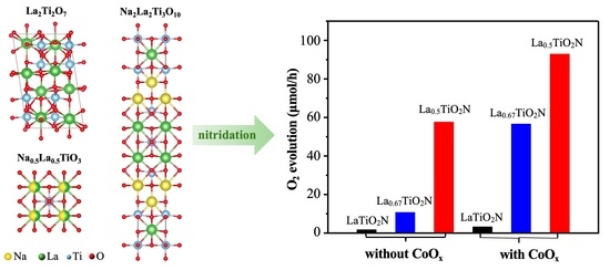
2N, respectively. Therefore, this method enables the precise and controllable synthesis of A-site deficient perovskites by in situ introducing A-site defects during conventional perovskite synthesis, without additional post-treatment or harsh chemical etching. Photocatalytic water-splitting experiments reveal that the reaction activities of those perovskite oxynitrides increase with the proportion of A-site defects, following the order: LaTiO
2N < La
0.67TiO
2N < La
0.5TiO
2N, achieving a maximum performance improvement of over 30 times. The performance boost is attributed to the lowered interfacial electron transfer barrier and the promoted charge separation caused by abundant A-site defects. These results confirm that constructing A-site deficient perovskites via alkali metal substitution and evaporation is an effective strategy, and that the resulting A-site defects can significantly enhance the photocatalytic activity of the perovskites. Moreover, this strategy can be extended to other perovskite materials, providing valuable insights for the rational design of A-site deficient perovskite catalysts for photocatalytic applications.
2. Results and Discussion
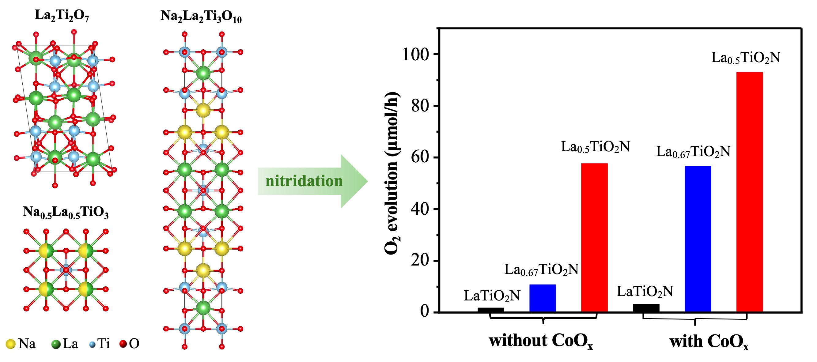
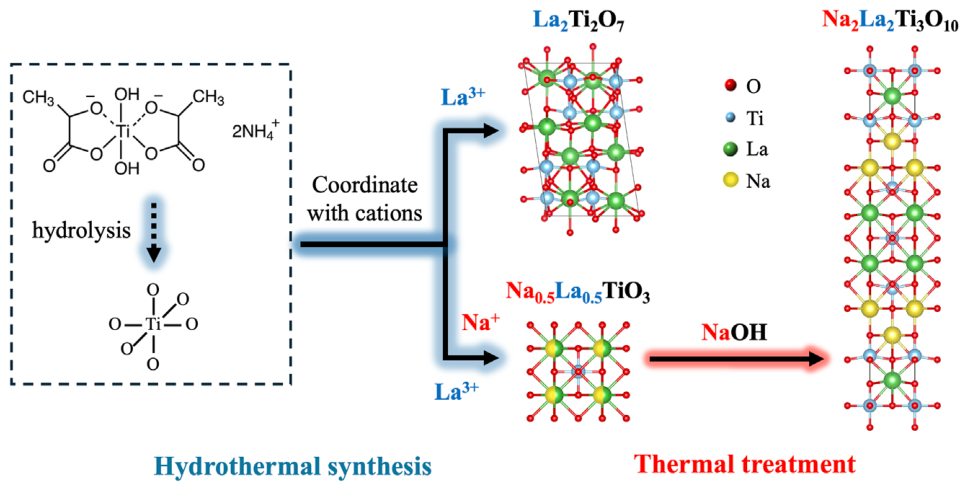
Figure 1 illustrates the synthesis methodology for several perovskite oxides, including La
2Ti
2O
7, Na
2La
2Ti
3O
10, and Na
0.5La
0.5TiO
3. Firstly, La
2Ti
2O
7 and Na
0.5La
0.5TiO
3 were synthesized using a hydrothermal route. A solution containing titanium(IV) bis(ammonium lactato) dihydroxide (TALH), La(NO
3)
3·6H
2O, and NaOH was homogeneously mixed and transferred into a stainless-steel autoclave for subsequent hydrothermal reactions. By tuning the hydrothermal synthesis conditions, the hydrolysis product (TiO
6 octahedra) derived from TALH could be engineered to coordinate with varying cations. For instance, the coordination of TiO
6 solely with La
3+ generated La
2Ti
2O
7, whereas its simultaneous coordination with both La
3+ and Na
+ produced Na
0.5La
0.5TiO
3. Subsequently, Na
2La
2Ti
3O
10 was obtained by mixing Na
0.5La
0.5TiO
3 with NaOH, followed by thermal treatment of the resultant mixture. During the treatment process, NaOH selectively extracted partial Ti-O layers from Na
0.5La
0.5TiO
3, leading to the formation of Na
2La
2Ti
3O
10.
As shown in
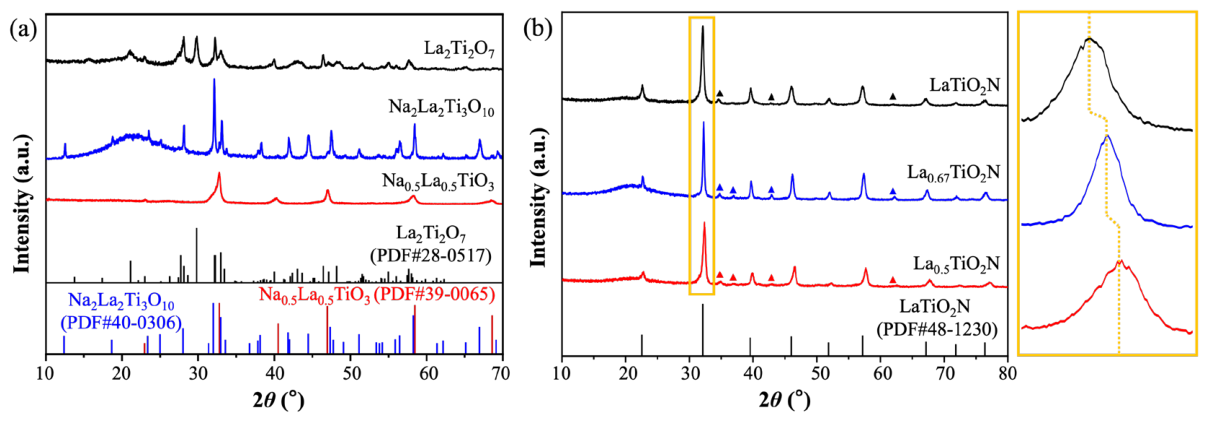
Figure 2a, the X-ray diffraction (XRD) patterns of the obtained perovskite oxides matched well with those of La
2Ti
2O
7 (PDF card No. 28-0517), Na
2La
2Ti
3O
10 (PDF card No. 40-0306), and Na
0.5La
0.5TiO
3 (PDF card No. 39-0065). No detectable impurities were found in any of the samples. Inductively coupled plasma optical emission spectrometry (ICP-OES) analyses were employed to determine the elemental ratios of Na, La, and Ti in the as-prepared samples (
Table 1). The results indicated that the Na:Ti ratios of La
2Ti
2O
7, Na
2La
2Ti
3O
10, and Na
0.5La
0.5TiO
3 were 0.01:1, 0.70:1, and 0.50:1, respectively, which were close to their nominal stoichiometric values of 0:1, 0.67:1, and 0.5:1. In contrast, the measured La:Ti ratios of these oxides were 1.50:1, 0.75:1, and 0.56:1, obviously higher than the ideal stoichiometric ratios (1:1, 0.67:1, and 0.5:1). Since no La-rich impurities were detected via XRD analysis, such elemental ratio deviations from theoretical stoichiometry might result from elemental interferences in this multielemental system [
26]. These experimental results supported the feasibility of the synthetic methodology depicted in
Figure 1, which enabled the controllable preparation of several perovskite oxides with varying Na contents without impurity formation.
Figure 1.
The synthesis strategies of perovskite oxides La
2Ti
2O
7, Na
2La
2Ti
3O
10, and Na
0.5La
0.5TiO
3. During the hydrothermal process, TALH underwent hydrolysis to form TiO
6 octahedra at high temperatures, while La
3+ ions incorporated into the Ti-O structure, leading to the formation of La
2Ti
2O
7. When the molar ratio of La(NO
3)
3·6H
2O to NaOH decreased to a certain threshold (e.g., 1:19), excess Na
+ ions could also coordinate with the TiO
6 octahedra. As a result, both Na
+ and La
3+ ions were incorporated into the Ti-O framework, leading to the formation of Na
0.5La
0.5TiO
3 [
27]. After further treatment of Na
0.5La
0.5TiO
3 with NaOH, partial Ti-O layers were extracted, resulting in the formation of Na
2La
2Ti
3O
10.
Figure 1.
The synthesis strategies of perovskite oxides La
2Ti
2O
7, Na
2La
2Ti
3O
10, and Na
0.5La
0.5TiO
3. During the hydrothermal process, TALH underwent hydrolysis to form TiO
6 octahedra at high temperatures, while La
3+ ions incorporated into the Ti-O structure, leading to the formation of La
2Ti
2O
7. When the molar ratio of La(NO
3)
3·6H
2O to NaOH decreased to a certain threshold (e.g., 1:19), excess Na
+ ions could also coordinate with the TiO
6 octahedra. As a result, both Na
+ and La
3+ ions were incorporated into the Ti-O framework, leading to the formation of Na
0.5La
0.5TiO
3 [
27]. After further treatment of Na
0.5La
0.5TiO
3 with NaOH, partial Ti-O layers were extracted, resulting in the formation of Na
2La
2Ti
3O
10.
Figure 2.
Crystal structure of the perovskite oxide precursors and oxynitrides. (a) XRD patterns and corresponding standard Powder Diffraction File (PDF) cards of La2Ti2O7, Na2La2Ti3O10, and Na0.5La0.5TiO3, where the PDF prefix was separated from the card number by the symbol #. (b) XRD patterns and the enlarged view (the yellow box in the right panel; the dashed lines inside the box marked the deviation of diffraction peak positions) of LaTiO2N, La0.67TiO2N, and La0.5TiO2N (the triangular markers belonged to the titanium nitride impurity phase, and the possible standard PDF cards were No. 87-0630 and No. 41-1352).
Figure 2.
Crystal structure of the perovskite oxide precursors and oxynitrides. (a) XRD patterns and corresponding standard Powder Diffraction File (PDF) cards of La2Ti2O7, Na2La2Ti3O10, and Na0.5La0.5TiO3, where the PDF prefix was separated from the card number by the symbol #. (b) XRD patterns and the enlarged view (the yellow box in the right panel; the dashed lines inside the box marked the deviation of diffraction peak positions) of LaTiO2N, La0.67TiO2N, and La0.5TiO2N (the triangular markers belonged to the titanium nitride impurity phase, and the possible standard PDF cards were No. 87-0630 and No. 41-1352).
Subsequently, the perovskite oxide precursors were converted into perovskite oxynitrides through high temperature ammonolysis. The XRD patterns of all the nitrided products closely matched that of orthorhombic LaTiO
2N (PDF card No. 48-1230), although a small amount of titanium nitride was observed in all cases (
Figure 2b). The relative proportion of each crystalline phase was determined by semi-quantitative analysis through integrating XRD diffraction peak areas and calculating their ratios. The phase fractions of titanium nitride in the nitrided products derived from La
2Ti
2O
7, Na
2La
2Ti
3O
10, and Na
0.5La
0.5TiO
3 were 2.85%, 4.71%, and 3.73%, respectively. These results indicated that all the precursors were converted into the target perovskite oxynitrides (LaTiO
2N or a similar structure) through high temperature nitridation. According to the ICP analysis (
Table 1), Na:La:Ti ratios of the nitrided products obtained from La
2Ti
2O
7, Na
2La
2Ti
3O
10, and Na
0.5La
0.5TiO
3 were 0.09:1.59:1, 0.07:0.67:1, and 0.01:0.55:1, respectively, which differed from those of the original precursors (0.01:1.50:1, 0.70:0.75:1 and 0.50:0.56:1). This indicated that during the high-temperature nitridation process, nearly all the Na in the Na-containing samples volatilized, yielding structures with La:Ti ratios close to those of the perovskite oxide precursors. Therefore, the La:Ti ratios of these perovskite oxynitrides should be 1:1, 0.67:1, and 0.5:1, which were consistent with those of the three precursors (La
2Ti
2O
7, Na
2La
2Ti
3O
10, and Na
0.5La
0.5TiO
3). Accordingly, the obtained perovskite oxynitrides were hereafter denoted as LaTiO
2N, La
0.67TiO
2N, and La
0.5TiO
2N, respectively.
As presented in
Figure 2b, two significant findings were identified by analyzing the XRD results. On the one hand, compared to LaTiO
2N, the peak positions of La
0.67TiO
2N and La
0.5TiO
2N shifted to higher angles, indicating significant lattice contraction in these samples, which was attributed to the presence of abundant A-site defects. The degree of peak position shift, or lattice contraction, was positively correlated with the number of A-site defects in the lattice, following the order: LaTiO
2N < La
0.67TiO
2N < La
0.5TiO
2N. This result was consistent with the observations obtained from transmission electron microscopy (TEM) characterization (
Figures S1–S3). High-resolution TEM (HRTEM) images of all three samples displayed clear and regular lattice fringes. Notably, La
0.67TiO
2N and La
0.5TiO
2N exhibited smaller interplanar distances of 0.275 nm and 0.274 nm, respectively, in comparison with the value of 0.277 nm for LaTiO
2N. All these lattice spacings corresponded to the (112) crystal plane of orthorhombic LaTiO
2N. On the other hand, the XRD peak intensities of La
0.5TiO
2N were markedly reduced in comparison with LaTiO
2N, indicating that abundant A-site defects led to decreased crystallinity. This conclusion was further supported by the Raman spectrum, in which the vibration peaks of La
0.5TiO
2N broadened and attenuated obviously due to the reduced crystallinity caused by abundant A-site defects (
Figure S4). In contrast, the XRD peak intensity and Raman vibration peak intensity of La
0.67TiO
2N were comparable to those of LaTiO
2N. This phenomenon could be ascribed to the additional high-temperature treatment employed during the synthesis of the precursor, Na
2La
2Ti
3O
10, thereby effectively improving the crystalline degree of the nitrided product, La
0.67TiO
2N. These experimental results confirmed that the highly A-site deficient perovskite oxynitrides (La
0.67TiO
2N and La
0.5TiO
2N) were successfully synthesized through high-temperature nitridation of Na-containing precursors (Na
2La
2Ti
3O
10 and Na
0.5La
0.5TiO
3). The A-site defects formed by Na evaporation during the high-temperature nitridation process did not alter the crystal structure of perovskite oxynitrides but induced significant lattice compression.
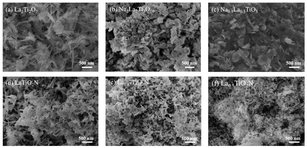
Morphological analysis was conducted by scanning electron microscopy (SEM) images. As displayed in
Figure 3a–c, all three perovskite oxides exhibited a nanosheet structure. Owing to the high specific surface area, the nanosheets in all samples tended to aggregate. After high temperature nitridation, the resultant perovskite oxynitrides retained the nanoscale structure closely resembling that of their perovskite oxide precursors. This phenomenon suggested that the nitridation treatment did not significantly alter the original nanostructure (
Figure 3d–f). One significant difference was that the perovskite oxynitrides exhibited a more porous structure, which might originate from nitrogen insertion, lattice distortion, defect formation, and gas-induced pore generation during the high-temperature nitridation process [
28,
29]. Furthermore, La
0.5TiO
2N was composed of numerous tiny and ultrathin nanosheets. Similarly, LaTiO
2N exhibited an ultrathin nanosheet structure but possessed larger lateral dimensions and an elongated lamellar morphology. In contrast, La
0.67TiO
2N tended to form thick granular aggregates (
Figure S5).
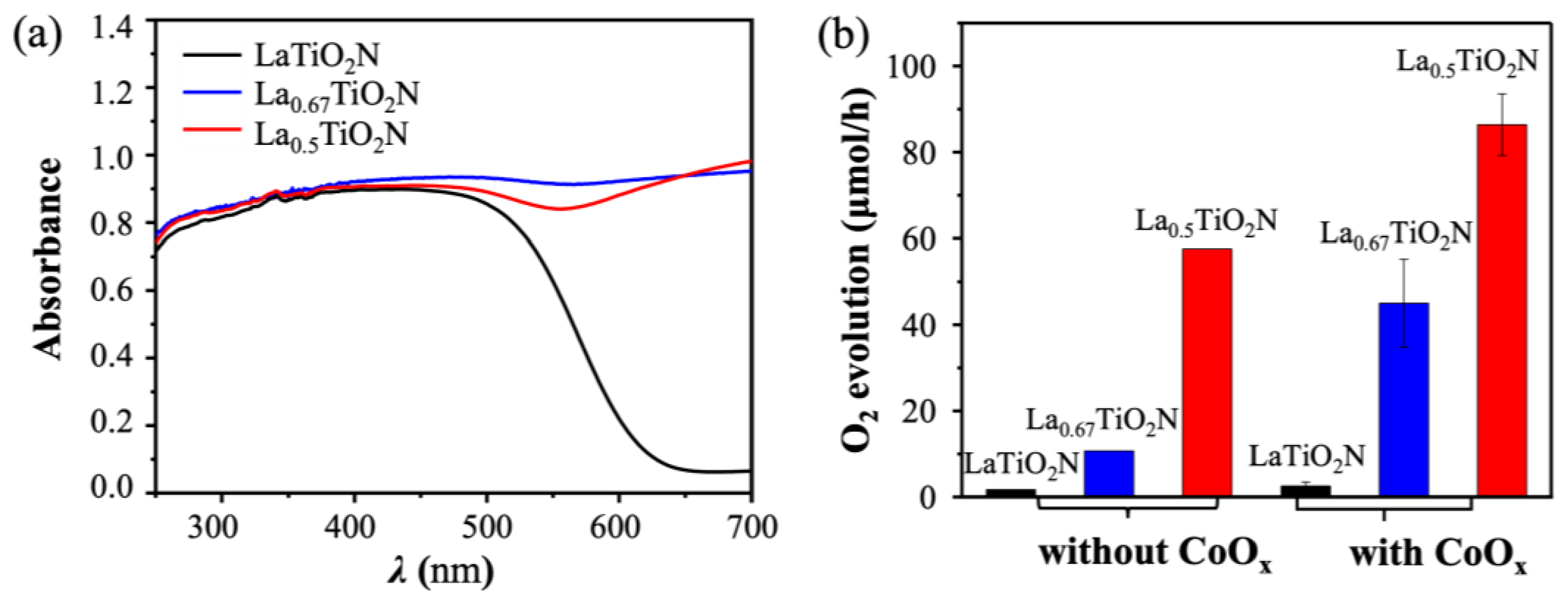
The optical absorption properties of these perovskite oxynitrides were investigated using UV-Vis diffuse reflectance spectroscopy (DRS), as presented in
Figure 4a. The results showed that after nitridation, the absorption range of the obtained perovskite oxynitrides were extended to around 600 nm, corresponding to a bandgap of 2.1 eV. This indicated that nitridation treatment effectively narrowed the bandgap, endowing these perovskites with visible-light responsiveness and thereby making them more suitable for subsequent photocatalytic reactions. Additionally, abundant A-site defects generated in La
0.67TiO
2N and La
0.5TiO
2N caused a characteristic tail-like absorption above 600 nm, resulting in a sample color change from bright orange to black. As shown in
Figure 4b, the photocatalytic water oxidation performance of these perovskite oxynitrides was assessed under visible light irradiation (λ > 420 nm), with silver nitrate employed as the sacrificial electron acceptor. The O
2 production rates for La
0.67TiO
2N and La
0.5TiO
2N were 10.7 μmol/h and 57.6 μmol/h, respectively, significantly higher than the 1.7 μmol/h observed for LaTiO
2N. After loading CoO
x as an O
2 evolution cocatalyst, the photocatalytic O
2 production activities of La
0.67TiO
2N and La
0.5TiO
2N were greatly enhanced to 45 μmol/h and 86.6 μmol/h, respectively, still markedly higher than the 2.55 μmol/h observed for LaTiO
2N. Further detailed data for the replicated samples were provided in
Table S1 and presented as the mean ± standard deviation in the figure. The discrepancies in sample performance might arise from several experimental variables, including batch-to-batch sample variation, distance between light source and sample, light spot position, optical power fluctuation, and stirring conditions during measurement. In addition,
Table S2 compares the A-site deficient perovskite oxynitrides with previously reported LaTiO
2N-based photocatalysts, demonstrating that A-site defect engineering delivered remarkable advantages over conventional A-site or B-site substitution strategies. These findings indicated that the presence of abundant A-site defects significantly enhanced the photocatalytic water oxidation activity of LaTiO
2N, with the activity improving as the concentration of A-site defects increased, following the order: LaTiO
2N < La
0.67TiO
2N < La
0.5TiO
2N.
The apparent quantum efficiency (AQE) of the most performant La
0.5TiO
2N was measured to investigate the wavelength dependence of its photocatalytic performance (
Table S3). The AQE exhibited a strong correlation with the incident photon wavelength, achieving a maximum value of 10.56% at 420 nm.
Figure S6 shows the time course of water oxidation reaction of La
0.5TiO
2N using AgNO
3 as the electron sacrificial agent and La
2O
3 as the pH buffer. Steady oxygen evolution was achieved over La
0.5TiO
2N under visible light irradiation. During the photocatalytic reaction, no obvious N
2 production was observed. After stability test, the crystal structure of La
0.5TiO
2N was well maintained (
Figure S7). Meanwhile, metallic Ag and La(OH)
3 coexisted on the catalyst surface. The formation of metallic Ag originated from the photoreduction of Ag
+ sacrificial reagent, while the generation of La(OH)
3 was ascribed to the spontaneous hydration of the La
2O
3 buffer species under aqueous reaction conditions. X-ray photoelectron spectroscopy (XPS) was further employed to analyze the variations in elemental chemical states of La
0.5TiO
2N before and after the stability test (
Figure S8). One notable change was observed in the O 1s spectra: the proportion of lattice oxygen (O
2− at 530.1 eV) decreased distinctly, while the content of oxygen vacancies (O
22−/O
− at 529.1 eV) increased significantly. This was attributed to the oxidation of lattice O
2− by photogenerated holes, demonstrating that lattice oxygen might serve as an active participant during water oxidation on La
0.5TiO
2N. The detailed mechanism had been discussed in our recent work.
The superior photocatalytic performance of La
0.5TiO
2N over LaTiO
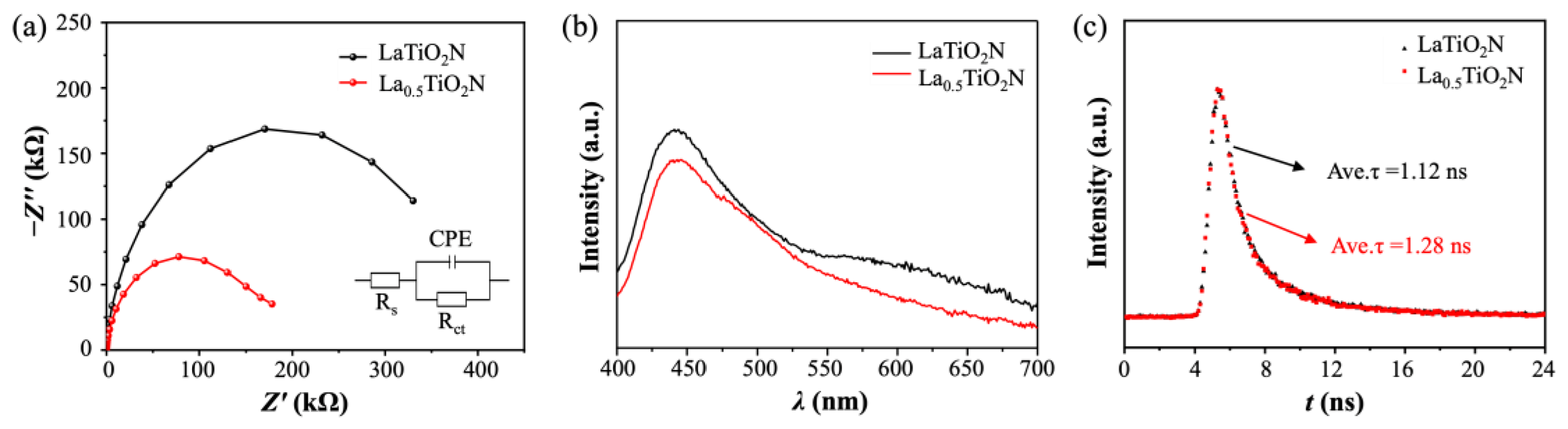
2N was clarified via electrochemical and photoluminescence analyses. As shown in
Figure 5a, electrochemical impedance spectroscopy (EIS) measurements were conducted under dark conditions, and the recorded Nyquist plots were fitted using the equivalent circuit presented in the figure. The charge-transfer resistance of LaTiO
2N and La
0.5TiO
2N were 101.3 kΩ/cm
2 and 46.34 kΩ/cm
2, respectively. In the absence of photogenerated electrons and holes, the impedance spectra were mainly governed by the transport behavior of majority charge carriers (electrons for n-type semiconductors) across the depletion regions of the semiconductor electrodes [
30]. Thus, the electron transport resistance in the depletion region of La
0.5TiO
2N was lower, indicating a smaller Schottky barrier for electron transfer at the semiconductor-electrolyte interface. The accelerated electron transfer behavior could be attributed to the strengthened B-O covalency induced by abundant A-site defects. This mechanism had been validated in previous studies of typical A-site deficient perovskite oxides, including La
1−xNiO
3, La
1−xFeO
3, La
1−xCoO
3, and La
1−xMnO
3 [
11,
15,
16,
19]. The relevant detailed mechanism had also been systematically elaborated in our recent work. Steady-state photoluminescence (PL) and time-resolved PL decay (TRPL) spectra were collected to elucidate the recombination behavior of photogenerated charge carriers (
Figure 5b). Compared with LaTiO
2N, La
0.5TiO
2N exhibited a reduced PL emission intensity. This result confirmed that A-site defects could effectively accelerate charge separation and suppress charge recombination. Accordingly, the average charge carrier lifetime of La
0.5TiO
2N was longer than that of LaTiO
2N (
Figure 5c). These findings indicated the superior photocatalytic activity of La
0.5TiO
2N originated from accelerated interfacial charge transfer and improved charge separation caused by abundant A-site defects.
3. Materials and Methods
3.1. Materials
Lanthanum(III) nitrate hexahydrate (La(NO3)3·6H2O, Macklin, 99.0%), Titanium(IV) bis(ammonium lactato) dihydroxide (TALH) (C6H18N2O8Ti, Aladdin, 50 wt% in water), La2O3 (Aladdin, 99.9%), Sodium hydroxide (NaOH, 99.9%), 5,5-Dimethyl-1-pyrroline N-oxide (DMPO) (C6H11NO, Aladdin, 97%), Silver nitrate (AgNO3, 99.7%) and Cobalt nitrate hexahydrate (Co(NO3)2·6H2O, 99.8%) were purchased from Sinopharm Chemical Reagent Co., Ltd., Shanghai, China. All chemicals were used as received without further purification.
3.2. Synthesis
Na0.5La0.5TiO3 was synthesized using a polymerized-complex (PC) method. The TALH solution was first diluted with deionized water to a concentration of 0.05 mol/L. Then, 1.8 mmol of La(NO3)3·6H2O was added to 48 mL of the diluted TALH solution and ultrasonically mixed until homogeneous. Subsequently, an aqueous solution of NaOH (7 mL, 5 mol/L) was added to the mixture. After stirring for 30 min, the solution was transferred into a 100 mL polytetrafluoroethylene (PTFE) vessel. The vessel was sealed and placed in a stainless-steel autoclave, which was heated in an oven to 200 °C at a rate of 5 °C/min and maintained at this temperature for 24 h. The resulting products were collected by centrifugation at 10,000 rpm for 10 min, washed with ethanol, and dried in an oven at 60 °C. La2Ti2O7 was also prepared by the PC method, with the only difference being that the amount of La(NO3)3·6H2O was increased to 4.8 mmol. Na2La2Ti3O10 was synthesized from Na0.5La0.5TiO3 using a high-temperature treatment method. In details, NaOH and Na0.5La0.5TiO3 powders were mixed in an agate mortar at a molar ratio of 2:1, and the mixture was ground for approximately 5 min in a N2-filled glovebox. The mixture was subsequently transferred into an alumina boat, placed in a furnace, heated to 950 °C at a rate of 10 °C/min, and maintained at this temperature for 2 h. Na0.5La0.5TiO3, Na2La2Ti3O10, and La2Ti2O7 precursors were subsequently nitrided in a high-temperature ammonia atmosphere (950 °C, 100 mL/min NH3 flow) for 5 h to obtain La0.5TiO2N, La0.67TiO2N, and LaTiO2N, respectively.
3.3. Characterization
Powder X-ray diffraction (XRD) patterns were recorded on a Bruker D8 Advance diffractometer (Bruker AXS SE, Karlsruhe, Germany) using Cu-Kα1 radiation (1.5406 Å). Raman spectra were collected using a HORIBA LabRAM HR Raman spectrometer (HORIBA Scientific, Palaiseau, France). X-ray photoelectron spectroscopy (XPS) measurements were performed on a PHI Quantum 2000 spectrometer (Physical Electronics, Chanhassen, MN, USA). UV-vis diffuse reflectance spectra (DRS) were recorded with a Varian Cary 500 Scan UV-visible spectrophotometer (Varian, Inc., Palo Alto, CA, USA). Inductively coupled plasma optical emission spectrometry (ICP-OES) analyses were carried out using a Perkin Elmer Avio 200 instrument (PerkinElmer, Inc., Waltham, MA, USA). Electrochemical impedance spectroscopy (EIS) analysis was conducted in a conventional three-electrode cell, with a Pt plate as the counter electrode and Ag/AgCl as the reference electrode, using a Bio-Logic VSP-300 electrochemical workstation (Bio-Logic Science Instruments SAS, Seyssinet-Pariset, France). Scanning electron microscopy (SEM) images were obtained with a Hitachi New Generation SU8010 field emission SEM (Hitachi High-Technologies, Tokyo, Japan). Transmission electron microscopy (TEM) images were acquired using a Thermo Fisher Scientific Talos F200S TEM (Thermo Fisher Scientific, Waltham, MA, USA). The probe current for energy-dispersive X-ray spectroscopy (EDS) mapping was set at 120 pA to generate sufficient X-ray signals. Bruker Dimension Icon atomic force microscopy (AFM, Bruker AXS SE, Karlsruhe, Germany) was utilized to observe the nanostructure dimensions. Photoluminescence (PL) spectra and time-resolved PL (TRPL) decay were recorded on an Edinburgh FLS980 spectrometer (Edinburgh Instruments Ltd., Livingston, West Lothian, UK) with an excitation wavelength of 380 nm. The PL decay curves were fitted with a tri-exponential model, and the mean decay lifetimes were calculated by the weighted average method.
3.4. Photocatalytic Performance Test
The photocatalytic water oxidation was performed in a Pyrex top-irradiation reaction vessel connected to a closed glass gas circulation system. 50 mg of La
0.5TiO
2N, La
0.67TiO
2N, or LaTiO
2N catalysts were dispersed in 150 mL of aqueous solution containing 20 mM of AgNO
3 as the electron sacrificial agent and 200 mg of La
2O
3 as the pH buffer agent. The suspension was then irradiated by a xenon lamp (300 W, full arc, LX300F, Shimadzu Co., Kyoto, Japan) equipped with a cutoff filter (λ > 420 nm) under vacuum conditions. The evolved gases were analyzed online by gas chromatography (GC-8A, Shimadzu Co., Kyoto, Japan), equipped with a thermal conductivity detector and 5 Å molecular sieve tubing, using argon as the carrier gas. During the reaction, the temperature of the reaction solution was maintained at room temperature by a continuous flow of cooling water. The CoO
x cocatalyst was deposited on the surface of photocatalyst using an impregnation method. Firstly, 50 mg of photocatalyst powder was dispersed in 20 mL of deionized water. Subsequently, an aqueous CoCl
2 solution containing 2 wt% Co was added to the above suspension. The resulting mixture was then stirred and heated in a water bath at 80 °C until the solvent was completely evaporated. The apparent quantum efficiency (AQE) for photocatalytic O
2 evolution was measured under monochromatic LED irradiation at wavelengths of 420, 450, 500, 550, and 600 nm, over an irradiation area of 4 cm
2. The amounts of gases produced during the photocatalytic reaction was measured by GC, and the AQE was then calculated using the following formula:
where
M is the amount of O
2 molecules (mol),
NA is the Avogadro constant (6.022 × 10
23/mol),
h is the Planck constant (6.626 × 10
−34 J·s),
c is the speed of light (3 × 10
8 m/s),
S is the irradiation area (cm
2),
P is the intensity of irradiation light (W/cm
2),
t is the photoreaction time (s),
λ is the wavelength of the monochromatic light (m).