Article

Discovery and Characterization of Novel 2-Phosphoglycerate Kinase and Cyclic 2,3-Diphosphoglycerate Synthase from Thermophilic (Meta)Genomes


1
Istituto di Scienze e Tecnologie Chimiche “G. Natta” (SCITEC), CNR, 20131 Milano, Italy
2
Department of Pharmaceutical Sciences, University of Milan, 20133 Milano, Italy
3
Henry Wellcome Building for Biocatalysis, Biosciences, Faculty of Health and Life Sciences, University of Exeter, Exeter EX4 4QD, UK
4
Institute of Environmental Sciences, Hebrew University of Jerusalem, Rehovot 7610001, Israel
5
Molecular Enzyme Technology and Biochemistry (MEB), Environmental Microbiology and Biotechnology (EMB), Centre for Water and Environmental Research (CWE), Faculty of Chemistry, University of Duisburg-Essen, 45141 Essen, Germany
*
Authors to whom correspondence should be addressed.
These authors contributed equally to this work.
Catalysts 2026, 16(4), 305; https://doi.org/10.3390/catal16040305
Submission received: 15 December 2025 / Revised: 20 March 2026 / Accepted: 20 March 2026 / Published: 1 April 2026
(This article belongs to the Special Issue Catalysis and Sustainable Green Chemistry)

Abstract

2-Phosphoglycerate kinase (2PGK) and cyclic 2,3-diphosphoglycerate synthase (cDPGS) are key enzymes involved in the biosynthesis of cyclic 2,3-diphosphoglycerate (cDPG), an extremolyte known to stabilize proteins in hyperthermophilic Archaea. Using bioinformatics approaches, two candidate genes for each enzyme were identified from a range of thermophilic bacterial and archaeal genomes and metagenomes. Significantly, one gene pair derived from the Taman mud volcano metagenome represents the first indication of a bacterial cDPG biosynthesis pathway. The recombinant expression and purification of these enzymes paved the way to their biochemical and structural characterization. One 2PGK candidate displayed predominant ATPase activity, while the newly identified cDPGS variants demonstrated cDPG synthase activity. Moreover, one of the latter biocatalysts, Ts-cDPGS from the hyperthermophilic archaeon Thermococcus sibiricus, demonstrated a notable thermostability and its 3D structure was resolved at a resolution of 2.2 Å. These findings broaden our understanding of extremophilic enzyme systems and lay the foundation for biotechnological applications involving extremolyte production.

1. Introduction

Osmolytes, or compatible solutes, are small organic molecules, including amino acid derivatives, sugars and polyols, that accumulate within cells to concentrations of up to 2 M in response to various stress conditions. They function by stabilizing and protecting nucleic acids, proteins, and cellular structures from the detrimental effects of extreme temperatures, dehydration, and osmotic stress [1,2].
Extremophilic organisms, which inhabit environments characterized by extreme physicochemical conditions, such as elevated hydrostatic pressure, extreme pH values, high salinity, or thermal extremes, synthesize a large variety of osmolytes including specific compounds termed extremolytes. These compounds have numerous biotechnological and industrial uses, including serving as stabilizing additives in the formulation of enzymes, drugs, and antibodies [3]. Additionally, some extremolytes, such as ectoine and hydroxyectoine, which act as chemical chaperones due to their strong protein-stabilizing properties, are produced at an industrial scale for use in skincare and medical applications [4].
The intracellular accumulation (up to 1.1 M) of the phosphorylated extremolyte cyclic 2,3-diphosphoglycerate (cDPG, Scheme 1) has been so far reported only in hyperthermophilic archaeal methanogens such as Methanothermus fervidus, Methanopyrus kandleri and Methanothermobacter thermoautotrophicus [5,6,7]. Its primary role is to stabilize proteins and enzymes by protecting them from thermal denaturation at temperatures often exceeding 80 °C. Additionally, cDPG may also play a role in maintaining intracellular osmotic balance and serving as a reservoir for phosphate and metabolic energy [8,9,10].
Concerning the synthesis of this extremolyte, two ATP-dependent key enzymes are involved in the biosynthetic pathway of cDPG in M. fervidus [11]. First, 2-phosphoglycerate (2-PG, Scheme 1) is phosphorylated to form 2,3-diphosphoglycerate (2,3-DPG) through the action of 2-phosphoglycerate kinase (2PGK). Then, the 2,3-DPG intermediate undergoes an intramolecular cyclization reaction, catalyzed by cyclic 2,3-diphosphoglycerate synthetase (cDPGS), resulting in the formation of cDPG.
Both M. fervidus enzymes (Mf-2PGK and Mf-cDPGS) were first isolated from its original producer and their molecular and catalytic properties analyzed [11]. Subsequently, the cloning of the corresponding genes and their recombinant expression in E. coli was reported, allowing some preliminary characterization [5,12].
Recently, we have improved the heterologous production of Mf-cDPGS in E. coli by codon optimization of its coding sequence along with a refinement of expression, isolation and storage conditions, thus leading to the establishment of an efficient enzymatic synthesis of cDPG from 2,3-DPG [13]. In addition, a new thermophilic host system was developed to produce cDPG in vivo with the bacterium Thermus thermophilus using the Mf-2PGK and Mf-cDPGS codon optimized genes within a modular BioBricks vector. Successful production of cDPG in this thermophilic host system was confirmed through mass spectrometry [14]. Moreover, the 3D structure of Mf-cDPGS has been recently solved at a high resolution both in its ligand-free form and in the ADP/Mg2+/2,3-DPG complex (1.7 Å and 2.2 Å, respectively) [15]. The Mf-cDPGS is highly thermostable with a melting temperature (Tm) above 95 °C. The structure shows a unique N-terminal domain (127 amino acids) with no known structural homologues, while the C-terminal domain resembles a P-loop kinase fold. The structural insights gained in this study also enabled the proposal of a detailed reaction mechanism, which may facilitate future biotechnological applications of cDPGS.
In this research, Mf-2PGK and Mf-cDPGS were employed as query sequences for in silico mining of (hyper)thermophilic Archaea and Bacteria (meta)genomic data. Using a combination of comparative amino acid sequence analysis and sequence-based phylogenetic reconstruction, novel candidates of 2PGKs and cDPGSs were identified. These putative enzymes were subsequently selected for expression trials and comprehensive biochemical characterization, encompassing both functional and structural aspects.

2. Results and Discussion

2.1. Identification of Putative 2PGKs and cDPGSs in Thermophilic (Meta)Genomes

To identify new 2PGKs and cDPGSs, in silico translated annotated genomes of eleven (hyper)thermophilic Archaea and thirteen thermophilic Bacteria (Supporting Information, Table S1), together with in silico translated annotated unbinned shotgun metagenomes of one terrestrial thermophilic hydrothermal spring located in Tyva (JGI GOLD Analysis Project ID Ga0395598 [16]) and one terrestrial mud volcano located in Taman (JGI GOLD Analysis Project ID Ga0369713 [17]), were screened using BLASTp (version 2.17.0). Biochemically characterized 2PGK from Methanothermus fervidus (Mf-2PGK, Uniprot accession: Q49156) and cDPGSs from M. fervidus (Mf-cDPGS, Uniprot accession: O93732) and Methanothermobacter thermautotrophicus (Uniprot accession: O26325) were used as the query sequences. Of the 24 genomes and two metagenomes, a total of 16 genes encoding for potential 2PGKs (one from the genomes set and five from the metagenomes set) and cDPGSs (seven from the genomes set and three from the metagenomes set) were identified. The corresponding protein sequences were then aligned using Mafft (version 7) (Supporting Information, Figure S2), and some of them were discarded as they turned out to be encoded by gene fragments. In addition, two potential 2PGK sequences were discarded as well because they had an unusually long N-terminal tail, while some potential cDPGS sequences were excluded because they had low alignment with the sequences of other predicted and biochemically characterized cDPGSs.
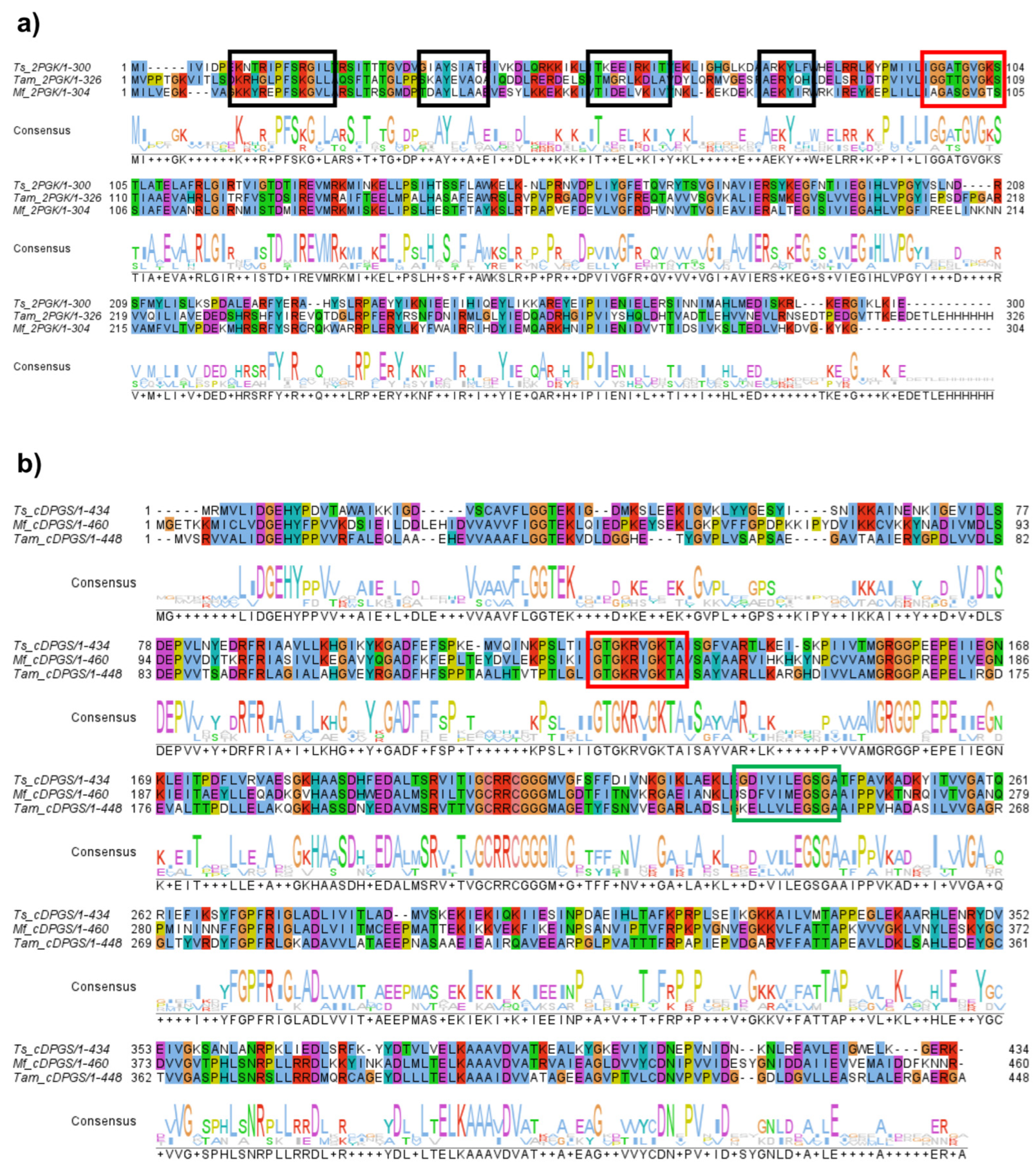
The remaining two potential 2PGK (Ga0369713_10007067, from this point forward named Tam-2PGK, and 644842203 TSIB_0159, i.e., Ts-2PGK) and two potential cDPGS (Ga0369713_10007068, i.e., Tam-cDPGS, and 644842204 TSIB_0160, i.e., Ts-cDPGS) sequences shared around 40% identity with Methanothermus fervidus 2PGK and cDPGS, respectively (Figure 1 and Table 1).
Both putative 2PGKs show the typical signatures of ATP-binding enzymes at the N-termini, such as conserved residues of the ATP-cone domain (Figure 1a, black boxes) [20], and of the phosphate binding loop (Walker A/P-loop, red box) [21]. The latter conserved region can be observed also in the putative cDPGS sequences (Figure 1b, red box), together with a Walker B motif downstream of the A-motif (green box). In silico predicted characteristics of the putative novel 2PGK and cDPGS sequences showed that they differ in hydrophobicity, solubility and isoelectric points (pI) (Table 1).
Table 1. Sequence analysis of identified putative 2PGKs and cDPGSs.
PropertiesLocus_Tag
Ga0369713_
10007067
644842203 TSIB_0159Ga0369713_
10007068
644842204 TSIB_0160
SourceTaman metagenomeT. sibiricus MM 739Taman metagenomeT. sibiricus MM 739
Putative activity2PGK2PGKcDPGScDPGS
Length (aa)317300447434
Mr (kDa)35.334.746.548
pI4.959.994.607.58
GRAVY 1−0.21−0.150.11−0.11
Solubility 10.730.260.320.51
IVYWREL 20.450.510.410.44
Swissprot best hit 3 (accession, % identity)M. fervidus DSM 2088 (Q49156, 41%)M. fervidus DSM 2088 (Q49156, 44%)M. fervidus DSM 2088 (O93732, 46%)M. fervidus DSM 2088 (O93732, 48%)
Uniprot best hit
(accession, % identity)
Clostridiales bacterium (A0A942JV59, 80%)Thermococcus sp. 40_45 (A0A101F0E8, 100%)Clostridiales bacterium (A0A942JV59, 78%)Thermococcus sp. 40_45 (A0A101F0E8, 100%)
1 Positive GRAVY score and Soluprot solubility prediction number < 0.5 points suggest poor protein solubility. 2 IVYWREL—the sum of the corresponding amino acids related to all amino acids in the protein or proteome—is a signature indicating that the protein or proteome belongs to a thermophilic microorganism [22]. According to [22], IVYWREL 0.41 corresponds to ~50–52 °C, 0.44 to 82 °C, 0.45 to ~100 °C, 0.51 to >110 °C. 3 Sequences with evidence at protein level.
Remarkably, one pair of these pgk2 and cdpgs genes comes from the genome of the hyperthermophilic archaeon Thermococcus sibiricus strain MM 739 [23,24], and another one from the Taman mud volcano metagenome [17]. More importantly, in both cases, the genes were co-located within their scaffolds (Supporting Information, Figure S3), and this gene context strongly supports the co-regulation of the genes and co-action of the respective proteins as parts of the same enzymatic pathway. These considerations suggested that both pairs of 2PGKs and cDPGSs are indeed parts of the cyclic 2,3-diphosphoglycerate (cDPG) synthesis pathway. Consequently, these candidates were selected for recombinant expression in E. coli.
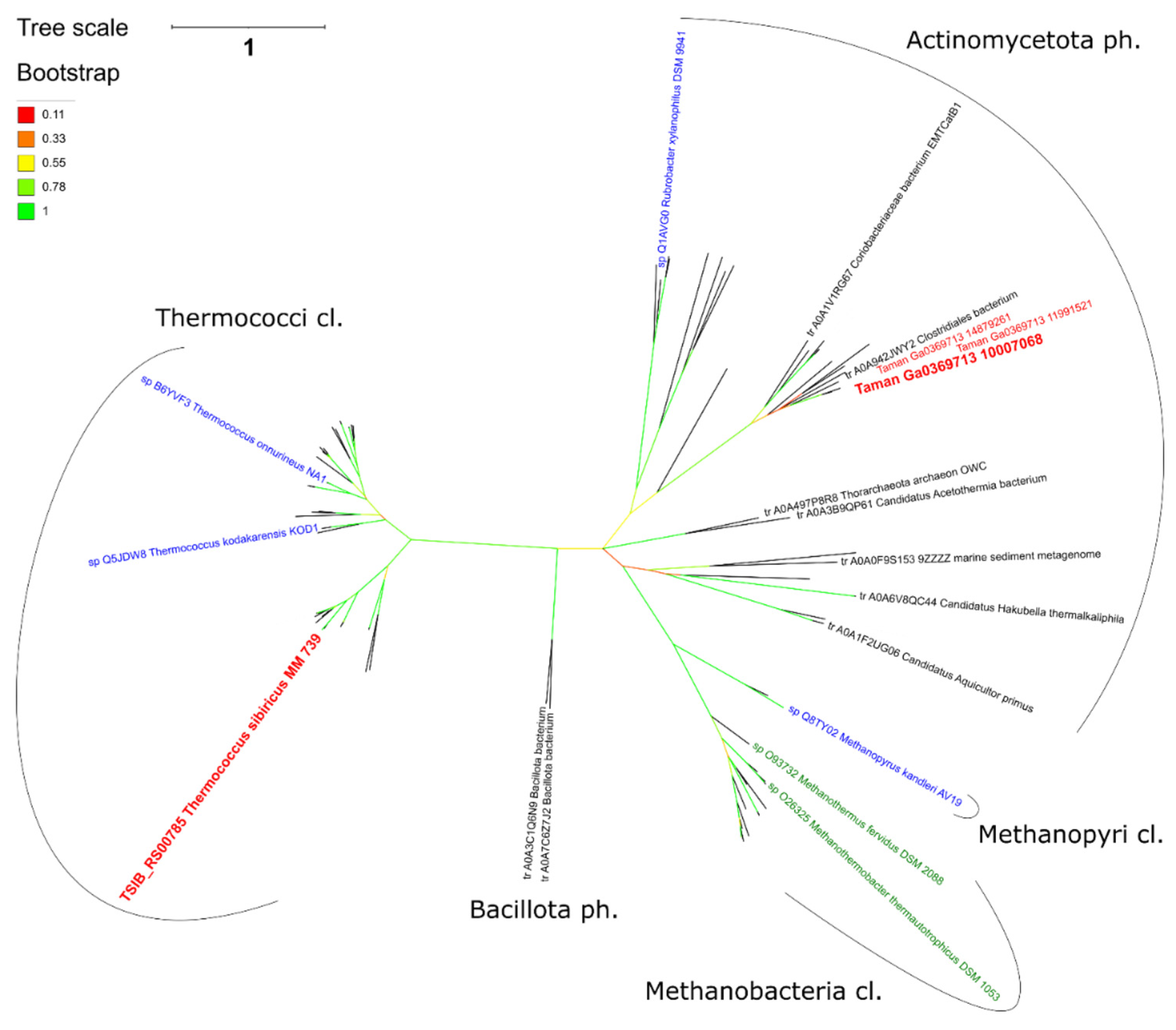
Proportion of IVYWREL amino acids [22] in the proteins suggests all of them came from (hyper)thermophilic microorganisms (Table 1), which is obvious in the case of the hyperthermophilic Thermococcus sibiricus, but not so evident for the proteins of a microorganism representing Taman microbiome, with the temperature at the sampling site of 20 °C [17]. The Taman unbinned metagenome scaffold containing pgk2 and cdpgs sequences was previously assigned to the metagenome assembled genome (MAG) of Coriobacteriia bacterium (Actinomycetota, genome assembly GenBank GCA_019429145.1) [17]. Since the nearest homologues of all 24 genes of the scaffold are from Coriobacteriia bacteria, no recent horizontal transfers occurred within this scaffold. At the same time, the nearest biochemically characterized relatives of Tam-2PGK and Tam-cDPGS encoded in this Coriobacteriia scaffold are archaeal proteins from M. fervidus (Q49156 and O93732 for 2PGK and cDPGS, respectively) and M. thermautotrophicus (O26325, cPDGS) which were used for initial search of these genes in the studied genomes and metagenomes. Surprisingly, both 2PGK and cDPGS sequences from a hyperthermophilic archaeon T. sibiricus and from a Coriobacteriia bacterium MAG were similarly identical (41–48% of sequence identity, Table 1) to the homologues from M. fervidus. However, phylogenetic analysis of cDPGS sequences (Figure 2) supports a rather distant relationship of Ts-cDPGS, Tam-cDPGS and Mf-cDPGS, with Ts-cDPGS falling into its own thermococci cluster, whereas Tam-cDPGS having a common root with methanoarchaeal cDPGS is located in its own actinomycete cluster. Given the sequence identity and phylogenetic analysis of cDPGSs, it could be assumed that the cDPG synthesis pathway was horizontally transferred to this Coriobacteriia bacterium or its ancestors from an unknown methanoarchaeon. However, this transfer was not recent and the fact that the horizontally transferred genes have not been removed by selection may suggest this pathway is operative in this bacterium. To the best of our knowledge, this is the first record of the occurrence of a cDPG synthesis pathway in Bacteria. At the same time, Ts-cDPGS is more phylogenetically distant to characterized Mf-cDPGS implying it might possess novel features which may expand our knowledge on the diversity of Archaea with respect to the specifics of extremolytes biosynthesis, as well as being relevant to biotechnology.

2.2. Protein Expression and Purification

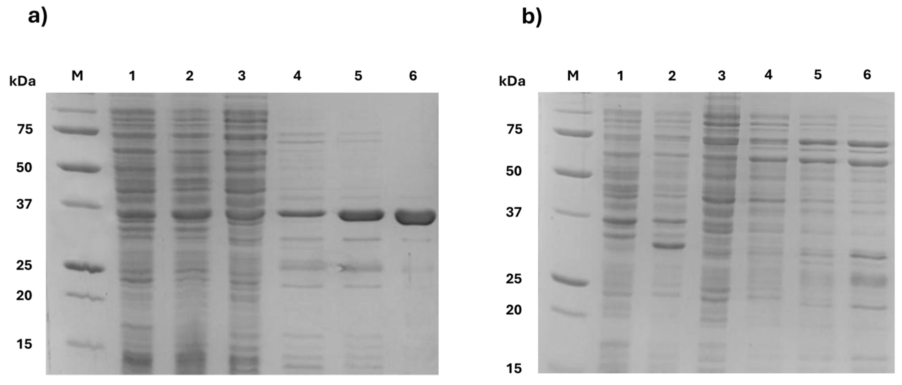
After codon optimization for recombinant expression in E. coli, the selected putative pgk2 and cdpgs genes were cloned into the expression vector pET28a in frame with a C-term His-tag, under control of an IPTG inducible promoter. Expression of putative 2PGKs was difficult as heterologous proteins formed inclusion bodies. First, expression trials were carried out by co-expressing the 2PGK genes with chaperones DnaJ/DnaK/GrpE and GroES/GroEL, but very low amounts of soluble protein were detected by SDS-PAGE (Supporting Information, Figure S1). In a second attempt, putative 2PGKs were expressed using an autoinducing medium (Figure 3) and this strategy resulted in successful production of potential Tam-2PGK with 36 mg of pure protein obtained from a 0.5 L culture. Unfortunately, again, almost no Ts-2PGK was detected by SDS-PAGE in the soluble fraction.
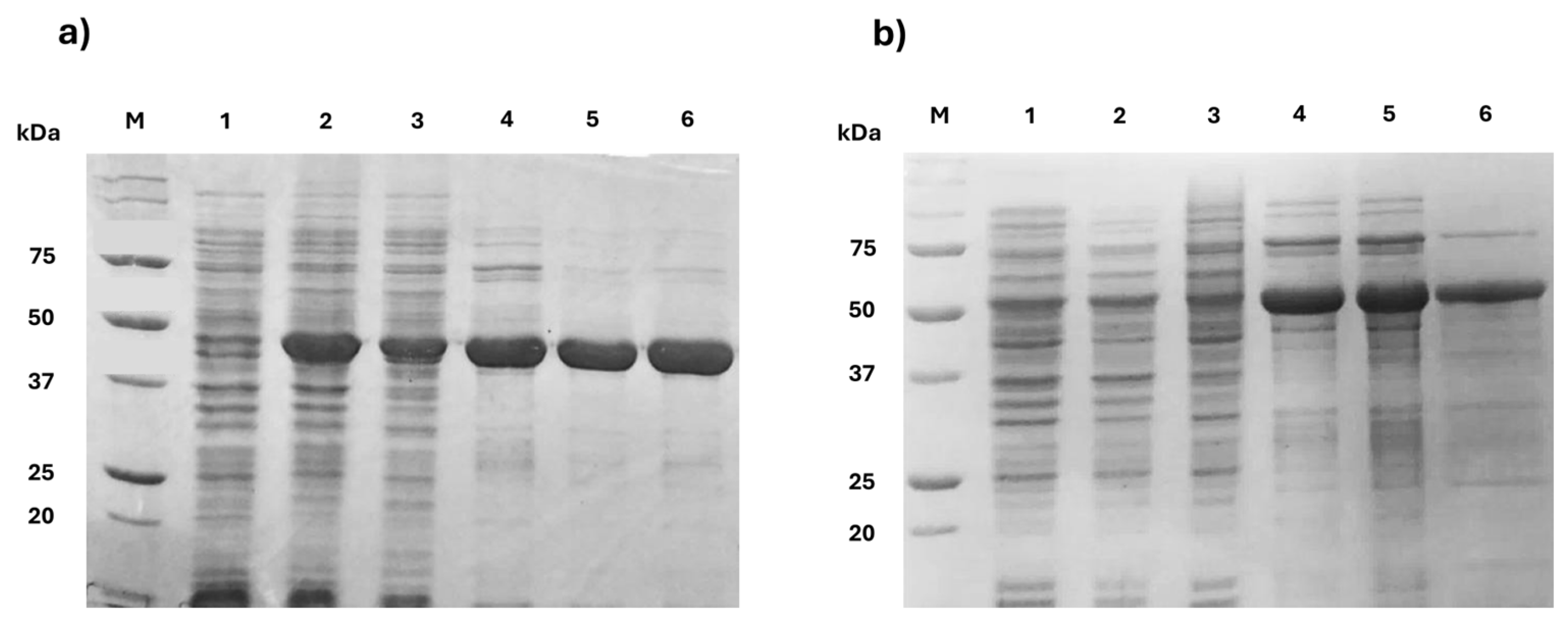
Concerning cDPGSs, the optimal expression conditions were obtained by incubating the cell cultures after IPTG induction for 72 h at 17 °C. 100 mg of Ts-cDPGS and 15 mg of Tam-cDPGS were obtained from 0.5 L culture, respectively (Figure 4). Notably, as observed with other cDPGS enzymes [9,11,13,15,25], replacing NaCl with KCl in the purification buffers and including KCl in the dialysis buffer in both cases effectively prevented protein precipitation.

2.3. Evaluation of Enzyme Activity and Synthesis of Cyclic 2,3-Diphosphoglycerate (cDPG)

The activity of the putative Tam-2PGK and both novel cDPGSs towards 2-phosphoglycerate (2-PG) and 2,3-diphosphoglycerate (2,3-DPG), respectively, was tested spectrophotometrically by setting up a 3-enzyme cascade that takes advantage of the ADP produced in the 2PGK and cDPGS reaction. The activity assay is based on the final oxidation of NADH to NAD+ by a lactate dehydrogenase (LDH) that can be detected as a decrease in the absorbance at λ = 340 nm (see Section 3.4 for details). Negative controls were carried out by performing the assay in the absence of the enzyme, or in the presence of the enzyme and ATP without the respective substrate 2-PG or 2,3-DPG.
Surprisingly, Tam-2PGK was found to be active in ATP hydrolysis even in the absence of 2-PG. Specifically, a specific activity of 1.78 mU mg−1 was measured in the presence of ATP. When both ATP and 2-PG were present, a small increase in specific activity was observed, reaching 2.68 mU mg−1. However, from these results, it cannot be conclusively determined that the enzyme exhibits 2PGK activity since the 2-PG-independent ATPase activity seems to be predominant.
On the other hand, both putative cDPGSs did not show ATPase activity in the absence of the substrate, while they were active in the presence of both 2,3-DPG and ATP. In particular, the specific activity of Tam-cDPGS and Ts-cDPGS resulted to be 190 mU mg−1 and 210 mU mg−1, respectively, under the tested experimental conditions.
To further confirm cDPGS activity, the in vitro synthesis of cDPG (2.5 mL of total volume) starting from 2,3-DPG was performed using 5 mM 2,3-DPG, 7.5 mM ATP, 0.5 mM MgCl2 and a biocatalyst loading of 1 mg mL−1. After 24 h, the reaction mixtures were lyophilized, resuspended in D2O and submitted to fast qualitative 31P-NMR analyses to evaluate the presence of cDPG comparing their spectra with the spectrum of commercially available cDPG (see NMR spectra in Supporting Information).
As expected, the 31P-NMR spectra of both Tam-cDPGS and Ts-cDPGS catalyzed reactions showed the formation of cDPG from 2,3-DPG by evidence of the characteristic product signals (multiplet at −9 and −10 ppm) (see Supporting Information for details).
Once the activity of the above-mentioned cDPGSs was verified, the performances of Tam-2PGK were evaluated in the cascade synthesis of cDPG from 2-PG using Ts-cDPGS as the partner enzyme. This approach overcame the limitations provided by the overlapping 31P signals of 2-PG and 2,3-DPG. Unfortunately, besides the ATPase activity of Tam-2PGK shown in the previously described spectrophotometric activity assays, no kinase activity was shown by this enzyme since cDPG was not formed in this case.

2.4. Thermal and Storage Stability of Novel cDPGSs

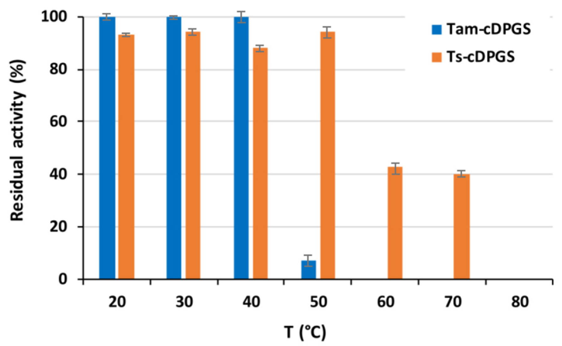
Since Tam-cDPGS was isolated from a metagenome derived from a hot environment and Ts-cDPGS originates from a hyperthermophilic archaeon, the thermal stability of these enzymes was evaluated. The two novel cDPGSs were incubated at pH 7.5 and different temperatures (20–80 °C), and their residual activity was assessed after 24 h. As shown in Figure 5, Ts-cDPGS exhibited significant thermostability, retaining approximately >40% of its activity after 24 h of incubation at temperatures up to 70 °C. Tam-cDPGS was less thermostable, most probably reflecting its possible origin from a moderate thermophilic bacterium.
Subsequently, the apparent melting temperatures (Tm) of both enzymes were determined by monitoring thermal events at 220 nm in circular dichroism (CD) analyses. For Ts-cDPGS, the Tm was found to be 84 °C, while for Tam-cDPGS it was 69 °C (Figure S4, Supporting Information).
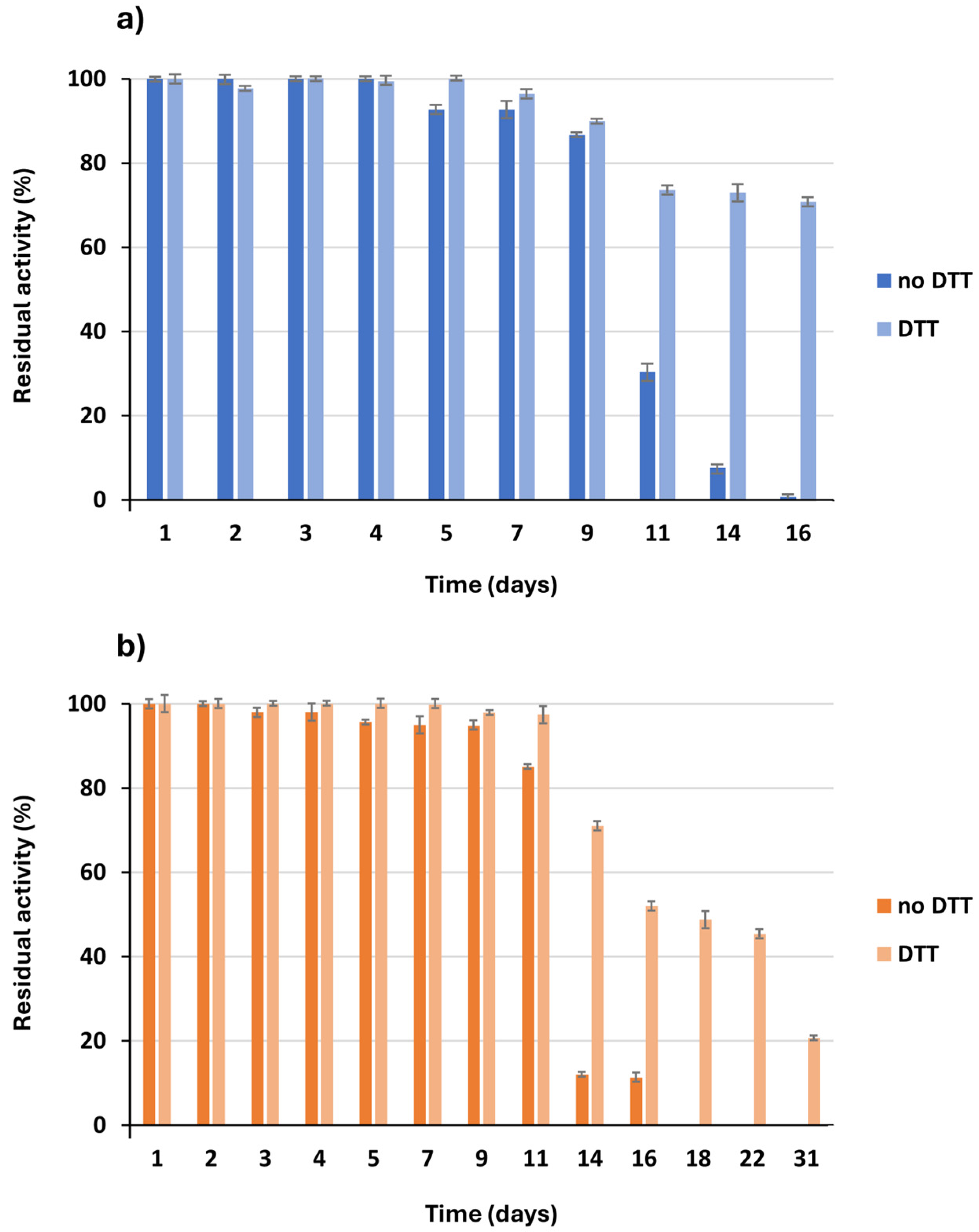
In addition, since it was shown that the reducing agent dithiothreitol (DTT) could stabilize the cDPGS from Methanothermus fervidus [13], the storage stability of Ts-cDPGS and Tam-cDPGS in the presence or absence of DTT (10 mM) was evaluated over the course of one month at room temperature in 50 mM HEPES buffer pH 7.5, 300 mM KCl. As shown in Figure 6, both cDPGS enzymes were indeed stabilized by the presence of DTT. In particular, in the absence of DTT the activity of Ts-cDPGS decreased markedly on the 14th day and it was almost completely lost after 18 days of incubation (Figure 6a). In contrast, in the presence of DTT, the activity began to decline after the eleventh day, but much more gradually, and it retained 20% of its activity by the end of the study.
Tam-cDPGS appeared to be slightly less stable than Ts-cDPGS and the effect of DTT in solution became significant already on the 11th day of incubation. The presence of DTT became even more essential on the 16th day of incubation where the DTT-free enzyme solution was already inactive, around 70% of activity was retained in DTT containing samples (Figure 6b).

2.5. Crystal Structure of Ts-cDPGS

While extensive crystallization trials were conducted for all four putative enzymes using various protein concentrations, multiple commercial and custom screening conditions, and seeding approaches with Ts-cDPGS crystals, only Ts-cDPGS yielded diffraction-quality crystals suitable for structure determination. The Ts-cDPGS crystallized readily in numerous screening conditions and the structure was solved by molecular replacement using the structure coordinates of Mf-cDPGS previously solved by our group (PDB: 8ORK). The structure was solved to 2.2 Å resolution and has been refined to R and Rfree values of 21.6% and 28.3%, respectively (Table 2). This table shows the overall quality of the protein structure and has been deposited in the Protein Data Base as PDB 9SXB.
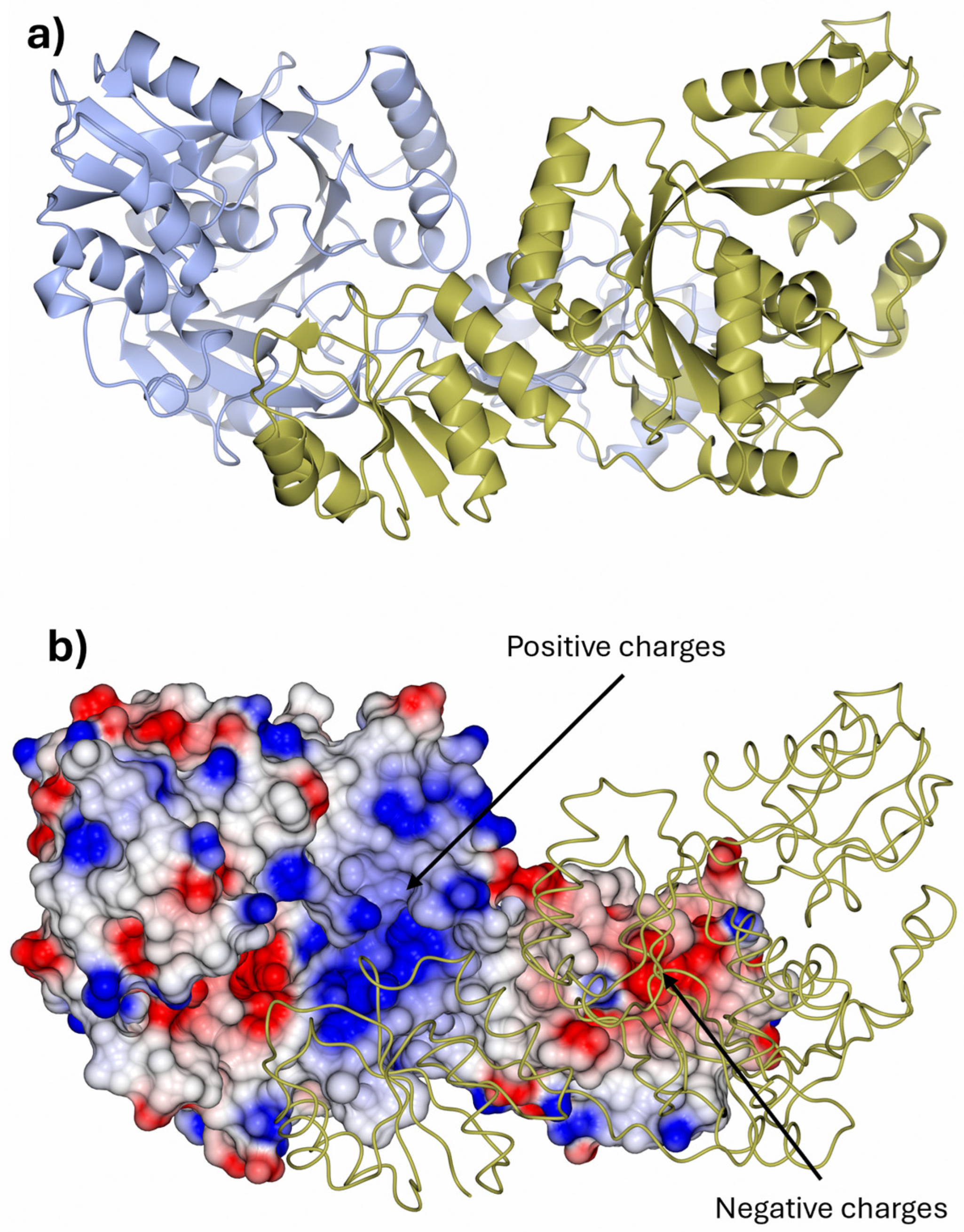
Despite its relatively low sequence identity supported by the distant location on the cDPGS phylogenetic tree, Ts-cDPGS showed high similarity to Mf-cDPGS with a RMSD of 2.6 Å over 432 residues. The overall structure is composed of a N-terminal domain and a distinct C-terminal domain responsible for the binding of the ATP. The Ts-cDPGS structure forms a tight dimer that buries a surface area of 2939.7 Å2 (Figure 7). PISA analysis [29] indicates that the interface is stabilized by 30 hydrogen bonds and seven salt bridges between the interacting subunits. The synergistic effect of these interactions stabilizes the protein quaternary structure by directly contributing to the high thermal stability observed in Ts-cDPGS, similarly to the Mf-cDPGS despite their sequence differences.
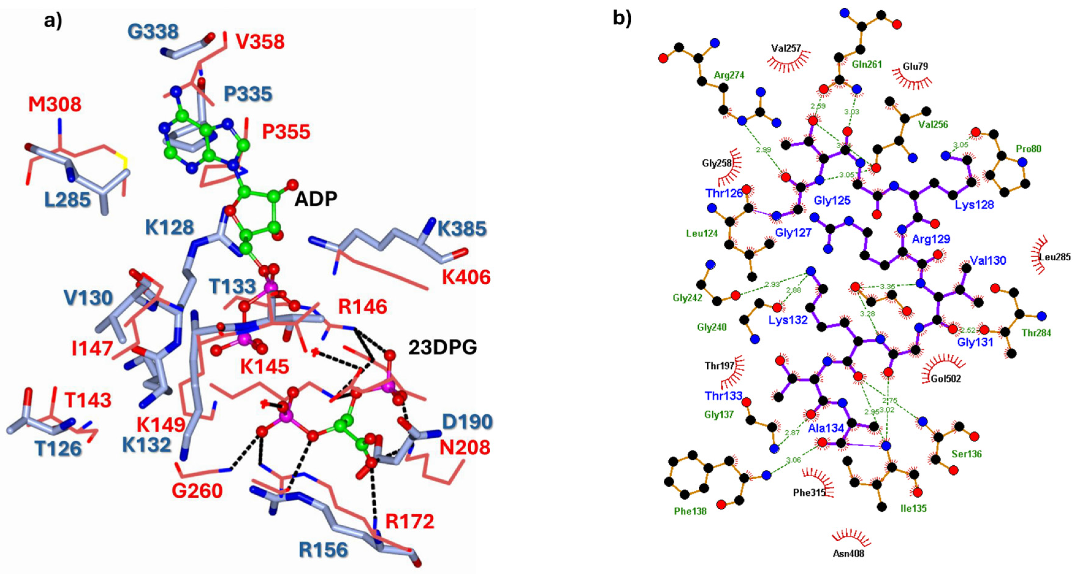
As for the Mf-cDPGS, the dimer interface of Ts-cDPGS clearly shows a large positively charged patch at the interface of the C-terminal domain that interacts with the negatively charged N-terminal domain of the opposing molecule (Figure 7b). The ionic interactions and hydrogen bonds between these patches contribute to both enzymes thermal stability. The multiple sequence alignments and the structural comparison revealed a number of highly conserved residues (Figure 8) that tend to cluster around the nucleotide and ligand binding sites, particularly a P-loop/Walker A motif GxxGxGK[T/S] [30] involved in the stabilization of the phosphate groups of phosphorylated ribonucleotides and in the catalysis of phosphoryl transfer [31].
Compared to the structure of Mf-cDPGS, differences are minimal despite a sequence similarity of only 48%. Remarkably, the active site geometry and chemistry of Ts-cDPGS are very similar to those observed in the apo form of Mf-cDPGS. Key active site residues K128 and R129 in Ts-cDPGS correspond to K145 and R146 in Mf-cDPGS. These residues adopt the “rest” conformation observed in the apo Mf-cDPGS structure (PDB: 8ORK). These positively charged residues play a crucial role in the proposed catalytic mechanism. They shield the negative charges on the phosphate groups of substrate 2,3-DPG, facilitating a close approach for phosphoryl transfer. Docking studies [32,33] of 2,3-DPG using a Ts-cDPGS model with a fixed ADP molecule, confirm an active site organization similar to that observed in the Mf-cDPGS structure. The docked complex exhibits fewer stabilizing interactions compared to Mf-cDPGS, which is explained by the unbound state of the Ts-cDPGS structure. The apo structure adopts a more relaxed and open conformation relative to the expected ligand-bound state (Figure S5, Supporting Information).
Figure 8. (a) Super-imposition of the active site residues of the Ts-cDPGS (cyan cylinders) on the Mf-cDPGS (red sticks) bound to ADP and 2,3-DPG shown as a ball-and-stick model (carbon, green; oxygen, red; nitrogen, blue; phosphorus, pink), active site stabilizing interactions are shown as black dashes. (b) Schematic overview of the stabilizing interactions of the Walker A and B loops. Hydrogen bonds are indicated with green dashes and their distances indicated (prepared with LigPlot+ (version 2.3) [34]).
Catalysts 16 00305 g008

3. Materials and Methods

3.1. General

All reagents and solvents were purchased from Merck KGaA (Darmstadt, Germany) unless otherwise stated. IPTG (isopropyl β-D-1-thiogalactopyranoside) (≥98% purity) was from VWR chemicals (Radnor, PA, USA). ZYM-5052 autoinducing medium was prepared as described in [35]. Escherichia coli BL21(DE3) cells were from Lucigen (Middleton, WI, USA).

3.2. In Silico Screening for Novel 2PGK and cDPGS Genes and Bioinformatic Analysis

In silico screening for 2PGK and cDPGS coding sequences in the studied (meta)genomes was carried out using BLASTp [36]. Multiple sequence alignment was performed in Mafft (version 7) [37] and Clustal Omega (https://www.ebi.ac.uk/Tools/msa/clustalo/, accessed on 21 October 2024). Phylogenetic analysis was made in MEGA X (version 10.2) [38] using the maximum likelihood method and Le-Gascuel model [39], and the tree was improved using iTOL (http://itol.embl.de/, accessed on 20 January 2025) [40]. The co-localization of putative pgk2 and cdpgs genes in the (meta)genome scaffolds was evaluated using IMG tools (version 5.0) [41] and Geneious Prime 2019 (https://www.geneious.com/features/prime, accessed on 4 February 2025). Whether the candidate genes were identified in the metagenomes belong to thermophilic microorganisms was predicted by calculating the amount of IVYWREL amino acids in the respective proteins [22], while solubility, hydrophobicity and other parameters of these proteins were predicted using Soluprot (version 1.0) [42] and GRAVI calculator (https://www.gravy-calculator.de/index.php, accessed on 5 March 2025).
The selected genes (pgk2, Ga0369713_10007067 and 644842203_TSIB0159; cdpgs, Ga0369713_10007068 and 644842204_ TSIB0160, see Supporting Information) were codon optimized for expression in E. coli, synthesized and cloned in the pET28a vector (pET28_Ts2PGK, pET28_Tam2PGK, pET28_TscDPGS, pET28_TamcDPGS) by Twist Bioscience (South San Francisco, CA, USA).

3.3. Expression and Purification of the Recombinant Proteins

E. coli BL21(DE3) competent cells were transformed with pET28_Ts2PGK, pET28_Tam2PGK, pET28_TscDPGS, pET28_TamcDPGS plasmids, respectively, using standard procedures [43]. Recombinant E. coli BL21(DE3) cells were grown overnight at 37 °C in 100 mL LB medium containing 30 µg mL−1 kanamycin (LBkan30). For expression of putative 2PGKs, cells were inoculated in 500 mL ZYM-5052 autoinducing medium with proper antibiotic supplementation to reach a theoretical starting OD600 = 0.2. After 24 h at 20 °C and 200 rpm, the cells were recovered by centrifugation (30 min, 4 °C, 4500× g) and resuspended in 20 mL of 100 mM Tris-HCl buffer, pH 8.0, containing 500 mM NaCl and 20 mM imidazole. Subsequently, cell lysis was performed by sonication (5 cycles, 30 s each at 40% of maximum power, followed by 15 s of rest, Omni Ruptor 250-Watt Ultrasonic Cell Disrupter, Kennesaw, GA, USA). The lysate fraction (around 20 mL) was incubated with 2.5 mL Protino® Ni-NTA Agarose (MACHEREY-NAGEL GmbH & Co, Dueren, Germany) for 90 min. For protein purification, five discrete elution steps were carried out with an elution buffer (100 mM Tris-HCl buffer, pH 8.0, containing 500 mM NaCl and increasing concentration of imidazole, namely 20 mM, 50 mM, 75 mM, 100 mM and 300 mM). Every step was made by mixing and inverting several times the incubated resin with the appropriate elution buffer (10 mL) and in between steps the resin–sample mix was centrifuged (2 min, 4 °C, 2000× g).
For putative cDPGSs expression, overnight cultures were inoculated in 500 mL fresh LBkan30 medium and enzyme expression was induced during the exponentially growth phase with IPTG (1 mM final concentration). The cells were maintained for 72 h at 17 °C and 200 rpm, then recovered by centrifugation (30 min, 4 °C, 4500× g), resuspended in 20 mL of 20 mM HEPES buffer, pH 7.5, containing 300 mM KCl and 20 mM imidazole, and lysed by sonication as described above. The obtained lysate was separated from the insoluble fraction by centrifugation (30 min, 4 °C, 7000× g) and incubated with 2.5 mL Ni-NTA resin for 90 min. Protein purification was obtained washing the resin loaded onto a glass column (10 × 110 mm) with 20 mM HEPES buffer, pH 7.5, containing 300 mM KCl and increasing imidazole concentration up to 300 mM.
Purified 2PGKs were dialyzed against 100 mM Tris-HCl buffer, pH 8.0, 100 mM NaCl, while purified cDPGSs were dialyzed against 20 mM HEPES buffer, pH 7.5, 300 mM KCl, at 4 °C for 24 h and stored at −80 °C. Protein content was measured using the Bio-Rad Protein Assay (Bio-Rad, Hercules, CA, USA) according to the method of Bradford and protein purity was verified by SDS-PAGE analysis (12% T, 2.6% C).
For crystallization studies, Ts-cDPGS was further purified: the protein was applied to a calibrated Superdex 200 pg HiLoad 16/600 size exclusion chromatography (SEC) column (Cytiva, Marlborough, MA, USA) and eluted with one column volume of 20 mM HEPES buffer, pH 7.5, 300 mM KCl, at 1.0 mL min−1 flow. The purity of the protein and the subunit molecular weight were analyzed by SDS-PAGE.

3.4. Enzyme Activity and Stability

The enzymatic activity of 2PGK and cDPGS was determined by coupling the ADP formation from ATP to the oxidation of NADH via pyruvate kinase (PK) and L-lactate dehydrogenase (LDH) (both from rabbit muscle, Merck, Darmstadt, Germany). The assay mixtures (0.25 mL) contained 50 mM HEPES buffer, pH 7.5, 1 mM MgCl2, 300 mM KCl, 1 mM ATP, 0.2 mM NADH, 3.7 µg of purified 2PGK or cDPGS, 8 U PK, 4 U LDH and 1 mM phosphoenolpyruvate. Reactions were initiated by the addition of either 5 mM 2-phosphoglycerate (2-PG, Scheme 1) to measure 2PGK activity, or 5 mM 2,3-diphosphoglycerate (2,3-DPG) to measure cDPGS activity. NADH oxidation was monitored at 35 °C using a Specord 210 UV/VIS spectrometer (Analytik Jena GmbH & Co. KG, Jena, Germany). All measurements were performed in triplicates. Control reactions were carried out to ensure that the coupling enzymes were not rate limiting. One unit (U) of enzyme activity is defined as the amount of enzyme that catalyzes the formation of 1 µmol of product (ADP) per minute under the above-described conditions.
The thermostability of cDPGSs was assessed by incubating the enzymes in 50 mM HEPES buffer, pH 7.5, containing 1 mM MgCl2 and 300 mM KCl, in a final volume of 1 mL, for 24 h in a thermomixer at the different temperatures (20–80 °C). Following incubation, enzyme activity was measured using the PK/LDH coupled assay at 35 °C. Similarly, the influence of the reducing agent dithiothreitol (DTT) on cDPGSs stability was evaluated by incubating the enzymes in 50 mM HEPES buffer, pH 7.5, with 1 mM MgCl2 and 300 mM KCl, either in the presence or absence of 10 mM DTT, at room temperature. Residual activity was monitored over the course of one month using the PK/LDH assay as previously described.
Enzyme apparent melting temperatures (Tm) were evaluated by circular dichroism (CD) analysis using a nitrogen-flushed Jasco J-1100 spectropolarimeter (Easton, MD, USA) equipped with a thermostatically controlled cell holder. Protein samples were diluted in degassed water to a final concentration of 0.15 mg mL−1 and analyzed in quartz cuvettes with a 0.1 cm path length. Apparent Tm values were estimated by monitoring the CD signal at 220 nm using the following temperature programs: from 20 °C to 60 °C at a rate of 5 °C min−1, data acquired every 2 °C with a 30 s hold; from 65 °C to 90 °C at 2.5 °C min−1, data acquired every 0.5 °C with a 30 s hold; and from 90 °C to 95 °C at 5 °C min−1, data acquired every 2 °C.

3.5. Biotransformations

In vitro synthesis of cDPG was performed in 20 mM HEPES buffer, pH 7.0, 300 mM KCl, 0.5 mM MgCl2 containing 5 mM 2,3-DPG (3.3 mg), 7.5 mM ATP, and 1 mg mL−1 cDPGS (2.5 mL total volume). The activity of the potential Taman metagenome 2PGK (Tam-2PGK) was evaluated in a cascade reaction for the synthesis of cDPG starting from 2-PG. The reaction was performed in the same buffer reported above, adding 5 mM 2-PG and 1 mg mL−1 Tam-2PGK. After 20 h of incubation, cDPGS was added to the reaction at 1 mg mL−1 final concentration.
The reaction mixtures were incubated at 35 °C and 150 rpm for 20 h. Then, they were subjected to lyophilization and redissolved in D2O (1 mL) for the 31P-NMR analysis. The 31P-NMR spectra were acquired at 162 MHz (25 °C) on a Bruker AV 400 MHz instrument (Bruker, Billerica, MA, USA). Commercially available standards of ATP, ADP, 2-PG, 2,3-DPG and cDPG were dissolved in D2O (1–2 mg mL−1) while biotransformations were lyophilized and taken up with 750 µL of D2O keeping the reaction buffer (HEPES) in the samples.

3.6. Crystal Structure

Purified Ts-cDPGS corresponding to the dimeric form was concentrated to about 20 mg mL−1 using a 10 kDa membrane Vivaspin (Sartorius, Göttingen, Germany). Microbatch crystallization trials were set up using an Oryx8 crystallization robot (Douglas Instruments, Berkshire, UK) using the Morpheus™ (Molecular Dimensions, Rotherham, UK) protein crystallization screens. Microbatch trials were set in hydrophobic plates (VB-SILVER-2, Douglas Instruments), with a final drop volume of 0.5 µL. The droplet contained a 50:50 ratio of protein solution to screen and was covered with Al’s oil (50:50 mix of silicon and paraffin oils) before being stored at 20 °C.
Ts-cDPGS crystals appeared within one week, the crystals were harvested directly from the crystallization droplet and plunged into liquid nitrogen.
The best diffracting crystals grew in condition A3 of the Morpheus screen consisting of 0.12 M divalents mix, 0.1 M Buffer System 1 pH 6.5 and 30% v/v Precipitant Mix 3 (https://calibrescientific.com/en/products/MDL-MD1-47, accessed on 3 February 2025).
Data were processed and scaled using XDS [44] and AIMLESS [45] in the Xia2 pipeline [46]. The structure was solved by molecular replacement software MOLREP [47] using the structure coordinates of Mf-cDPGS previously solved by our group (PDB: 8ORK). All further data and model manipulations were carried out using the CCP4 suite of programs [48,49]. The resulting structure was subjected to refinement in REFMAC5 [50] and rebuilding in COOT (version 0.9.x) [51]. The PISA software [29] was used for oligomeric state analysis of the cDPGS models. Furthermore, 2D representations of the active site interactions, including hydrogen bonds and hydrophobic contacts, were generated using LigPlot+ (version 2.3) [34].

4. Conclusions

In this study, we identified, expressed, and characterized novel enzymes of the cyclic 2,3-diphosphoglycerate (cDPG) biosynthetic pathway from thermophilic genomes and metagenomes. Notably, our findings provide the first evidence of a bacterial cDPG biosynthesis pathway, as indicated by the discovery of co-localization of pgk2 and cdpgs genes in the Taman mud volcano metagenome. While the Tam-2PGK exhibited predominantly ATPase activity with only limited evidence of kinase function, both Tam-cDPGS and Ts-cDPGS were confirmed as functional cDPG synthases. Ts-cDPGS demonstrated remarkable thermostability, retaining activity after prolonged incubation at elevated temperatures, consistent with its hyperthermophilic archaeal origin.
The high-resolution crystal structure of Ts-cDPGS further revealed strong similarity to the archaeal Mf-cDPGS, including the conservation of key catalytic residues. Together, these results broaden the known phylogenetic distribution of cDPG metabolism and underline the potential of thermophilic (meta)genomes as sources of novel biocatalysts.
Overall, this work not only advances knowledge of extremolyte biosynthesis in thermophiles but also identifies new biocatalysts with favorable properties for future biotechnological applications, thereby providing a foundation for exploiting novel enzymes and their activities in extremolyte synthesis.

Supplementary Materials

The following supporting information can be downloaded at: https://www.mdpi.com/article/10.3390/catal16040305/s1, DNA sequences; Protein sequences; 2PGK expression trials; Figure S1: SDS-PAGE of the resulting samples from Ts_p-2PGK (a) and Tam_p-2PGK (b) co-expression with chaperones (Takara system) and purification via affinity chromatography; Table S1: List of (hyper)thermophilic (meta)genomes considered in this work; Figure S2: Multiple sequence alignment of genes encoding for potential 2PGKs and cDPGSs; Figure S3: Co-localization of novel putative 2PGK and cDPGS sequences in their corresponding (meta)genomes; Figure S4: Melting temperature curves obtained by circular dichroism spectroscopic analysis at 220 nm; 31P-NMR spectra; Figure S5. 2,3-Diphosphoglycerate docking studies performed with the SwissDock server using AutoDock Vina (version 1.2.3).

Author Contributions

S.P.: Investigation, Data curation, Writing—original draft. S.A.D.R.: Investigation, Data curation, Writing—original draft, Writing—review and editing. M.N.I.: Investigation, Data curation, Writing—original draft, Writing—review and editing. I.V.K.: Investigation, Data curation, Conceptualization, Writing—original draft, Writing—review and editing. I.M.A.: Investigation, Data curation. S.R.: Writing—review and editing. I.B.: Investigation, Data curation, Writing—original draft. E.D.: Investigation, Data curation. C.S.: Writing—original draft, Writing—review and editing. B.S.: Writing—original draft, Writing—review and editing, Funding acquisition. E.E.F.: Investigation, Data curation, Conceptualization, Writing—original draft, Writing—review and editing. J.A.L.: Conceptualization, Writing—original draft, Writing—review and editing, Funding acquisition. D.M.: Conceptualization, Writing—original draft, Writing—review and editing, Funding acquisition. All authors have read and agreed to the published version of the manuscript.

Funding

C.S. acknowledges funding by an Evonik Industries AG Scholarship. C.S. and B.S. acknowledge funding by the German Federal Ministry of Education and Research (BMBF) grant HotSolute, 031B0612A within the ERA CoBioTech funding initiative. HotSolute has received funding from the European Union’s Horizon 2020 research and innovations program under grant agreement No [722361]. J.L., S.A.R., and M.N.I. acknowledge Biotechnology and Biological Sciences Research Council BB/R02166X/1 grant for funding within the ERA CoBioTech funding initiative. They would also like to thank funding from the Diamond Synchrotron Light Source for access to beamline I03, I04-1 and I04 (proposal Nos. mx22563) and the beamline scientists for assistance. D.M. acknowledges funding from the Italian Ministry of Education and Research (MIUR), Fondo per le agevolazioni alla ricerca “First 2016” (Decreto n. 110/2019). In addition, we would like to extend our gratitude to Bitop AG (Dortmund, Germany) for generously providing cDPG.

Data Availability Statement

The original contributions presented in this study are included in the article and Supplementary Materials. Further inquiries can be directed to the corresponding authors.

Conflicts of Interest

The authors declare that this study received funding from Evonik Industries AG, Bitop AG. The funder was not involved in the study design, collection, analysis, interpretation of data, the writing of this article or the decision to submit it for publication.

References

  1. Empadinhas, N.; da Costa, M.S. Diversity and Biosynthesis of Compatible Solutes in Hyper/Thermophiles. Int. Microbiol. 2006, 9, 199–206. [Google Scholar] [CrossRef]
  2. Gunde-Cimerman, N.; Plemenitaš, A.; Oren, A. Strategies of Adaptation of Microorganisms of the Three Domains of Life to High Salt Concentrations. FEMS Microbiol. Rev. 2018, 42, 353–375. [Google Scholar] [CrossRef] [Scilit] [PubMed]
  3. Becker, J.; Wittmann, C. Microbial Production of Extremolytes—High-Value Active Ingredients for Nutrition, Health Care, and Well-Being. Curr. Opin. Biotechnol. 2020, 65, 118–128. [Google Scholar] [CrossRef] [Scilit]
  4. Liu, M.; Liu, H.; Shi, M.; Jiang, M.; Li, L.; Zheng, Y. Microbial Production of Ectoine and Hydroxyectoine as High-Value Chemicals. Microb. Cell Factories 2021, 20, 76. [Google Scholar] [CrossRef] [Scilit]
  5. Matussek, K.; Moritz, P.; Brunner, N.; Eckerskorn, C.; Hensel, R. Cloning, Sequencing, and Expression of the Gene Encoding Cyclic 2,3-Diphosphoglycerate Synthetase, the Key Enzyme of Cyclic 2,3-Diphosphoglycerate Metabolism in Methanothermus fervidus. J. Bacteriol. 1998, 180, 5997–6004. [Google Scholar] [CrossRef] [Scilit]
  6. Shima, S.; Hérault, D.A.; Berkessel, A.; Thauer, R.K. Activation and Thermostabilization Effects of Cyclic 2,3-Diphosphoglycerate on Enzymes from the Hyperthermophilic Methanopyrus kandleri. Arch. Microbiol. 1998, 170, 469–472. [Google Scholar] [CrossRef] [Scilit]
  7. Hensel, R.; König, H. Thermoadaptation of Methanogenic Bacteria by Intracellular Ion Concentration. FEMS Microbiol. Lett. 1988, 49, 75–79. [Google Scholar] [CrossRef]
  8. Sastry, M.V.K.; Robertson, D.E.; Moynihan, J.A.; Roberts, M.F. Enzymatic Degradation of Cyclic 2,3-Diphosphoglycerate to 2,3-Diphosphoglycerate in Methanobacterium thermoautotrophicum. Biochemistry 1992, 31, 2926–2935. [Google Scholar] [CrossRef] [Scilit]
  9. van Alebeek, G.-J.W.M.; Tafazzul, G.; Kreuwels, M.J.J.; Keltjens, J.T.; Vogels, G.D. Cyclic 2,3-Diphosphoglycerate Metabolism in Methanobacterium thermoautotrophicum (Strain ΔH): Characterization of the Synthetase Reaction. Arch. Microbiol. 1994, 162, 193–198. [Google Scholar] [CrossRef]
  10. Lentzen, G.; Schwarz, T. Extremolytes: Natural Compounds from Extremophiles for Versatile Applications. Appl. Microbiol. Biotechnol. 2006, 72, 623–634. [Google Scholar] [CrossRef] [Scilit] [PubMed]
  11. Lehmacher, A.; Vogt, A.B.; Hensel, R. Biosynthesis of Cyclic 2,3-Diphosphoglycerate. Isolation and Characterization of 2-Phosphoglycerate Kinase and Cyclic 2,3-Diphosphoglycerate Synthetase from Methanothermus fervidus. FEBS Lett. 1990, 272, 94–98. [Google Scholar] [CrossRef] [Scilit]
  12. Lehmacher, A.; Hensel, R. Cloning, Sequencing and Expression of the Gene Encoding 2-Phosphoglycerate Kinase from Methanothermus fervidus. Mol. Gen. Genet. 1994, 242, 163–168. [Google Scholar] [CrossRef] [Scilit] [PubMed]
  13. Stracke, C.; Meyer, B.H.; De Rose, S.A.; Ferrandi, E.E.; Kublanov, I.V.; Isupov, M.N.; Harmer, N.J.; Monti, D.; Littlechild, J.; Müller, F.; et al. Establishment of an Efficient One-Step Enzymatic Synthesis of Cyclic-2,3-Diphosphoglycerate. Front. Microbiol. 2025, 16, 1601972. [Google Scholar] [CrossRef] [Scilit] [PubMed]
  14. De Rose, S.A.; Finnigan, W.; Harmer, N.J.; Littlechild, J.A.; The HotSolute consortium. Production of the Extremolyte Cyclic 2,3-Diphosphoglycerate Using Thermus thermophilus as a Whole-Cell Factory. Front. Catal. 2021, 1, 803416. [Google Scholar] [CrossRef] [Scilit]
  15. De Rose, S.A.; Isupov, M.N.; Worthy, H.L.; Stracke, C.; Harmer, N.J.; Siebers, B.; Littlechild, J.A. Structural Characterization of a Novel Cyclic 2,3-Diphosphoglycerate Synthetase Involved in Extremolyte Production in the Archaeon Methanothermus fervidus. Front. Microbiol. 2023, 14, 1267570. [Google Scholar] [CrossRef] [Scilit]
  16. Korzhenkov, A.A.; Teplyuk, A.V.; Lebedinsky, A.V.; Khvashchevskaya, A.A.; Kopylova, Y.G.; Arakchaa, K.D.; Golyshin, P.N.; Lunev, E.A.; Golyshina, O.V.; Kublanov, I.V.; et al. Members of the Uncultured Taxon OP1 (“Acetothermia”) Predominate in the Microbial Community of an Alkaline Hot Spring at East-Tuvinian Upland. Microbiology 2018, 87, 783–795. [Google Scholar] [CrossRef] [Scilit]
  17. Merkel, A.Y.; Chernyh, N.A.; Slobodkin, A.I.; Pimenov, N.V.; Bonch-Osmolovskaya, E.A. Diversity and Metabolic Potential of the Terrestrial Mud Volcano Microbial Community with a High Abundance of Archaea Mediating the Anaerobic Oxidation of Methane. Life 2021, 11, 953. [Google Scholar] [CrossRef] [Scilit]
  18. Madeira, F.; Madhusoodanan, N.; Lee, J.; Eusebi, A.; Niewielska, A.; Tivey, A.R.N.; Lopez, R.; Butcher, S. The EMBL-EBI Job Dispatcher Sequence Analysis Tools Framework in 2024. Nucleic Acids Res. 2024, 52, W521–W525. [Google Scholar] [CrossRef] [Scilit]
  19. Waterhouse, A.M.; Procter, J.B.; Martin, D.M.A.; Clamp, M.; Barton, G.J. Jalview Version 2—A Multiple Sequence Alignment Editor and Analysis Workbench. Bioinformatics 2009, 25, 1189–1191. [Google Scholar] [CrossRef] [Scilit]
  20. Aravind, L.; Wolf, Y.I.; Koonin, E. V The ATP-Cone: An Evolutionarily Mobile, ATP-Binding Regulatory Domain JMMB Correspondence. J. Mol. Microbiol. Biotechnol. 2000, 301, 191–194. [Google Scholar]
  21. Saraste, M.; Sibbald, P.R.; Wittinghofer, A. The P-Loop—A Common Motif in ATP- and GTP-Binding Proteins. Trends Biochem. Sci. 1990, 15, 430–434. [Google Scholar] [CrossRef] [Scilit]
  22. Zeldovich, K.B.; Berezovsky, I.N.; Shakhnovich, E.I. Protein and DNA Sequence Determinants of Thermophilic Adaptation. PLoS Comput. Biol. 2007, 3, e5. [Google Scholar] [CrossRef] [Scilit] [PubMed]
  23. Miroshnichenko, M.L.; Hippe, H.; Stackebrandt, E.; Kostrikina, N.A.; Chernyh, N.A.; Jeanthon, C.; Nazina, T.N.; Belyaev, S.S.; Bonch-Osmolovskaya, E.A. Isolation and Characterization of Thermococcus sibiricus Sp. Nov. from a Western Siberia High-Temperature Oil Reservoir. Extremophiles 2001, 5, 85–91. [Google Scholar] [CrossRef] [Scilit] [PubMed]
  24. Mardanov, A.V.; Ravin, N.V.; Svetlitchnyi, V.A.; Beletsky, A.V.; Miroshnichenko, M.L.; Bonch-Osmolovskaya, E.A.; Skryabin, K.G. Metabolic Versatility and Indigenous Origin of the Archaeon Thermococcus sibiricus, Isolated from a Siberian Oil Reservoir, as Revealed by Genome Analysis. Appl. Environ. Microbiol. 2009, 75, 4580–4588. [Google Scholar] [CrossRef] [Scilit] [PubMed]
  25. van Alebeek, G.-J.W.M.; Klaassen, C.; Keltjens, J.T.; van der Drift, C.; Vogels, G.D. ATP Synthesis from 2,3-Diphosphoglycerate by Cell-Free Extract of Methanobacterium thermoautotrophicum (Strain ΔH). Arch. Microbiol. 1991, 156, 491–496. [Google Scholar] [CrossRef] [Scilit]
  26. Karplus, P.A.; Diederichs, K. Linking Crystallographic Model and Data Quality. Science 2012, 336, 1030–1033. [Google Scholar] [CrossRef] [Scilit]
  27. Vaguine, A.A.; Richelle, J.; Wodak, S.J. SFCHECK: A Unified Set of Procedures for Evaluating the Quality of Macromolecular Structure-Factor Data and Their Agreement with the Atomic Model. Acta Crystallogr. D Biol. Crystallogr. 1999, 55, 191–205. [Google Scholar] [CrossRef] [Scilit]
  28. Chen, V.B.; Arendall, W.B.; Headd, J.J.; Keedy, D.A.; Immormino, R.M.; Kapral, G.J.; Murray, L.W.; Richardson, J.S.; Richardson, D.C. MolProbity: All-Atom Structure Validation for Macromolecular Crystallography. Acta Crystallogr. D Biol. Crystallogr. 2010, 66, 12–21. [Google Scholar] [CrossRef] [Scilit]
  29. Krissinel, E.; Henrick, K. Inference of Macromolecular Assemblies from Crystalline State. J. Mol. Biol. 2007, 372, 774–797. [Google Scholar] [CrossRef] [Scilit]
  30. Walker, J.E.; Saraste, M.; Runswick, M.J.; Gay, N.J. Distantly Related Sequences in the Alpha- and Beta-Subunits of ATP Synthase, Myosin, Kinases and Other ATP-Requiring Enzymes and a Common Nucleotide Binding Fold. EMBO J. 1982, 1, 945–951. [Google Scholar] [CrossRef] [Scilit]
  31. Romero Romero, M.L.; Yang, F.; Lin, Y.-R.; Toth-Petroczy, A.; Berezovsky, I.N.; Goncearenco, A.; Yang, W.; Wellner, A.; Kumar-Deshmukh, F.; Sharon, M.; et al. Simple yet Functional Phosphate-Loop Proteins. Proc. Natl. Acad. Sci. 2018, 115, E11943–E11950. [Google Scholar] [CrossRef] [Scilit]
  32. Bugnon, M.; Röhrig, U.F.; Goullieux, M.; Perez, M.A.S.; Daina, A.; Michielin, O.; Zoete, V. SwissDock 2024: Major Enhancements for Small-Molecule Docking with Attracting Cavities and AutoDock Vina. Nucleic Acids Res. 2024, 52, W324–W332. [Google Scholar] [CrossRef] [Scilit]
  33. Eberhardt, J.; Santos-Martins, D.; Tillack, A.F.; Forli, S. AutoDock Vina 1.2.0: New Docking Methods, Expanded Force Field, and Python Bindings. J. Chem. Inf. Model. 2021, 61, 3891–3898. [Google Scholar] [CrossRef] [Scilit]
  34. Laskowski, R.A.; Swindells, M.B. LigPlot+: Multiple Ligand–Protein Interaction Diagrams for Drug Discovery. J. Chem. Inf. Model. 2011, 51, 2778–2786. [Google Scholar] [CrossRef] [Scilit] [PubMed]
  35. Studier, F.W. Protein Production by Auto-Induction in High-Density Shaking Cultures. Protein Expr. Purif. 2005, 41, 207–234. [Google Scholar] [CrossRef] [Scilit]
  36. Altschul, S.F.; Gish, W.; Miller, W.; Myers, E.W.; Lipman, D.J. Basic Local Alignment Search Tool. J. Mol. Biol. 1990, 215, 403–410. [Google Scholar] [CrossRef] [PubMed]
  37. Katoh, K.; Standley, D.M. MAFFT Multiple Sequence Alignment Software Version 7: Improvements in Performance and Usability. Mol. Biol. Evol. 2013, 30, 772–780. [Google Scholar] [CrossRef] [Scilit]
  38. Kumar, S.; Stecher, G.; Li, M.; Knyaz, C.; Tamura, K. MEGA X: Molecular Evolutionary Genetics Analysis across Computing Platforms. Mol. Biol. Evol. 2018, 35, 1547–1549. [Google Scholar] [CrossRef] [Scilit]
  39. Le, S.Q.; Gascuel, O. An Improved General Amino Acid Replacement Matrix. Mol. Biol. Evol. 2008, 25, 1307–1320. [Google Scholar] [CrossRef] [Scilit] [PubMed]
  40. Letunic, I.; Bork, P. Interactive Tree of Life v2: Online Annotation and Display of Phylogenetic Trees Made Easy. Nucleic Acids Res. 2011, 39, 475–478. [Google Scholar] [CrossRef] [Scilit]
  41. Chen, I.-M.A.; Chu, K.; Palaniappan, K.; Pillay, M.; Ratner, A.; Huang, J.; Huntemann, M.; Varghese, N.; White, J.R.; Seshadri, R.; et al. IMG/M v.5.0: An Integrated Data Management and Comparative Analysis System for Microbial Genomes and Microbiomes. Nucleic Acids Res. 2019, 47, D666–D677. [Google Scholar] [CrossRef] [Scilit]
  42. Hon, J.; Marusiak, M.; Martinek, T.; Kunka, A.; Zendulka, J.; Bednar, D.; Damborsky, J. SoluProt: Prediction of Soluble Protein Expression in Escherichia coli. Bioinformatics 2021, 37, 23–28. [Google Scholar] [CrossRef] [Scilit]
  43. Sambrook, J.; Russell, D.W. Molecular Cloning: A Laboratory Manual, 3rd ed.; Cold Spring Harbor Laboratory Press: Cold Spring Harbor, NY, USA, 2001. [Google Scholar]
  44. Kabsch, W. XDS. Acta Crystallogr. D Biol. Crystallogr. 2010, 66, 125–132. [Google Scholar] [CrossRef] [Scilit] [PubMed]
  45. Evans, P.R.; Murshudov, G.N. How Good Are My Data and What Is the Resolution? Acta Crystallogr. D Biol. Crystallogr. 2013, 69, 1204–1214. [Google Scholar] [CrossRef] [Scilit] [PubMed]
  46. Winter, G.; Lobley, C.M.C.; Prince, S.M. Decision Making in Xia2. Acta Crystallogr. D Biol. Crystallogr. 2013, 69, 1260–1273. [Google Scholar] [CrossRef] [Scilit]
  47. Vagin, A.; Teplyakov, A. Molecular Replacement with MOLREP. Acta Crystallogr. D Biol. Crystallogr. 2010, 66, 22–25. [Google Scholar] [CrossRef] [Scilit] [PubMed]
  48. Winn, M.D.; Ballard, C.C.; Cowtan, K.D.; Dodson, E.J.; Emsley, P.; Evans, P.R.; Keegan, R.M.; Krissinel, E.B.; Leslie, A.G.W.; McCoy, A.; et al. Overview of the CCP4 Suite and Current Developments. Acta Crystallogr. D Biol. Crystallogr. 2011, 67, 235–242. [Google Scholar] [CrossRef] [Scilit]
  49. Agirre, J.; Atanasova, M.; Bagdonas, H.; Ballard, C.B.; Baslé, A.; Beilsten-Edmands, J.; Borges, R.J.; Brown, D.G.; Burgos-Mármol, J.J.; Berrisford, J.M.; et al. The CCP4 Suite: Integrative Software for Macromolecular Crystallography. Acta Crystallogr. D Struct. Biol. 2023, 79, 449–461. [Google Scholar] [CrossRef] [Scilit]
  50. Murshudov, G.N.; Skubák, P.; Lebedev, A.A.; Pannu, N.S.; Steiner, R.A.; Nicholls, R.A.; Winn, M.D.; Long, F.; Vagin, A.A. REFMAC5 for the Refinement of Macromolecular Crystal Structures. Acta Crystallogr. D Biol. Crystallogr. 2011, 67, 355–367. [Google Scholar] [CrossRef] [Scilit]
  51. Emsley, P.; Lohkamp, B.; Scott, W.G.; Cowtan, K. Features and Development of Coot. Acta Crystallogr. D Biol. Crystallogr. 2010, 66, 486–501. [Google Scholar] [CrossRef] [Scilit]
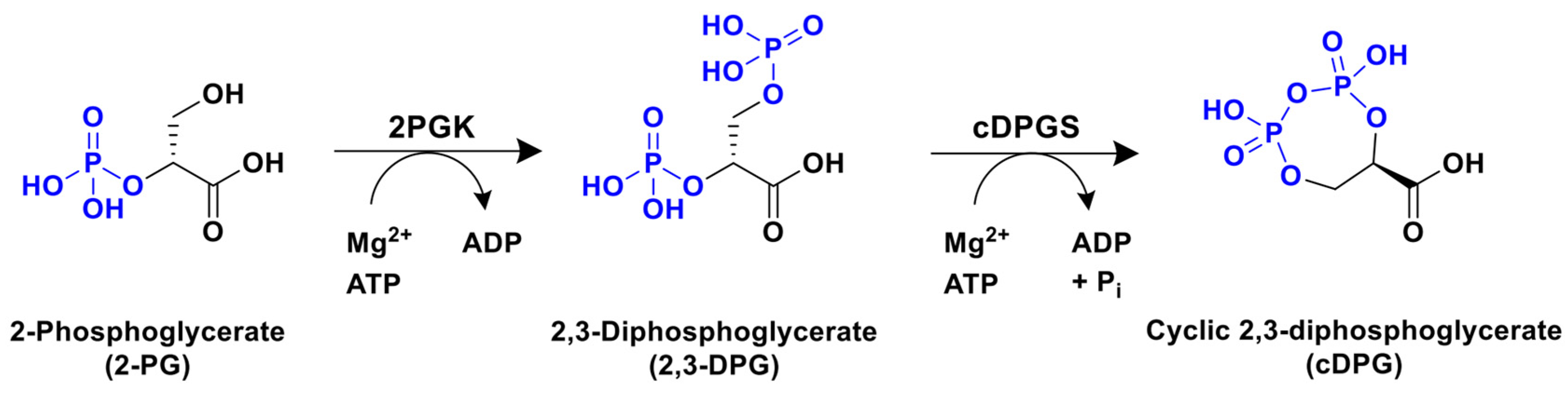
Scheme 1. Biocatalytic synthesis of cyclic 2,3-diphosphoglycerate (cDPG) from 2-phosphoglycerate (2-PG) by the sequential action of 2-phosphoglycerate kinase (2PGK) and cyclic 2,3-diphosphoglycerate synthase (cDPGS) enzymes.
Catalysts 16 00305 sch001
Figure 1. Sequence alignments of putative 2PGKs (a) and cDPGSs (b) with the corresponding enzymatically characterized homologues from Methanothermus fervidus (Mf-2PGK and Mf-cDPGS, respectively). Alignments were carried out using the Clustal Omega tool (version 1.2.4) [18] and visualized with Jalview (version 2.11.5.1) [19].
Catalysts 16 00305 g001
Figure 2. Maximum likelihood phylogenetic tree of cDPGS. The percentage of trees in which the associated taxa clustered together (bootstrap, 1000 replications) is shown using the heat lines with the scale, shown at the top left of the figure. This analysis is based on the multiple sequence alignment (MSA) of 104 amino acid sequences and 427 positions after all positions with less than 95% site coverage were discarded. The tree is drawn to scale, with branch lengths corresponding to the number of substitutions per site according to the model used. Colors of the labels are as follows: green—biochemically characterized cDPGSs from Swissprot database; blue—cDPGSs from Swissprot database without biochemically confirmed activity; red—cDPGSs from this study. Class or phylum level taxonomy of most of the clade’s representatives is indicated next to the clades.
Catalysts 16 00305 g002
Figure 3. Heterologous expression in E. coli and purification of putative Tam-2PGK (a) and Ts-2PGK (b). M, MW marker; 1, before-inoculum in ZYM medium cell sample; 2, after-inoculum in ZYM medium cell sample; 3, cell extract; 4–6, purified fractions. 2PGK expected MW: around 35 kDa.
Catalysts 16 00305 g003
Figure 4. Heterologous expression in E. coli and purification of putative Tam-cDPGS (a) and Ts-cDPGS (b). M, MW marker; 1, before-induction cell sample; 2, after-induction cell sample; 3, cell extract; 4–6, purified fractions. cDPGS expected MW: around 50 kDa.
Catalysts 16 00305 g004
Figure 5. Stability of the two cDPGS enzymes after incubation at different temperatures for 24 h. At each temperature, enzymes were incubated in 50 mM HEPES buffer, pH 7.5, containing 1 mM MgCl2 and 300 mM KCl. Experiments were performed at least in triplicate; standard deviation of residual activity was below 5%.
Catalysts 16 00305 g005
Figure 6. Long-term stability at room temperature of the novel cDPGSs (a), Ts-cDPGS; (b), Tam-cDPGS) in the presence or in the absence of 10 mM dithiothreitol (DTT). Experiments were performed at least in triplicate; standard deviation of residual activity was below 5%.
Catalysts 16 00305 g006
Figure 7. (a) Cartoon representation of the Ts-cDPGS dimer with the two protomers in cyan and gold. (b) A cartoon diagram showing hydrophobic interactions at the dimer interface of Ts-cDPGS. For clarity, one of the protomers is shown in space filling mode with electrostatic surface potential and the other as a thin gold tube. The areas of positive charge are shown in blue, with the areas of negative charge in red. The hydrophobic surfaces are represented in white. The two major charge patches involved in the dimer interface are highlighted by black arrows.
Catalysts 16 00305 g007
Table 2. Ts-cDPGS data collection and refinement statistics.
Data Collection
Wavelength0.954
Space groupP 21 21 2
Cell dimensionsa = 162.46, b = 80.56, c = 82.74, α = β = γ = 90.00
Resolution range (Å) 173.73–2.28
Total reflections 1684,808
Unique reflections 150,337
Completeness (%)100
Multiplicity13.7
Rmeas (%) 1,20.115
<I>/<σ(I)> 111.6
CC1/2 1,31
Wilson B-factor 42)75.79
MODEL PROPERTIES
Number of models1
Number of chains6
Overall number of atoms (non-H)6710
in macromolecules6677
in ligands12 GOL/4 EDO/1 CA
in solvent16
REFINEMENT
Resolution73.73–2.28
Reflections in refinement49,939
Reflections in free set2339
Rwork0.196
Rfree0.266
FSC average0.922
RMSD bonds (Å)0.0117
RMSD angles2.893
Ramachandran favored (%) 593.5
Ramachandran allowed (%) 55.8
Ramachandran outliers (%) 50.7
Rotamer outliers8
Clash score 52.7
MolProbity score2.17
Average B-factor (Å2)64.4
for macromolecules (Å2)64.3
for ligands (Å2)113.4 GOL/92.1 EDO/120.5 CA
for solvent (Å2)74.9
PDB accession code9SXB
1 Values for the highest resolution shell are given in parentheses. 2 Rmeas = Σh [m/(m − 1)]1/2 Σi|Ih,I − <Ih>|/Σh ΣiIh,i. 3 CC1/2 is defined in [26]. 4 Wilson B-factor was estimated by SFCHECK (version 7.0.4) [27]. 5 The Ramachandran statistics and clashscore statistics were calculated using MOLPROBITY (version 4.02-528) [28].
Disclaimer/Publisher’s Note: The statements, opinions and data contained in all publications are solely those of the individual author(s) and contributor(s) and not of MDPI and/or the editor(s). MDPI and/or the editor(s) disclaim responsibility for any injury to people or property resulting from any ideas, methods, instructions or products referred to in the content.