Article

Recyclable TiO2–Fe3O4 Magnetic Composites for the Photocatalytic Degradation of Paracetamol: Comparative Effect of Pure Anatase and Mixed-Phase P25 TiO2

1
Faculty of Physics, Babeș-Bolyai University, M. Kogălniceanu St. 1, 400084 Cluj-Napoca, Romania
2
Nanostructured Materials and Bio-Nano-Interfaces Center, Interdisciplinary Research Institute on Bio-Nano-Sciences, Babeș-Bolyai University, Treboniu Laurian St. 42, 400271 Cluj-Napoca, Romania
3
Faculty of Chemistry and Chemical Engineering, Babeș-Bolyai University, Arany János St. 11, 400028 Cluj-Napoca, Romania
4
Department of Applied and Environmental Chemistry, University of Szeged, Rerrich Béla Sqr. 1, 6720 Szeged, Hungary
5
Department of Molecular Sciences, Faculty of Medicine, Iuliu Haţieganu University of Medicine and Pharmacy, Victor Babeș St. 8, 400012 Cluj-Napoca, Romania
6
Contrast Agents and Specific Therapeutics Center—INSPIRE Platform, Interdisciplinary Research Institute in Bio-Nano-Sciences, Babeș-Bolyai University, Treboniu Laurian St. 42, 400271 Cluj-Napoca, Romania
7
Electron Microscopy Center, Faculty of Biology and Geology, Babeș-Bolyai University, Clinicilor St. 5-7, 400006 Cluj-Napoca, Romania
8
National Institute for Research and Development of Isotopic and Molecular Technologies, Donat St. 67-103, 400293 Cluj-Napoca, Romania
9
INSPIRE Research Platform InfoBioNano4Health & Biomedical Imaging, Babeș-Bolyai University, Arany János St. 11, 400084 Cluj-Napoca, Romania
10
Laboratory for Advanced Materials and Applied Technologies, Institute of Research-Development-Innovation in Applied Natural Sciences, Babes-Bolyai University, Fântânele St. 30, 400327 Cluj-Napoca, Romania
*
Authors to whom correspondence should be addressed.
Catalysts 2025, 15(9), 839; https://doi.org/10.3390/catal15090839 (registering DOI)
Submission received: 31 May 2025 / Revised: 26 August 2025 / Accepted: 28 August 2025 / Published: 1 September 2025
(This article belongs to the Special Issue TiO2 Photocatalysts: Design, Optimization and Application)

Abstract

Magnetically separable TiO2-based composite photocatalysts have gained significant interest in the past two decades; however, the optimization of their synthesis and the stabilization of the magnetic iron oxide within the composite is still an open challenge. The present study investigates the photocatalytic behavior and recyclability of TiO2-Fe3O4 composites, with emphasis on a possible correlation between pollutant degradation efficiency, recyclability, iron oxide stability, and the phase composition of the chosen TiO2 base. Magnetite nanoparticles were synthesized under varied temperature and alkaline conditions to identify optimal parameters for achieving the desirable magnetic properties. The magnetic nanoparticles were integrated into composite systems with either commercial TiO2 (Evonik Aeroxide P25 with anatase–rutile mixed phase) or a hydrothermally synthesized anatase TiO2. The P25-based composite removed 99% paracetamol from aqueous solutions under UV-A irradiation and demonstrated successful recyclability, maintaining 96% paracetamol degradation efficiency after four uses. In contrast, the anatase TiO2-based magnetic composite exhibited a lower performance (70%) and a significantly hindered recyclability (45% after four cycles). The difference in performance was attributed to variations in the phase composition of the employed TiO2 in the composites and, consequently, in their charge separation mechanisms.

1. Introduction

Advanced catalytic processes, including the widely recognized advanced oxidation processes (AOPs) [1] and the increasingly explored advanced reduction processes (ARPs) [2,3], represent powerful alternatives for wastewater remediation. These approaches are particularly effective in degrading persistent organic pollutants and emerging contaminants in complex aqueous environments. Moreover, they accommodate complex catalytic systems, such as multi-material composites [4,5], magnetic composites [5,6], catalyst immobilization [7,8], metal–organic frameworks (MOFs) [9,10], carbon-based materials [11,12], and aerogel-based supports [13,14], facilitating enhanced recyclability, selectivity, and overall catalytic efficiency.
Among AOPs, heterogeneous photocatalysis stands out as one of the most extensively studied methods for environmental remediation, owing to several advantages, including the separability of the photocatalyst from the pollutant-containing solution after the photocatalytic process.
However, identifying and optimizing separation methods remains a persistent challenge in the field. In 1999, Chen et al. were the first to apply a magnetically removable TiO2–iron oxide composite system in photocatalysis, specifically TiO2 combined with γ-Fe2O3 [15]. Although considerable progress has been made over the past two decades, the method remains in need of further research, largely due to the ongoing challenges in terms of stabilizing the magnetic iron oxide in composites through facile methods and enhancing both the photocatalytic and magnetic properties of the composite.
Among iron oxides, magnetite (Fe3O4) is one of the favored materials applied in photocatalytic composites due to its superior magnetic properties [16]. However, magnetite is also the iron oxide that is most susceptible to oxidation due to its high Fe2+ content [17]. This intrinsic instability poses difficulties not only in the case of applications but also during synthesis. The synthesis of magnetite (Fe3O4) nanoparticles can result in various by-products, such as maghemite (γ-Fe2O3) [18], hematite (α-Fe2O3) [19], or goethite (α-FeOOH) [20], depending on the controlled and uncontrolled synthesis parameters. Magnetite can also undergo changes when applied in photocatalytic systems together with TiO2, due to the changes in the reaction environment [21]. Alternatives for the stabilization of magnetic nanoparticles in composites include coatings with SiO2 [22,23] or polymers [24,25]. Nevertheless, these strategies increase the complexity of the synthesis method and introduce additional chemicals, potentially hindering not only the feasibility but also the environmental sustainability of the process. As a result, researchers in the field continue to explore binary composite systems, focusing on optimizing the properties of the individual components to achieve improved efficiency and stability without the need for additional materials.
For the photocatalytic counterpart of the composite, TiO2 is one of the most popular choices, as it is a widely applied photocatalyst with favorable properties [26]. Two alternatives are predominantly employed—the commercial Evonik Aeroxide P25 TiO2 [27] or TiO2 synthesized by various methods such as sol–gel [28] or hydrothermal synthesis [29]. One of the most important differences between these two relates to their crystal phase composition. While P25 contains both anatase and rutile phases in a carefully engineered ratio, TiO2 prepared by alternative synthesis methods typically results in a material with a pure anatase crystal phase.
Numerous studies have successfully demonstrated the incorporation of both types of TiO2 into composite materials and their application in photocatalysis. Among the most popular applications was the removal of dyes [30] and phenol [31], but some investigations also focused on the photocatalytic degradation of pharmaceuticals such as metronidazole [29] or on evaluating the antibacterial properties of the composites [32]. While the photocatalytic performance of most P25-Fe3O4 composites does not surpass that of pristine P25 [21], Harifi et al. (2014) were the first to report a highly efficient and recyclable magnetic TiO2 composite that shows superior performance compared to bare P25 [33]. The enhanced efficiency was attributed to the optimized Fe2+/TiO2 ratio within the composite structure. A similar observation was made in the case of anatase TiO2-Fe3O4 composites [29]. However, in many of these studies, either no reference TiO2 material is employed [34] or the commercial P25 is applied as a benchmark [31].
No reports have been found in the literature that directly compare the two types of magnetic composites, i.e., those incorporating anatase–rutile mixed-phase TiO2 and those containing exclusively anatase, under identical experimental conditions. Such a comparative study would offer valuable insights into the behavior of these composite systems and would highlight the influence of the applied TiO2 crystal phase on pollutant degradation efficiency. Motivated by this idea, the present study investigates the photocatalytic removal of paracetamol from aqueous solutions under UV-A irradiation, employing two TiO2-Fe3O4 magnetic composites—one based on mixed-phased Evonik Aeroxide P25 TiO2 and the other on pure anatase TiO2. Paracetamol, also known as acetaminophen, was chosen as a model pollutant due to its widespread use, its toxicological effect on the environment and human health at long-term exposure, and its rise as an emerging pollutant in recent years [35,36].
In addition, the influence of synthesis parameters on the properties of magnetic iron oxide nanoparticles is explored, and an optimal parameter set is chosen for composite preparation. The aim of the work is to map the differences in the photocatalytic behavior and recyclability of composites as a function of compositional and structural properties, as well as to gain deeper insight into the stabilization mechanism of magnetite within such composite systems.

2. Results and Discussion

To identify an iron oxide nanoparticle (IONP) with suitable properties for magnetic composite preparation, four IONP samples were synthesized under different experimental conditions and were thoroughly characterized in terms of structure and composition. The investigations that led to the selection of the appropriate IONP are presented in detail in the Supplementary Materials.

2.1. Structural and Optical Characterization of the TiO2 Support Materials and Their Composites

After the decision regarding the magnetic nanoparticles was made, their photocatalytic counterpart in the composite was prepared. For this purpose, a titanium dioxide sample was prepared employing a hydrothermal synthesis method (hereafter, sample TH), and a commercially available titania sample, the Evonik Aeroxide ® TiO2 P25 (Evonik Industries AG, Essen, Germany), was chosen as a material for comparison (hereafter, P25). In the final step, these titania samples were introduced into composites with IONP_0.8_75 (see Table 1 and Table S1) via the precipitation method, yielding the composites named TH/IONP and P25/IONP, respectively. Similarly to the case of the IONP samples, the preparation phase was succeeded by material characterization.
The XRD measurements revealed that the titania sample TH is pure anatase (TiO2-JCPDS card no. 21-1272) (Figure 1), with a calculated primary crystallite size of 9.2 nm (Table 1). In parallel, the characteristics of the TiO2 P25 were also verified. The recorded diffractogram confirms the presence of both anatase and rutile crystal phases in P25, with mean crystallite sizes of 24 nm (anatase) and 36 nm (rutile). The XRD patterns of the composites predominantly display the reflections of the TH anatase sample and the anatase and rutile reflections of P25, respectively; however, the characteristic and most intense reflections of Fe3O4 (IONP) are also present in the diffractogram (Figure 1). In the case of both composites, the components display larger primary crystallite sizes compared to their bare counterparts. In the case of TH/IONP, the primary crystallite size of anatase particles was assessed to be 9.9 nm, while for the IONP, this value was 52.3 nm. Similarly, for the P25/IONP composites, the size of the anatase crystallites is assessed to be 25 nm, the size of rutile is around 41 nm, and the primary crystallite size of the IONP_0.8_75 is also larger, at 65.2 nm. The above-mentioned phenomenon usually occurs in alkaline media, where the solubilization and crystallization of titania (or any other oxide) might occur simultaneously. The insolubilized particles act as seeds; thus, precipitation and subsequent crystallization occur on the surface, leading to an increase in crystallite size [37]. The success of the composite preparation method is confirmed by the presence of both components—titania base and magnetic IONP—in the samples.
To reveal the formation of new chemical bonds in the composites and to further investigate any structural changes, Fourier transform infrared spectroscopy measurements were carried out, including for the IONP sample (Figure S5). The FT-IR spectrum of the IONP sample displays the main bands of Fe3O4: Fe-O stretching vibrations at 582 and 632 cm−1 [38,39], while the spectrum of TiO2 includes bands in the 500–700 cm−1 spectral region, which are characteristic of Ti-O-Ti stretching vibrations [40,41]. The spectra of the TiO2-Fe3O4 composites match the TiO2 bands and confirm the presence of Fe3O4, faintly displaying a band in the 580 cm−1 region, corresponding to Fe-O stretching vibrations [38]. In the spectra of both composites, a new, additional band appears in the 1340 cm−1 region, which, considering the synthesis method, could indicate the presence of CH3-COO- acetate ions adsorbed on the surface of the composite [42]. The FT-IR spectra of the composites show no clear evidence of the formation of new chemical bonds between TiO2 and Fe3O4.
To investigate the morphology and particle size of the samples, TEM was performed. The TEM micrographs revealed that both TH and P25 consist of agglomerates made up of a mixture of angular and roughly spherical to slightly irregular particles (Figure S6). In the TH-IONP sample, the TiO2 particles were found to be in the range of approximately 10–15 nm in size, while in the P25-IONP composite, the P25 TiO2 particles exhibited larger dimensions, ranging from about 20 to 40 nm. These findings are in good correlation with the primary crystallite size estimates obtained from X-ray diffraction using the Scherrer equation, as presented in Table 1.
Upon a more in-depth investigation into the composites’ structural and qualitative properties, the following two properties related to their applicability were also investigated: magnetic properties for facile separation and reusability, as well as their optical properties for photocatalytic application.
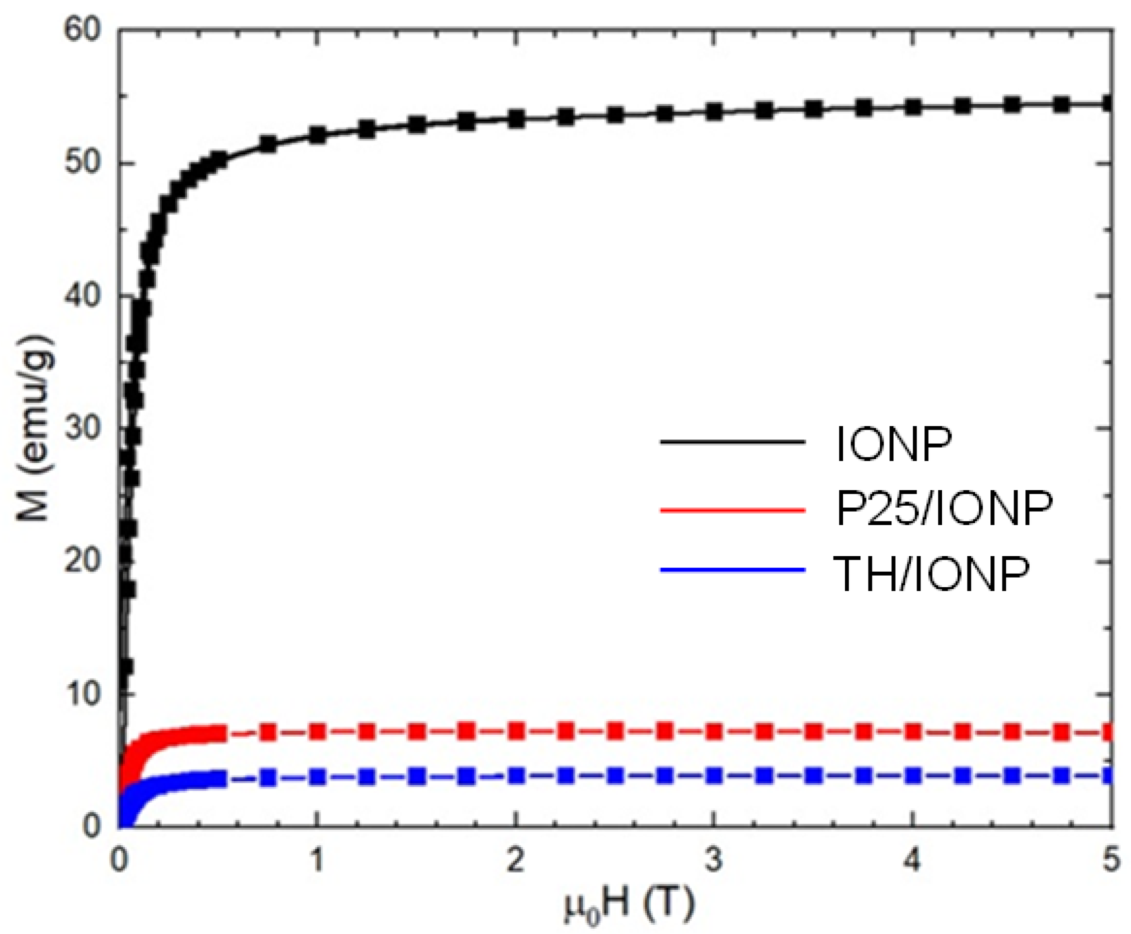
The external magnetic field dependences of magnetization measured at 300 K are presented in Figure 2. It can be observed that the saturation is not attended in a 5 T external magnetic field in the case of the IONP_0.8_75 sample, while in the composite samples, the saturation is attended at around 0.8 T. The saturation magnetization (Ms) presented in Table 2 has a value of 54.2 emu/g for the pristine sample, while in the case of composite samples, it decreases strongly, being around 7.5 emu/g for P25/IONP and 4.2 emu/g for the TH/IONP samples.
The high decrease in saturation magnetization in the case of the composites can be explained by the dilution, with only 10% being magnetite. The obtained values are very close to the results reported by Kubiak in the case of TiO2-10%Fe3O4 composites, with Ms being between 6 emu/g and 9 emu/g, depending on the preparation method [29]. The saturation magnetization of the pristine sample is somewhat lower than what was reported earlier [17], probably due to the preparation conditions, the nanoparticle’s size, and its crystallinity degree.
The hysteresis loops were measured at 300 K in external magnetic fields between −2 and 2 T; they are shown in Figure 3.
Very small values of the coercive field (Hc) and remanent magnetizations (Mr) were found in all studied samples (Table 2). These small values indicate that the studied samples show weak ferrimagnetic properties and a superposition of superparamagnetic properties due to the presence of nanoparticles with low diameters.
Previously, it was shown that the presence or absence of the different types of inter-grain exchanges is determined by the amount of the squareness ratio, Mr/Ms, being in the range between 0 and 1 [43]. It was shown that in the case of R < 0.5, the particle interacts by magnetostatic interaction, while for R = 0.5, an assembly of noninteracting and randomly oriented particles is present in agreement with the Stoner–Wohlfarth theory. In the case in which R > 0.5, the existence of exchange coupling particles is revealed. In line with the model proposed by Stoner–Wohlfarth, the value for noninteracting single-domain particles that have the easy axis randomly oriented is 0.5 for uniaxial anisotropy and 0.832 for cubic anisotropy [44]. The obtained Mr/Ms values at room temperature for the studied samples are less than 0.5 (Table 2), showing that the studied nanoparticles are in a single-domain regime, which interacts by magnetostatic forces [45]. Considering the magnetic properties of the composites, it is possible to apply these materials for magnetic separation in different technical applications.
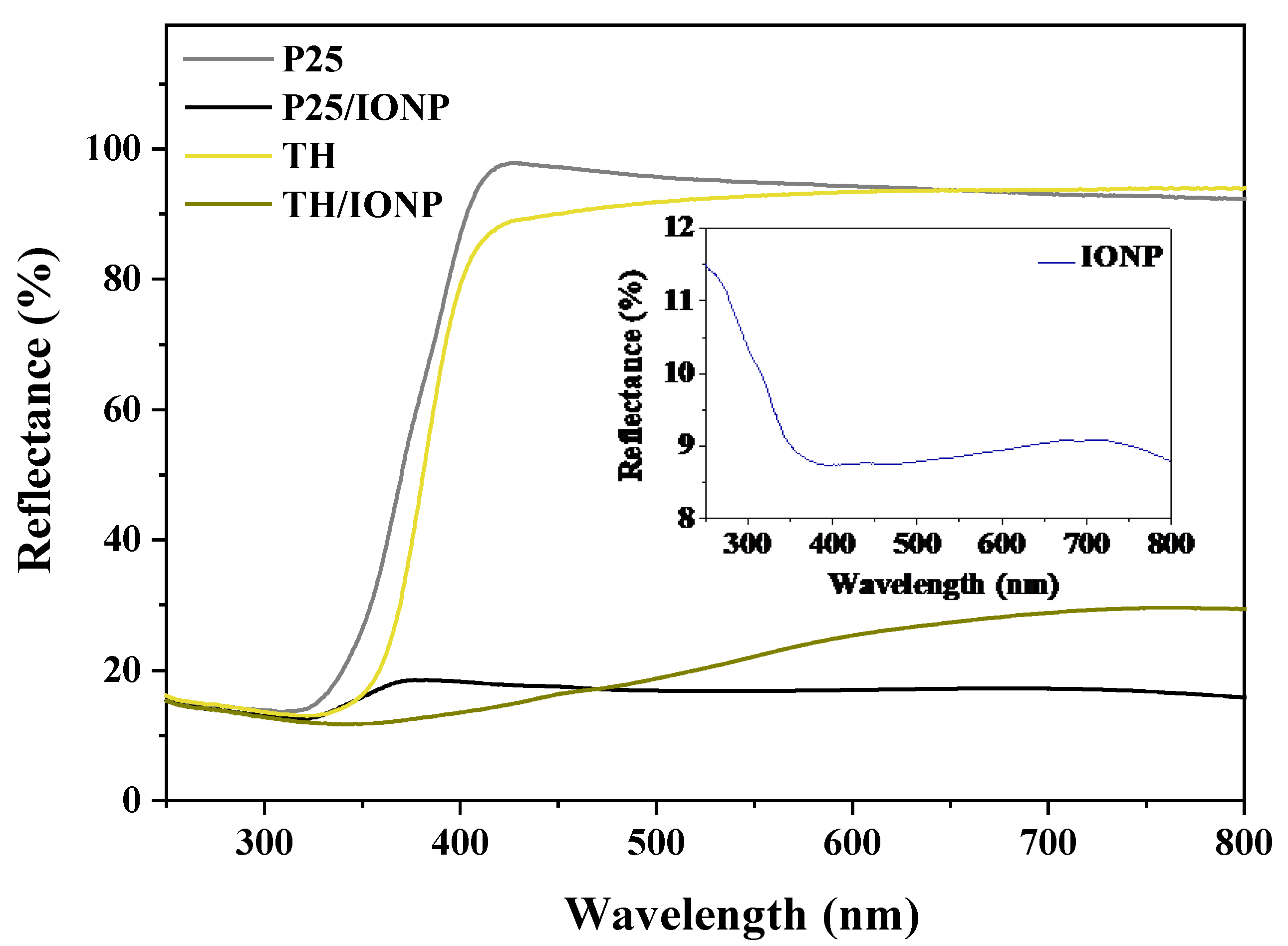
The planned application of the titania samples and the titania-based composites was to drive photocatalytic degradation reactions. Therefore, their optical properties also needed to be evaluated. The results of the performed DRS measurements and the band gap energy values calculated from this investigation support the findings of the XRD analysis for both the TH sample and P25. The band gap energy of TH is 3.1 eV, which coincides with the band gap energy of the anatase crystal phase of the TiO2 (Figure S7a,b) [46,47]. In the case of commercial P25, the band gap values are 3.2 eV and 3.0 eV for anatase and rutile, respectively, which are also consistent with the literature findings (Figure S7c,d) [48]. Considering its anatase crystal phase and foreshadowed light absorption properties in the UV region, the TH sample alone could be a promising photocatalyst, in the same way that P25 already is. Introducing the titania samples in composite with IONP_0.8_75, the dark-colored iron oxide content decreased the reflectance intensities of the P25 and TH samples by 79% and 75%, respectively, as shown in Figure 4.
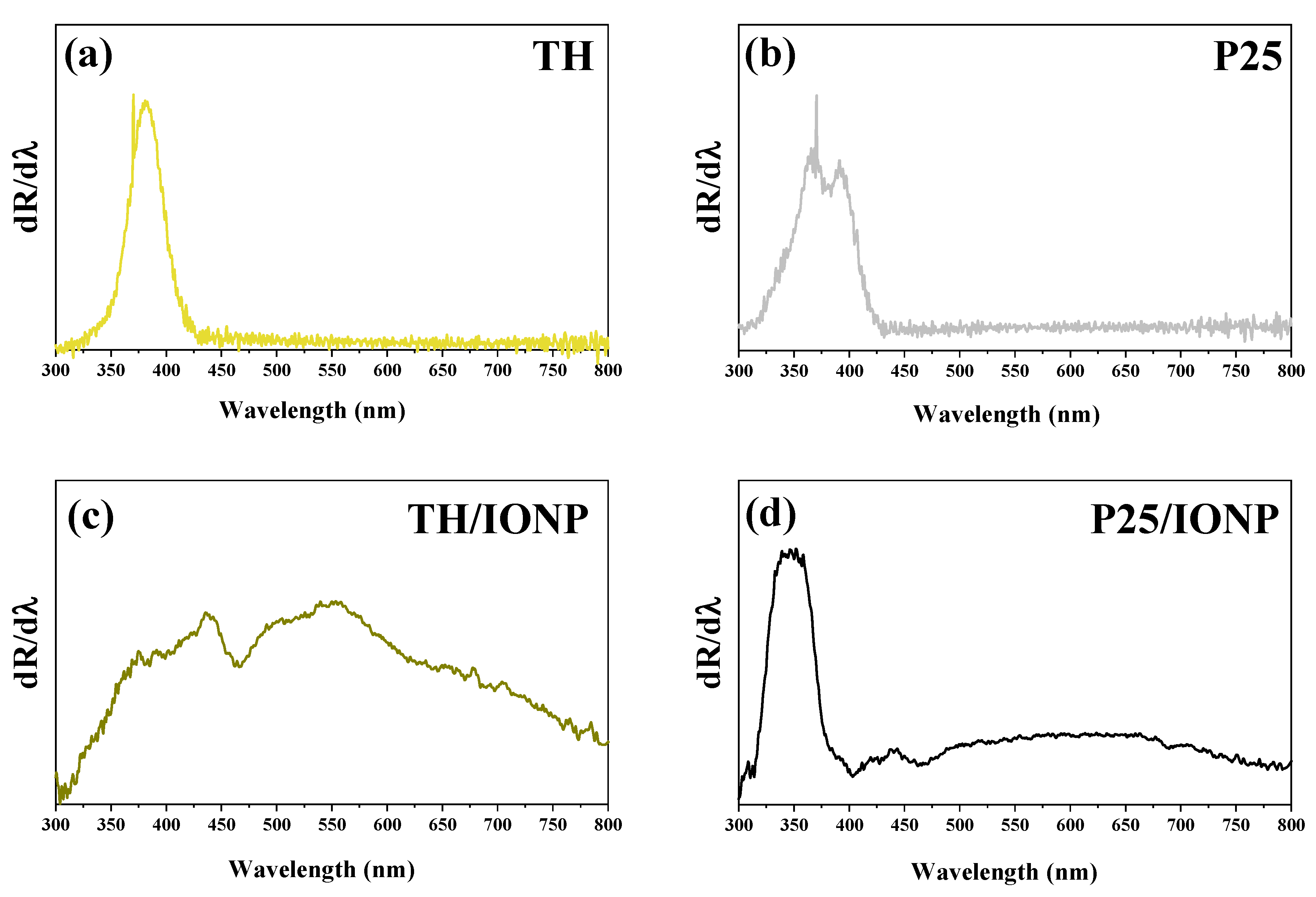
In the case of the TH/IONP composite, a broader non-linear region was observed, with a shift in the adsorption edge towards the visible region. Owing to the inaccuracies caused by the low reflectance values, the application of the classical Kubelka–Munk equation for the band gap energy calculation of the composites and of the IONP was avoided, and the optical properties were further explored by analyzing the first derivative spectra of the samples (Figure 5).
Comparing the first derivative of the DRS spectrum of the bare P25 sample with its composite, the absorption edge of both the anatase and rutile components appears slightly shifted to the UV region at 350 and 390 nm, respectively (~3.5 and 3.1 eV). Considering the correlation between optical properties and the primary crystallite size of a sample, these changes can be accounted for by the slight growth in particle size when introducing them into composites. The peak of the magnetite absorption edge is observable at 440 nm and corresponds to a 2.8 eV photon energy, which is in good accordance with the literature, considering the alkaline media of its synthesis (Figure 5d) [49]. On the derivative of the TH/IONP composite spectra, the adsorption edges of anatase (376 nm, i.e., 3.3 eV) and magnetite (440 nm, i.e., 2.8 eV) are faintly distinguishable but in agreement with the previous literature (Figure 5c). A broad unidentified band is also observed in the 500–550 nm region, which could suggest the appearance of another form of iron oxide on the sample surface, such as maghemite (γ-Fe2O3), which is known to have band gap values in the mentioned region [50]. Although these optical properties of the TH/IONP composite suggest a low band gap energy and potential for visible light activation, these characteristics originate from the iron oxide component; it is known in the literature that Fe3O4 lacks intrinsic photocatalytic properties [51].

2.2. Investigation of the Photocatalytic Performance of the TiO2 Bases and Composites

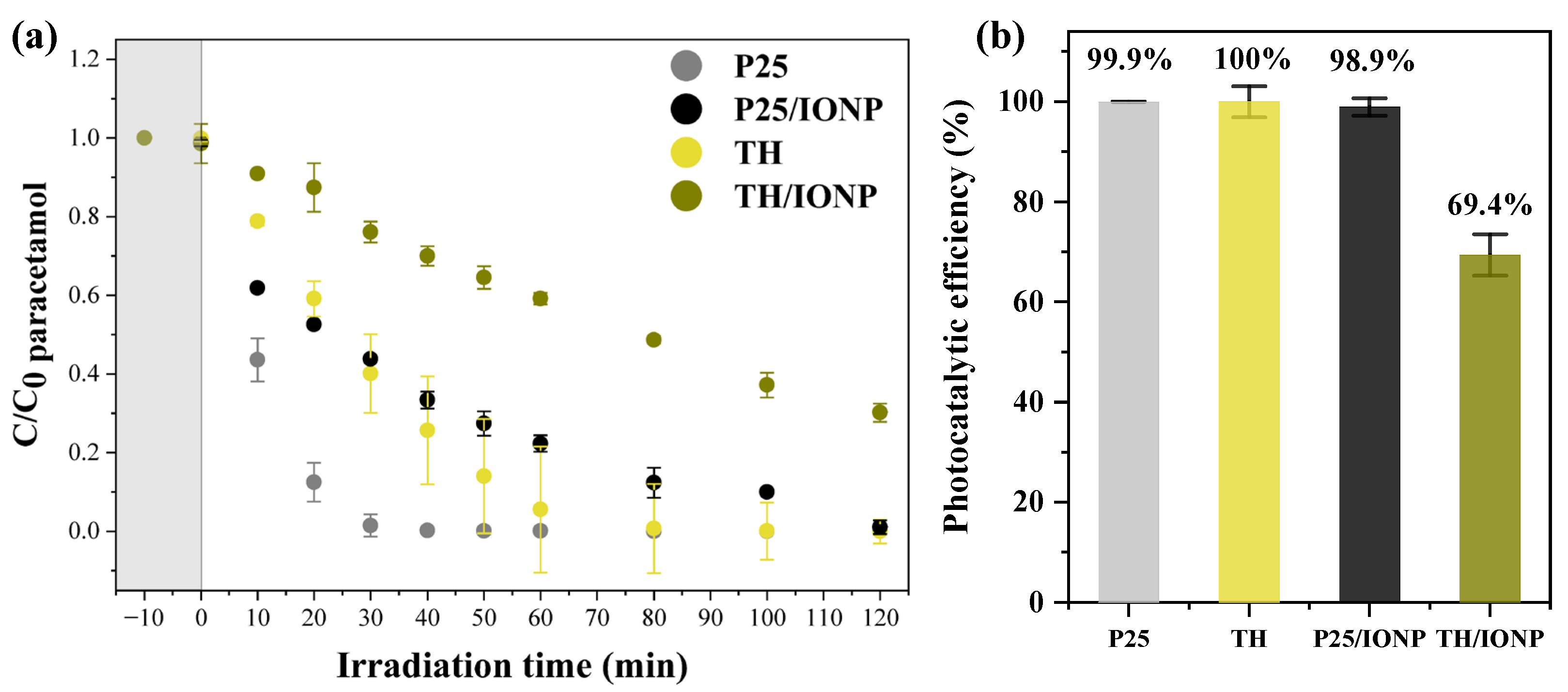
Following a more in-depth investigation of the structural and optical characteristics of both bare samples and composites, it was time to implement them in heterogeneous photocatalysis (Figure 6). For this purpose, the removal of paracetamol from aqueous solutions under UV-A irradiation was carried out.
As expected, bare commercial P25 TiO2 decomposed nearly all of the paracetamol solution in the first hour, reaching a final degradation efficiency of 99.9%. The synthesized TH anatase exhibited a similarly high photocatalytic performance, removing 100% of the paracetamol after 2 h of irradiation. However, in contrast to P25, the standard deviations for the measurement points of TH were larger during most of the photocatalytic reaction time, indicating a variability in its degradation profile. This suggests that while TH has a high efficiency with good repeatability (Figure 6), its intermediate photocatalytic behavior is less consistent.
Upon incorporation into a composite with IONP, the efficiency of both titania decreased. The P25/IONP composite showed slightly reduced activity compared to bare P25, achieving 98.9% degradation after 2 h. The TH/IONP composite proved to be the least effective among the four tested samples, with 69.4% efficiency after 2 h of irradiation. This value can still be considered a reasonable efficiency in comparison to the performance of other related catalytic systems presented in previous studies (Table S2). Similar decreases in photocatalytic activity compared to pure TiO2 have been reported in the literature, with photocatalytic performance strongly depending on the optimum TiO2:Fe3O4 ratio [29]. Thus, a plausible explanation of the efficiency decrease could be the partial shielding of active titania sites by the IONP, where junctions occurred in the composite. However, two other highly important aspects of magnetite presence should be considered in pursuit of an explanation. First, the stability of IONPs during repetitive photocatalytic runs must be studied, as iron is a variable oxidation state metal, with possible sensitivity towards various photochemical parameters in the system. Second, the possible role of magnetite, if any, in the charge transfer or photocatalytic processes should be investigated.

2.3. Recycling Tests of the Photocatalysts

To evaluate the main purpose of the magnetic composites, namely their reusability in photocatalytic degradation processes, and to tackle the first question regarding the stability of the iron oxide during recycling, all four samples were reused three additional times. After each cycle, the photocatalysts were recovered from the suspensions and dried at 50 °C. For simplicity and to better simulate real-life applications, the samples were not washed before recycling. This approach is not only practically more feasible but also reflects conditions closer to those in scalable or field-use scenarios. Before each catalytic cycle, adsorption–desorption tests were conducted, with the gained adsorption values being summarized in Table 3. Table 3 also presents the photocatalytic efficiencies of each photocatalytic test during the four consecutive recycling tests, along with corresponding apparent reaction rate constants calculated based on pseudo-first-order kinetics.
Adsorption values prior to the first photocatalytic test were generally low (<2%). However, after recycling, adsorption increased in some cases across all samples, reaching moderate values in the 5–10% range, without following a consistent trend. Notably, during one of the repetitions of the second photocatalytic runs of the TH sample, a significant concentration drop was observed after the adsorption phase, more exactly, 26.7% of the paracetamol was adsorbed prior to irradiation (Figure S8). Additionally, the presence of degradation intermediates was observed on the chromatogram of the sample taken after adsorption. The freshly prepared paracetamol solution is stable at room temperature and in the dark and does not decompose spontaneously. Considering this, the only explanation for paracetamol degradation products in the second, fresh solution is the desorption of these from the surface of the unwashed, recycled photocatalyst. Degradation intermediates on the photocatalyst’s surface can increase the photocatalyst’s hydrophilicity, thus enhancing its adsorption properties, which explains the occasional high adsorption values when the photocatalyst is not washed between cycles.
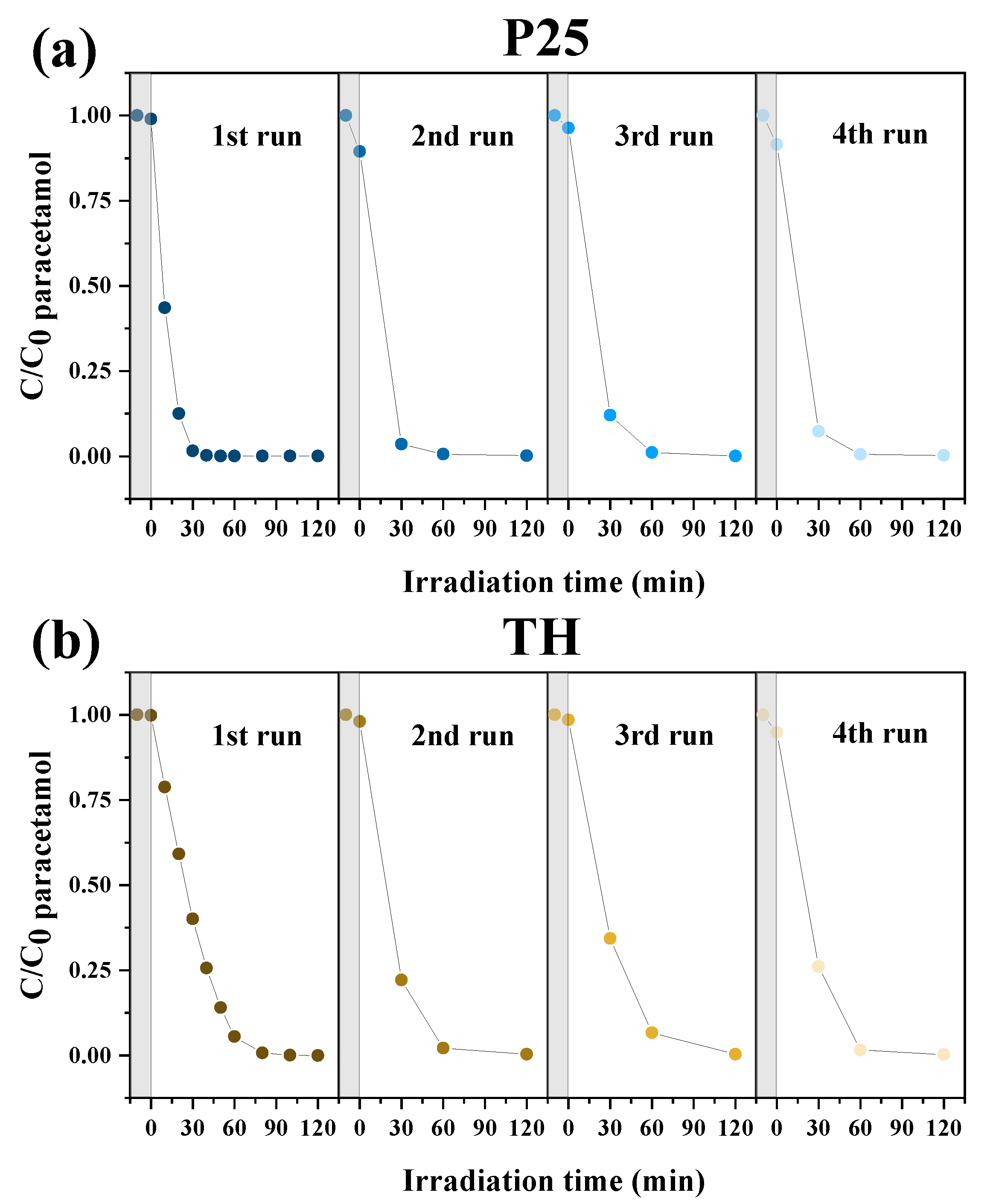
During repeated photocatalytic cycles, the bare P25 sample maintained exceptionally high efficiency, almost completely removing the paracetamol in all four runs (Table 3, Figure 7a). The TH anatase sample also demonstrated excellent recyclability, as well as an overall efficiency comparable to that of P25 (Table 3, Figure 7b).
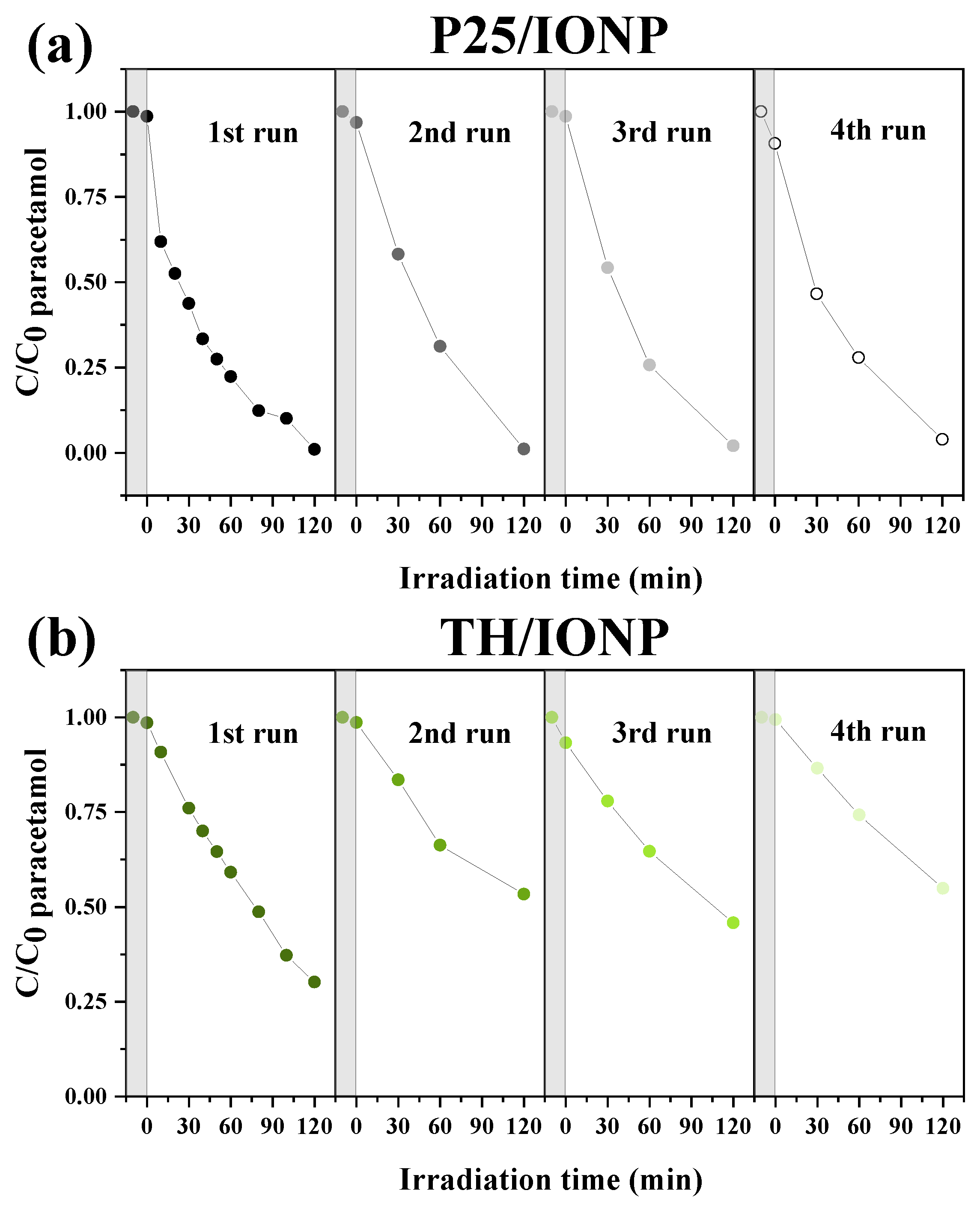
In contrast, the P25/IONP composite showed a gradual, but moderate, decline in its performance. Although it maintained its high efficiency, a small downward trend was noticeable by the fourth cycle, with a decrease from 98.9% efficiency to 95.7%. However, the apparent reaction rate constants showed no significant change, indicating that the composite retained good recyclability and photocatalytic activity over repeated use (Table 3, Figure 8a).
The composite prepared from TH and IONP exhibited not only the lowest efficiencies in paracetamol removal but also the poorest recyclability. After the initial degradation efficiency of 69.4%, its performance significantly dropped in the subsequent cycles, reaching 44.8% by the fourth cycle (Table 3, Figure 8b).
The apparent reaction rate constants generally remained stable across the photocatalytic cycles, showing a similar trend to the photocatalytic degradation efficiencies. While the kapp values belonging to P25 and TH were consistently higher, the composites containing IONP exhibited lower rate constants overall, with TH/IONP showing the smallest values. These results suggest that incorporating iron oxide in the composites, particularly in the TH-based composite, reduces the photocatalytic reaction rate and affects the stability of the degradation process during repeated use.
Studying only the recorded chromatograms and degradation curves of the samples during the repeated photocatalytic runs does not give significant information about the factors contributing to the reduced efficiency. As it is clear that this observation is linked to the introduction of iron oxide in the composites, the investigations led to additional structural and compositional characterizations of the samples after the photocatalytic tests.

2.4. Characterization of Photocatalysts After Degradation Cycles

To investigate any modifications in the crystal structure of the samples during photocatalytic runs and to verify the presence of IONP in the composites after recycling, their diffractograms were recorded before and after photocatalytic runs. The two bare TiO2 bases, P25 and TH, did not exhibit any modification in terms of structure and composition, both retaining their anatase–rutile and pure anatase crystal phases, respectively (Figure S9). Studying the diffraction patterns of the two composites, the characteristic reflection belonging to the crystal plane (311) of cubic Fe3O4 can be observed around 35.5 2θ°, even after the fourth photocatalytic run—confirming the presence of magnetite in the composites during and after reusability tests (Figure 9) in both composites. However, the XRD pattern of TH/IONP after the 4th run shows the appearance of an additional reflection, around 33 2θ°. This reflection is attributed to the (104) plane of hematite (JCPDS car no. 33-0664), indicating the gradual oxidation of bulk IONP magnetite to hematite in the TH/IONP composite, as the recycling advances.
After establishing the bulk composition of the composites, the investigation was focused on their surface properties, as they play a key role in photocatalytic processes. For this purpose, XPS measurements were performed.
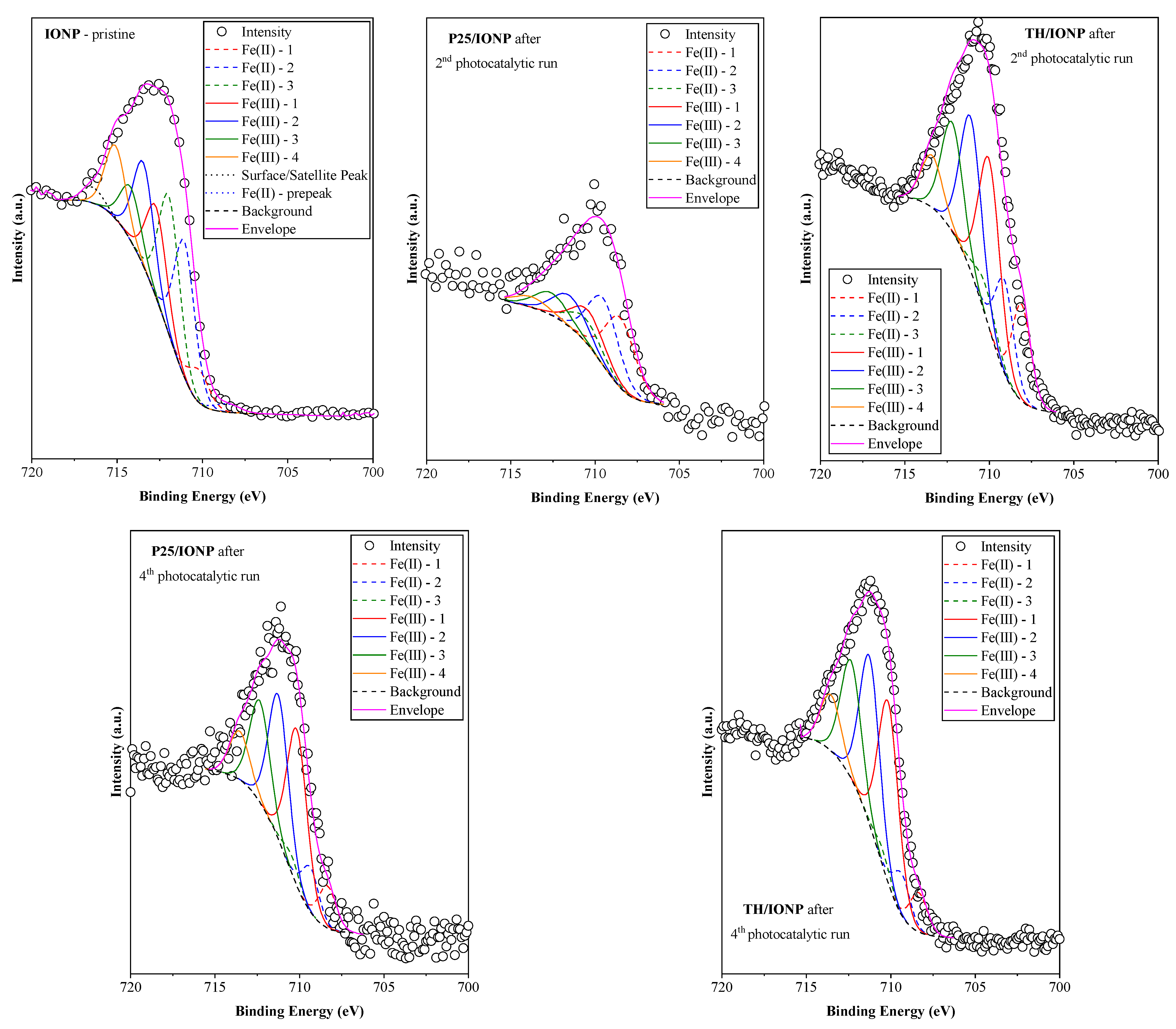
The main goal of the XPS investigation was to determine whether the iron in the magnetite samples suffered any modifications following the catalytic process. First, the pristine IONP was analyzed (Figure 10) considering its Fe 2p XPS spectra (more specifically, Fe 2p3/2). The identification of the components was carried out according to Grosvenor et al. [52]. Four Fe3+ signals (712.8, 713.5, 714.4, and 715.1 eV), three Fe2+ signals (710.3, 711.2, and 712.1 eV), a surface/satellite signal (716.5 eV), and a Fe2+ pre-signal (709.1 eV) were detected (please note that the latter two may or may not be present at all times). The order, ratio, and distance between the signals are in accordance with the Fe2p spectra of maghemite (1:1.1 Fe2+:Fe3+ ratio) [53]. This observation is surprising at first glance. However, XPS provides information regarding the surface of the particles. The other measurement techniques showed evidence towards magnetite, meaning that the bulk of the particles is composed of magnetite, while its surface shows signs of maghemite. It should be noted that these signals may slightly shift and change in intensity when the magnetic particles are included in composites.
Following the first two photocatalytic runs with paracetamol as the model pollutant, sample P25/IONP revealed no significant alterations in the Fe 2p spectrum (Figure 10), as the same components were successfully identified as the ones in the case of pristine IONP. This points out that the magnetic nanoparticles are somewhat stable alongside P25. The opposite was observed for sample TH/IONP, whereby the Fe3+ content increased significantly (1:2 Fe2+:Fe3+ ratio), suggesting an intense oxidation process after the second photodegradation (Figure 10). Following further catalytic runs, the Fe2p spectral components’ ratio values of TH/IONP changed even more, reaching the 1:4 Fe2+:Fe3+ ratio (Figure 10).

2.5. Linking Structural and Photocatalytic Data

As was observed and discussed previously, the different samples show various behaviors and interesting anomalies during the evaluation of photocatalytic activity, thus requiring an individual assessment.
P25 is one of the most efficient photocatalytic materials on the market. Its high-efficiency versatility can be attributed to its special anatase–rutile ratio [54]. In the present case, P25 showed the highest photocatalytic activity towards paracetamol. After four recycling experiments, the same activity was observed, emphasizing its superior and stable photocatalytic nature. TH, a pure anatase-containing material, showed a slightly lower performance in paracetamol degradation than P25, with lower apparent rate constants of the degradation reaction, as expected, due to the absence of anatase–rutile synergy. Interestingly, the degradation profile of paracetamol was not constant when TH was applied, but at the end of the 120 min irradiation, it demonstrated a consistently high efficiency. This observation is likely due to the adsorption of polar degradation intermediates, which increased its surface hydrophilicity and, in turn, modified its efficiency in the different stages of the degradation.
P25/IONP also achieved considerable photocatalytic activity, even though 10 w/w% of this composite was the magnetic component of the sample and had a lower photocatalytic material content. This was a trade-off between two properties—gaining removability and losing some photoactivity. During the recycling experiments, the composite suffered a small loss in activity; however, its overall performance was not affected significantly. This is notable given the known oxidative instability of magnetite if not properly protected. In contrast, the TH/IONP composite was the least photoactive in the degradation of paracetamol. Although it was expected that, similarly to the TH sample, a decent activity might be observed here, the results show the opposite of this. The composite TH/IONP lost a significant portion of its already poor activity during recycling and proved, overall, to have the lowest apparent rate constants (Table 3).
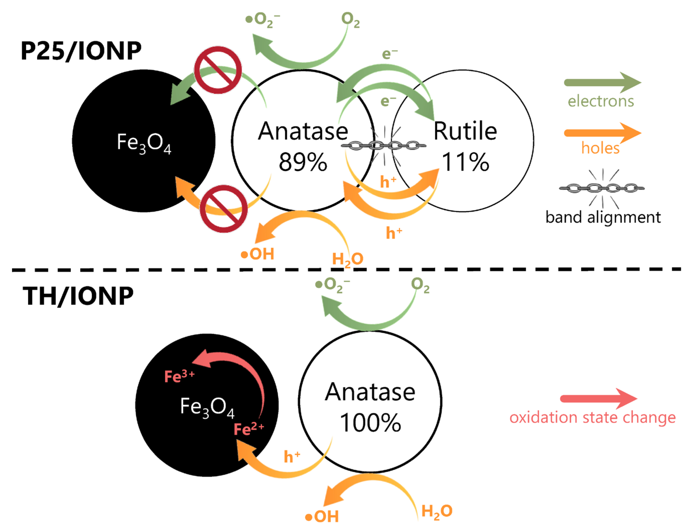
The differences observed in activity and anomalies are approachable if two important charge transfer-related phenomena are considered (Figure 11). The first is that anatase and rutile have a special synergy between them. If both are present, it is known that a band alignment occurs among the phases, facilitating the transfer of electrons from anatase to rutile and consequently permitting an efficient charge separation mechanism [26]. This way, all the photogenerated charges can migrate nearly unhindered between the particles of the two crystal phases, conferring high activity to the material. If the system contains magnetite particles (P25/IONP), they remain unaffected mostly by the photogenerated charges, as those are distributed among the TiO2 particles. Hence, magnetite may remain stable for a longer period. Alternatively, it has been reported that Fe3O4 can act as electron sinks, modulating the recombination rate of charge carriers, enhancing the photocatalytic activity of TiO2 [33]. The presence of an energetically favorable alignment in anatase–rutile–magnetite composites allows this mechanism by the migration of electrons from anatase to rutile and, consequently, from rutile to magnetite.
The second important charge transfer mechanism occurs when only anatase is present alongside magnetite (TH/IONP). Since the TH sample is pure anatase, the previously mentioned photogenerated charge distribution cannot occur. This way, the photogenerated charges are used directly, while some of the holes are free to oxidize magnetite, more precisely Fe2+ to Fe3+, which was reinforced by XPS measurements. Photogenerated electrons do not reach the magnetite nanoparticles at this point, as they are consumed by the dissolved O2 present in the system. Additionally, TH generates more charge carriers over time, which are partially consumed in the oxidation of the magnetic nanoparticles, transforming Fe2+ to Fe3+, rather than participating in pollutant degradation. Moreover, previous studies have shown that Fe3+ ions can act as recombination centers in TiO2/Fe3+ systems, hindering photocatalytic efficiency [55,56]; this is a mechanism that could occur in parallel if some of the photogenerated electrons of anatase reach the surface of IONP. After the 4th photocatalytic run, in the TH/IONP sample, the oxidation of Fe2+ to Fe3+ was observable not only on the surface of the IONP (indicated by XPS measurements) but also in the bulk, as its diffraction pattern showed signs of hematite in its structure (Figure 9).
However, it is known that during photocatalysis, TiO2, meaning both TH and P25, produces hydroxyl radicals (·OH) and superoxide anions (·O2), with ROS playing a major role in pollutant degradation [29,30,57]. This means that as several recycling experiments are carried out, even the P25-based composite may suffer changes in the Fe2+:Fe3+ ratio of the IONP component, if affected by these ROS. This was revealed by the XPS measurements of P25/IONP after the 4th photocatalytic run, as shown in Figure 10.

3. Materials and Methods

3.1. Chemicals

Iron (II) chloride tetrahydrate (FeCl2·4H2O, 99.99% trace metals basis), titanium (IV) isopropoxide (C12H28O4Ti, min. 98%), and sodium hydroxide (NaOH, analytical-grade pellets) were purchased from Sigma-Aldrich, Merck KGaA, Darmstadt, Germany. Commercial TiO2, namely Evonik Aeroxide P25, was supplied by Evonik Industries AG, Essen, Germany. Glacial acetic acid (C2H4O2, min. 99.5%) was purchased from Chempur, Sp. z o.o., Piekary Śląskie, Poland. Absolute ethanol (C2H5OH, min. 99.5%) was purchased from Chimreactiv S.R.L., Bucharest, Romania. All chemicals mentioned were used as received, without further purification. For the photocatalytic degradation tests, paracetamol tablets (500 mg active substance) were purchased from Terapia—SUN PHARMA company, Cluj-Napoca, Romania and processed according to “Section 3.6.1.”.

3.2. Synthesis of Iron Oxide Nanoparticles

A modified co-precipitation method was employed to synthesize iron oxide nanoparticles (IONPs), with different variations in synthesis parameters (Table S1). In total, 50 mL of NaOH solution with a specific concentration was prepared and kept at a constant temperature. The Fe2+ precursor solution of 0.2 M concentration was freshly prepared, adding 50 mL of distilled water to the FeCl2·4H2O. This second solution was added dropwise to the NaOH, keeping the previously adjusted temperature and the stirring constant, after which the synthesis solution was mixed for 1 h. The pH of the solution was monitored after the precursor addition and after the 1 h mixing process in order to ensure that the pH exceeded the 11.5 value. Following the stirring process, the obtained black suspension was allowed to cool down if necessary, and it was centrifuged and washed several times with distilled water and EtOH. The final step was drying for 12 h at 50 °C. The obtained black powder responded clearly to the proximity of a simple hand-held magnet, providing visual confirmation of the samples’ magnetic behavior during preliminary screening.

3.3. Synthesis of Anatase TiO2 Sample

Titanium (IV) isopropoxide (TTIP) was vigorously mixed with anhydrous ethanol (EtOH), followed by the gradual addition of a solution of acetic acid (AcOH) and water at ambient temperature under continuous agitation. The resulting solution was immediately added into a Teflon-lined stainless-steel autoclave and heated at 180 °C for 2 h. After the hydrothermal treatment, the autoclave was naturally chilled to ambient temperature. After centrifuging the resultant material, it was rinsed with distilled water a minimum of three times until the particle suspension had a pH value of 7 and no longer had a sour odor. Lastly, the as-synthesized white material was dried for 12 h at 80 °C. The molar ratio of TTIP:EtOH:AcOH:H2O was 1:22.75:2.33:1.35. The as-gained sample was named TH.

3.4. Preparation of Magnetic Composites of Synthetic Anatase TiO2 and P25

The synthesis of the magnetic composites was similar to the synthesis of IONPs described previously, with two main differences. As the first step in the preparation method, either commercial TiO2 P25 or the synthesized TiO2 was added to a 50 mL solution of 0.8 M NaOH under vigorous stirring. The temperature of the solution was adjusted only afterward. The second difference was the concentration of the aqueous FeCl2·4H2O solution, which was adjusted to 0.05 M, maintaining its volume at 50 mL. The total volume of the synthesis and every other step of the preparation method of IONPs was unmodified. The quantity of the TiO2 and the Fe2+ precursor was calculated to allow the final composite to contain 10 w/w% IONP.

3.5. Methods and Instrumentation

The crystal phase identification and the primary mean crystallite size values were determined using a Shimadzu XRD 6000 diffractometer (Shimadzu Corporation, Kyoto, Japan) using CuKα radiation (λ = 1.54 Å) with a Ni filter, in a 2θ range of 3–80°, with 0.2°/min speed for the IONPs, as well as a 20–80° range, with 2°/min speed, for the composites. The crystal phases of the synthesized materials were identified using the Powder Diffraction File database. The primary crystallite sizes were determined via the Scherrer equation [58], and the crystallinity degrees were determined with the help of the FullProf software suite Version 7.95, applying the following equation:
D e g r e e   o f   c r y s t a l l i n i t y   % = I c r y s t a l l i n e I t o t a l × 100
where Icrystalline is the area under the crystalline peaks, and Itotal is the area under all peaks.
To evaluate their structural stability, the untested and the recycled samples were also analyzed with a Rigaku MiniFlex II diffractometer (Rigaku Corporation, Tokyo, Japan) equipped with a graphite monochromator and functioning with Cu-Kα radiation (λ = 1.54 Å, 30 kV, 15 mA). The scanning speed was 2°/min, and the XRD diffractograms were recorded in the 2θ° range from 20 to 80°.
The reflectance spectra of the samples were recorded with a Jasco-V650 (Jasco Corporation, Wien, Austria) spectrophotometer in the 250–800 nm region, using an ILV-724 integration sphere for the transformation of the optical signal. The band gap energy (Eg) values were estimated based on the measured spectra using the Kubelka–Munk function [59]. However, for samples where this method proved unreliable, a differential approach was applied by analyzing the first derivative of the reflectance spectra (dR/dλ) to locate the inflection point corresponding to the absorption edge.
The sample’s FT-IR absorption spectra were obtained using a Jasco 6000 spectrometer (Jasco Corporation, Tokyo, Japan) in reflection mode in the wavelength range of 400 to 4000 cm−1 with a resolution of 4 cm−1. All materials examined were prepared as pellets using potassium bromide (KBr) in a hydraulic press.
Raman spectra were recorded with a multilaser confocal Renishaw inVia Reflex Raman spectrometer (Renishaw, Gloucestershire, UK) equipped with a RenCam CCD detector. The 633 nm laser was applied as an excitation source, and the Raman spectra were collected using a 0.9 NA objective of 100× magnification. For all spectra, the integration times were 10 s, 1800 lines/mm grating, and 10% of the laser maximum intensity—laser power was 20 mW. The spectral resolution was about 4 cm−1.
The samples’ morphology was investigated via Scanning Electron Microscopy (SEM), employing a Hitachi SU 8230 CFEG SEM (Hitachi High-Tech Corporation, Tokyo, Japan) operating at 30 kV accelerating voltage, as well as via Transmission Electron Microscopy (TEM), employing a cold field emission Hitachi HD2700 STEM (Hitachi High-Tech Corporation, Tokyo, Japan) operated at 200 kV.
Magnetic measurements were performed using a vibrating sample magnetometer, 12 T VSM (Cryogenic Limited, London, UK), at 300 K, with external magnetic fields up to 5 T. The prepared powders were sealed in epoxy resin for measurements. The saturation magnetizations (Ms) were determined from magnetization isotherms according to the approach to saturation law [60].
XPS spectra of samples before photocatalytic testing were recorded using a SPECS instrument featuring a PHOIBOS 150 MCD 9 hemispherical electron energy analyzer, in FAT mode (Specs GmbH, Berlin, Germany). The system utilized a monochromatic Al-K source (1486.6 eV), operated at 14 kV and 20 mA. The X-ray source was operated at a power of 200 W. High-resolution spectra for all detected elements were acquired with an analyzer pass energy of 20 eV, employing 0.05 eV steps for the analyzed samples.
After the photocatalytic runs, the XPS spectra of samples were collected with a Kratos XSAM 800 instrument (Kratos Analytical Ltd., Manchester, UK), with the Al Kα radiation source operated with 120 W (12 kV) of power. A low-energy electron flood gun was operated during data acquisition to negate any surface charging. The survey spectra were collected with a pass energy of 160 eV and a 1 eV step size. The high-resolution spectra were collected with a pass energy of 40 eV and a 0.1 eV step size; the following transitions were monitored: C 1s, O 1s, Ti 2p, and Fe 2p. In both cases, the high-resolution spectra were evaluated with the CasaXPS software package (version 2.3.26rev1.2Y) [61]. All high-resolution spectra were corrected with a Shirley background. The aliphatic component of the C 1s spectra at 284.8 eV was used as an inner reference. All peaks were fit with a Gauss–Lorentzian product function, where the Lorentzian contribution is 0.3.

3.6. Photocatalytic Experiments

3.6.1. Preparation of Paracetamol Solution from Tablets

The purity of paracetamol tablets concerning N-acetyl-para-aminophenol was 90.25%; this was calculated based on the weight of three tablets, each with 500 mg active substance content. The tablets were crushed and mixed with the agate mortar and pestle method. A 2 mM stock solution was prepared using room-temperature distilled water, which was filtered to remove the tablet’s insoluble fillers from the mixture. For the photocatalytic tests, a 0.1 mM paracetamol solution was freshly made each time from the above-mentioned stock solution; its content and concentration were verified with UV-Vis spectroscopy.

3.6.2. Method of Photocatalytic Activity Determination

The photocatalytic degradation experiments of the 0.1 mM paracetamol solutions were carried out in a Pyrex glass reactor with a cooling jacket circulating water at room temperature and surrounded by 6 × 6 W in the emission range of 315–400 nm. The emission maximum of the lamps was λmax ≈ 365 nm, with an irradiation intensity measured at the reactor position of 9.53 W/m2. The tested photocatalyst was added into the solution in a ccatalyst = 1 g·L−1 suspension concentration. The irradiation lasted for 2 h, during which the suspension was continuously stirred at 500 rpm and air was introduced into the system through bubbling.
Before each photocatalytic investigation, all suspensions were stirred in the dark for 10 min under the above-described conditions to reach an adsorption–desorption equilibrium.
During the adsorption and photocatalytic tests, the concentration changes in the paracetamol solution were followed with a high-performance liquid chromatograph (Hitachi D-7000 HPLC, Hitachi Ltd., Tokyo, Japan) with a Poroshell 120 C18 column using a 25:75 (V/V) methanol/water mixture eluent, a 0.7 mL·min−1 flow rate, and a λ = 243 nm detection wavelength. The sampling time was 10 min in the first hour and 20 min in the second hour.
Following the first photocatalytic run, the photocatalysts were recycled for three additional cycles, using the same photocatalytic testing procedure, with aliquots taken after the adsorption period and following 30, 60, and 120 min of irradiation. After each cycle, the photocatalysts were recovered, dried at 50 °C, and reused without a washing step in order to simulate real-life operating conditions where continuous use and minimal post-treatment are preferred for feasibility.

4. Conclusions

There is an abundance of studies relating to the combination of magnetite nanoparticles, in which synthesis, stability, and reusability aspects are the main research driving forces. However, relative stability during recycling is rarely investigated. In the present work, magnetite nanoparticles were prepared in order to obtain composite systems with two different, highly active titania photocatalysts. The obtained magnetite nanoparticles were pure and highly crystalline, making them adequate for the proposed investigation. By adding magnetite nanoparticles, the photoactivity towards paracetamol decreased in all cases; however, for the pure anatase sample, the activity decrease was significant, especially after multiple photocatalytic runs. This was explained by the different charge separation mechanisms occurring in P25 between anatase and rutile, as well as the lack of this mechanism in the anatase-containing sample, opening the pathway for the photooxidation of Fe2+ to Fe3+. These results also give insight into how magnetite can remain relatively stable for longer recycling times, without the need to introduce a third component into the composite to act as an insulating material, if an appropriate photocatalyst is chosen, such as P25.

Supplementary Materials

The following supporting information can be downloaded at https://www.mdpi.com/article/10.3390/catal15090839/s1. S2.1. Structural and morphological characterization of IONPs. Table S1. Overview of properties of IONPs synthesized in various conditions. Figure S1. XRD patterns of IONPs synthesized using various NaOH concentrations and temperatures. Figure S2. Raman spectra of the IONP samples. Figure S3. UV-Vis reflectance spectra of IONP samples. Figure S4. SEM images of iron oxide nanoparticles prepared under various experimental conditions: IONP_0.4_25—NaOH 0.4 M and 25 °C, IONP_0.4_75—NaOH 0.4 M and 75 °C, IONP_0.8_25—NaOH 0.8 M and 25 °C, and IONP_0.8_75—NaOH 0.8 M and 75 °C. Figure S5. FT-IR spectra of pristine IONP, TiO2, and their composites. Figure S6. TEM micrographs of pristine TiO2 TH, P25, and their composites. Figure S7. (a) UV-Vis spectra of synthesized TH TiO2, (b) band gap energy calculation of synthesized TH TiO2, (c) UV-Vis spectra of P25 TiO2, (d) band gap energy calculation of P25 TiO2. Figure S8. Adsorption properties and photocatalytic activity of the TH sample after the first two photocatalytic runs (repeated experimental set). Figure S9. XRD pattern of P25 (a) and TH (b) after synthesis, as well as after the 1st, 2nd, and 4th photocatalytic runs. Table S2. Comparison of the performance of the composite photocatalysts of the present study with the findings of the literature on related catalytic systems. References [28,29,30,49,57,62,63,64,65,66,67] are cited in the supplementary materials.

Author Contributions

Conceptualization: K.S. and Z.P.; methodology: Z.P., L.-C.P., K.M., and L.B.; validation: K.S., S.G., and L.S.; formal analysis: K.S., Z.P., R.T., and M.M.-P.; investigation: K.S., S.G., L.S., I.S., R.T., M.T., Á.S., L.B.-T., and K.M.; resources: Z.P. and L.B.; data curation: K.S.; writing—original draft preparation: K.S., R.T., and Z.P.; writing—review and editing: L.-C.P., Z.P., L.B., and K.M.; visualization: K.S. and Z.P.; supervision: Z.P., L.-C.P., and L.B.; project administration: Z.P. and L.B.; funding acquisition: Z.P. and L.B. All authors have read and agreed to the published version of the manuscript.

Funding

This research was funded by UEFISCDI (the Executive Unit for Higher Education, Research, Development and Innovation Funding), Romania, project number PN−III-P1-1.1-TE-2019-1318, in the framework of PNCDI III, Romania.

Data Availability Statement

The original contributions presented in this study are included in the article/Supplementary Materials. Further inquiries can be directed to the corresponding author.

Acknowledgments

K. Saszet is grateful for the funding received from the Collegium Talentum scholarship, provided by the Sapientia Hungariae Foundation. The authors gratefully acknowledge the contribution of Laura Vivien Lakatos in performing the photocatalytic degradation analysis that supported this study.

Conflicts of Interest

The authors declare no conflicts of interest.

References

  1. Umair, M.; Kanwal, T.; Loddo, V.; Palmisano, L.; Bellardita, M. Review on Recent Advances in the Removal of Organic Drugs by Advanced Oxidation Processes. Catalysts 2023, 13, 1440. [Google Scholar] [CrossRef]
  2. Hegedus, M.; Lacina, P.; Plotěný, M.; Lev, J.; Kamenická, B.; Weidlich, T. Fast and efficient hydrodehalogenation of chlorinated benzenes in real wastewaters using Raney alloy. J. Water Process Eng. 2020, 38, 101645. [Google Scholar] [CrossRef]
  3. Cardoso, I.M.F.; da Silva, L.P.; da Silva, J.C.G.E. Nanomaterial-Based Advanced Oxidation/Reduction Processes for the Degradation of PFAS. Nanomaterials 2023, 13, 1668. [Google Scholar] [CrossRef] [PubMed]
  4. Ma, D.; Xue, Q.; Liu, Y.; Liang, F.; Li, W.; Liu, T.; Zhuang, C.; Zhao, Z.; Li, S. Manipulating interfacial charge redistribution in Mn0.5Cd0.5S/N-rich C3N5 S-scheme heterojunction for high-performance photocatalytic removal of emerging contaminants. J. Mater. Sci. Technol. 2026, 243, 265–274. [Google Scholar] [CrossRef]
  5. Chi, S.W.; Din, A.T.M.; Abdullah, A.Z. Critical review on magnetic separable molecularly imprinted photocatalyst: Synthesis, reaction mechanism and outlook for practical applications. Mater. Sci. Semicond. Process 2025, 189, 109292. [Google Scholar] [CrossRef]
  6. Hao, Y.; Xiao, Y.; Liu, X.; Ma, J.; Lu, Y.; Chang, Z.; Luo, D.; Li, L.; Feng, Q.; Xu, L.; et al. A Novel SnO2/ZnFe2O4 Magnetic Photocatalyst with Excellent Photocatalytic Performance in Rhodamine B Removal. Catalysts 2024, 14, 350. [Google Scholar] [CrossRef]
  7. Liang, Y.; Yin, Y.; Deng, Q.; Jiao, S.; Liang, X.; Huo, C.; Luo, Y. Efficient and easily recyclable photocatalytic reduction of Se(IV) from wastewater using stable TiO2/BiOBr/cloth: Mechanism insight and machine learning modeling. Sep. Purif. Technol. 2025, 352, 128021. [Google Scholar] [CrossRef]
  8. Ibrahim, I.; Elseman, A.M.; Sadek, H.; Eliwa, E.M.; Abusaif, M.S.; Kyriakos, P.; Belessiotis, G.V.; Mudgal, M.M.; Abdelbasir, S.M.; Elsayed, M.H.; et al. Membrane-Based Photocatalytic and Electrocatalytic Systems: A Review. Catalysts 2025, 15, 528. [Google Scholar] [CrossRef]
  9. Yu, Z.-F.; Yang, Y.; Zhuang, H.-F.; Shan, S.-D.; Beldean-Galea, M.-S.; Xue, Q.-Q.; Shen, X.-F.; Li, S.-J. In-situ growth of MIL-53 (Fe) on charcoal sponge as a highly efficient and recyclable photocatalyst for removal of Cr(VI). Rare Met. 2024, 43, 4344–4355. [Google Scholar] [CrossRef]
  10. Li, S.; Wu, F.; Lin, R.; Wang, J.; Li, C.; Li, Z.; Jiang, J.; Xiong, Y. Enabling photocatalytic hydrogen production over Fe-based MOFs by refining band structure with dye sensitization. Chem. Eng. J. 2022, 429, 132217. [Google Scholar] [CrossRef]
  11. Bamba, D.; Coulibaly, M.; Fort, C.I.; Coteţ, C.L.; Pap, Z.; Vajda, K.; Zoro, E.G.; Yao, N.A.; Danciu, V.; Robert, D. Synthesis and characterization of TiO 2 /C nanomaterials: Applications in water treatment. Phys. Status Solidi 2015, 252, 2503–2511. [Google Scholar] [CrossRef]
  12. Mendonça, T.A.P.; Giroto, A.S.; Chambi, J.T.; Cuffini, S.L.; Vieira, N.C.S.; Gonçalves, M. Efficient photocatalyst based on activated carbon/graphene oxide/TiO2 synthesized under acidic conditions for environmental remediation. J. Photochem. Photobiol. A Chem. 2025, 462, 116244. [Google Scholar] [CrossRef]
  13. Yang, X.-C.; Zhao, J.-T. Aerogel for Highly Efficient Photocatalytic Degradation. Gels 2024, 10, 100. [Google Scholar] [CrossRef] [PubMed]
  14. Baia, L.; Vulpoi, A.; Radu, T.; Karácsonyi, É.; Dombi, A.; Hernádi, K.; Danciu, V.; Simon, S.; Norén, K.; Canton, S.E.; et al. TiO2/WO3/Au nanoarchitectures’ photocatalytic activity “from degradation intermediates to catalysts’ structural peculiarities” Part II: Aerogel based composites—Fine details by spectroscopic means. Appl. Catal. B 2014, 148–149, 589–600. [Google Scholar] [CrossRef]
  15. Chen, F.; Zhao, J. Preparation and photocatalytic properties of a novel kind of loaded photocatalyst of TiO2/SiO2/γ-Fe2O3. Catal Lett. 1999, 58, 245–247. [Google Scholar] [CrossRef]
  16. Kalidass, J.; Reji, M.; Sivasankar, T. Synthesis of Fe3O4 nanoparticles with enhanced properties via sonoelectrochemical approach: A comparative study with electrochemical and hydrothermal method. Chem. Eng. Process.—Process Intensif. 2024, 197, 109690. [Google Scholar] [CrossRef]
  17. Nguyen, M.D.; Tran, H.-V.; Xu, S.; Lee, T.R. Fe3O4 Nanoparticles: Structures, Synthesis, Magnetic Properties, Surface Functionalization, and Emerging Applications. Appl. Sci. 2021, 11, 11301. [Google Scholar] [CrossRef]
  18. Demortière, A.; Panissod, P.; Pichon, B.P.; Pourroy, G.; Guillon, D.; Donnio, B.; Bégin-Colin, S. Size-dependent properties of magnetic iron oxidenanocrystals. Nanoscale 2011, 3, 225–232. [Google Scholar] [CrossRef]
  19. Srivastava, N.; Srivastava, M.; Alhazmi, A.; Mohammad, A.; Khan, S.; Pal, D.B.; Haque, S.; Singh, R.; Mishra, P.K.; Gupta, V.K. Sustainable green approach to synthesize Fe3O4/α-Fe2O3 nanocomposite using waste pulp of Syzygium cumini and its application in functional stability of microbial cellulases. Sci. Rep. 2021, 11, 24371. [Google Scholar] [CrossRef]
  20. Nyirő-Kósa, I.; Rečnik, A.; Pósfai, M. Novel methods for the synthesis of magnetite nanoparticles with special morphologies and textured assemblages. J. Nanoparticle Res. 2012, 14, 1150. [Google Scholar] [CrossRef]
  21. López, J.; Rey, A.; Viñuelas-Zahinos, E.; Álvarez, P.M. Preparation of a new green magnetic Fe3O4 @TiO2-P25 photocatalyst for solar advanced oxidation processes in water. J. Environ. Chem. Eng. 2023, 11, 109999. [Google Scholar] [CrossRef]
  22. Li, Z.-D.; Wang, H.-L.; Wei, X.-N.; Liu, X.-Y.; Yang, Y.-F.; Jiang, W.-F. Preparation and photocatalytic performance of magnetic Fe3O4@TiO2 core–shell microspheres supported by silica aerogels from industrial fly ash. J. Alloys Compd. 2016, 659, 240–247. [Google Scholar] [CrossRef]
  23. Cendrowski, K.; Sikora, P.; Zielinska, B.; Horszczaruk, E.; Mijowska, E. Chemical and thermal stability of core-shelled magnetite nanoparticles and solid silica. Appl. Surf. Sci. 2017, 407, 391–397. [Google Scholar] [CrossRef]
  24. Korina, E.; Stoilova, O.; Manolova, N.; Rashkov, I. Polymer fibers with magnetic core decorated with titanium dioxide prospective for photocatalytic water treatment. J. Environ. Chem. Eng. 2018, 6, 2075–2084. [Google Scholar] [CrossRef]
  25. Wittmar, A.S.M.; Fu, Q.; Ulbricht, M. Photocatalytic and Magnetic Porous Cellulose-Based Nanocomposite Films Prepared by a Green Method. ACS Sustain. Chem. Eng. 2017, 5, 9858–9868. [Google Scholar] [CrossRef]
  26. Guo, Q.; Zhou, C.; Ma, Z.; Yang, X. Fundamentals of TiO 2 Photocatalysis: Concepts, Mechanisms, and Challenges. Adv. Mater. 2019, 31, 1901997. [Google Scholar] [CrossRef]
  27. Ohtani, B.; Prieto-Mahaney, O.O.; Li, D.; Abe, R. What is Degussa (Evonik) P25? Crystalline composition analysis, reconstruction from isolated pure particles and photocatalytic activity test. J. Photochem. Photobiol. A Chem. 2010, 216, 179–182. [Google Scholar] [CrossRef]
  28. Tang, P.; Ding, Y.; Chen, H.; Wang, Y. Preparation of TiO2/Fe3O4 composite by sol-gel method and its photocatalytic activity for removal of Rhodamine B from water. Ferroelectrics 2020, 562, 66–72. [Google Scholar] [CrossRef]
  29. Kubiak, A. Comparative study of TiO2–Fe3O4 photocatalysts synthesized by conventional and microwave methods for metronidazole removal. Sci. Rep. 2023, 13, 12075. [Google Scholar] [CrossRef]
  30. Madima, N.; Kefeni, K.K.; Mishra, S.B.; Mishra, A.K.; Kuvarega, A.T. Fabrication of magnetic recoverable Fe3O4/TiO2 heterostructure for photocatalytic degradation of rhodamine B dye. Inorg. Chem. Commun. 2022, 145, 109966. [Google Scholar] [CrossRef]
  31. Lendzion-Bieluń, Z.; Wojciechowska, A.; Grzechulska-Damszel, J.; Narkiewicz, U.; Śniadecki, Z.; Idzikowski, B. Effective processes of phenol degradation on Fe3O4–TiO2 nanostructured magnetic photocatalyst. J. Phys. Chem. Solids 2020, 136, 109178. [Google Scholar] [CrossRef]
  32. Kubiak, A.; Kubacka, M.; Gabała, E.; Dobrowolska, A.; Synoradzki, K.; Siwińska-Ciesielczyk, K.; Czaczyk, K.; Jesionowski, T. Hydrothermally Assisted Fabrication of TiO2-Fe3O4 Composite Materials and Their Antibacterial Activity. Materials 2020, 13, 4715. [Google Scholar] [CrossRef]
  33. Harifi, T.; Montazer, M. A novel magnetic reusable nanocomposite with enhanced photocatalytic activities for dye degradation. Sep. Purif. Technol. 2014, 134, 210–219. [Google Scholar] [CrossRef]
  34. Chang, J.; Zhang, Q.; Liu, Y.; Shi, Y.; Qin, Z. Preparation of Fe3O4/TiO2 magnetic photocatalyst for photocatalytic degradation of phenol. J. Mater. Sci. Mater. Electron. 2018, 29, 8258–8266. [Google Scholar] [CrossRef]
  35. Pandey, B.; Pandey, A.K.; Bhardwaj, L.; Dubey, S.K. Biodegradation of acetaminophen: Current knowledge and future directions with mechanistic insights from omics. Chemosphere 2025, 372, 144096. [Google Scholar] [CrossRef] [PubMed]
  36. Wang, Z.; Chen, H.; Rong, C.; Li, A.; Hua, X.; Dong, D.; Liang, D.; Liu, H. Photocatalytic Degradation of Acetaminophen in Aqueous Environments: A Mini Review. Toxics 2023, 11, 604. [Google Scholar] [CrossRef]
  37. Imran, M.; Riaz, S.; Naseem, S. Synthesis and Characterization of Titania Nanoparticles by Sol-gel Technique. Mater. Today Proc. 2015, 2, 5455–5461. [Google Scholar] [CrossRef]
  38. Nalbandian, L.; Patrikiadou, E.; Zaspalis, V.; Patrikidou, A.; Hatzidaki, E.; Papandreou, C.N. Magnetic Nanoparticles in Medical Diagnostic Applications: Synthesis, Characterization and Proteins Conjugation. Curr. Nanosci. 2016, 12, 455–468. [Google Scholar] [CrossRef]
  39. Ghalamchi, L.; Rasoulifard, M.H. Immobilization of Fe3O4/TiO2 nanocomposite thin layer on the glass tubes in a component parabolic collector for the treatment of DR23. Int. J. Environ. Sci. Technol. 2019, 16, 7509–7522. [Google Scholar] [CrossRef]
  40. Younis, A.B.; Milosavljevic, V.; Fialova, T.; Smerkova, K.; Michalkova, H.; Svec, P.; Antal, P.; Kopel, P.; Adam, V.; Zurek, L.; et al. Synthesis and characterization of TiO2 nanoparticles combined with geraniol and their synergistic antibacterial activity. BMC Microbiol. 2023, 23, 207. [Google Scholar] [CrossRef]
  41. Thamir, A.D.; Haider, A.J.; Ali, G.A. Preparation of NanostructureTiO2 at Different Temperatures by Pulsed Laser Deposition as Solar Cell. Eng. Technol. J. 2016, 34, 193–204. [Google Scholar] [CrossRef]
  42. Anselmi, C.; Mosconi, E.; Pastore, M.; Ronca, E.; De Angelis, F. Adsorption of organic dyes on TiO2 surfaces in dye-sensitized solar cells: Interplay of theory and experiment. Phys. Chem. Chem. Phys. 2012, 14, 15963. [Google Scholar] [CrossRef] [PubMed]
  43. Kurtan, U.; Topkaya, R.; Baykal, A.; Toprak, M.S. Temperature dependent magnetic properties of CoFe2O4/CTAB nanocomposite synthesized by sol–gel auto-combustion technique. Ceram. Int. 2013, 39, 6551–6558. [Google Scholar] [CrossRef]
  44. Stoner, E.C.; Wohlfarth, E.P. A mechanism of magnetic hysteresis in heterogeneous alloys, Philosophical Transactions of the Royal Society of London. Series A. Math. Phys. Sci. 1948, 240, 599–642. [Google Scholar] [CrossRef]
  45. Alzoubi, G.M.; Masadeh, A.S.; Shatnawi, M.T.M. Investigation of the structural, morphological, and magnetic properties of small crystalline Co–Cu ferrite nanoparticles in the single-domain regime. AIP Adv. 2022, 12. [Google Scholar] [CrossRef]
  46. Muthukrishnan, S.; Vidya, R.; Sjåstad, A.O. Band gap engineering of anatase TiO2 by ambipolar doping: A first principles study. Mater. Chem. Phys. 2023, 299, 127467. [Google Scholar] [CrossRef]
  47. Shi, H.; Lin, Y.; Jiang, Z.; Su, Y.; Ding, X.; Zhang, X.; Zhu, H.; Zhang, R. Enhanced optical absorption and photocatalytic activity of anatase TiO2 through C Nd-codoped: A DFT+ U calculations. J. Phys. Chem. Solids 2017, 109, 70–77. [Google Scholar] [CrossRef]
  48. Hossain, M.K.; Mortuza, A.A.; Sen, S.K.; Basher, M.K.; Ashraf, M.W.; Tayyaba, S.; Mia, M.N.H.; Uddin, M.J. A comparative study on the influence of pure anatase and Degussa-P25 TiO2 nanomaterials on the structural and optical properties of dye sensitized solar cell (DSSC) photoanode. Optik 2018, 171, 507–516. [Google Scholar] [CrossRef]
  49. Compeán-Jasso, M.E.; Ruiz, F.; Martínez, J.R.; Herrera-Gómez, A. Magnetic properties of magnetite nanoparticles synthesized by forced hydrolysis. Mater. Lett. 2008, 62, 4248–4250. [Google Scholar] [CrossRef]
  50. Mirza, I.M.; Ali, K.; Sarfraz, A.K.; Ali, A.; Ul Haq, A. A study of dielectric, optical and magnetic characteristics of maghemite nanocrystallites. Mater. Chem. Phys. 2015, 164, 183–187. [Google Scholar] [CrossRef]
  51. Bui, H.T.; Im, S.M.; Kim, K.; Kim, W.; Lee, H. Photocatalytic degradation of phenolic compounds of defect engineered Fe3O4: An alternative approach to solar activation via ligand-to-metal charge transfer. Appl. Surf. Sci. 2020, 509, 144853. [Google Scholar] [CrossRef]
  52. Grosvenor, A.P.; Kobe, B.A.; Biesinger, M.C.; McIntyre, N.S. Investigation of multiplet splitting of Fe 2p XPS spectra and bonding in iron compounds. Surf. Interface Anal. 2004, 36, 1564–1574. [Google Scholar] [CrossRef]
  53. Akbar, A.; Riaz, S.; Bashir, M.; Naseem, S. Effect of Fe3+/Fe2+ Ratio on Superparamagnetic Behavior of Spin Coated Iron Oxide Thin Films. IEEE Trans. Magn. 2014, 50, 1–4. [Google Scholar] [CrossRef]
  54. Žerjav, G.; Žižek, K.; Zavašnik, J.; Pintar, A. Brookite vs. rutile vs. anatase: What`s behind their various photocatalytic activities? J. Environ. Chem. Eng. 2022, 10, 107722. [Google Scholar] [CrossRef]
  55. Kanakaraju, D.; Abdullah, M.A.B.; Chin, L.Y. TiO2/PKSAC functionalized with Fe3O4 for efficient concurrent removal of heavy metal ions from water. Colloid. Interface Sci. Commun. 2021, 40, 100353. [Google Scholar] [CrossRef]
  56. Komaraiah, D.; Radha, E.; Sivakumar, J.; Reddy, M.V.R.; Sayanna, R. Influence of Fe3+ ion doping on the luminescence emission behavior and photocatalytic activity of Fe3+, Eu3+-codoped TiO2 thin films. J. Alloys Compd. 2021, 868, 159109. [Google Scholar] [CrossRef]
  57. Zhang, Q.; Yu, L.; Xu, C.; Zhang, W.; Chen, M.; Xu, Q.; Diao, G. A novel method for facile preparation of recoverable Fe3O4@TiO2 core-shell nanospheres and their advanced photocatalytic application. Chem. Phys. Lett. 2020, 761, 138073. [Google Scholar] [CrossRef]
  58. Hossain, M.S.; Ahmed, S. Easy and green synthesis of TiO2 (Anatase and Rutile): Estimation of crystallite size using Scherrer equation, Williamson-Hall plot, Monshi-Scherrer Model, size-strain plot, Halder- Wagner Model. Results Mater. 2023, 20, 100492. [Google Scholar] [CrossRef]
  59. Landi, S.; Segundo, I.R.; Freitas, E.; Vasilevskiy, M.; Carneiro, J.; Tavares, C.J. Use and misuse of the Kubelka-Munk function to obtain the band gap energy from diffuse reflectance measurements. Solid. State Commun. 2022, 341, 114573. [Google Scholar] [CrossRef]
  60. Zhang, H.; Zeng, D.; Liu, Z. The law of approach to saturation in ferromagnets originating from the magnetocrystalline anisotropy. J. Magn. Magn. Mater. 2010, 322, 2375–2380. [Google Scholar] [CrossRef]
  61. Fairley, N.; Fernandez, V.; Richard-Plouet, M.; Guillot-Deudon, C.; Walton, J.; Smith, E.; Flahaut, D.; Greiner, M.; Biesinger, M.; Tougaard, S.; et al. Systematic and collaborative approach to problem solving using X-ray photoelectron spectroscopy. Appl. Surf. Sci. Adv. 2021, 5, 100112. [Google Scholar] [CrossRef]
  62. Haavik, C.; Stølen, S.; Fjellvåg, H.; Hanfland, M.; Häusermann, D. Equation of state of magnetite and its high-pressure modification: Thermodynamics of the Fe-O system at high pressure. American Mineralogist 2000, 85, 514–523. [Google Scholar] [CrossRef]
  63. Kim, W.; Suh, C.-Y.; Cho, S.-W.; Roh, K.-M.; Kwon, H.; Song, K.; Shon, I.-J. A new method for the identification and quantification of magnetite–maghemite mixture using conventional X-ray diffraction technique. Talanta 2012, 94, 348–352. [Google Scholar] [CrossRef] [PubMed]
  64. Qu, B.; Bonifacio, C.S.; Majidi, H.; van Benthem, K. Stabilization of metal(II)oxides on the nanoscale. Mater. Res. Lett. 2020, 8, 41–47. [Google Scholar] [CrossRef]
  65. Testa-Anta, M.; Ramos-Docampo, M.A.; Comesaña-Hermo, M.; Rivas-Murias, B.; Salgueiriño, V. Raman spectroscopy to unravel the magnetic properties of iron oxide nanocrystals for bio-related applications. Nanoscale Adv. 2019, 1, 2086–2103. [Google Scholar] [CrossRef]
  66. Drummond, A.L.; Feitoza, N.C.; Duarte, G.C.; Sales, M.J.A.; Silva, L.P.; Chaker, J.A.; Bakuzis, A.F.; Sousa, M.H. Reducing Size-Dispersion in One-Pot Aqueous Synthesis of Maghemite Nanoparticles. J. Nanosci. Nanotechnol. 2012, 12, 8061–8066. [Google Scholar] [CrossRef]
  67. Khasawneh, O.F.S.; Palaniandy, P.; Ahmadipour, M.; Mohammadi, H.; Hamdan, M.R.B. Removal of acetaminophen using Fe2O3-TiO2 nanocomposites by photocatalysis under simulated solar irradiation: Optimization study. J. Environ. Chem. Eng. 2021, 9, 104921. [Google Scholar] [CrossRef]
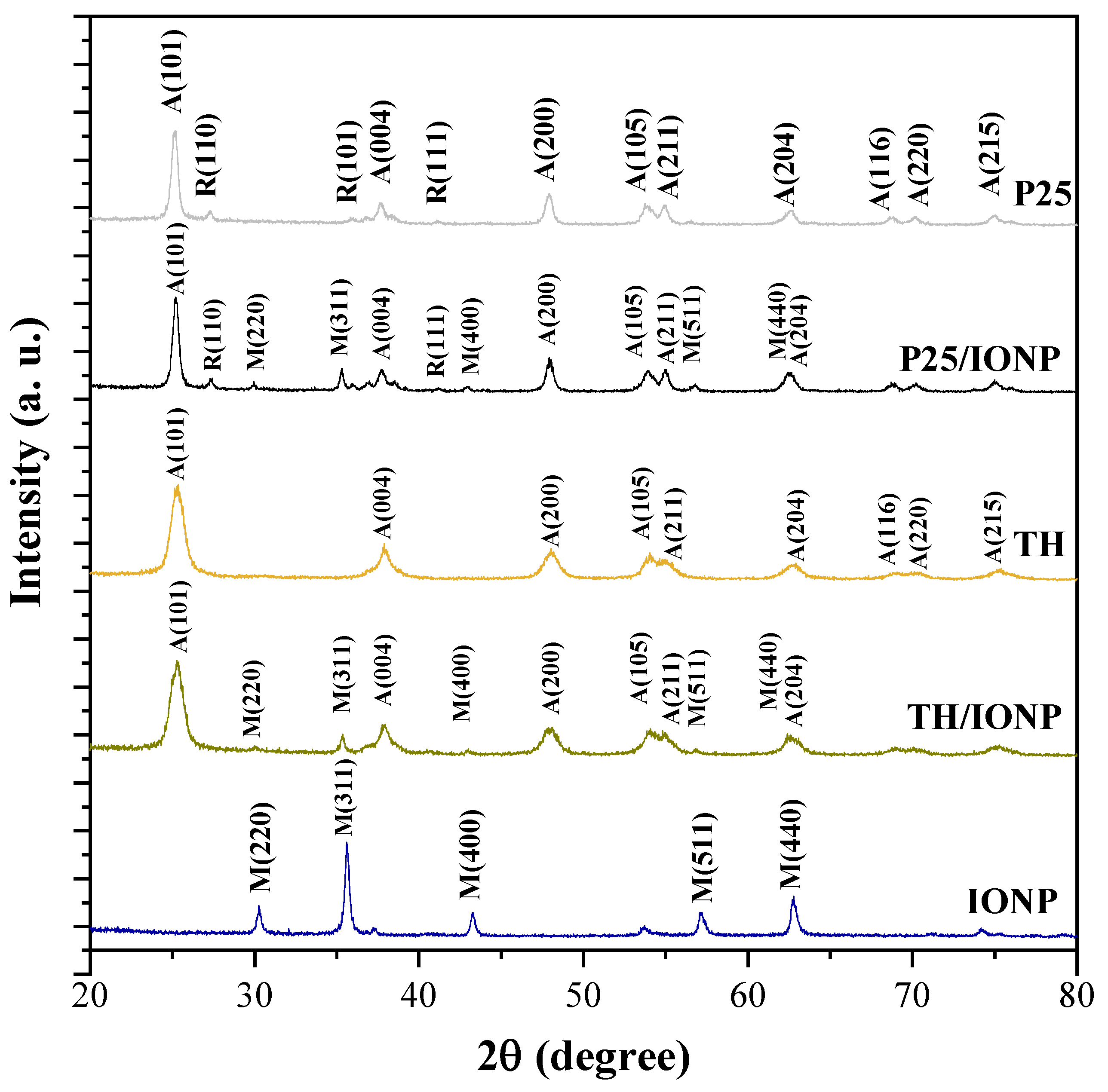
Figure 1. XRD patterns of the synthesized TiO2 sample (TH), the commercial TiO2 (P25), the chosen IONP, and their composites.
Catalysts 15 00839 g001
Figure 2. Magnetization isotherms measured at 300 K for IONP_0.8_75, P25/IONP, and TH/IONP.
Catalysts 15 00839 g002
Figure 3. Hysteresis loops measured at 300 K (a); close-up around 0 T (b).
Catalysts 15 00839 g003
Figure 4. UV-Vis spectra of the synthesized TiO2 sample (TH), the commercial TiO2 (P25), synthesized IONP, and their composites.
Catalysts 15 00839 g004
Figure 5. First order derivative of UV-Vis spectra of TH (a), P25 (b), TH/IONP (c), and P25/IONP (d).
Catalysts 15 00839 g005
Figure 6. Photocatalytic investigation of the synthesized TiO2 sample (TH), the commercial TiO2 (P25), and their composites prepared with IONP_0.8_75, using paracetamol as a model pollutant (a). Paracetamol degradation efficiency of the samples (b).
Catalysts 15 00839 g006
Figure 7. Comparison of the photocatalytic activity of the samples P25 (a) and TH (b) during the four photocatalytic runs.
Catalysts 15 00839 g007
Figure 8. Comparison of the photocatalytic activity of the samples P25/IONP (a) and TH/IONP (b) during the four photocatalytic runs.
Catalysts 15 00839 g008
Figure 9. XRD pattern of P25/IONP (a) and TH/IONP (b) after synthesis and after the 1st, 2nd, and 4th photocatalytic runs.
Catalysts 15 00839 g009
Figure 10. XPS core spectra of Fe2p in the pristine IONP, as well as in P25/IONP and TH/IONP, following paracetamol degradation.
Catalysts 15 00839 g010
Figure 11. The differences in the charge transfer mechanism of the two composites with magnetite content.
Catalysts 15 00839 g011
Table 1. Overview of synthesis conditions and crystalline properties of IONP_0.8_75 and TiO2 samples and their composites.
Sample Name NaOH
Concentration
[M]
Synthesis
Temperature
[°C]
Hydrothermal Treatment [°C] Crystal Phase Primary Crystallite Size [nm]
IONP_0.8_750.875-magnetite46.4
P25-25-anatase24
rutile36
TH-25180anatase9.2
P25/IONP0.875-anatase25
rutile41
magnetite65.2
TH/IONP0.875-anatase9.9
magnetite52.3
Table 2. Saturation magnetizations (Ms), coercive fields (μ0Hc), remanent magnetization (Mr), and the squareness ratio (Mr/Ms).
Sample NameMs (emu/g)μ0Hc (mT)Mr (emu/g)Mr/Ms
IONP_0.8_7554.2 (2)149.380.17
P25/IONP7.51 (1)151.970.26
TH/IONP4.22 (7)141.10.26
Table 3. Adsorption rates, degradation efficiency, apparent rate constant (kapp), and coefficient of determination (R2) of photocatalysts P25, TH, P25/IONP, and TH/IONP in each recycling run.
Photocatalyst Cycle Adsorption (%) Paracetamol Degradation Efficiency (%) kapp (min−1) R2
P2511.099.90.1320.9566
210.599.90.0820.9677
33.799.90.0750.9983
48.499.70.0850.9999
TH10.1100.00.0460.9470
21.999.70.0640.9838
31.599.60.0450.9842
45.299.70.0680.9563
P25/IONP11.498.90.0230.9799
23.298.80.0190.9966
31.497.90.0220.9961
49.395.70.0200.9944
TH/IONP11.469.40.0090.9998
21.345.90.0070.9911
36.750.90.0060.9999
40.644.80.0050.9991
Disclaimer/Publisher’s Note: The statements, opinions and data contained in all publications are solely those of the individual author(s) and contributor(s) and not of MDPI and/or the editor(s). MDPI and/or the editor(s) disclaim responsibility for any injury to people or property resulting from any ideas, methods, instructions or products referred to in the content.