Article

Synergistic Effects of UV Radiation and H2O2 on Chloramphenicol Degradation by REE-Based Catalysts

by
Alice Cardito
1,
Mariateresa Lettieri
2,
Lorenzo Saviano
3,
Olga Sacco
1,
Giusy Lofrano
4,*,
Vincenzo Vaiano
5,
Giovanni Libralato
3,
Marco Guida
3 and
Maurizio Carotenuto
1
1
Department of Chemistry and Biology “A. Zambelli”, University of Salerno, Via Giovanni Paolo II 132, 84084 Fisciano, Italy
2
CNR-SPIN (National Research Council—Institute for SuPerconductors, INnovative Materials, and Devices), c/o University of Salerno, Via Giovanni Paolo II 132, 84084 Fisciano, Italy
3
Department of Biology, University of Naples Federico II, Via Vicinale Cupa Cinthia 26, 80126 Naples, Italy
4
Department of Psychology and Health Science, Pegaso University, Centro Direzionale Isola F2, 80126 Naples, Italy
5
Department of Industrial Engineering, University of Salerno, Via Giovanni Paolo II 132, 84084 Fisciano, Italy
*
Author to whom correspondence should be addressed.
Catalysts 2025, 15(8), 776; https://doi.org/10.3390/catal15080776
Submission received: 11 July 2025 / Revised: 5 August 2025 / Accepted: 12 August 2025 / Published: 14 August 2025

Abstract

The persistent occurrence of antibiotics like chloramphenicol (CAP) in aquatic systems poses serious environmental and public health risks. This study investigates the photocatalytic degradation of CAP using cerium oxide (CeO2), lanthanum oxide (La2O3), and lanthanum-doped cerium oxide (CexLayO2−δ), synthesized via co-precipitation. The catalysts were tested under a solar simulator, UV-A, and UV-C radiation, both with and without hydrogen peroxide (H2O2). Structural characterization confirmed successful synthesis of nanometric catalysts, with La doping causing lattice expansion in CeO2 and a reduction in crystallite size (from 27 nm in CeO2 to ~20 nm in doped samples). Photolysis alone achieved limited CAP removal (~34–35%), while photocatalysis with La2O3 under UV-A and UV-C improved removal up to 58% and 55%, respectively. Complete degradation was obtained with La2O3 under UV-C in the presence of H2O2 within 15 min. Pareto analysis highlighted the dominant effect of the interaction between radiation and H2O2 (43%), while the catalyst type contributed minimally (0.23%). These findings confirm the potential of REE oxides, especially La2O3, in advanced oxidation processes and underscore the importance of light source and radical generation over catalyst selection alone.

1. Introduction

Over the last few decades, the contamination of aquatic ecosystems has emerged as a critical global concern. One of the most pressing issues in this context is the presence of contaminants of emerging concern (CECs) in water bodies, derived from domestic and industrial activities. CECs are defined as “naturally occurring, manufactured or anthropogenic chemicals or materials that have recently been detected or are suspected to be present in various environmental compartments”, and “are believed to pose potential risks to human health or the environment but are not yet regulated by established standards or guidelines” [1]. This broad classification includes pharmaceuticals and personal care products, endocrine-disrupting compounds, flame retardants, and pesticides. Among pharmaceuticals, antibiotics are the most frequently detected compounds [2]. Global antibiotic consumption increased by 65% between 2000 and 2015, and it is predicted to rise to 200% by 2030 relative to 2015 levels [3]. Antibiotic consumption is directly linked to the emergence of antibiotic resistance bacteria, which poses a serious and persistent threat to public health, with significant associated morbidity and mortality [4].
Conventional wastewater treatment plants, based on activated sludge processes, are generally unable to completely remove CECs, resulting in their continuous release into the aquatic environment [5]. In this context, advanced oxidation processes (AOPs) have gained attention as promising alternatives for the degradation of these persistent pollutants, allowing non-selective oxidation of organic contaminants in wastewater. Among AOPs, photocatalysis, based on the use of semiconductors under light irradiation, has proven to be an effective method for removing a wide range of CECs from aqueous matrices due to the generation of hydroxyl radicals, which are powerful oxidants able to degrade a wide range of organic contaminants [6].
In recent years, oxides of rare earth elements (REEs) have attracted significant attention as promising materials for enhancing photocatalytic efficiency. Due to their unique physicochemical characteristics, such as high surface area, reactivity, and redox potential, REEs are suitable for catalyzing the formation of reactive oxygen species (ROS) under UV irradiation [7]. Among REEs, cerium (Ce) has been the most extensively studied, followed by lanthanum (La). Cerium oxide (CeO2) and lanthanum oxide (La2O3) are novel catalytic materials that exhibit remarkable photocatalytic performance due to their unique electronic and optical properties, particularly related to their 4f electronic transitions [8,9]. The reported band gaps of CeO2 and La2O3 are 3.2 eV and 5.2 eV, respectively [10,11]. Their high photocatalytic activity is primarily attributed to the presence of oxygen vacancies on the surface and the efficient redox cycling between Ce4+/Ce3+ and La3+/La2+, which facilitates the formation and elimination of oxygen defects [12,13]. The general photocatalytic degradation mechanism of pollutants by REE oxides can be described by the following reactions [14]:
R E E   o x i d e s + h v R E E   o x i d e s ( e C B + h V B + )
H O + h ( V B ) + H O
O 2 + e ( C B ) O 2
O 2 + H + H O 2
H O 2 + H O 2 O 2 + H 2 O 2
H 2 O 2 + O 2 H O + H O + O 2
H O + O r g a n i c   p o l l u t a n t s I n t e r m e d i a t e s C O 2 + H 2 O + P r o d u c t s
Additionally, both Ce and La oxides can act as photo Fenton-like catalysts in the presence of hydrogen peroxide, promoting H2O2 decomposition and the generation of radicals that further enhance pollutant degradation [15,16].
Doping CeO2 with La has been reported to increase photocatalytic activity. The incorporation of La3+ ions into the CeO2 lattice (replacing Ce4+ ions) generates additional oxygen vacancies to maintain charge balance, thereby enhancing catalytic performance [17].
In this study, CeO2, La2O3, and lanthanum-doped cerium oxide (CexLayO2−δ) were synthesized via a simple precipitation method and evaluated for the photocatalytic degradation of chloramphenicol (CAP), a broad-spectrum antibiotic commonly used to treat various bacterial infections ranging from conjunctivitis to meningitis. CAP has been detected in municipal sewage, wastewater treatment plant effluents, river water, and river sediment worldwide [18]. It is known for its high toxicity, with well-documented adverse effects on aquatic organisms and algae [19]. Several studies dealing with the photocatalytic degradation of CAP have been performed in the last few decades, as shown in Table S1. To the best of our knowledge, only one previous study investigated CAP removal using CeO2 [20], and this work represents the first investigation of CAP photocatalytic degradation using La2O3 and CexLayO2−δ.

2. Results and Discussion

2.1. Characterization of REE Oxides

The SEM analysis of the CeO2 sample revealed a compact material with a regular morphology characterized by flat regions (Figure 1A,D). The La2O3 samples exhibited a homogenous morphology (Figure 1B), showing the typical powder-like appearance (Figure 1E) reported in a previous study [21]. The morphology of CexLayO2−δ (Figure 1C) was very similar to that observed in CeO2; however, some porous regions, indicative of particle aggregation, were also observed.
The results of the EDX analyses are reported in Table 1. In the CeO2 sample, cerium and oxygen were the most abundant elements, with a Ce/O atomic ratio of 0.8 ± 0.4, which is higher than the expected stoichiometric value of 0.5. For the La2O3 samples, lanthanum and oxygen were the predominant elements, although the measured La/O ratio (0.5 ± 0.1) was lower than the expected stoichiometric value of 0.67. EDX analysis of the CexLayO2−δ sample revealed high concentrations of cerium and oxygen, along with a small amount of lanthanum. The stoichiometric coefficients were calculated using the elemental composition (as atomic percentage—At%) measured from the EDX analysis. More in detail, starting from the formula CexLayO2−δ, (i.e., the general formula of the lanthanum-doped CeO2), the following equation was applied: y = (La At%)/(Ce At% + La At%) and x = 1 − y. The coefficient for oxygen was calculated as (2 − δ) = (100 − (Ce At% + La At%))/(O At%). The calculation was repeated for each of the spot EDX analyses performed on the sample, the results were averaged, and the standard deviations were determined. The stoichiometry coefficients resulted in x = 0.96 ± 0.01, y = 0.04 ± 0.01, and δ = 0.82 ± 0.04.
EDX analyses were performed on distinct morphological features of CexLayO2−δ, such as smooth areas and particulate (Figure 2). The composition of the flat regions was consistent with the average composition of the entire sample, whereas the particle aggregates displayed higher oxygen content and slightly lower cerium content. In all samples, the detected silver and carbon were attributed to the conductive silver coating and to the carbon tape used for SEM preparation, respectively.
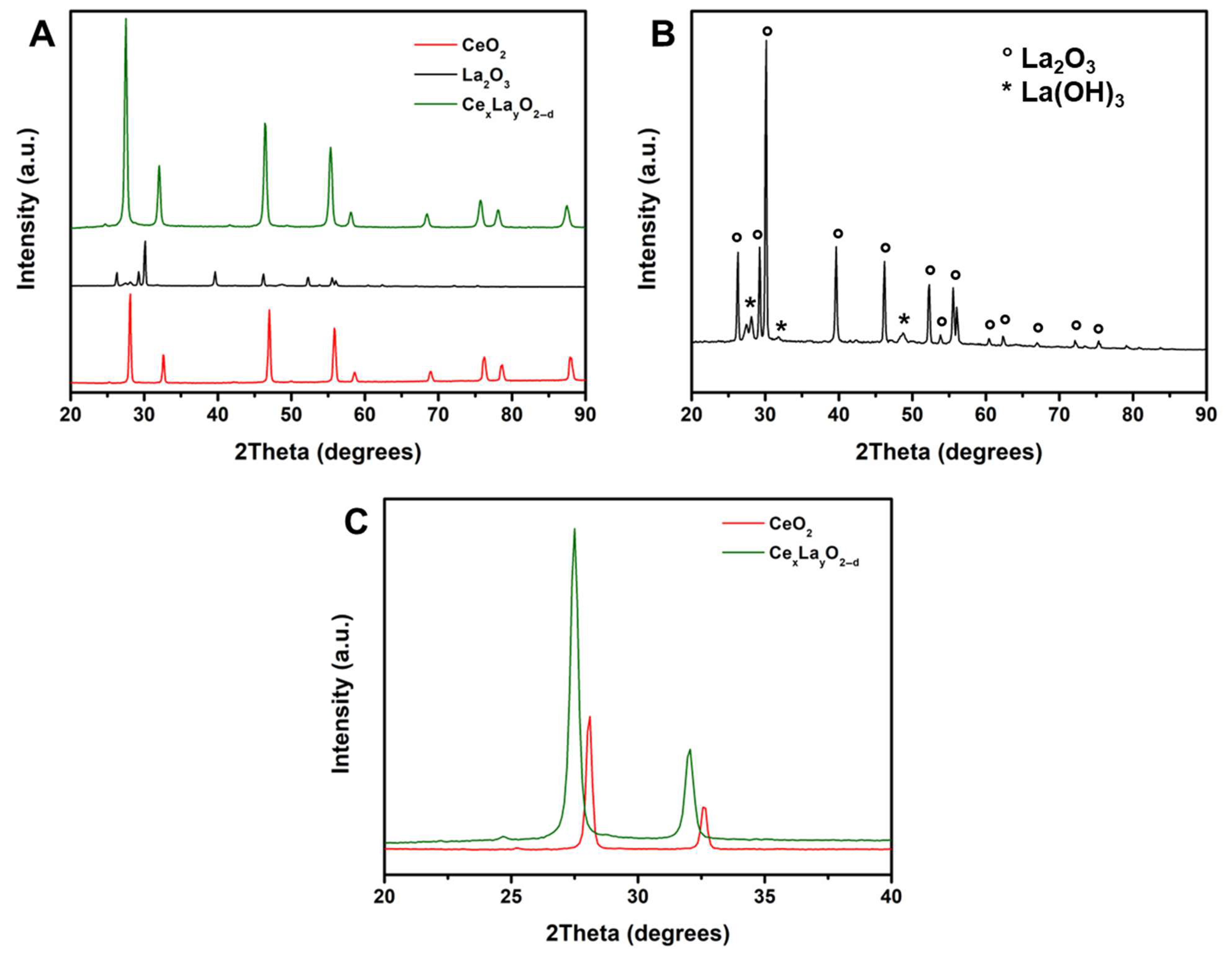
XRD patterns of the synthesized REE oxides powders are shown in Figure 3A. The CeO2 sample exhibited characteristic diffraction peaks at 28.51°, 33.07°, 47.43°, 56.33°, 59.06°, 69.38°, 76.66°, 79.09°, and 88.70° [22]. The XRD pattern of La2O3 displayed peaks corresponding to the presence of La(OH)3 [23] (Figure 3B). The CexLayO2−δ sample exhibited a diffraction pattern similar to that of CeO2; however, a slight shift in the peaks towards lower angles was observed (Figure 3C), attributed to the substitution of Ce4+ ions (ionic radius: 87 pm) by larger La3+ ions (ionic radius: 103 pm), resulting in lattice expansion [24].
The average crystallite size of CeO2 was estimated to be 27 nm, while that of La2O3 and CexLayO2−δ waws approximately 20 nm. These values are consistent with previous studies employing co-precipitation method for the synthesis of cerium and lanthanum oxides [17,25,26].

2.2. Photolysis and Photocatalytic Degradation of CAP

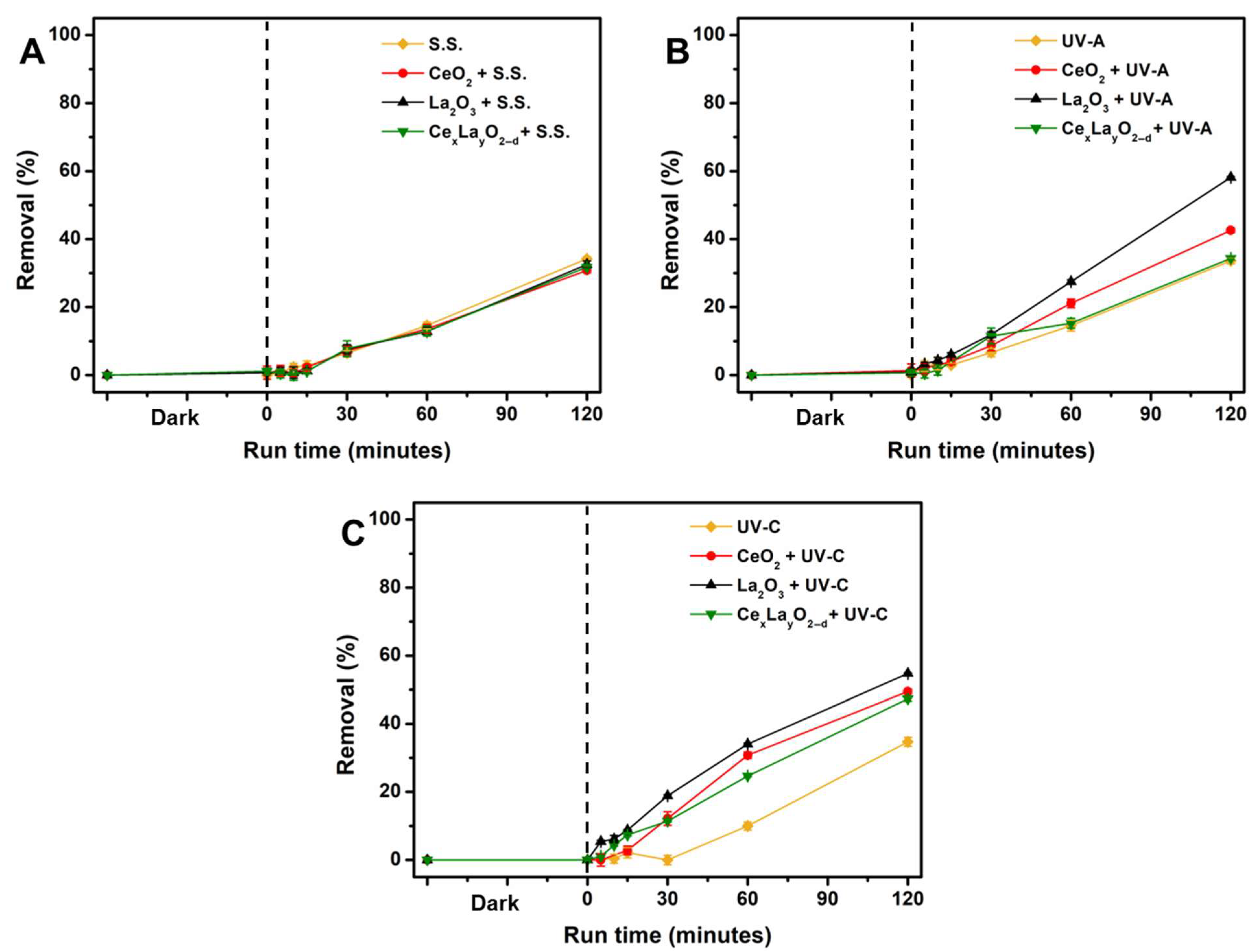
The results of CAP photolysis under solar, UV-A, and UV-C radiation are reported in Figure 4. After 120 min, all photolysis experiments resulted in similar CAP removal efficiencies, with overall removal of 34–35%, regardless of the radiation source.
Degradation of CAP by direct photolysis is known to be challenging due to its low quantum yield (approximately 3%, independent of wavelength) [27,28]. For example, da Rocha et al. [29] reported only ~5% and ~25% removal of CAP after 2 h under solar and UV-C irradiation, respectively, using a 20 mg/L solution. Higher removal efficiencies (up to 21.8% with solar and 83.3% with UV-C) were achieved only after extended photolysis duration (up to 12 h). Table 2 summarizes relevant literature data on CAP photolysis. The removal observed in this study, using UV-C, is comparable with those observed in studies [30,31,32], where UV-C lamps were also used and the same treatment time was established. It is worth noting that the initial concentration was relatively higher (25 and 9.69 mg/L, respectively).
Figure 4 shows the CAP degradation results using REE oxides under various radiation sources. No significant CAP adsorption on the catalysts was detected during the initial 60 min dark phase.
Under solar irradiation, CAP removal reached approximately 31–33% after 120 min, suggesting that photolysis was the dominant process (Figure 4A). This observation aligns with prior reports stating that CeO2 and La2O3 mainly absorb UV light [26].
Under UV-A irradiation, photocatalytic treatment led to CAP removals of 43% with CeO2, 58% with La2O3, and 34% with lanthanum-doped cerium oxide after 120 min (Figure 4B). Photocatalysis under UV-C resulted in CAP removals of 49%, 55%, and 47% with CeO2, La2O3, and CexLayO2−δ, respectively (Figure 4C).
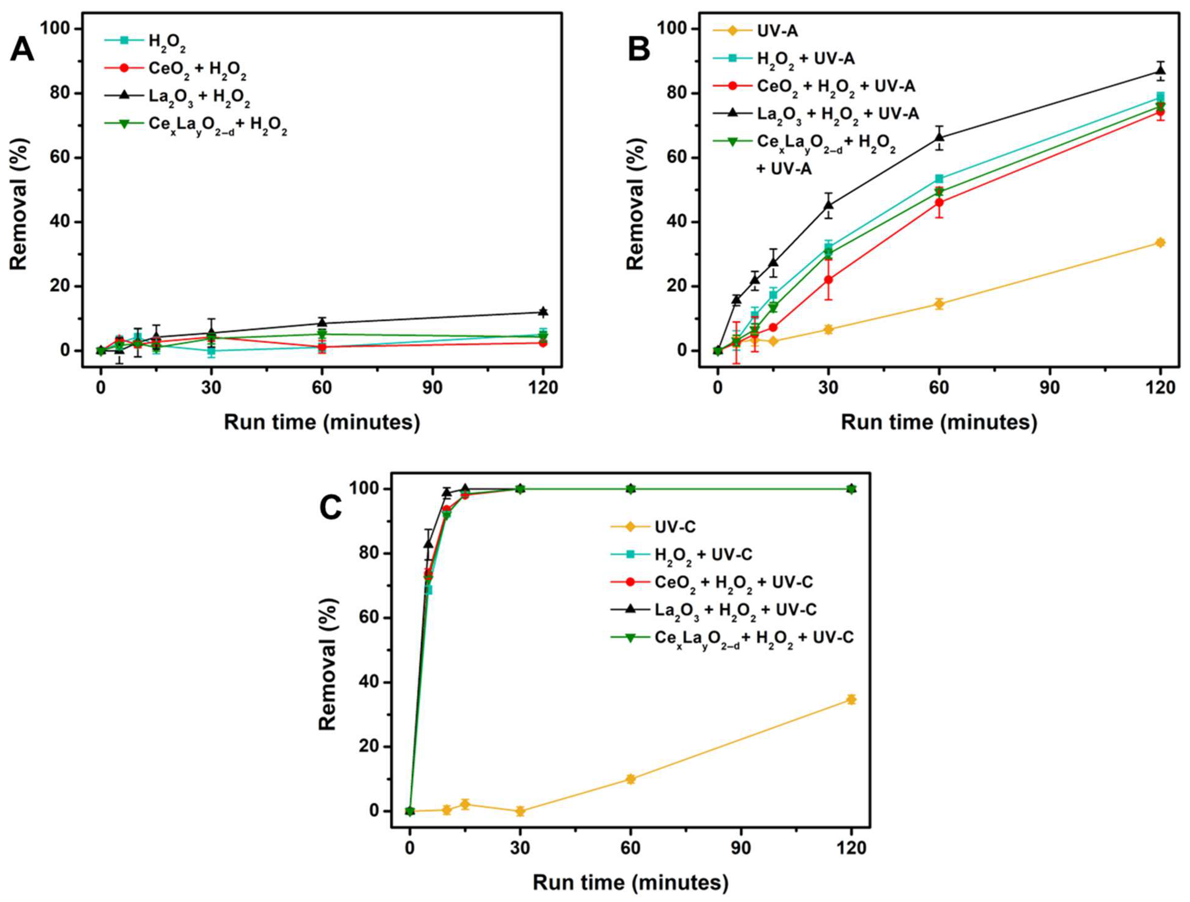
Figure 5 presents the results obtained by combining REE oxides with H2O2.
In dark conditions, minimal CAP degradation was observed after 120 min, with removal rates ranging from 2% (H2O2/CeO2) to 12% (H2O2/La2O3) (Figure 5A), consistent with previous findings [30,36].
Under UV irradiation, H2O2 photolysis generates hydroxyl radicals (HO), enhancing CAP degradation:
H 2 O 2 + h v 2 H O
In UV-A experiments, CAP removals reached 79% with H2O2 alone, and 74%, 87%, and 76% with H2O2 combined with CeO2, La2O3, and lanthanum-doped cerium oxide, respectively (Figure 5B). The presence of H2O2 significantly enhanced CAP removal, improving photocatalytic efficiency by 29–42%.
Under UV-C irradiation, rapid CAP degradation occurred within 30 min in the presence of H2O2 (Figure 5C). The higher efficiency under UV-C is due to the stronger absorption of H2O2 at shorter wavelengths (200–350 nm), promoting rapid photolysis [37]. Notably, complete CAP degradation was achieved within 15 min using the H2O2/UV-C/ La2O3 system.
A previous study [17] showed that a synergistic effect was observed for La-doped cerium oxide in terms of removal of methylene blue when the La percentage was 10% (removal of 99.99% after 180 min). In our case, the doping level of 10% was not achieved, suggesting that further optimization of the doping level may be needed to enhance photocatalytic efficiency.
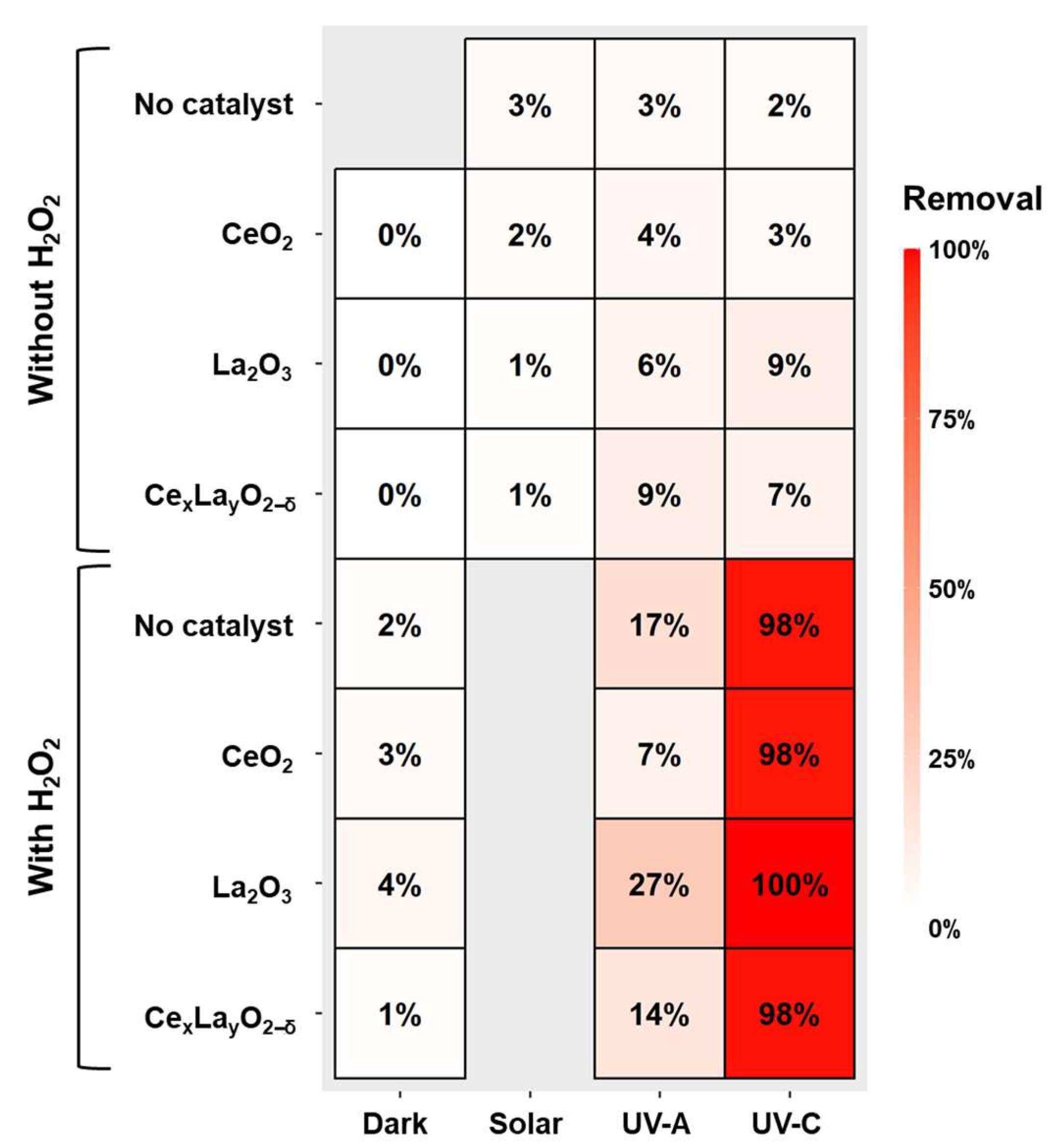
The removal results for all treatments after 15 min are summarized in the heatmap shown in Figure 6.
In most cases, CAP degradation followed pseudo-first-order kinetics, described by:
l n C t C 0 = k o b s t
where C t and C 0 are the CAP concentration at time t and 0, respectively, and k o b s is the observed rate constant for the photodecomposition reaction. The k o b s values, obtained from the slope of the linear plots, are reported in Table 3.
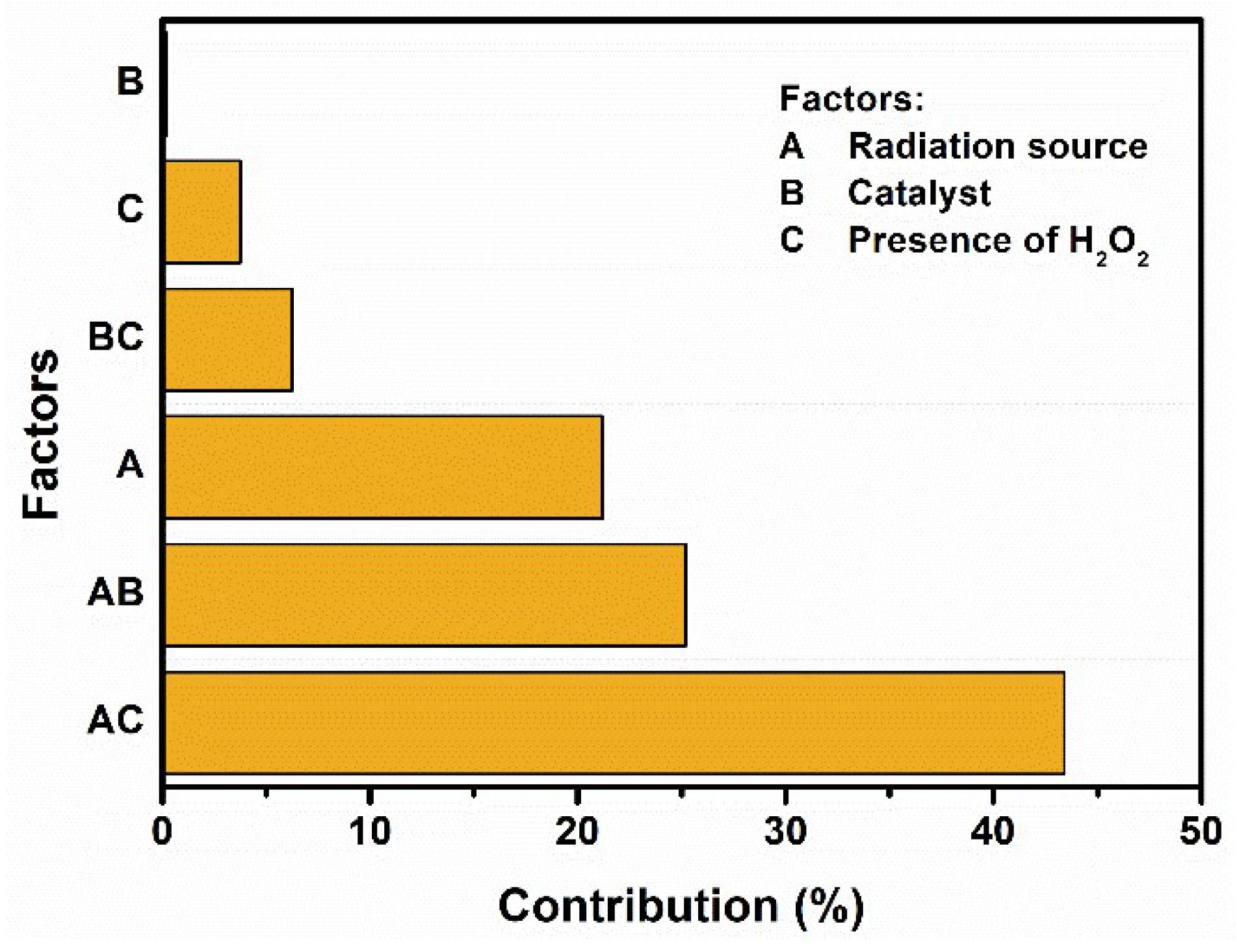
The relative contribution of each factor and their interactions on CAP degradation was evaluated from through Pareto chart (Figure 7). The most influential factor was the interaction between the radiation source (A) and the H2O2 addition (C), accounting for 43% of the observed variance. This reflects the high effectiveness of UV-C combined with H2O2, whereas limited degradation occurred under dark conditions. The catalyst type exhibited the least significant effect (0.23%), confirming similar performances among the tested oxides.

3. Materials and Methods

3.1. Synthesis of RRE Oxides

The REE oxides were synthetized following a co-precipitation method, as described by Ref. [38]. Cerium(III) nitrate hexahydrate (Ce(NO3)3 · 6H2O, CAS number: 10294-41-4, purchased by Merck) and lanthanum(III) nitrate hexahydrate (La(NO3)3 · 6H2O, CAS number: 10277-43-7, purchased by Merck) were individually dissolved in 50 mL of deionized water (ρ > 5 MΩ cm, obtained by Milli-Q Elix®, Millipore, Merck, Milan, Italy) to achieve a final concentration of 0.2 mol/L. To prepare the lanthanum-doped CeO2 (CexLayO2−δ), both precursors were mixed, with Ce(NO3)3 accounting for 90% and La(NO3)3 for 10% of the total molar concentration. A doping percentage of 10% was previously reported as optimal for enhancing photocatalytic activity in methylene blue degradation [17]. The solutions were stirred with a magnetic stirrer for 30 min at room temperature. Aqueous ammonia was then added dropwise until the pH reached between 8 and 9, initiating the precipitation of the metal oxides. Stirring continued for an additional 15 min after the precipitation began. The resulting precipitate was collected in centrifuge tubes and centrifuged at 3000 rpm for 10 min, followed by three washing cycles with deionized water. The washed samples were then dried in an oven at 110 °C for 8 h and subsequently ground into fine powders. Finally, the powders were calcined at 850 °C for 2 h in air.
The surface morphology of the synthesized oxides was analyzed using a scanning electron microscope (SEM) (LEO Evo 50, Carl Zeiss AG, Oberkochen, Germany). The elemental composition of the samples was determined using an energy-dispersive X-ray spectroscopy (EDX) system (7650 INCA x-sight, Oxford Instruments, High Wycombe, UK), coupled with the SEM. Before the SEM-EDX analysis, the samples were mounted on aluminum stubs using a double-stick carbon tape and sputter-coated with silver to enhance surface conductivity and minimize charging effects. All SEM analyses were performed at an acceleration voltage of 20 kV, with a working distance of 8.5 mm and a probe current of 348 pA. SEM images were acquired using secondary electron detection. EDX data acquisition and processing were conducted using Oxford Inca software (v. 4.14). Elemental quantification was performed via spot analysis at seven different locations on each sample, and the results were averaged. The results of the EDX analyses were also used to calculate the stoichiometry coefficients of the investigated CexLayO2−δ compound.
X-ray diffraction (XRD) analysis was conducted to characterize the crystalline structure of the REE oxides using a D2 Phaser diffractometer (Bruker, Milan, Italy), with CuKα radiation (λ = 1.5418 Å), 2θ range of 20–90°, voltage of 30 kV, and current of 10 mA. The average crystallite size was calculated using the Scherrer equation (Equation (2)):
D = K λ β cos θ
where D is the average crystallite size (nm), K is the particle shape factor (taken as 0.89), λ is the X-ray wavelength corresponding to the Cu-Kα irradiation, β is the full width at half maximum (FWHM) of the diffraction peaks (in radians), and θ is the Bragg diffraction angle [25,26].

3.2. Degradation Experiments

For each test, 100 mL of a 1 mg/L CAP solution were placed under stirring in a double walled beaker, with a continuous flow of water to maintain a constant temperature and prevent evaporation. Three different light sources were employed to provide irradiation: a solar simulator (I = 250 W/m2, λ = 300–800 nm), a UV-A lamp (I = 28 W/m2, λ = 370 nm), and a UV-C lamp (I = 5 W/m2, λ = 253.7 nm). A schematic representation of the photocatalytic reactor setup is presented in Figure S1. In photolysis experiments, the solution was irradiated for 120 min without any catalysts. For photocatalytic experiments, 0.5 g/L of either CeO2, La2O3, or CexLayO2−δ was added to the solution. Initially, the suspension was kept in the dark for 60 min to establish adsorption–desorption equilibrium, after which the selected light source was switched on. For experiments involving hydrogen peroxide (H2O2 30%, CAS number: 7722-84-1, purchased by Merck), H2O2 was added to achieve a final concentration of 340 mg/L.
Tests were carried out using H2O2 alone, or in combination with 0.5 g/L of either CeO2, La2O3, or CexLayO2−δ, under both dark and irradiated (UV-A and UV-C) conditions. Each experiment was conducted for 120 min. At predetermined time intervals (5, 10, 15, 30, 60, and 120 min), 1.5 mL aliquots were withdrawn and immediately filtered through a syringe filter with a pore size of 0.45 μm (Millipore, Merck, Milan, Italy). Sodium thiosulfate pentahydrate (Na2S2O3· 5H2O, CAS number: 10102-17-7, Sigma Aldrich, Milan, Italy) was used to quench residual H2O2 in the samples.
CAP concentration was analyzed using High Performance Liquid Chromatography (HPLC) (UltiMate™ 3000 Basic Automated System, Thermo Scientific, Monza, Italy), equipped with a reversed-phase C18 analytical column (Luna®, 5 μm, 150 × 4 mm, Phenomenex, Castel Maggiore, Bologna, Italy) and a spectrophotometer as a detector. The chromatographic conditions were as follows: mobile phase consisting of 40% of an A solution (H2O + H3PO4 0.2%) and 60% of a B solution (CH3OH), flow rate of 1.5 mL/min, column temperature of 25 °C, detection wavelength at 275 nm, injection volume of 50 µL, and a total run time of 5 min.
The effects of each factor (i.e., radiation source, catalyst, and presence of H2O2), as well as their interactions, were evaluated through Pareto analysis, which determined the relative importance of each factor in the removal of the target compound. The percentage contribution of each factor was calculated according to Equation (3).
P i = b i 2 b i 2 × 100   ( if   i 0 )
where Pi is the percentage contribution of factor i, and bi represents the estimated effect of each factor i [39].

4. Conclusions

This work demonstrated the potential of rare earth element (REE) oxides for the photocatalytic degradation of chloramphenicol (CAP), particularly under UV-assisted advanced oxidation conditions. The synthesized materials—CeO2, La2O3, and CexLayO2–δ—were characterized by SEM-EDX and XRD. Doping CeO2 with La resulted in smaller crystallite size and lattice expansion, confirming structural modification of the host lattice.
Photocatalytic tests revealed that photolysis alone was insufficient for CAP degradation (<35%), while UV-A and UV-C irradiation in the presence of REEs enhanced removal efficiency. Among the tested REEs, La2O3 under UV-C in the presence of H2O2 was the most effective, achieving complete CAP degradation within 15 min. However, La doping did not lead to a synergistic effect under the tested conditions, suggesting that further optimization of the La/Ce ratio is necessary.
Kinetic analysis confirmed pseudo-first-order degradation in most treatments, and Pareto analysis clearly indicated that the combination of UV radiation and H2O2 addition was the primary driver of CAP degradation, while the catalyst type had only a marginal effect. These findings suggest that operational parameters such as light wavelength and oxidant presence may outweigh the influence of catalyst composition—though the optimization of nanoengineered materials can play a significant role in fine-tuning the performance of AOPs.

Supplementary Materials

The following supporting information can be downloaded at: https://www.mdpi.com/article/10.3390/catal15080776/s1, Figure S1: Schematic representation of the photocatalytic reactor setup; Table S1: Previous studies on photocatalysis of chloramphenicol. References [20,29,31,40,41,42,43,44] are cited in the supplementary materials.

Author Contributions

A.C.: investigation, data curation, formal analysis, writing—original draft; M.L.: investigation, writing-original draft, writing—review and editing; L.S.: investigation, data curation, writing—original draft; O.S.: investigation, data curation; G.L. (Giusy Lofrano): methodology, conceptualization, supervision, writing—original draft; V.V.: methodology, writing—original draft; G.L. (Giovanni Libralato): validation, writing—review and editing, funding acquisition; M.G.: methodology, conceptualization, supervision, writing—original draft; M.C.: conceptualization, formal analysis, review and editing. All authors have read and agreed to the published version of the manuscript.

Funding

This research received no external funding.

Data Availability Statement

Data will be made available on request.

Conflicts of Interest

The authors declare that they have no known competing financial interests or personal relationships that could have appeared to influence the work reported in this paper.

References

  1. Li, X.; Shen, X.; Jiang, W.; Xi, Y.; Li, S. Comprehensive review of emerging contaminants: Detection technologies, environmental impact, and management strategies. Ecotoxicol. Environ. Saf. 2024, 278, 116420. [Google Scholar] [CrossRef] [PubMed]
  2. Papaioannou, C.; Geladakis, G.; Kommata, V.; Batargias, C.; Lagoumintzis, G. Insights in pharmaceutical pollution: The prospective role of eDNA metabarcoding. Toxics 2023, 11, 903. [Google Scholar] [CrossRef]
  3. Ehalt Macedo, H.; Lehner, B.; Nicell, J.A.; Khan, U.; Klein, E.Y. Antibiotics in the global river system arising from human consumption. PNAS Nexus 2025, 4, pgaf096. [Google Scholar] [CrossRef]
  4. Barrocas, B.T.; Fernandes, S.M.; Alcobia, T.; Lourenço, M.C.; Oliveira, M.C.; Marques, A.C. Optimization of TiO2 loaded sol-gel derived MICROSCAFS® for enhanced minocycline removal from water and real wastewater. J. Sol-Gel Sci. Technol. 2025, 1–16. [Google Scholar] [CrossRef]
  5. Salimi, M.; Esrafili, A.; Gholami, M.; Jonidi Jafari, A.; Rezaei Kalantary, R.; Farzadkia, M.; Kermani, M.; Sobhi, H.R. Contaminants of emerging concern: A review of new approach in AOP technologies. Environ. Monit. Assess. 2017, 189, 414. [Google Scholar] [CrossRef]
  6. Yadav, D.; Rangabhashiyam, S.; Verma, P.; Singh, P.; Devi, P.; Kumar, P.; Mustansar Hussain, C.; Gaurav, G.K.; Kumar, K.S. Environmental and health impacts of contaminants of emerging concerns: Recent treatment challenges and approaches. Chemosphere 2021, 272, 129492. [Google Scholar] [CrossRef]
  7. Saviano, L.; Brouziotis, A.A.; Padilla Suarez, E.G.; Siciliano, A.; Spampinato, M.; Guida, M.; Trifuoggi, M.; Del Bianco, D.; Carotenuto, M.; Romano Spica, V.; et al. Catalytic activity of rare earth elements (REEs) in advanced oxidation processes of wastewater pollutants: A review. Molecules 2023, 28, 6185. [Google Scholar] [CrossRef]
  8. Li, Q.; Song, L.; Liang, Z.; Sun, M.; Wu, T.; Huang, B.; Luo, F.; Du, Y.; Yan, C.H. A review on CeO2-based electrocatalyst and photocatalyst in energy conversion. Adv. Energy Sustain. Res. 2021, 2, 2000063. [Google Scholar] [CrossRef]
  9. Khan, A.A.; Partho, A.T.; Arnab, M.H.; Khyam, M.A.; Kumar, N.; Tahir, M. Recent advances in Lanthanum-based photocatalysts with engineering aspects for photocatalytic hydrogen production: A critical review. Mater. Sci. Semicond. Process. 2024, 184, 108809. [Google Scholar] [CrossRef]
  10. Khan, M.M.; Ansari, S.A.; Pradhan, D.; Han, D.H.; Lee, J.; Cho, M.H. Defect-induced band gap narrowed CeO2 nanostructures for visible light activities. Ind. Eng. Chem. Res. 2014, 53, 9754–9763. [Google Scholar] [CrossRef]
  11. Pandey, A.; Jain, G.; Vyas, D.; Irusta, S.; Sharma, S. Nonreducible, Basic La2O3 to Reducible, Acidic La2–xSbxO3 with Significant Oxygen Storage Capacity, Lower Band Gap, and Effect on the Catalytic Activity. J. Phys. Chem. C 2017, 121, 481–489. [Google Scholar] [CrossRef]
  12. Rehman, Y.; Morlando, A.; Chaki Borras, M.; Sluyter, R.; Wang, X.; Huang, X.F.; Konstantinov, K. Defect-rich La2O3 nanoparticles with antioxidant activity for human keratinocytes. ACS Appl. Nano Mater. 2021, 4, 6345–6356. [Google Scholar] [CrossRef]
  13. Hu, J.; Wu, B.; Chen, L.; Song, C.; Yang, H.; Long, F.; Sun, J.; Chi, R.A. Influences of CeO2 morphology on enhanced performance of electro-Fenton for wastewater treatment. J. Rare Earths 2022, 40, 1870–1877. [Google Scholar] [CrossRef]
  14. Zinatloo-Ajabshir, S.; Sayyar, Z. Rare-Earth-Based Materials for Heterogeneous Photocatalysis. In Concepts of Semiconductor Photocatalysis; IntechOpen: Rijeka, Croatia, 2019. [Google Scholar]
  15. Bokare, A.D.; Choi, W. Review of iron-free Fenton-like systems for activating H2O2 in advanced oxidation processes. J. Hazard. Mater. 2014, 275, 121–135. [Google Scholar] [CrossRef]
  16. Alobi, N.O.; Ita, B.I.; Odey, M.T.; Nyong, B.E. The activity of γ–Al2O3 and La2O3 in peroxide decomposition. J. Ind. Technol. 2016, 1, 69–72. [Google Scholar]
  17. Singh, K.; Kumar, K.; Srivastava, S.; Chowdhury, A. Effect of rare-earth doping in CeO2 matrix: Correlations with structure, catalytic and visible light photocatalytic properties. Ceram. Int. 2017, 43, 17041–17047. [Google Scholar] [CrossRef]
  18. Cardito, A.; Albarano, L.; Sacco, O.; Vaiano, V.; Lettieri, M.; Libralato, G.; Lofrano, G.; Carotenuto, M. Removal and toxicity effects of chloramphenicol and acid orange solutions using zero-valent iron nanoparticles. J. Water Process Eng. 2025, 69, 106868. [Google Scholar] [CrossRef]
  19. Nguyen, L.M.; Nguyen, N.T.T.; Nguyen, T.T.T.; Nguyen, T.T.; Nguyen, D.T.C.; Tran, T.V. Occurrence, toxicity and adsorptive removal of the chloramphenicol antibiotic in water: A review. Environ. Chem. Lett. 2022, 20, 1929–1963. [Google Scholar] [CrossRef]
  20. Iannaco, M.C.; Mancuso, A.; Mottola, S.; Pipolo, A.; Vaiano, V.; De Marco, I. Visible-Light-Driven Degradation of Chloramphenicol Using CeO2 Nanoparticles Prepared by a Supercritical CO2 Route: A Proof of Concept. Nanomaterials 2025, 15, 102. [Google Scholar] [CrossRef]
  21. Razali, N.A.; Conte, M.; McGregor, J. The role of impurities in the La2O3 catalysed carboxylation of crude glycerol. Catal. Lett. 2019, 149, 1403–1414. [Google Scholar] [CrossRef]
  22. Jayakumar, G.; Irudayaraj, A.A.; Raj, A.D. Particle size effect on the properties of cerium oxide (CeO2) nanoparticles synthesized by hydrothermal method. Mech. Mater. Sci. Eng. J. 2017, 9. [Google Scholar] [CrossRef]
  23. Singh, A.; Palakollu, V.; Pandey, A.; Kanvah, S.; Sharma, S. Green synthesis of 1, 4-benzodiazepines over La2O3 and La(OH)3 catalysts: Possibility of Langmuir–Hinshelwood adsorption. RSC Adv. 2016, 6, 103455–103462. [Google Scholar] [CrossRef]
  24. Chahal, S.; Singh, S.; Kumar, A.; Kumar, P. Oxygen-deficient lanthanum doped cerium oxide nanoparticles for potential applications in spintronics and photocatalysis. Vacuum 2020, 177, 109395. [Google Scholar] [CrossRef]
  25. Farahmandjou, M.; Zarinkamar, M.; Firoozabadi, T.P. Synthesis of Cerium Oxide (CeO2) nanoparticles using simple CO-precipitation method. Rev. Mex. De Física 2016, 62, 496–499. [Google Scholar]
  26. Saviano, L.; Mancuso, A.; Cardito, A.; Sacco, O.; Vaiano, V.; Carotenuto, M.; Libralato, G.; Lofrano, G. Photocatalytic Degradation of Levofloxacin and Inactivation of Enterococci Levofloxacin-Resistant Bacteria Using Pure Rare-Earth Oxides. Separations 2024, 11, 272. [Google Scholar] [CrossRef]
  27. Lin, J.; Zhang, K.; Jiang, L.; Hou, J.; Yu, X.; Feng, M.; Ye, C. Removal of chloramphenicol antibiotics in natural and engineered water systems: Review of reaction mechanisms and product toxicity. Sci. Total Environ. 2022, 850, 158059. [Google Scholar] [CrossRef]
  28. Belikov, Y.A.; Snytnikova, O.A.; Sheven, D.G.; Fedunov, R.G.; Grivin, V.P.; Pozdnyakov, I.P. Laser flash photolysis and quantum chemical studies of UV degradation of pharmaceutical drug chloramphenicol: Short-lived intermediates, quantum yields and mechanism of photolysis. Chemosphere 2024, 351, 141211. [Google Scholar] [CrossRef]
  29. da Rocha, O.R.S.; Pinheiro, R.B.; Duarte, M.M.B.; Dantas, R.F.; Ferreira, A.P.; Benachour, M.; da Silva, V.L. Degradation of the antibiotic chloramphenicol using photolysis and advanced oxidation process with UVC and solar radiation. Desalin. Water Treat. 2013, 51, 7269–7275. [Google Scholar] [CrossRef]
  30. Trovó, A.G.; de Paiva, V.A.; Machado, A.E.; de Oliveira, C.A.; Santos, R.O. Degradation of the antibiotic chloramphenicol by photo-Fenton process at lab-scale and solar pilot plant: Kinetic, toxicity and inactivation assessment. Sol. Energy 2013, 97, 596–604. [Google Scholar] [CrossRef]
  31. Lofrano, G.; Libralato, G.; Adinolfi, R.; Siciliano, A.; Iannece, P.; Guida, M.; Volpi Ghirardini, A.; Carotenuto, M. Photocatalytic degradation of the antibiotic chloramphenicol and effluent toxicity effects. Ecotoxicol. Environ. Saf. 2016, 123, 65–71. [Google Scholar] [CrossRef]
  32. Tan, C.; Fu, D.; Gao, N.; Qin, Q.; Xu, Y.; Xiang, H. Kinetic degradation of chloramphenicol in water by UV/persulfate system. J. Photochem. Photobiol. A Chem. 2017, 332, 406–412. [Google Scholar] [CrossRef]
  33. Nie, M.; Yan, C.; Xiong, X.; Wen, X.; Yang, X.; Dong, W. Degradation of chloramphenicol using a combination system of simulated solar light, Fe2+ and persulfate. Chem. Eng. J. 2018, 348, 455–463. [Google Scholar] [CrossRef]
  34. Marson, E.O.; Paniagua, C.E.; Costa-Serge, N.M.; Sousa, R.M.; Silva, G.D.; Becker, R.W.; Sirtori, C.; Starling, M.C.V.M.; Carvalho, S.R.; Trovó, A.G. Chemical and toxicological evaluation along with unprecedented transformation products during photolysis and heterogeneous photocatalysis of chloramphenicol in different aqueous matrices. Environ. Sci. Pollut. Res. 2021, 28, 23582–23594. [Google Scholar] [CrossRef]
  35. Qu, X.; Wu, H.; Zhang, T.; Liu, Q.; Wang, M.; Yateh, M.; Tang, Y. Degradation of chloramphenicol using UV-LED based advanced oxidation processes: Kinetics, mechanisms, and enhanced formation of disinfection by-products. Water 2021, 13, 3035. [Google Scholar] [CrossRef]
  36. Zuorro, A.; Fidaleo, M.; Fidaleo, M.; Lavecchia, R. Degradation and antibiotic activity reduction of chloramphenicol in aqueous solution by UV/H2O2 process. J. Environ. Manag. 2014, 133, 302–308. [Google Scholar] [CrossRef] [PubMed]
  37. Moreno Andrés, J.; Tierno-Galán, M.; Romero Martínez, L.; Acevedo Merino, A.; Nebot Sanz, E. Inactivation of the waterborne marine pathogen Vibrio alginolyticus by photo-chemical processes driven by UV-A, UV-B, or UV-C LED combined with H2O2 or HSO5. Water Res. 2023, 232, 119686. [Google Scholar] [CrossRef]
  38. Singh, K.; Kumar, R.; Chowdhury, A. Synthesis of La-doped ceria nanoparticles: Impact of lanthanum depletion. J. Mater. Sci. 2016, 51, 4134–4141. [Google Scholar] [CrossRef]
  39. Akhtar, A.; Akram, K.; Aslam, Z.; Ihsanullah, I.; Baig, N.; Bello, M.M. Photocatalytic degradation of p-nitrophenol in wastewater by heterogeneous cobalt supported ZnO nanoparticles: Modeling and optimization using response surface methodology. Environ. Prog. Sustain. Energy 2023, 42, e13984. [Google Scholar] [CrossRef]
  40. Zhao, L.; Chen, Y.; Liu, Y.; Luo, C.; Wu, D. Enhanced degradation of chloramphenicol at alkaline conditions by S (-II) assisted heterogeneous Fenton-like reactions using pyrite. Chemosphere 2017, 188, 557–566. [Google Scholar] [CrossRef]
  41. Chatzitakis, A.; Berberidou, C.; Paspaltsis, I.; Kyriakou, G.; Sklaviadis, T.; Poulios, I. Photocatalytic degradation and drug activity reduction of chloramphenicol. Water Res. 2008, 42, 386–394. [Google Scholar] [CrossRef] [PubMed]
  42. Palma, T.L.; Vieira, B.; Nunes, J.; Lourenço, J.P.; Monteiro, O.C.; Costa, M.C. Photodegradation of chloramphenicol and paracetamol using PbS/TiO2 nanocomposites produced by green synthesis. J. Iran. Chem. Soc. 2020, 17, 2013–2031. [Google Scholar] [CrossRef]
  43. Balamurugan, K.S.; Rohini, V.; Minnam Reddy, V.R.; Kim, W.K.; Afzal, M. Effective photocatalytic degradation of antibiotic chloramphenicol and anionic direct violet 51 dye using g-C3N4 embedded NiO nanocomposite. Ionics 2024, 30, 4245–4255. [Google Scholar] [CrossRef]
  44. Xu, Q.; Song, Z.; Ji, S.; Xu, G.; Shi, W.; Shen, L. The photocatalytic degradation of chloramphenicol with electrospun Bi2O2CO3-poly (ethylene oxide) nanofibers: The synthesis of crosslinked polymer, degradation kinetics, mechanism and cytotoxicity. RSC Adv. 2019, 9, 29917–29926. [Google Scholar] [CrossRef] [PubMed]
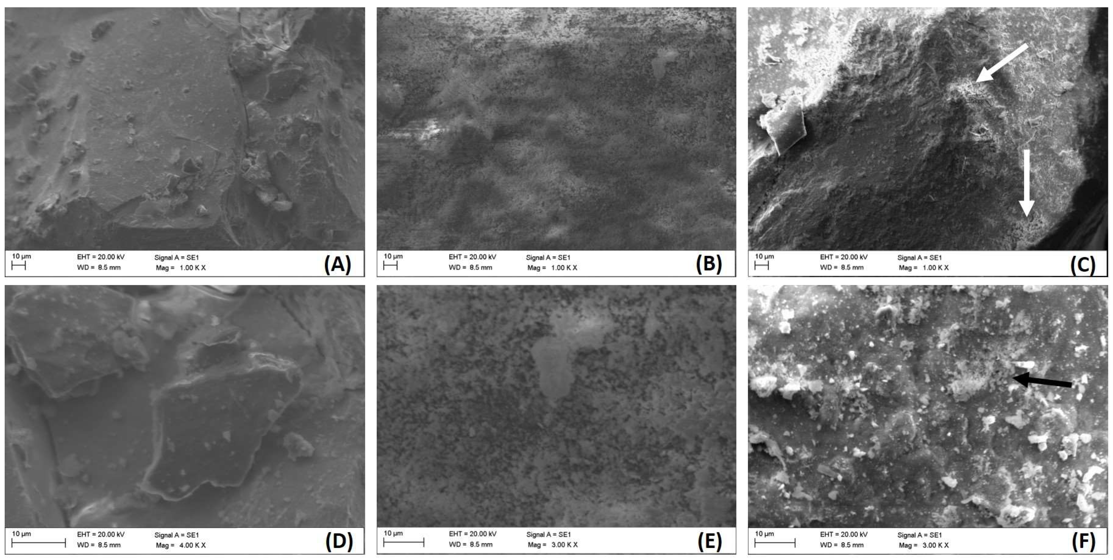
Figure 1. SEM images taken on CeO2 (A,D), La2O3 (B,E), and CexLayO2−δ (C,F) at 1000 magnification (AC) and at higher magnification (DF). The arrows in (C,F) highlight porous regions.
Catalysts 15 00776 g001
Figure 2. SEM image (at 3000 magnification) on Ce0.96±0.01La0.04±0.01O1.18±0.04 (B); EDX spectra on a flat area (A) and on particle aggregates (C); the tables in the insets show the elemental compositions by atomic percentage. The arrows indicate where the EDX spectra were acquired.
Catalysts 15 00776 g002
Figure 3. XRD patterns of the synthesized oxides: (A) all diffractograms; (B) La2O3 diffractogram with lanthanum hydroxide signals; (C) zoomed-in comparison of CeO2 and lanthanum-doped CeO2, highlighting peak shifts.
Catalysts 15 00776 g003
Figure 4. Removal of CAP during photolysis and photocatalysis experiments with REE oxides (solar radiation (A), UV-A (B), UV-C (C)) (S.S.: solar simulator). Dash line indicates the end of the dark phase.
Catalysts 15 00776 g004
Figure 5. Removal of CAP during catalysis experiments with REE oxides and H2O2 (dark (A), UV-A (B), UV-C (C)).
Catalysts 15 00776 g005
Figure 6. Heatmap of CAP removal (%) after 15 min for each treatment.
Catalysts 15 00776 g006
Figure 7. Pareto chart showing the effects of process factors and their interactions on CAP removal after 120 min of treatment.
Catalysts 15 00776 g007
Table 1. Elemental composition from EDX analysis: The results are expressed as atomic percentage (At%); standard deviations are reported for each value.
CeOLaCAg
CeO239 ± 1153 ± 11---6 ± 22.3 ± 0.5
La2O3---57 ± 428 ± 614 ± 61.6 ± 0.3
CexLayO2−δ37 ± 753 ± 81.4 ± 0.27 ± 22.1 ± 0.9
Table 2. CAP removal by photolysis reported in the literature.
Initial CAP Concentration
(mg/L)
RadiationTime
(min)
RemovalReference
20Solar72021.8%[29]
UV-C83.3%[29]
200Solar6034%[30]
25UV-C12040%[31]
9.69UV-C12020.3%[32]
16.16Solar10021.9%[33]
0.969UV-A3020%[34]
5UV6040%[35]
1Solar simulator12034%This study
UV-A34%
UV-C35%
Table 3. Pseudo-first order kinetics constant for the performed experiments.
Processkobs (min−1)R2
Solar0.00320.98
UV-A0.00320.98
UV-C0.00300.89
Solar/CeO20.00290.99
Solar/La2O30.00300.96
Solar/CexLayO2−δ0.00300.97
UV-A/CeO20.00440.99
UV-A/La2O30.00670.98
UV-A/CexLayO2−δ0.00340.98
UV-C/CeO20.00570.99
UV-CLa2O30.00671.00
UV-C/CexLayO2−δ0.00520.99
UV-A/H2O20.12801.00
UV-A/H2O2/CeO20.10900.99
UV-A/H2O2/La2O30.01741.00
UV-A/H2O2/CexLayO2−δ0.01171.00
UV-C/H2O20.26691.00
UV-C/H2O2/CeO20.26831.00
UV-C/H2O2/La2O30.41880.99
UV-C/H2O2/CexLayO2−δ0.27001.00
Disclaimer/Publisher’s Note: The statements, opinions and data contained in all publications are solely those of the individual author(s) and contributor(s) and not of MDPI and/or the editor(s). MDPI and/or the editor(s) disclaim responsibility for any injury to people or property resulting from any ideas, methods, instructions or products referred to in the content.