Article

N-Doped Porous Graphene Film Decorated with Palladium Nanoparticles for Enhanced Electrochemical Detection of Hydrogen Peroxide

by
Yue Zhang
,
Shi Zheng
*,
Jian Xiao
and
Jiangbo Xi
*
School of Chemistry and Environmental Engineering, Hubei Key Laboratory of Novel Reactor and Green Chemical Technology, School of Chemical Engineering and Pharmacy, Wuhan Institute of Technology, Wuhan 430205, China
*
Authors to whom correspondence should be addressed.
Catalysts 2025, 15(4), 298; https://doi.org/10.3390/catal15040298
Submission received: 10 January 2025 / Revised: 10 March 2025 / Accepted: 17 March 2025 / Published: 21 March 2025
(This article belongs to the Section Electrocatalysis)

Abstract

:
Graphene film has excellent electrical conductivity and flexibility, with which it can be used as a versatile substrate to load active species to construct free-standing electrochemical sensors. In this work, Pd nanoparticle-decorated N-doped porous graphene film (Pd/NPGF) was prepared by a simple and mild strategy to enhance the electrochemical behavior of graphene film-based free-standing electrodes. The morphological structure and surface component of the Pd/NPGF were characterized by scanning electron microscopy, transmission electron microscopy, Raman spectra and X-ray photoelectron spectroscopy measurements. The results revealed that the Pd/NPGF contained abundant pores and uniformly dispersed Pd nanoparticles, which could bring a favorable electrochemical response. Due to the synergetic effects of abundant pores, uniform Pd nanoparticles and the substitutional doping of the graphene framework with N, the novel free-standing Pd/NPGF electrode provides a high active site exposure, a high specific area and fast electron/mass diffusion during electrochemical reactions. Considering the favorable flexibility and excellent electrical conductivity of Pd/NPGF, we selected hydrogen peroxide, a significant biomarker, as a model to investigate its electrochemical performance in neutral conditions. The electrochemical biosensor based on the Pd/NPGF electrode exhibited enhanced activity relative to the NPGF and porous graphene film (PGF) with different concentrations of H2O2. The Pd/NPGF electrode displayed a high sensitivity (176.7 μA·mM−1·cm−2), a large linear range from 5 μM to 36.3 mM, a low limit of detection (LOD) of 2.3 μM, excellent stability and a short response time, all of which qualify the Pd/NPGF electrode for a promising sensor for H2O2 sensing.

Graphical Abstract

1. Introduction

Hydrogen peroxide (H2O2) is a key redox signaling agent in living cells and plays a key role in basic biological processes [1]. However, excessive accumulation may cause an abnormal concentration of H2O2 in blood plasma and may lead to various diseases, such as cancer, Alzheimer’s disease, neurodegeneration and cardiovascular blockage [2,3,4]. Therefore, H2O2 can be used as a biomarker for clinical diagnosis and the study of related diseases. Moreover, H2O2 is widely used in the modern chemical industry, water treatment and domestic settings [5,6,7]. Thus, it is tremendously important to develop rapid, sensitive and accurate methods to detect the concentration of H2O2.
In the past few decades, electrochemical quantification of H2O2 has received significant attention due to its advantages that include simple operation, economical desirability, portability and excellent sensitivity [8]. Nevertheless, a high overpotential is often needed when detecting H2O2 directly with bare electrodes [9]. To overcome this obstacle, several natural enzymes, such as horseradish peroxidase, have been adopted to increase the electron transfer and thus increase the electrochemical sensing performance [10]. However, the properties of these enzymes, such as the high price, environmental instability and easy denaturation, do not suit the application of enzyme-based sensors. An alternative strategy is to develop enzyme-free electrochemical sensors using various electrode materials such as inorganic nanomaterials [11,12], conducting polymers [13] and composite materials [14].
Graphene, a two-dimensional sheet of single-atom-thick carbon, has received considerable attention in the synthesis of nanocomposites for H2O2 sensing [15]. Relying on its large specific surface area, excellent conductivity, flexibility and mechanical strength, graphene is often used as a versatile substrate to load active species to construct free-standing electrochemical sensors [16]. Owing to the stacking configurations inside fabricated graphene film, the inner part has no contribution to the electrochemical performance and may be an obstacle to mass transfer processes. To prepare a porous structure is vital to enhancing the active electrochemical surface area and mass transfer [15]. Prepared film-like self-supported graphene could be modified by various metal or alloy nanoparticles to construct electrochemical sensors [17]. The active metal nanoparticles could provide high electrocatalytic activity toward H2O2 redox to enhance the electron transfer. In addition, the interaction between the metal and support may affect the catalytic performance of the electrode materials. The chemical doping of heteroatoms (e.g., B, N, S and P) into graphene can improve its physical and chemical properties and create new active sites [18,19]. For example, the doping of graphene with nitrogen atoms alters the hydrophilic properties that are best suited for the construction of H2O2 sensors of practical utility [20,21]. Otherwise, the interaction between the metal and support can be enhanced by the N-dopants in graphene, benefitting the anchoring or loading of active metal [22,23,24].
In this work, Pd nanoparticle-decorated N-doped porous graphene film (Pd/NPGF) was prepared by a simple and mild strategy to enhance the electrochemical behavior of graphene film-based free-standing electrodes. Morphology analysis data revealed that the Pd/NPGF contained abundant pores and uniformly dispersed Pd nanoparticles, which could bring a favorable electrochemical response. When employed as a working electrode for detecting H2O2, the Pd/NPGF electrode displayed a high sensitivity (176.7 μA·mM−1·cm−2), a large linear range from 5 μM to 36.3 mM, a low limit of detection (2.3 μM), excellent stability and a short response time, all of which qualify the Pd/NPGF electrode for a promising sensor for H2O2 sensing.

2. Results and Discussion

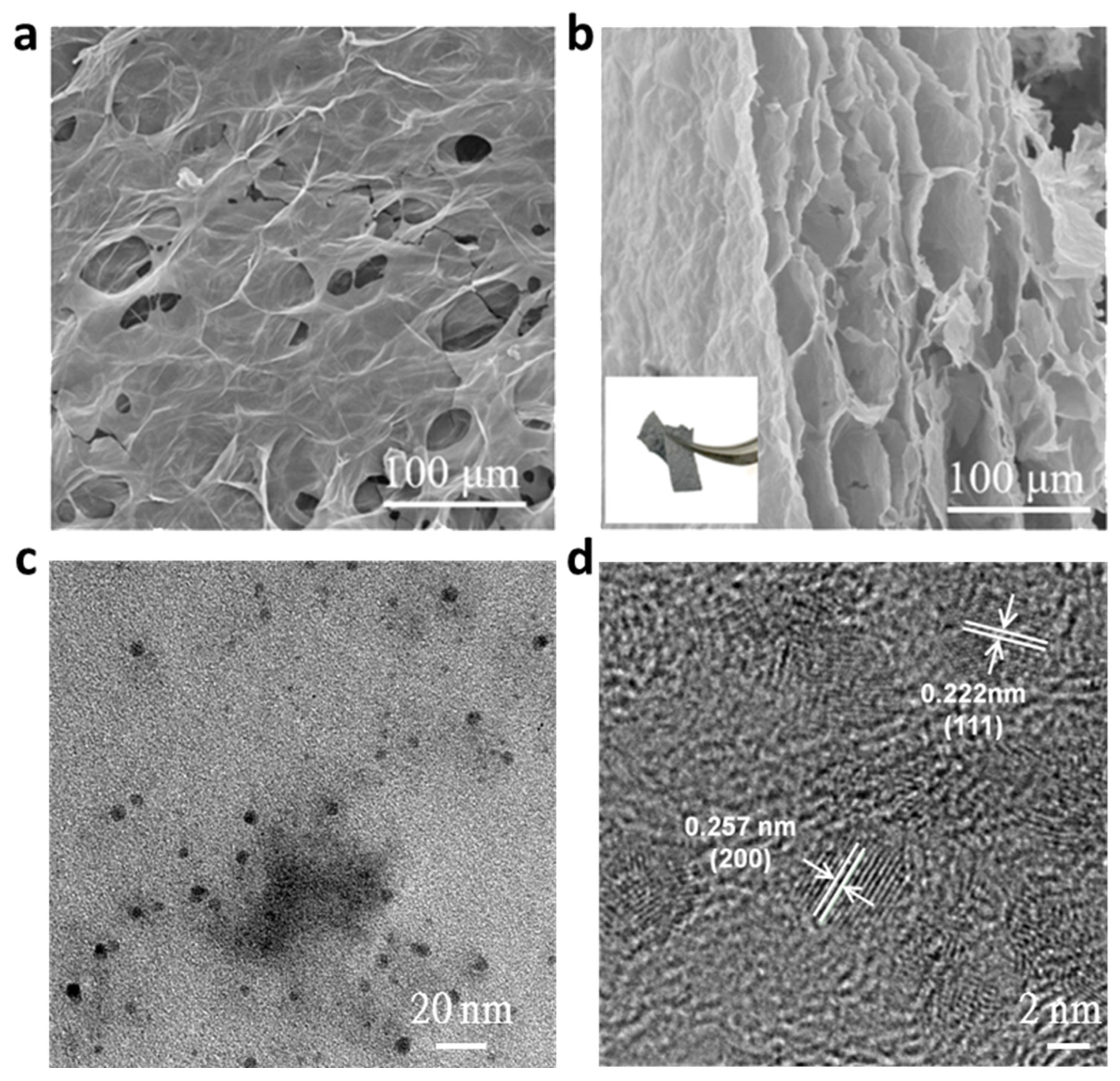
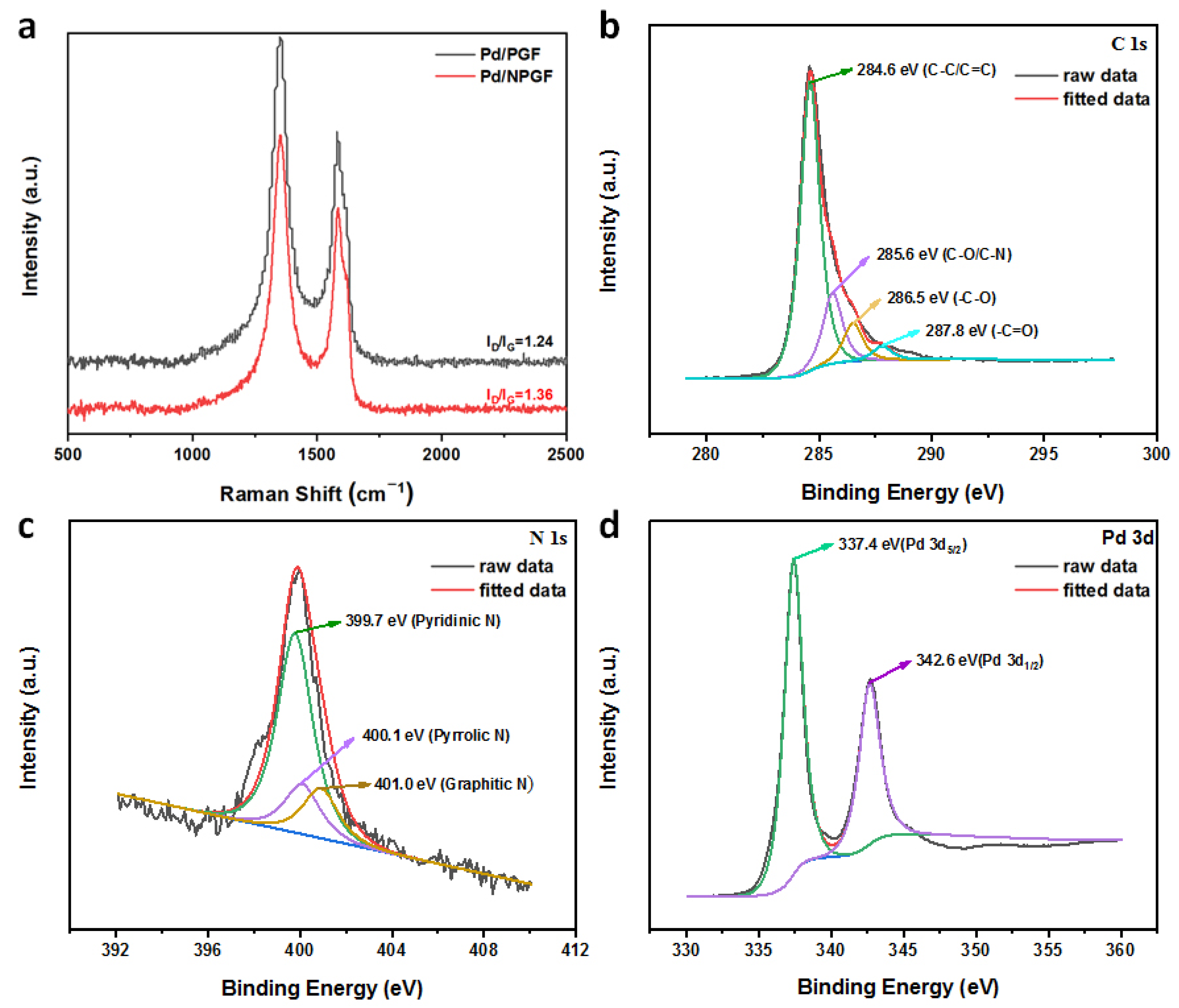
2.1. Morphological and Structural Characterization

The preparation process of the Pd/NPGF is given in Figure 1. Firstly, graphene oxide (GO) is used as a raw material and NaCl is used as the template or pore-forming agent to prepare the NaCl/GO composite film. After reduction with hydroiodic acid (HI) and removing of the NaCl template, a flexible free-standing porous reduced graphene oxide film (PGF) is obtained. Then, the obtained porous graphene film is immersed in saturated urea aqueous solution. The urea is introduced to both the inner pores and surface of the PGF after drying. Next, the resultant urea–graphene composite film is further annealed at a high temperature, during which the urea is decomposed and consumed as a nitrogen source and pore-forming agent, leading to the formation of the N-doped porous graphene film (NPGF). After a spontaneous redox between the Pd precursor (K2PdCl4) and NPGF [25], the pores, interfaces and surface of the NPGF are impregnated with Pd nanoparticles. Benefitting from the rich dopants or defect sites (i.e., oxygen- and nitrogen-containing functional groups) and pores in NPGF, Pd nanoparticles can more easily be anchored and dispersed on the NPGF. Finally, a flexible free-standing Pd/NPGF composite material is obtained.
The used amount of NaCl has an important effect on the morphology and performance of the NPGF material. In order to examine the surface microstructural characteristics of the NPGF, SEM was carried out. As shown in Figure 2 and Figure S2, the pore is uniformly dispersed in the entire film and the number of pores increase with the increasing used amount of NaCl. According to the images of NPGF20, the pore size is approximately 100 μm. The cross profile of NPGF20 (Figure 2b) presents stacked morphology and the destruction of the laminar structure after the annealing process. In addition, the illustration in Figure 2b shows the elastomer properties of NPGF20, which demonstrates that an increase in defect sites may not decrease the high mechanical strength of the electrode material. Thus, the optimum pore-forming agent amount of 20 mg NaCl was chosen for the subsequent experiments.
High-resolution transmission electron microscopy (TEM) images (Figure 2c,d) demonstrate that the Pd nanoparticles are uniformly dispersed in the surface area of the laminar structure, with an average diameter size of ~2.8 nm. The spacings between the adjacent crystal faces of the nanoparticles are 0.257 nm and 0.222 nm, attributed to the (200) [26] and (111) [27] crystal faces of the Pd.
Wettability is a key surface property for electrodes. Figure S3 displays the contact angle of the water droplets formed on the Pd/NPGF. The left side and right side of the contact angle are 83.449° and 78.853°, respectively, so the measured water contact angle is 81.151°, which means Pd/NPGF has a limited wettability. However, it is higher than the water contact angle of pristine graphene, with an average value of ≈30° [28]. This can be attributed to the loss of hydrophilic groups during reduction with HI. Figure 3a shows the Raman spectroscopy of the Pd/NPGF and Pd/PGF samples without pore-forming agents. The peak at 1344 cm−1 (D-band) represents the disorder or defect of the carbon domains, while the peak at 1580 cm−1 (G-band) indicates the degree of crystallinity of the graphitic carbon [29]. The degree of defect in a carbon structure can be compared by the ratio between the D and G band intensities (ID/IG). The ID/IG value of the Pd/NPGF (1.36) is higher than that of Pd/PGF (1.24), which indicates an increase in active sites by the doping of the N atoms [30].
The chemical states and elemental composition on the Pd/NPGF surface were investigated by X-ray photoelectron spectroscopy (XPS). The results showed that the Pd/NPGF was composed of C, O (35.65 at. %), N (3.32 at. %) and Pd. The C 1s spectrum (Figure 3b) presented four peaks of C−C/C=C (284.6 eV), C−O/C−N (285.6 eV), C−O (286.5 eV) and C=O (288.2 eV), where the presence of the C−O and C−N peaks indicated the formation of N and O co-doped graphene species [31,32]. The N 1s spectrum (Figure 3c) presented three peaks of pyridinic N (399.7 eV), pyrrolic N (400.1 eV) and graphitic N (401.0 eV) [33,34]. The Pd 3d spectrum (Figure 3d) presented two peaks of Pd5/2 (337.4 eV) and Pd3/2 (342.6 eV) [35,36]. In addition, the Pd concentration in the Pd/NPGF was 0.06 wt. %, determined by ICP-MS.

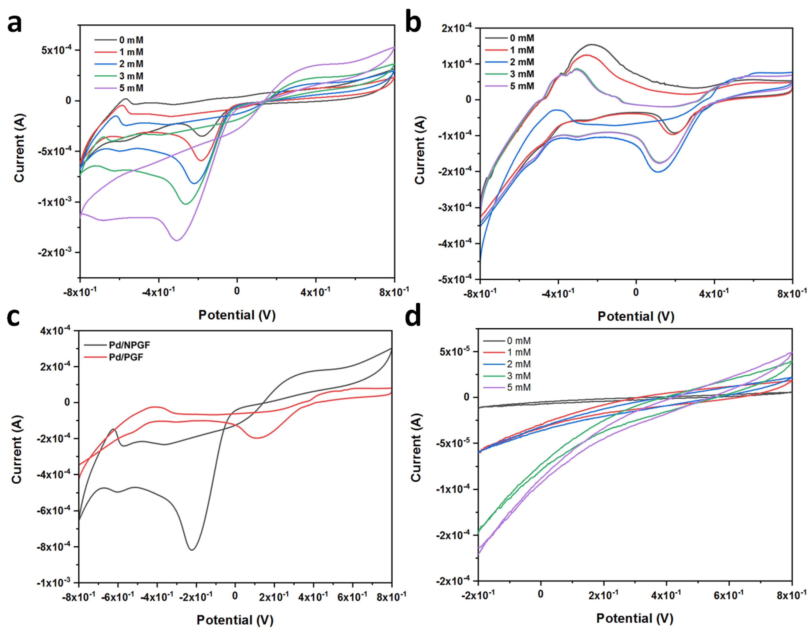
2.2. Electrochemical Behaviors of the Pd/NPGF Modified Electrodes

The electrocatalytic activity of the Pd/NPGF and Pd/PGF toward H2O2 were estimated by cyclic voltammetry (CV) curves in 1M PBS containing different H2O2 concentrations over a potential range from −0.8 to 0.8 V. According to the CV curves in Figure 4a,b, with the increasing H2O2 concentrations from 0 to 5 mM, the reduction peak current density and the effective area rise dramatically. Additionally, the peak potential gradually shifts negatively, indicating the Pd/NPGF and Pd/PGF electrode process’ application potential in the detection of H2O2. As shown in Figure 4c, compared to the Pd/PGF, the Pd/NPGF has a larger effective area, which can be attributed to the abundant pores and the substitutional doping of the graphene framework with N. When NPGF (Figure 4d) is used to detect H2O2 concentrations, no redox peak is observed. However, the sensor exhibits a proportional response in the examined concentration range of 0–5 mM. Thus, the Pd/NPGF electrode process has outstanding properties in the electrochemical detection of H2O2. Furthermore, only a minority of Pd nanoparticles in the used Pd/NPGF electrode material displayed increased size compared with the freshly prepared one (Figure S5), demonstrating a favorable stability due to the strong metal–support interactions [22].
The interfacial properties of the Pd/NPGF and NPGF electrodes were further investigated by electrochemical impedance spectroscopy (EIS) using Fe(CN)63−/4− as the probe. As shown in Figure S4, based on the Randles equivalent circuit, the charge transfer resistances (Rct) of the Pd/NPGF and NPGF were calculated to be 206.7 Ω and 329 Ω, respectively. This result demonstrates that the Pd/NPGF electrode exhibits higher electron transfer efficiency than the NPGF [15].
The amperometric current–time (I-t) curve of the Pd/NPGF is shown in Figure 5a. The current response of the Pd/NPGF was investigated by the successive addition of H2O2 with different concentrations into 0.1 M PBS under optimum conditions at the applied potential of −0.2 V. The detection linear interval of the H2O2 was from 5 μM to 36.3 mM. The response time of the Pd/NPGF was less than 3 s, as shown in Figure 5a. The Pd/NPGF electrode displayed a high sensitivity (176.7 μA·mM−1·cm−2). The limit of detection was calculated to be 2.3 μM based on the criterion that the signal-to-noise ratio is 3 in Figure 5b. Different from some former results, the linear range obtained by the Pd/NPGF was significantly improved [37].
Electroactive species such as dopamine (DA), glucose, L-ascorbic acid (AA) and uric acid (UA) were generally present in the biological samples. Selective detection of H2O2 is critically important for Pd/NPGF electrodes to detect H2O2 in vivo and in vitro. Based on Figure 5c, when DA, GLU, AA and UA with concentrations of 0.2 mM were added into a PBS solution, a consistent amperometric response was observed with the subsequent addition of 0.5 mM H2O2. This result shows that the amperometric current response caused by common electroactive species is negligible compared to H2O2. To assess the reliability of the Pd/NPGF electrode for H2O2 detection, the relative standard deviation (RSD) of the amperometric response from one Pd/NPGF electrode was 0.73% in six replicate measurements. Thus, compared to some previously reported electrodes (Table 1), the Pd/NPGF electrode shows high selectivity, competitive detection (LOD) and favorable electrocatalytic performance.

3. Materials and Methods

3.1. Reagents and Materials

Natural graphite powder was purchased from Alfa Aesar (Ward Hill, MA, USA); potassium tetrachloropalladate (K2PdCl4) was procured from Aladdin Chemistry Co., Ltd. (Shanghai, China); and hydrogen peroxide (H2O2), sodium chloride (NaCl), urea, hydroiodic acid (HI) and hydrochloric acid (HCl) were purchased from Sinopharm Chemical Reagent Co. (Shanghai, China) and were used directly without further purification. All other chemicals used were of analytical reagent grade. Ultrapure water (18 MΩ·cm) for solution preparation was produced using an Aquapro System (Chongqing, China).

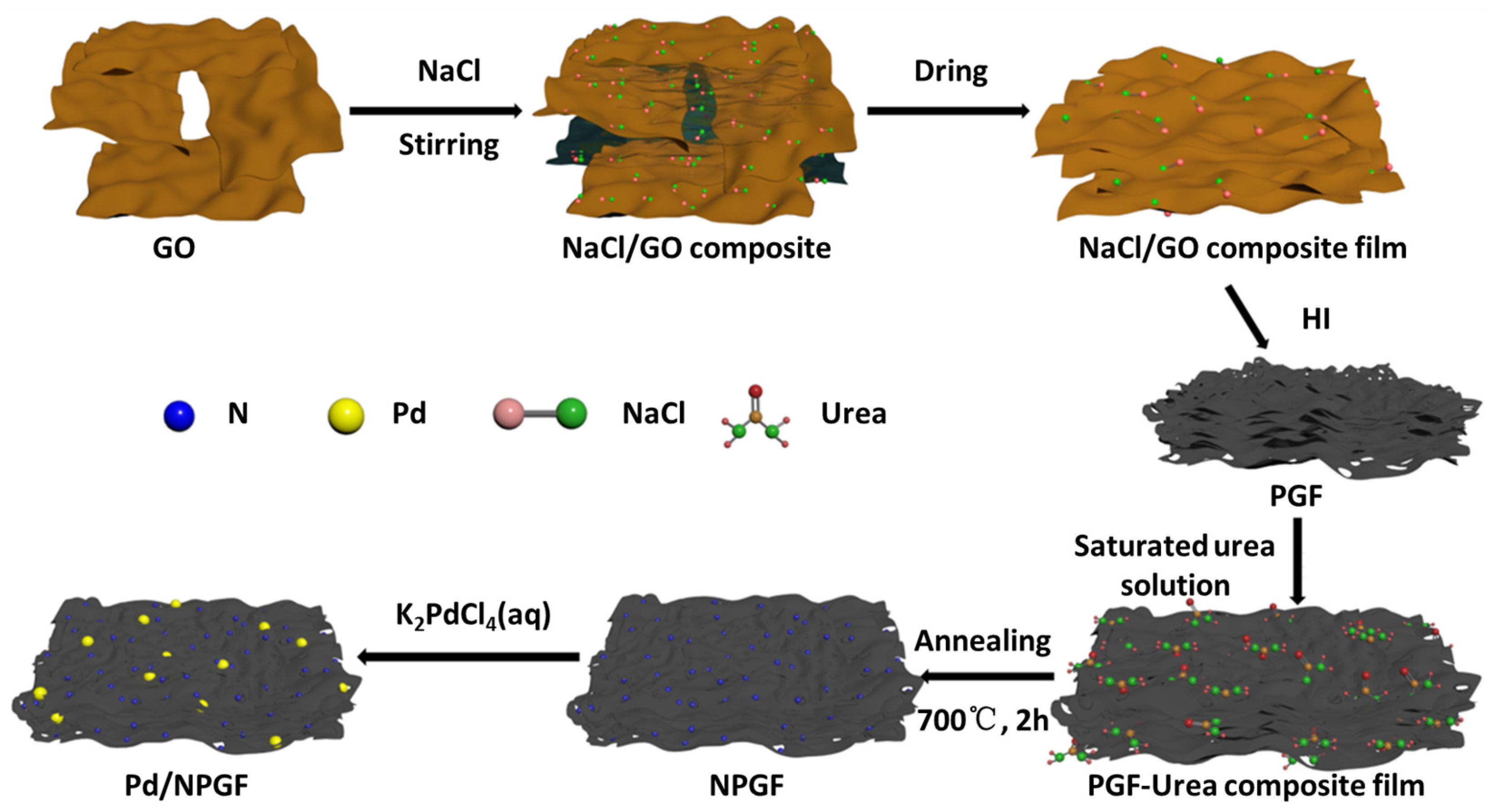
3.2. Preparation of N-Doped Porous Graphene Film (NPGF)

A modified Hummer method [24] was used to synthesize a graphene oxide (GO) aqueous solution. Amounts of 0 mg, 2 mg, 10 mg, 15 mg and 20 mg of NaCl were added respectively to the solution of 10 mL of GO (10 mg·mL−1). We kept stirring at room temperature for approximately 30 min and then sonicated for 5 min to obtain a homogeneous GO aqueous dispersion. Then, 2.5 mL of the mixture was coated on the surface of a filter membrane with a diameter of 50 mm. Drying with room-temperature air, the NaCl/GO composite film was prepared. Next, the film was immersed in 5 mL of HI and placed in a dark place at room temperature for 8 h. Finally, the obtained PGF was washed with ethanol and ultrapure water until colorless and then dried at room temperature. The prepared NPGF support materials were denoted as NPGF0, NPGF2, NPGF10, NPGF15 and NPGF, respectively.
A stock solution of saturation urea was prepared in ultrapure water. The obtained PGF was immersed in 10 mL of the saturation urea solution and placed under room temperature for 3 h. After drying, the obtained PGF–urea composite was annealed at 700 °C for 2 h under nitrogen flow. The weights of the NPGFs were 9.5 mg, 12.4 mg, 14.3 mg and 17.0 mg, respectively. The control NPGF sample was fabricated by the same method, with 20 mg of NaCl but without the addition of urea. The weight of the control sample was 6.6 mg.

3.3. Preparation of Pd Nanoparticle-Decorated N-Doped Porous Graphene Film (Pd/NPGF)

A stock solution of K2PdCl4 was prepared in ultrapure water in an ice-water bath. The obtained NPGF was immersed in 10 mL of K2PdCl4 solution (the ratio of Pd/NPGF was 2 wt. %) and kept in a dark place at room temperature for 3 h. Finally, the obtained Pd/NPGF was washed with deionized water for several times to remove the residual metal precursor. The obtained Pd/NPGF was freeze-dried overnight for use in subsequent experiments.

3.4. Structure and Electrochemical Characterization

The morphology of the NPGF was characterized by scanning electron microscopy (SEM) (Vega3, Tescan Co., Ltd., Brno, Czech Republic). The structure of the Pd/NPGF was observed by high-resolution transmission electron microscopy (TEM) (TECNAI G2-20 U-Twin instrument, FEI, Ermelo, The Netherlands) with a 200 kV accelerating voltage. Raman spectroscopy was characterized using a Raman spectrometer (Thermo Fisher, Waltham, MA, USA). X-ray photoelectron spectroscopy (XPS) was conducted on a VG ESCALAB 250 spectrometer (Altrincham, UK) with Al Kα X-ray radiation (hν = 1486 eV) to analyze the chemical state and composition. A CHI1800D electrochemical workstation (CH Instruments Co., Shanghai, China) was carried out to measure the cyclic voltammetry test (CV), electrochemical impedance (EIS) and timing current response (I-T) of the samples to analyze the electrochemical state of the Pd/NPGF. The electrochemical tests were performed in a three-electrode cell with the Pd/NPGF (0.6 cm × 1 cm in effective area) as the working electrodes, Pt as the counter electrode and Ag/AgCl as the reference electrode. All electrochemical measurements were carried out with 1 mol·L−1 H2O2 at 25 °C. A phosphate buffer solution (PBS, 1 mol·L−1) of pH 7.4 was used as a supporting electrolyte.

4. Conclusions

In summary, Pd nanoparticle-decorated N-doped porous graphene film was prepared by a simple and mild strategy to enhance the electrochemical behavior of the graphene film-based free-standing electrodes. The as-prepared Pd/NPGF electrode exhibited superb electrochemical performance, with a wide linear range from 5 μM to 36.3 mM, a high sensitivity (176.7 μA·mM−1·cm−2), a low limit of detection (2.3 μM), excellent stability, a short response time and good selectivity. The excellent sensing performance can be attributed to the synergetic effects of the abundant pores, the uniform Pd nanoparticles and the substitutional doping of the graphene framework with N. Thus, the novel free-standing Pd/NPGF electrode provides high active site exposure, a high specific area and fast electron/mass diffusion during electrochemical reactions. More importantly, this study has provided a promising sensing tool for detecting H2O2 in vitro and in vivo, with a potential future applications of being applicable to the clinical diagnosis and surveillance of various diseases.

Supplementary Materials

The following supporting information can be downloaded at: https://www.mdpi.com/article/10.3390/catal15040298/s1, Figure S1. Physical images of NPGF film obtained by different pore-making agent dosage: (a) NPGF2; (b) NPGF10; (c) NPGF15; (d) NPGF20. Figure S2. SEM images of NPGF prepared with different pore-forming agent quality. (a) NPGF0; (b) NPGF2; (c) NPGF10; (d) NPGF15. Figure S3. Contact angle of Pd/NPGF. Figure S4. Electrochemical Impedance Spectroscopy (EIS) at open circuit potential. Figure S5. Dark-field TEM image of used Pd/NPGF.

Author Contributions

Conceptualization, J.X. (Jiangbo Xi) and S.Z.; methodology, J.X. (Jiangbo Xi) and Y.Z.; software, J.X. (Jiangbo Xi) and Y.Z.; validation, Y.Z. and J.X. (Jian Xiao); formal analysis, J.X. (Jian Xiao); investigation, J.X. (Jian Xiao); resources, J.X. (Jiangbo Xi); data curation, Y.Z.; writing—original draft preparation, S.Z.; writing—review and editing, S.Z.; visualization, Y.Z.; supervision, J.X. (Jiangbo Xi) and S.Z.; project administration, J.X. (Jian Xiao); funding acquisition, J.X. (Jiangbo Xi). All authors have read and agreed to the published version of the manuscript.

Funding

This research was financially supported by the Key Research and Development Program of Hubei Province (No. 2022BAA026), the Open Project of the Hubei Key Laboratory of Novel Reactor and Green Chemical Technology (No. NRGC202203), the Open/Innovation Project of the Engineering Research Center of Phosphorus Resources Development and Utilization of the Ministry of Education (No. LCX202203), the Open/Innovation Project of the Key Laboratory of Novel Biomass-Based Environmental and Energy Materials in the Petroleum and Chemical Industry (No. 2022BEEA06), the Open Project of the Key Laboratory of the Green Chemical Engineering Process of the Ministry of Education (No. GCX2022005), the Young and Middle-Aged Talent Program of the Hubei Provincial Department of Education (Q20181507) and the Scientific Research Foundation of the Wuhan Institute of Technology (K201718).

Data Availability Statement

The data are contained within the article and Supplementary Materials.

Conflicts of Interest

The authors declare no conflicts of interest.

References

  1. Sies, H.; Jones, D.P. Reactive oxygen species (ROS) as pleiotropic physiological signalling agents. Nat. Rev. Mol. Cell Biol. 2020, 21, 363–383. [Google Scholar]
  2. Cheignon, C.; Tomas, M.; Bonnefont-Rousselot, D.; Faller, P.; Hureau, C.; Collin, F. Oxidative stress and the amyloid beta peptide in Alzheimer’s disease. Redox Biol. 2018, 14, 450–464. [Google Scholar] [PubMed]
  3. Li, H.G.; Xia, N. The role of oxidative stress in cardiovascular disease caused by social isolation and loneliness. Redox Biol. 2020, 37, 101585. [Google Scholar]
  4. Rhee, S.G. H2O2, a necessary evil for cell signaling. Science 2006, 312, 1882–1883. [Google Scholar] [PubMed]
  5. Kurowska, E.; Brzózka, A.; Jarosz, M.; Sulka, G.D.; Jaskuła, M. Silver nanowire array sensor for sensitive and rapid detection of H2O2. Electrochim. Acta 2013, 104, 439–447. [Google Scholar]
  6. Guascito, M.R.; Filippo, E.; Malitesta, C.; Manno, D.; Serra, A.; Turco, A. A new amperometric nanostructured sensor for the analytical determination of hydrogen peroxide. Biosens. Bioelectron. 2008, 24, 1057–1063. [Google Scholar]
  7. Chen, H.; Zhang, H.; Chi, K.; Zhao, Y. Pyrimidine-containing covalent organic frameworks for efficient photosynthesis of hydrogen peroxide via one-step two electron oxygen reduction process. Nano Res. 2024, 17, 9498–9506. [Google Scholar]
  8. Zhu, C.; Yang, G.; Li, H.; Du, D.; Lin, Y. Electrochemical sensors and biosensors based on nanomaterials and nanostructures. Anal. Chem. 2015, 87, 230–249. [Google Scholar]
  9. Salimi, A.; Hallaj, R.; Soltanian, S.; Mamkhezri, H. Nanomolar detection of hydrogen peroxide on glassy carbon electrode modified with electrodeposited cobalt oxide nanoparticles. Anal. Chim. Acta 2007, 594, 24–31. [Google Scholar]
  10. López Marzo, A.M.; Mayorga-Martinez, C.C.; Pumera, M. 3D-printed graphene direct electron transfer enzyme biosensors. Biosens. Bioelectron. 2020, 151, 111980. [Google Scholar]
  11. Bai, J.; Jiang, X. A facile one-pot synthesis of copper sulfide-decorated reduced graphene oxide composites for enhanced detecting of H2O2 in biological environments. Anal. Chem. 2013, 85, 8095–8101. [Google Scholar] [CrossRef] [PubMed]
  12. Xiang, M.; Wu, J.; Li, L.; Ren, F.; Dai, R.; Dai, F.; Dong, S.; Yang, Z. High-loading Bi-MOF nanoparticles anchored on biomass waste-derived carbon for sensitive non-enzymic nitrite sensing. Microchim Acta 2025, 192, 125. [Google Scholar] [CrossRef]
  13. Tian, F.; Xu, B.; Zhu, L.; Zhu, G. Hydrogen peroxide biosensor with enzyme entrapped within electrodeposited polypyrrole based on mediated sol-gel derived composite carbon electrode. Anal. Chim. Acta 2001, 443, 9–16. [Google Scholar]
  14. Luo, L.; Li, F.; Zhu, L.; Zhang, Z.; Ding, Y.; Deng, D. Non-enzymatic hydrogen peroxide sensor based on MnO2-ordered mesoporous carbon composite modified electrode. Electrochim. Acta 2012, 77, 179–183. [Google Scholar]
  15. Xi, J.; Zhang, Y.; Ye, T.; Xiao, J.; Fang, J.; Han, M.; Zhao, A.; Zhang, Y. Self-supported electrochemical sensor based on uniform palladium nanoparticles functionalized porous graphene film for monitoring H2O2 released from living cells. Anal. Bioanal. Chem. 2024, 416, 6995–7006. [Google Scholar]
  16. Wan, Y.; Wang, Y.; Wu, J.; Zhang, D. Graphene oxide sheet-mediated silver enhancement for application to electrochemical biosensors. Anal. Chem. 2011, 83, 648–653. [Google Scholar]
  17. Yao, Y.; Ping, J. Recent advances in graphene-based freestanding paper-like materials for sensing applications. TrAC Trends Anal. Chem. 2018, 105, 75–88. [Google Scholar]
  18. Liang, J.; Jiao, Y.; Jaroniec, M.; Qiao, S.Z. Sulfur and nitrogen dual-doped mesoporous graphene electrocatalyst for oxygen reduction with synergistically enhanced performance. Angew. Chem. Int. Ed. 2012, 51, 11496–11500. [Google Scholar]
  19. Liu, Y.; Li, M.; Zhou, B.; Xuan, X.; Li, H. Flexible B, N co-doped graphene electrodes for electrochemical detection of serotonin in bodily fluids. Electrochim. Acta 2023, 457, 142494. [Google Scholar]
  20. Xi, J.B.; Zhang, Y.; Wang, Q.J.; Xiao, J.; Chi, K.; Duan, X.M.; Chen, J.; Tang, C.Y.; Sun, Y.M.; Xiao, F.; et al. Multi-element doping design of high-efficient carbocatalyst for electrochemical sensing of cancer cells. Sens. Actuators B Chem. 2018, 273, 108–117. [Google Scholar]
  21. Kaushal, S.; Kaur, M.; Kaur, N.; Kumari, V.; Singh, P.P. Heteroatom-doped graphene as sensing materials: A mini review. RSC Adv. 2020, 10, 28608–28629. [Google Scholar] [CrossRef] [PubMed]
  22. Bai, S.; Jia, A.; Song, J.; Cao, S.; Wang, N.; Liu, X. Metal-support interactions in heterogeneous catalytic hydrogen production of formic acid. Chem. Eng. J. 2023, 474, 145612. [Google Scholar]
  23. Huang, J.; Li, X.; Xie, R.-H.; Tan, X.; Xi, J.; Tian, F.; Liu, P.; Willum Hansen, T.; Bai, Z.-W. Defect anchoring of atomically dispersed Pd on nitrogen-doped holey carbon nanotube for catalytic hydrogenation of nitroarenes. Appl. Surf. Sci. 2023, 615, 156344. [Google Scholar]
  24. Zhao, J.; Zhang, A.; Li, Y.; Hu, H.; Xi, J. Pd nanoparticles decorated N-doped holey graphene assembled on aluminum silicate fibers agglomerate for catalytic continuous-flow reduction of nitroarenes. Chem. Eng. Sci. 2024, 286, 119656. [Google Scholar]
  25. Chen, X.; Wu, G.; Chen, J.; Chen, X.; Xie, Z.; Wang, X. Synthesis of “clean” and well-dispersive Pd nanoparticles with excellent electrocatalytic property on graphene oxide. J. Am. Chem. Soc. 2011, 133, 3693–3695. [Google Scholar] [CrossRef]
  26. Meku, E.; Du, C.; Wang, Y.; Du, L.; Sun, Y.; Kong, F.; Yin, G. Concentration gradient Pd-Ir-Ni/C electrocatalyst with enhanced activity and methanol tolerance for oxygen reduction reaction in acidic medium. Electrochim. Acta 2016, 192, 177–187. [Google Scholar]
  27. Zheng, G.; Altman, E.I. The oxidation of Pd(111). Surf. Sci. 2000, 462, 151–168. [Google Scholar]
  28. Zhang, J.; Jia, K.; Huang, Y.; Liu, X.; Xu, Q.; Wang, W.; Zhang, R.; Liu, B.; Zheng, L.; Chen, H.; et al. Intrinsic wettability in pristine graphene. Adv. Mater. 2022, 34, 2103620. [Google Scholar]
  29. Zhao, A.; She, J.; Xiao, C.; Xi, J.; Xu, Y.; Manoj, D.; Sun, Y.; Xiao, F. Green and controllable synthesis of multi-heteroatoms co-doped graphene fiber as flexible and biocompatible microelectrode for in situ electrochemical detection of biological samples. Sens. Actuators B Chem. 2021, 335, 129683. [Google Scholar]
  30. Hu, C.; Dai, L. Doping of carbon materials for metal-free electrocatalysis. Adv. Mater. 2019, 31, 1804672. [Google Scholar] [CrossRef]
  31. Zhu, Y.; Wu, J.; Han, L.; Wang, X.; Li, W.; Guo, H.; Wei, H. Nanozyme sensor arrays based on heteroatom-doped graphene for detecting pesticides. Anal. Chem. 2020, 92, 7444–7452. [Google Scholar] [PubMed]
  32. Hu, H.; Liu, P.; Cao, S.; You, L.; Zhang, N.; Xi, J.; Guo, S.; Zhou, K. Single metal atoms anchored on N-doped holey graphene as efficient dual-active-component catalysts for nitroarene reduction. Adv. Funct. Mater. 2024, 34, 2307162. [Google Scholar]
  33. Ma, X.; Wang, J.; Zhu, Z.; Wang, N.; Wang, C.; Nie, G. A two-pronged strategy to boost the capacitive deionization performance of nitrogen-doped porous carbon nanofiber membranes. Desalination 2025, 594, 118293. [Google Scholar]
  34. Du, S.; Cao, S.; Chen, W.; Xi, J. Fibrous catalyst based on atomic Pd and N-doped holey graphene functionalized cotton fiber for continuous-flow reaction. Int. J. Biol. Macromol. 2024, 280, 136049. [Google Scholar]
  35. Wang, M.; Liang, L.; Liu, X.; Sun, Q.; Guo, M.; Bai, S.; Xu, Y. Selective semi-hydrogenation of alkynes on palladium-selenium nanocrystals. J. Catal. 2023, 418, 247–255. [Google Scholar]
  36. Li, J.; Cai, T.; Feng, Y.; Liu, X.; Wang, N.; Sun, Q. Subnanometric bimetallic Pt-Pd clusters in zeolites for efficient hydrogen production and selective tandem hydrogenation of nitroarenes. Sci China Chem 2024, 67, 2911–2917. [Google Scholar] [CrossRef]
  37. Hu, J.; Li, F.; Wang, K.; Han, D.; Zhang, Q.; Yuan, J.; Niu, L. One-step synthesis of graphene-AuNPs by HMTA and the electrocatalytical application for O2 and H2O2. Talanta 2012, 93, 345–349. [Google Scholar] [CrossRef] [PubMed]
  38. Peng, C.; Zhou, S.; Zhang, X.; Zeng, T.; Zhang, W.; Li, H.; Liu, X.; Zhao, P. One pot synthesis of nitrogen-doped hollow carbon spheres with improved electrocatalytic properties for sensitive H2O2 sensing in human serum. Sens. Actuators B Chem. 2018, 270, 530–537. [Google Scholar]
  39. Yao, H.; Zhang, W.-T.; Yan, T.-Y.; Li, X.-Q.; Wang, X.-F. Electrochemical sensor for detection of hydrogen peroxide based on Cu-doped ZIF-8 material modified with chitosan and cytochrome c. Int. J. Electrochem. Sci. 2022, 17, 220654. [Google Scholar]
  40. Mathivanan, D.; Shalini Devi, K.S.; Sathiyan, G.; Tyagi, A.; da Silva, V.A.O.P.; Janegitz, B.C.; Prakash, J.; Gupta, R.K. Novel polypyrrole-graphene oxide-gold nanocomposite for high performance hydrogen peroxide sensing application. Sens. Actuators A Phys. 2021, 328, 112769. [Google Scholar]
  41. Li, H.; Zhao, H.; He, H.; Shi, L.; Cai, X.; Lan, M. Pt-Pd bimetallic nanocoral modified carbon fiber microelectrode as a sensitive hydrogen peroxide sensor for cellular detection. Sens. Actuators B Chem. 2018, 260, 174–182. [Google Scholar] [CrossRef]
  42. Chen, D.; Zhuang, X.; Zhai, J.; Zheng, Y.; Lu, H.; Chen, L. Preparation of highly sensitive Pt nanoparticles-carbon quantum dots/ionic liquid functionalized graphene oxide nanocomposites and application for H2O2 detection. Sens. Actuators B Chem. 2018, 255, 1500–1506. [Google Scholar] [CrossRef]
  43. Liu, J.; Zheng, J.; Sheng, Q. One-step synthesis of Au@Pt-Graphene nanocomposites and their electrochemical properties. J. Nanosci. Nanotechnol. 2019, 19, 5546–5553. [Google Scholar] [CrossRef]
  44. Yang, Q.; Li, N.; Li, Q.; Chen, S.; Wang, H.-L.; Yang, H. Amperometric sarcosine biosensor based on hollow magnetic Pt-Fe3O4@C nanospheres. Anal. Chim. Acta 2019, 1078, 161–167. [Google Scholar] [CrossRef] [PubMed]
  45. He, F.-G.; Yin, J.-Y.; Sharma, G.; KUmar, A.; Stadler, F.J.; Du, B. Facile fabrication of hierarchical rGO/PANI@PtNi nanocomposite via microwave-assisted treatment for non-enzymatic detection of hydrogen peroxide. Nanomaterials 2019, 9, 1109. [Google Scholar] [CrossRef]
  46. Zhang, Y.; Liu, Y.; Sun, F.; Yang, N. 3D heteroatom-doped graphene-wrapped flexible carbon fiber microsensor for real-time hydrogen peroxide detection in live cancer cells. Appl. Surf. Sci. 2023, 611, 155655. [Google Scholar] [CrossRef]
  47. Qi, C.; Luo, Y.; Dong, Y. Synergistic effects of Fe-Se dual single-atom sites for boosting electrochemical nonenzymatic H2O2 sensing. Appl. Surf. Sci. 2023, 637, 157900. [Google Scholar] [CrossRef]
  48. Wang, X.; Wang, Y.; Liu, Y.; Cao, X.; Zhang, F.; Xia, J.; Wang, Z. MOF-derived porous carbon nanozyme-based flexible electrochemical sensing system for in situ and real-time monitoring of H2O2 released from cells. Talanta 2024, 266, 125132. [Google Scholar] [CrossRef]
  49. Lee, G.-G.; Hong, H.-G. Catalytically synthesized prussian blue by a one-step copolymerization of polydopamine-polypyrrole for electrochemical sensing of hydrogen peroxide. Electrochim. Acta 2023, 465, 142949. [Google Scholar] [CrossRef]
  50. Nasir, A.; Khalid, S.; Mazare, A.; Yasin, T. Non-enzymatic hydrogen peroxide detection on a novel nanohybrid composite of chitosan and grafted graphene oxide. Mater. Res. Bull. 2023, 167, 112427. [Google Scholar] [CrossRef]
  51. Li, G.; Zheng, Y.; Hu, G.; Chen, B.; Gu, Y.; Yang, J.; Yang, H.; Hu, F.; Li, C.; Guo, C. Boosting photo-electro-fenton process via atomically dispersed iron sites on graphdiyne for invitro hydrogen peroxide detection. Small 2023, 19, 2301540. [Google Scholar]
  52. Cheng, D.; Wu, H.; Feng, C.; Ding, Y.; Mei, H. Bifunctional photoelectrochemical sensor based on Bi/Bi2S3/BiVO4 for detecting hexavalent chromium and hydrogen peroxide. Sens. Actuators B Chem. 2022, 353, 131108. [Google Scholar] [CrossRef]
  53. Du, Y.; Jia, M.; Zhang, X.; Ma, M.; Zhang, X.; Li, C.; Wang, S.; Zhang, J.; Li, D.; He, W.; et al. Graphene oxide covered WO3 nanosheet arrays as photoelectrode for photoelectrochemical detection of trace H2O2. ACS Appl. Nano Mater. 2024, 7, 7896–7905. [Google Scholar]
  54. Murugan, P.; Sundramoorthy, A.K.; Nagarajan, R.D.; Atchudan, R.; Shanmugam, R.; Ganapathy, D.; Arya, S.; Alothman, A.A.; Ouladsmane, M.; Murthy, H.C.A. Electrochemical detection of H2O2 on graphene nanoribbons/cobalt oxide nanorods-modified electrode. J. Nanomater. 2022, 2022, 1–10. [Google Scholar] [CrossRef]
  55. Ju, J.; Chen, W. In situ growth of surfactant-free gold nanoparticles on nitrogen-doped graphene quantum dots for electrochemical detection of hydrogen peroxide in biological environments. Anal. Chem. 2015, 87, 1903–1910. [Google Scholar]
  56. Xu, F.; Sun, Y.; Zhang, Y.; Shi, Y.; Wen, Z.; Li, Z. Graphene-Pt nanocomposite for nonenzymatic detection of hydrogen peroxide with enhanced sensitivity. Electrochem. Commun. 2011, 13, 1131–1134. [Google Scholar]
  57. Bhaduri, S.N.; Ghosh, D.; Chatterjee, S.; Biswas, R.; Bhaumik, A.; Biswas, P. Fe(iii)-incorporated porphyrin-based conjugated organic polymer as a peroxidase mimic for the sensitive determination of glucose and H2O2. J. Mater. Chem. B 2023, 11, 8956–8965. [Google Scholar]
Figure 1. Schematic illustration of the synthesis of the Pd/NPGF free-standing electrode.
Catalysts 15 00298 g001
Figure 2. SEM images of NPGF prepared with different pore-forming agent qualities: (a) NPGF; (b) cross profile of NPGF (inset is a photograph of folded NPGF) and (c,d) TEM of Pd/NPGF.
Catalysts 15 00298 g002
Figure 3. (a) Raman spectra of Pd/PGF and Pd/NPGF. High-resolution XPS survey spectra of Pd/NPGF: (b) C 1s; (c) N 1s and (d) Pd 3d.
Catalysts 15 00298 g003
Figure 4. Cyclic voltammetry test of different concentrations of H2O2 solution with different electrodes: (a) Pd/NPGF and (b) Pd/PGF. (c) Cyclic voltammetry test of 2 mM hydrogen peroxide with Pd/NPGF and Pd/PGF and (d) cyclic voltammetry test of NPGF.
Catalysts 15 00298 g004
Figure 5. (a) Amperometric current response of Pd/NPGF electrode to the continuous addition of different concentrations of H2O2 in stirred 0.1 M PBS; (b) linear calibration curve for amperometric current densities of Pd/NPGF versus the H2O2 concentrations; (c) amperometric current response of Pd/NPGF electrode to the addition of 0.2 mM H2O2 and 0.2 mM UA, DA, AA and glucose in stirred 0.1 M PBS; and (d) amperometric current response of Pd/NPGF electrode tested under the same conditions toward 0.2 mM H2O2; applied potential: −0.20 V.
Catalysts 15 00298 g005
Table 1. Comparison of analytical performance with some other sensors for detection of H2O2.
Electrode MaterialLinear Range (mM)Sensitivity
(μA·mM−1·cm−2)
LOD (μM)Reference
N-HCS/GCE0.05–29.5, 29.5–47.5-0.00002[38]
Cu-doped ZIF-8/Chitosan0.01–5.2-3.7[39]
PPy-GO-AuNPs/GCE2.5–2541.355[40]
Pt-Pd/CFME0.005–3.9211.60.42[41]
PtNPs-CDs/IL-GO/GCE0.001–0.940.30.1[42]
Au@Ptgraphene0.5–22.3-0.2[43]
Pt-Fe3O4@C0.0005–0.0648.80.43[44]
rGO/PANI@Pt0.1–0.126-1.1[45]
CF@3D-NBG0.0005–4.252330.1[46]
Fe1Se1/NC0.02–131508.611.5[47]
PCNSs0.001–2041.20.76[48]
PB-PDA-PPY/GCE0.005–11.61123.64[49]
CS/GOP0.0005–0.20.7717.3[50]
Fe-GDY0.0001–48.160-0.033[51]
Bi-BSV0.001–130-0.089[52]
GO/WO30.1 to 2.08.14100[53]
GNR/Co3O40.01–0.20051001.27[54]
Au NPs−N-GQDs0.00025–13.327186.220.12[55]
Graphene-Pt0.002–0.71-0.5[56]
Fe-DMP-POR0.005–2947.673.16[57]
Pd/NPGF0.005–36.3176.72.3This work
Disclaimer/Publisher’s Note: The statements, opinions and data contained in all publications are solely those of the individual author(s) and contributor(s) and not of MDPI and/or the editor(s). MDPI and/or the editor(s) disclaim responsibility for any injury to people or property resulting from any ideas, methods, instructions or products referred to in the content.

