Article

Polyol Formation of Silver@Metal Oxides Nanohybrid for Photocatalytic and Antibacterial Performance

1
Department of Chemistry, Mirpur University of Science and Technology (MUST), Mirpur 10250, Pakistan
2
Department of Chemistry, Government College University Faisalabad, Faisalabad 38000, Pakistan
3
National Center for Physics (NCP), Shahdra Valley Road, P.O. Box 2141, Islamabad 44000, Pakistan
4
Department of Chemistry, The University of Lahore, Sargodha Campus, Sargodha 40100, Pakistan
5
Department of Pharmaceutics, College of Pharmacy, King Saud University, Riyadh 11451, Saudi Arabia
6
Department of Chemistry and Chemical Biology, Northeastern University, Boston, MA 02115, USA
*
Authors to whom correspondence should be addressed.
Catalysts 2025, 15(3), 283; https://doi.org/10.3390/catal15030283
Submission received: 28 January 2025 / Revised: 27 February 2025 / Accepted: 10 March 2025 / Published: 17 March 2025

Abstract

:
The polyol method under a single pot has successfully produced a coating of CuO, TiO2, and the combination of CuO/TiO2 around Ag NWs under sequential addition. The Ag NWs and their coating with a pure metal oxide and a hybrid of metal oxide were characterized using X-ray diffraction (XRD), scanning electron microscopy (SEM) coupled with EDX, X-ray photoelectron spectroscopy (XPS), UV–Visible, photoluminescent (PL) spectroscopy, and cyclic voltammetry (CV). The formation of ultra-thin NWs was also been seen in the presence of the TiO2 coating. The ultra-thin and co-axial coating of each metal oxide and their hybrid form preserved the SPR of the Ag NWs and demonstrated photon harvesting from the 400–800 nm range. The band gap hybridization was confirmed by CV for the Ag@CuO/TiO2 design, which made the structure a reliable catalyst. Therefore, the material expresses excellent photocatalytic activities for carcinogenic textile dyes such as turquoise blue (TB), sapphire blue (SB), and methyl orange (MO), with and without the reagent H2O2. The hybrid form (i.e., Ag@CuO/TiO2) exhibited degradation within 6 min in the presence of H2O2. Additionally, the material showed antibacterial activities against various bacteria (Pseudomonas aeruginosa, Escherichia coli, Staphylococcus aureus, Bacillus subtilis, and Bacillus pumilus) when assayed using broth media. Therefore, the materials have established degrading and disinfection roles suitable for environmental perspectives. The role of coating with each metal oxide and their hybrid texture further improved the growth of Ag NWs. The preparatory route possibly ensued metal–metal oxide and metal–hybrid metal oxide Schottky junctions, which would expectedly transform it into a diode material for electronic applications.

1. Introduction

Fabricating asymmetrical and anisotropic metals like Ag nanostructures, their co-axial coating, and the hybridization of coating elements is a leading area of research in the current era due to the unification of diversity in a single system [1,2]. Metal–metal or metal–metal oxide core@shell structures are the best expression of this system [3]. Such designs make it possible to maximize the number of atoms on their respective surfaces, existing as a sheath [4,5]. The sheath provides an ideal binding site for organic pollutants, sufficiently traps the reagents, and eventually becomes a reliable catalyst as an environmental remedy [6,7]. Therefore, it is an auspicious system that ensures the habitats of all flora and fauna along the industrial catchment area. Besides the role of catalysis, it is also desirable for designing photovoltaic cells due to the combinations of different band gaps in a single overlapping mode. The photovoltaic cells of nano Si, Ge, Sn, Zn, Ti, Cu, and their oxides are normally assembled, and their transistors are constituted [8,9]. Moreover, the nanostructures of these metals and their different interactions have also been employed as disinfectants for food and food-containing materials [10]. Similarly, core-shell nanostructures are also employed as antimicrobial agents to inhibit their growth in vulnerable areas. Their synthesis is achieved through sonochemical, hydrothermal, galvanic replacement, sol–gel, and laser ablation [11,12]. Although these methods have their merits, they are insufficient to produce core@shell co-axial nanocables. The polyol, being versatile, reliable, and accurate, is efficiently employed to prepare diverse nanosystems in a single experimental setup [11]. It has also been further proven to be beneficial in preparing core@shell nanocables of metal@metal, and metal@metal oxide nanostructures, imbuing their semi-conducting nature [13]. Its versatility has been demonstrated in the synthesis of noble metal nanoparticles and quantum dots from semiconducting metal oxides and combining them in a single hybrid system [14].
Core@shell nanostructures are key components of many emerging nanodevices; furthermore, these are frequently used in catalysis for many organic and polymer reactions [15]. They demonstrate strong potential to degrade many pollutants and act as environmental remedial agents to preserve a clean and green ecosystem [16]. The role of core@shell structures is significantly impacted by the degradation of carcinogenic dyes, which once discharged from the textile or print industry, mix with water and become a risk to health for all flora and fauna inhabited the area [17]. These dyes also cause widespread arthritis and neurovascular diseases, along with cancer [18]. Therefore, various metal@metal/metal oxide core shells are being prepared and employed. Alongside being pollutant degrading agents, these materials are also antimicrobial [19,20]. The selected pathogens are the usual contaminants found in water, which are also antibiotic-resistant strains. Therefore, their anti-pathogenic nature is further appealing in the fabrication of new structures and improving the existing ones. Both properties (i.e., catalysis as well as antipathogenic) exist in nano CuO/TiO2/ZnO, CuO/TiO2, Ag/MgO, and Ag/Pd, according to published reports [21,22]. The work on these metals has mainly focused on their nanoparticles [23], while a few reports are related to CuO, TiO2, and Si NWs and their application in dye-sensitized solar cells, catalysis, photoelectrochemical cells, and photovoltaic cells. For each system, the band hybridization of binary or ternary systems was explored and related to exceptional catalytic and photovoltaic applications.
In this study, core@shell nanostructures comprising Ag NWs, Ag@CuO, Ag@TiO2, and Ag@CuO/TiO2 nanocables were fabricated through the polyol method. The linear structures of Ag and metal oxides in their pure and hybrid forms developed overlapping band gaps, produced many discrete energy levels for a high degree of electron resonance, and ensured the interaction with visible electromagnetic radiation and polyaromatic dyes. XRD, SEM-EDX, and XPS techniques were used to explore the structure, morphology, and elemental coating composition of the fabricated nanomaterials. Additionally, UV–Vis, PL spectroscopy, and CV were used to understand the surface plasmon resonance, light-harvesting behavior, and electrochemical activities of the synthesized materials. The applications of these nanocables in catalysis, specifically in degrading carcinogenic dyes like turquoise blue (TB), sapphire blue (SB), and methyl orange (MO) without any reagent as well as with H2O2, were analyzed under a tungsten bulb. The results showed that these catalysts can degrade dyes without any reagent and rapidly degrade in the presence of reagents. Furthermore, the antimicrobial behavior of the as-synthesized material was also evaluated against P. aeruginosa, E. coli, S. aureus, B. subtilis, and B. pumilus. It was observed that their ability against microbes increased significantly in successive systems. This study reports new versions of hybrid systems around Ag, highlighting their catalytic and microbial contributions. The findings can be useful for analyzing other aspects of catalysis, evaluating against other microbes and fungi as well as for photovoltaic and electronic applications.

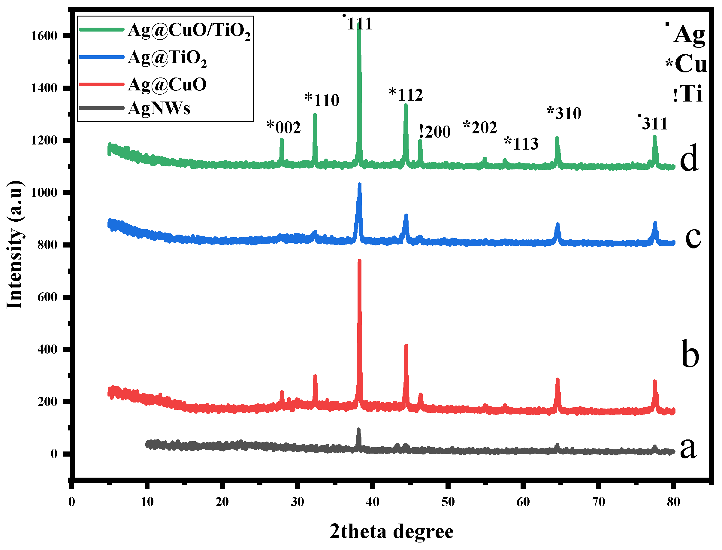
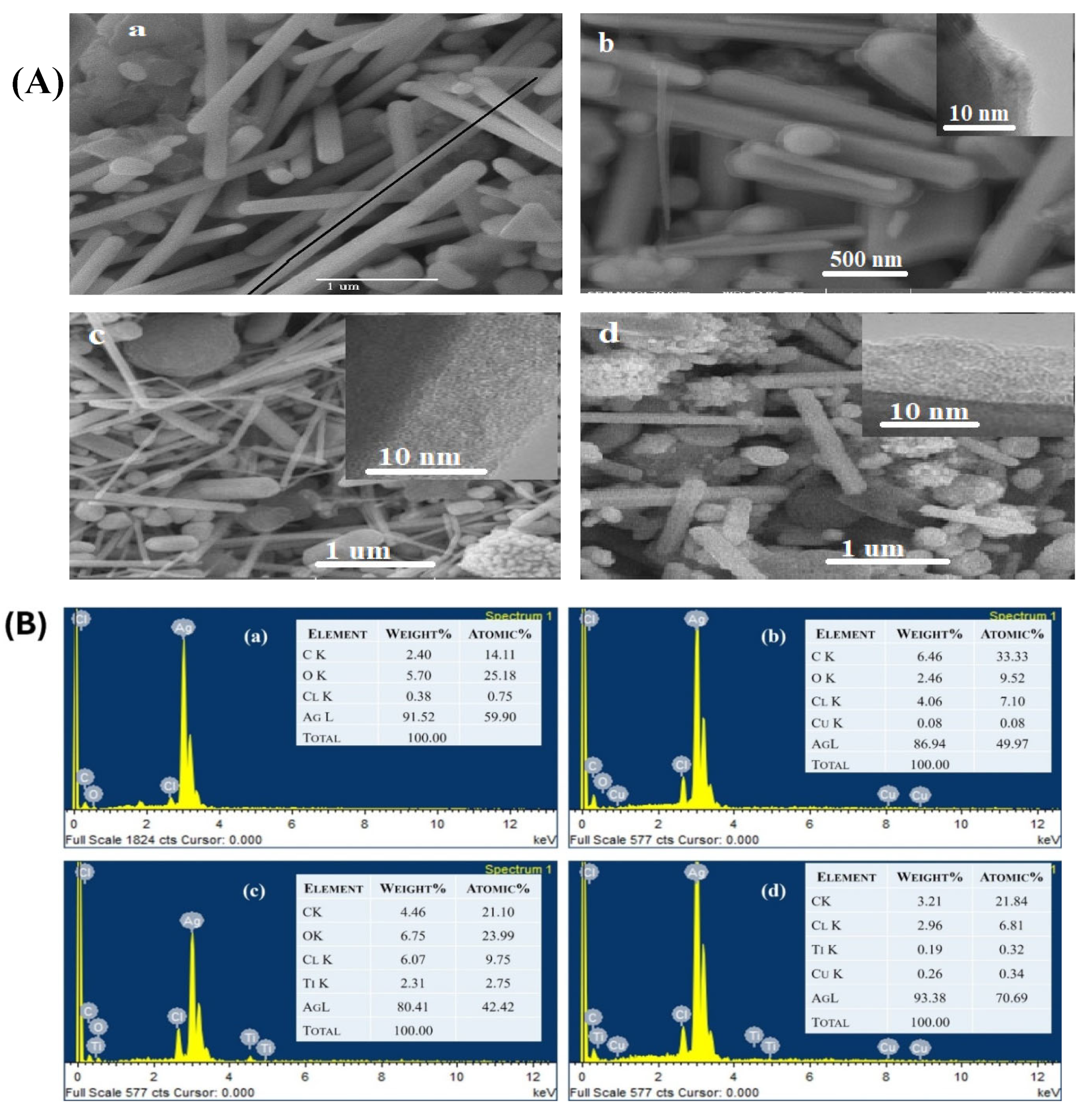
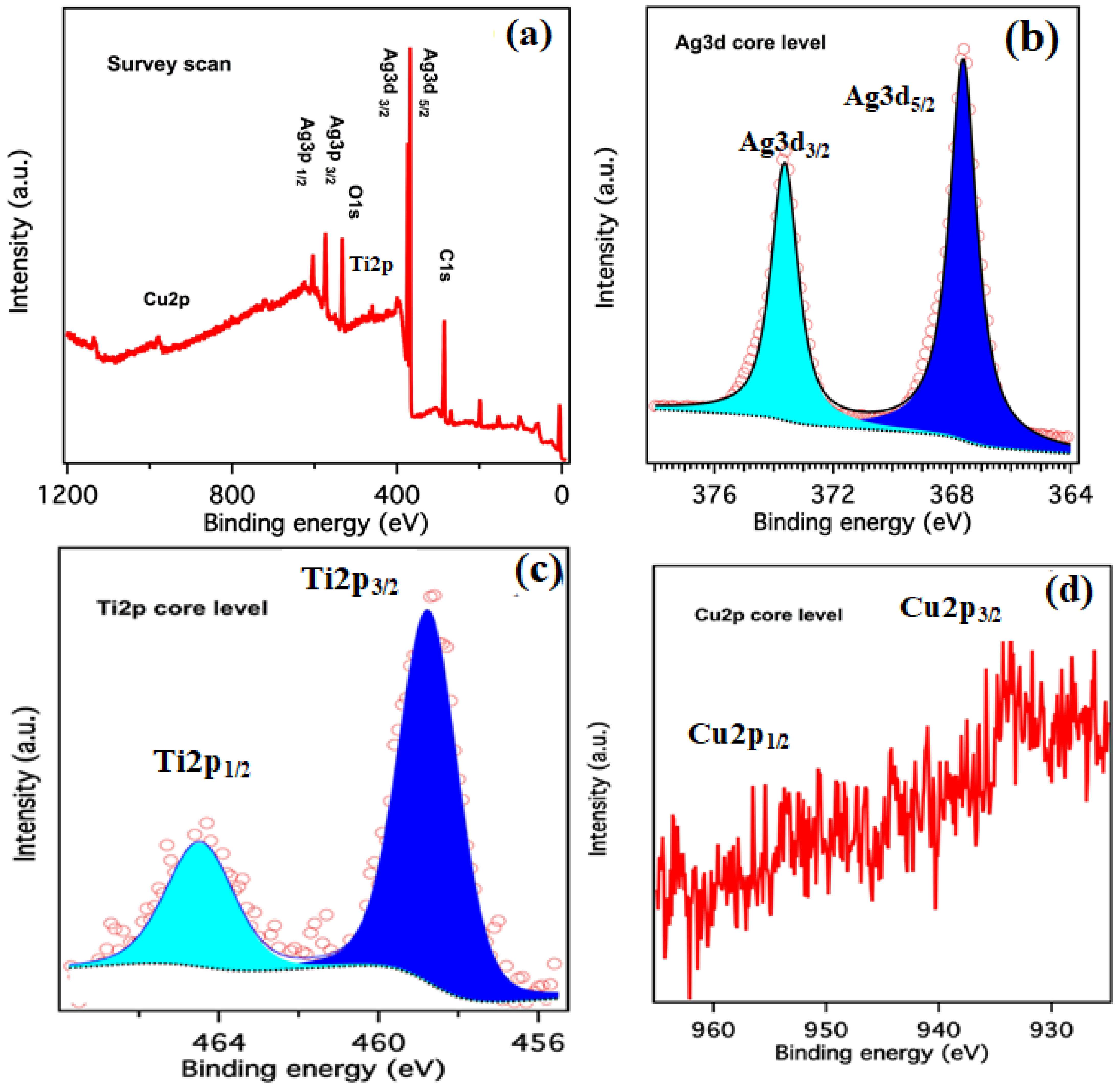
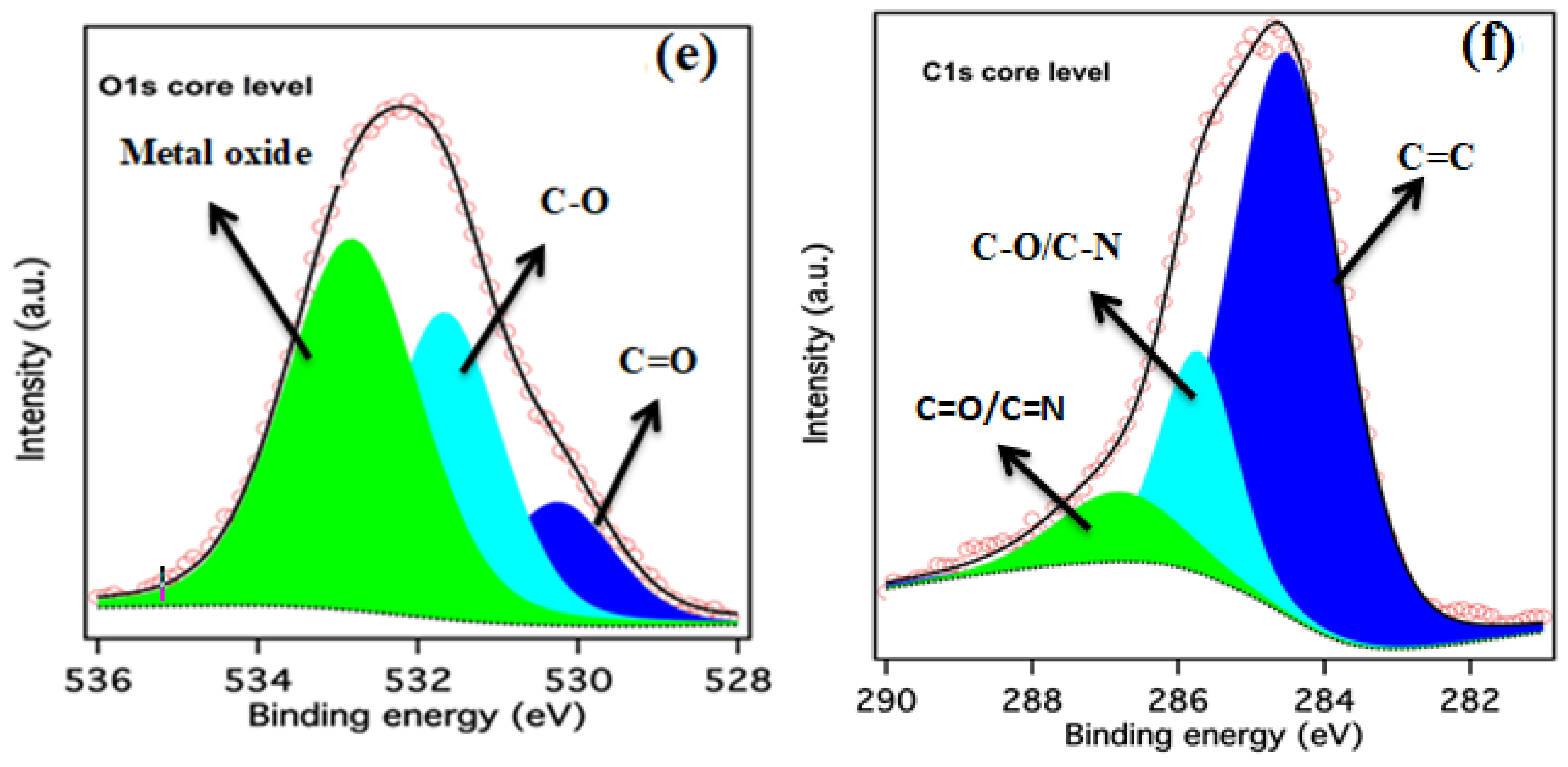
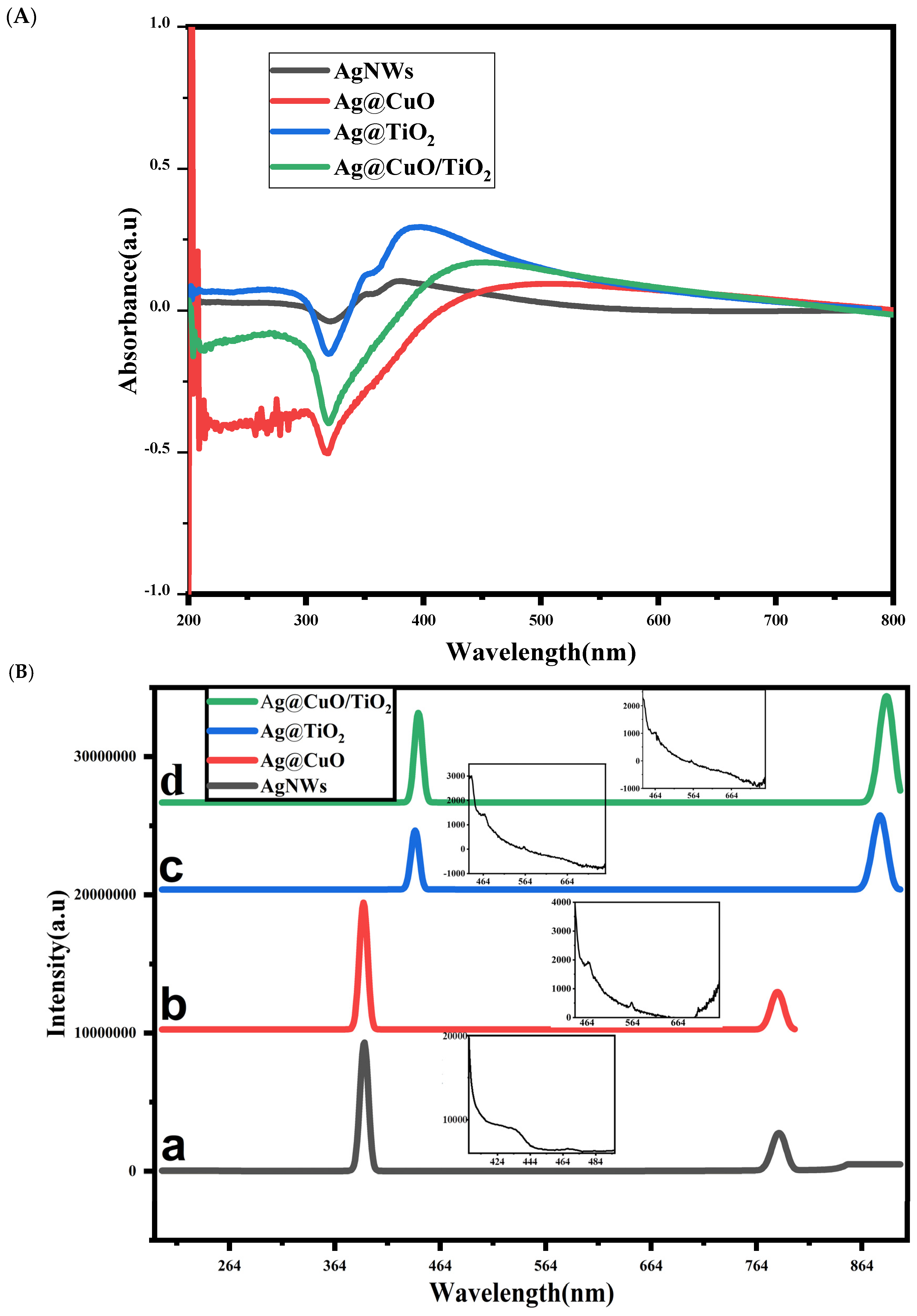
2. Results and Discussion

The samples prepared for this study were characterized using XRD, SEM-EDX, XPS, UV–Vis, a PL spectrophotometer, and CV. The results are discussed with the help of ongoing work related to core@shell aspects of nanomaterials. The crystal structures and constituents of the AgNWs, Ag@CuO, Ag@TiO2, and Ag@CuO/TiO2 were examined using X-ray diffraction. The major diffraction peak positions of the samples indicated that each sample contained the same amount of the primary component (Ag) in the composites. Figure 1a shows the distinctive peaks of the Ag crystal planes (111), (200), (220), and (311) represented by 2θ at 38.1, 44.2, 64.4, and 77.3, respectively (JCPDS No. 87-0597). Figure 1b indicates a strong crystalline diffraction peak for CuO (110) and a significant mass proportion of Ag@CuO. The diffraction peaks at 2θ = 28.5, 32.5, 38.6, 44.8, 48.8, 56.5, and 58.2 were respectively assigned to CuO crystal planes (002), (110), (111), (112), (113), (202), and (310), respectively (JCPDS 48-154). Figure 1c shows diffraction peaks at 31.2°, 38.1°, 45.5°, 64.3°, and 78.1°, representing the crystal planes (121), (004), (200), (204), and (311), respectively, for Ag@TiO2. The peak at 65.3° indicates the presence of the anatase phase of TiO2. Diffraction peaks at 28.2°, 32.1°, 38.1°, 44.5°, 48.2°, 56.1°, 58.3°, 65.3°, and 79.1° representing crystal planes (002), (110), (111), (112), (200), (202), (113), (310), and (311), respectively, were observed for Ag@CuO/TiO2, as shown in Figure 1d. The crystal planes (111) and (311) indicated silver, while the crystal planes (002), (110), (112), (202), (113), and (310) corresponded to CuO, and crystal planes (200) and (204) signified TiO2 in the ternary nanodesign. The presence of the characteristic peaks of both moieties (CuO and TiO2), along with the representative peaks of silver in the XRD diffractogram of Ag@CuO/TiO2, are helpful for expecting the successful and simultaneous coating of CuO and TiO2 over the Ag NWs.
The SEM images, supported by the inset images of TEM, are presented in Figure 2A(a–d), showing the morphology and surface topography of the pure Ag NWs, Ag@CuO, Ag@TiO2 nanocables, and Ag@CuO/TiO2 nanocables with a hybridized shell of both metal oxides. The inset images, which were focused on to analyze the coating thicknesses of CuO, TiO2, and mix of these two metal oxides, are presented in Figure 2A(b–d). In Figure 2A(a), the Ag NWs were produced as a dominant product having diameters between 70 and 80 nm and lengths in the µm scale, consistent with the reported data [24]. The surface of the Ag NWs was smooth, with the appearance of a truncated pentagonal texture. During coating by the pure metal oxides and their hybridized form, the diameter and length of the NWs were affected due to the interference in the mass transfer of the Ag nuclei over growing Ag NWs. The smooth surface of the NWs was coated with an ultra-thin sheath of CuO, reducing the truncated pentagonal texture and converting the whole surface as a round. This information is well-disclosed in Figure 2A(b). Figure 2A(b) also depicts the rod-like structure smoothly coated with a CuO ultra-thin nanosheath. The thickness of the nanosheath is expressed in the inset of same figure, which is related to the TEM observation and clearly differentiated the core from the surface coating. The thickness of the surface coating was analyzed from 5 to 8 nm. In Figure 2A(c), the SEM micrograph shows the Ag@TiO2 nanostructures, with the Ag NWs having an average diameter of 60–70 nm. This figure further exposes the remarkable structures of ultra-thin wires, which in the presence of TiO2 decreased the reduction of the Ag cation and maintained the slow deposition, leading to longer cables with a small diameter [25]. Herein, the coating developed a rough surface. It further shows the cubical- and cylindrical-shaped structures along with agglomerated fine particles that are supposed to be TiO2. However, the inset of the same figure, which is again a TEM image, showed a uniform surface coating with a thickness around 10 nm. Figure 2A(d) displays the SEM micrograph of the Ag@CuO/TiO2 ternary system, where both metal oxides are in their hybridized form. The diameter and length of the Ag NWs are likely to be in this form, as these were coated with pure metal oxides, as shown in Figure 2A(b,c). However, in this case, the roughness around Ag was more pronounced. Similarly, the inset of the same figure displays the two phases where one is related to the core, and the other phase is related to the surface coating, with an approximate measurement of 8–12 nm. This morphology, when compared with the respective XRD data given in Figure 1, further elucidates the increase in grain boundaries around the linear Ag NWs. Therefore, the clearly manifested surface coating phases did not look like compact structures when compared with the core metals. Finally, the formation of NPs was also seen in every case as it is a limitation of the polyol method, and the study of these particles is useful to understand the bottom–up growth of metal NWs and the co-axial growth of the coating metal or metal oxide [26]. Therefore, XRD, SEM, and TEM confirmed the formation of metal oxide-based nanocables of Ag NWs and the hybridization of metal oxides around Ag NWs. Thus, binary and ternary nanodesigns were fabricated between metal NWs and semiconducting metal oxide quantum wells. A Schottky diode type of material is expected to be produced by combining the nano and quantum domains [27].
The EDX analysis of the Ag NWs, Ag@CuO, Ag@TiO2, and Ag@CuO/TiO2 is presented to demonstrate the elemental composition of the materials, as presented in Figure 2B(a–d). The qualitative EDX patterns display the elemental composition, atomic percentages, and weight percentages of these samples. For all of the prepared samples, the weight and atomic percentage of silver were significantly higher than those of the other elements. The values for silver were 91.52%, 86.94%, 80.41%, and 93.38%, respectively, as shown in Figure 2B(a–d). In CuO/TiO2, the hybridized shell and the compactness of the surface decreased, as seen in Figure 2B(d), which increased the exposure of the Ag core to the EDX scan and so its percentage was reflected as higher [28]. The weight percentages of Cu and Ti around the Ag NWs were observed to be 0.08% and 2.31%, respectively, as shown in the EDX patterns for Ag@CuO and Ag@TiO2, and given in Figure 2B(b,c). Similarly, in the ternary nanodesign, Ag@CuO/TiO2, both Cu and Ti were observed to have weight percentages of 0.26% and 0.19%, respectively, confirming the presence of CuO and TiO2 in these samples. Additionally, all samples had varying weight percentages of carbon (2–6%) due to the leftover capping agent during synthesis, which was trapped and could not be removed during washing. Elemental mapping revealed that carbon from the capping agent shielded the Ag NWs. This is a significant finding, as carbon might exist at the interface between the core and the shell, which can isolate the nano and quantum domains, creating a physical gap and barrier between them [29].
The chemical state of each element in the Ag@CuO/TiO2 ternary nanodesign was examined using XPS analysis, as presented in Figure 3. The binding energy for the C 1s peak at 284.6 eV was used as the reference for calibration. Figure 3a illustrates the survey scan spectrum of Ag@CuO/TiO2, which confirmed the presence of Ag 3d, Ti 2p, Cu 2p, O 1s, and C 1s in the ternary Ag@CuO/TiO2 nanodesign and is reflected in Figure 3b–f, respectively. The presence of the metallic state of Ag was demonstrated by the two Ag 3d characteristic peaks of Ag 3d5/2 at 367 eV and Ag 3d3/2 at 373.5 eV (Figure 3b), with a spin energy separation of 6.0 eV in metallic Ag [30]. The HR peaks in the Ti 2p spectra indicated the presence of Ti4+ (Figure 3c), consistent with the reported value for Ti4+ in anatase TiO2 [31]. The values of the binding energies appeared typically at 459 and 464.5 eV. In Figure 3d, the 935 eV and 952 eV binding energies were ascribed to Cu 2p3/2 and Cu 2p1/2 of Cu2+ in CuO, respectively [32]. The characteristic shakeup satellite peak around 543.71 eV and 562.21 eV further confirmed the oxidation state of Cu as Cu(II). The O 1s spectrum (Figure 3e) at 534.00 V was attributed to O in TiO2 and CuO, while a broad shoulder at a higher binding energy region may be attributed to oxygen in the hydroxyl groups [33]. The peaks fitted for 532 and 533 eV were ascribed for O, which was present as a mixed oxide form. XPS was the most reliable surface probe technique, thus confirming the metal oxide and mix of the metal oxide around the Ag NWs. The source of C was the capping agent, which was used in the form of PVP and is displayed in Figure 3f. Therefore, C in the oxidized form and graphene like existed, as indicated by peak splitting around the core of Ag, as previously discussed.
The surface plasmon resonance (SPR) phenomenon was demonstrated by the metal nanostructures including the Ag NWs, Ag@CuO, Ag@TiO2, and Ag@CuO/TiO2 nanodesigns, as expressed in Figure 4A. The whole prepared sample was concentrated in ethanol within 5 mL, and just one drop was taken in the cuvette and diluted with ethanol to the level of its cap to record the UV–Vis observation. Figure 4 shows a typical view of Ag NWs comprising its transverse and longitudinal growth. The longitudinal mode exhibited a SPR at 393 nm, while the transverse pattern showed a SPR at 357 nm. The shoulder peak at 357 nm and the peak at 393 nm were attributed to the out-of-plane quadrupole resonance and the out-of-plane dipole resonance of the Ag NWs, respectively [34]. These nanowires either had a lower plasmon concentration or moved away from the UV–Vis radiation scan after being subjected to surface coating, and the SPR bands decreased because they were less sweeping. This effect was observed for the Ag@CuO nanocables, which shows that when Ag NWs are coated with CuO, the SPR results decrease. The transverse SPR of the Ag NWs at 357 nm was suppressed, a result consistent with CuO deposited on the surface of wires [35]. A broader peak for the TiO2 coating appeared in the Ag@TiO2, but the possibility of Ag SPR was similarly decreased. Moreover, a similar impact with a slight red shift was observed for the Ag@CuO/TiO2 nanohybrid, as shown in Figure 4A. These results prove that the SPR is a surface property of metal nanostructures; in the case of Ag, this phenomenon was quite strong. Metal oxides like CuO and TiO2 did not have this property, but their coating did not affect the intrinsic SPR of metal, however, the coating decreased the innate intensity.
Photoluminescence (PL) spectroscopy was used to analyze the light-harvesting abilities of the prepared samples [36]. The fluorescence in metal–metal oxide core@shell nanoheterostructures depends on the emission and excitation wavelengths. Figure 4B(a–d) shows the PL spectra of the Ag NWs, Ag@CuO, Ag@TiO2, and Ag@CuO/TiO2 nanodesign, and these spectra are helpful for a better understanding of the charge separation, charge recombination, and behavior of electrons and holes in the prepared heteronanostructures. The rate of electron-hole recombination is directly related to the peak intensity [37]. When excited at 393 nm, the Ag NWs showed a visible emission peak at 786 nm (Figure 4B(a)). Similarly, the Ag@CuO nanohybrid with a 396 nm excitation displayed a broad emission band at 792 nm (Figure 4B(b)). Likewise, Ag@TiO2 and Ag@CuO/TiO2 had excitation at wavelengths of 424 nm and 434 nm and displayed large emission bands at 848 nm and 868 nm, respectively (Figure 4B(c,d)). These specific emission bands at 786 nm, 792 nm, 848 nm, and 868 nm are thought to be the result of the radiative recombination of a photo-generated hole and an electron filling the oxygen vacancy. Moreover, the Ag@CuO/TiO2 ternary nanodesign displayed a comparatively more intense fluorescence emission, and the strong fluorescence emission determines the photostability of the fluorophore. This PL behavior largely contributes to stabilizing free radicals and improves the photocatalytic behavior for the degradation of inert organic dyes. Consequently, stabilizing such radicals is expected to enhance their catalytic process. Moreover, the inset of each spectrum exposed the vivid energy levels between 500 and 600 nm. The presence of such energy levels further retains the electron by harvesting the given excitation. This light-harvesting trend is more prominent in ternary nanodesign, where the hybridization of metal oxides could be seen through the XPS analysis compared with single metal oxide coating.
Figure 5 depicts the cyclic voltammograms (CVs) of four different nanostructures, namely Ag NWs, Ag@CuO, Ag@TiO2, and Ag@CuO/TiO2, which were used as working electrodes in a 0.1 M KOH aqueous solution at a scan rate of 5 mV/s. The CV curve of the Ag NWs exhibited nearly symmetrical peaks around the zero-current axis, indicating typical redox processes involving Ag. The reduction and oxidation peaks were observed at a potential window range of −0.5 V to +0.5 V, suggesting the reduction of Ag+ ions to Ag and the oxidation of Ag to Ag+ ions, respectively. The relatively moderate current response of ~20 µA indicates that the Ag NWs exhibited good but not exceptional electrochemical activity in the KOH solution. Likewise, the CV graph for the Ag@TiO2 nanostructures displayed distinct and well-defined redox peaks compared with the Ag NWs, indicating enhanced electrochemical activity. The reduction and oxidation peaks around −0.7 V and +0.7 V were attributed to the processes involving TiO2 and Ag, respectively. The increased current density (~25 µA) highlights the enhanced activity due to the TiO2 coating, which improved the electron transfer kinetics and overall electrochemical behavior. The CV curve for the CuO nanostructures showed much a lower current response of ~0.4 µA, indicating significantly lower electrochemical activity. Reduction and oxidation peaks were very subtle, suggesting minimal redox reactions. The flat regions and low current densities indicate poor conductivity and limited electrochemical performance, making CuO less effective as a pristine electrode material. The CV curve for the Ag@CuO/TiO2 nanostructures showed the most complex and highest current response of ~50 µA, indicating superior electrochemical activity. A broader reduction peak around −0.8V, involving the reduction of the Ag and CuO/TiO2 composites, and the multiple oxidation peaks observed up to +1.5V, suggest overlapping redox processes of Ag, CuO, and TiO2. The high current density and multiple redox peaks demonstrate the synergistic effect of combining CuO and TiO2 with Ag, significantly enhancing the overall electrochemical performance. These findings suggest that the Ag@CuO/TiO2 composite could be a promising candidate for photocatalytic applications that require high electrochemical activity. The enhanced performance can be attributed to the synergistic effects of the individual components, which improve the conductivity, stability, and overall redox behavior. Such electrochemical activity is quite helpful in producing HO* and ROS from H2O2 and the environment [38]. The band gap can sustain the e-h pair, where electrons and holes can activate dyes and reagents and impact the degradation process [39]. The same mechanism operates for the antimicrobial process; therefore, the material is expected to be reliable in degradation and antimicrobial cases.

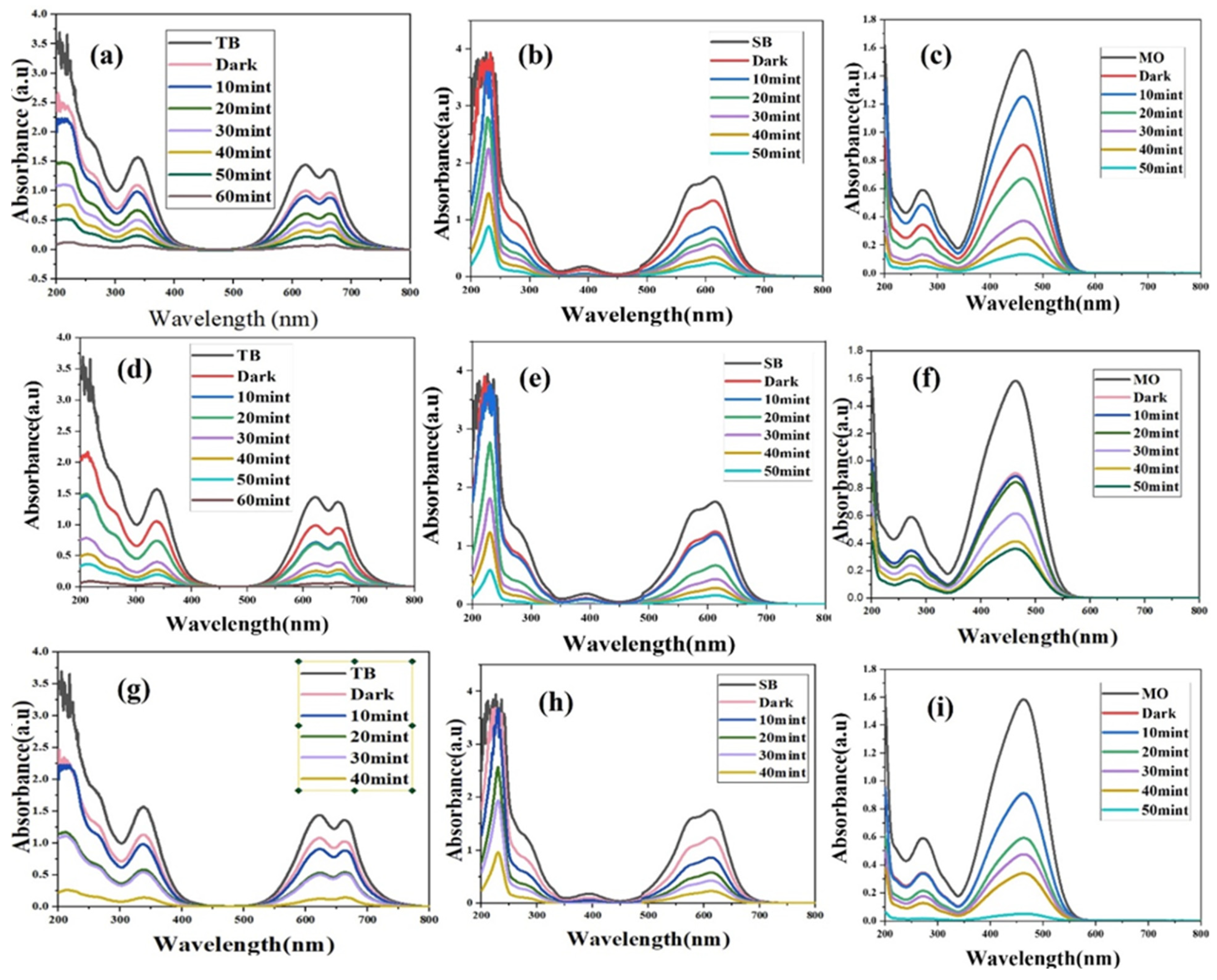
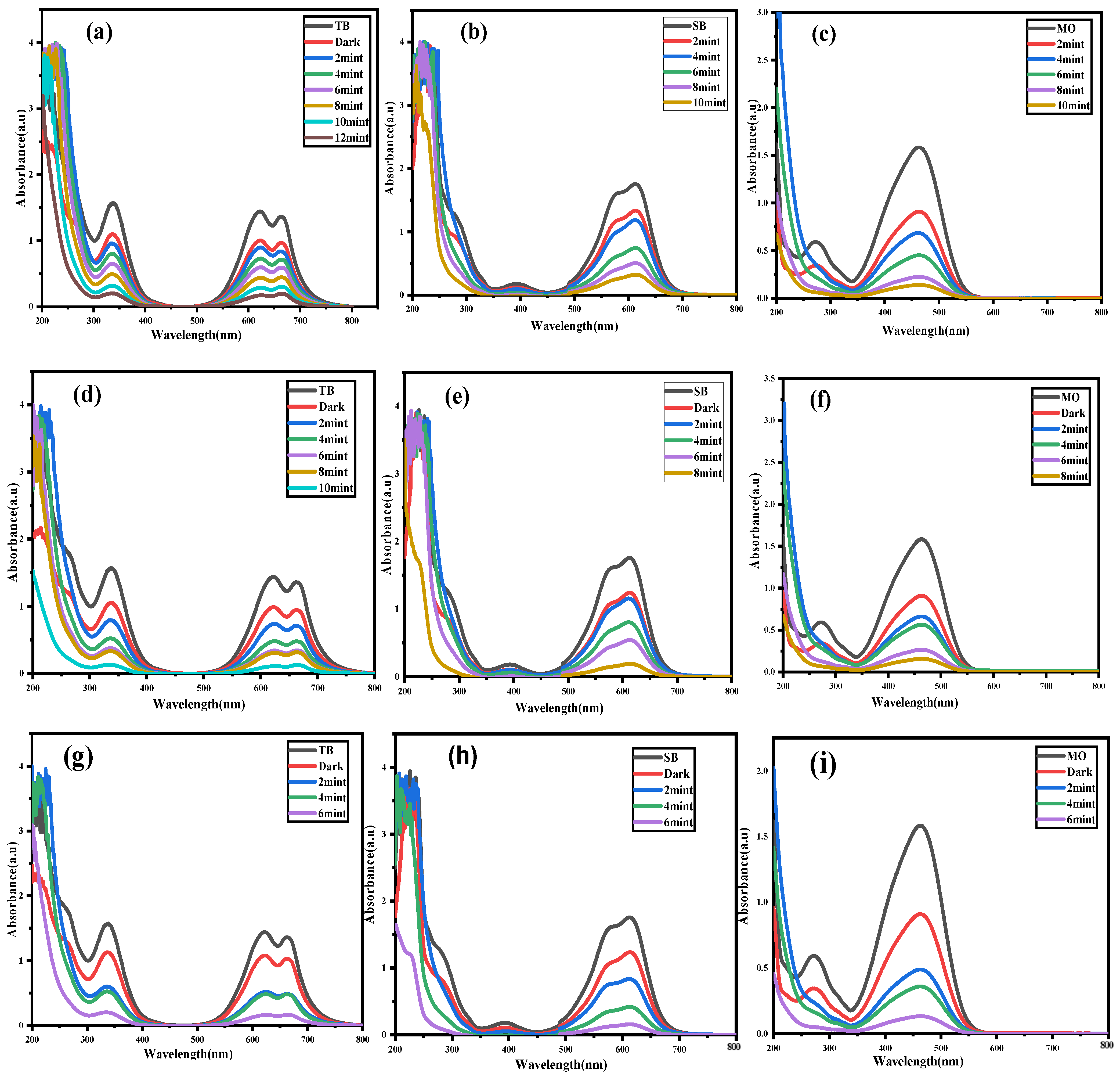
2.1. Photocatalytic Activities

The photocatalytic activity of Ag@CuO, Ag@TiO2, and Ag@CuO/TiO2 coated and hybridized-coated material against dyes such as TB, SB, and MO were monitored using a UV–Vis spectrophotometer. The catalysis phenomenon depends on the catalyst’s ability to capture or remove oxygen radicals [40,41]. In this study, the efficiency of catalysis was established with and without H2O2. The UV spectra of the prepared material against various dye degradation in the absence of H2O2 are shown in Figure 6. The binary core-shell, Ag@CuO, and Ag@TiO2 materials exhibited photocatalytic degradation against the TB, SB, and MO dyes within 50–60 min, as shown in Figure 6a–f. Therefore, 20–25% of unaffected dye still existed. However, in the case of Ag@CuO/TiO2, a hybridized coating of two different metal oxides, the photocatalytic activity improved where these selected dyes were almost fully degraded within 40 min, as presented in Figure 6g–i. These findings suggest that the tertiary nanocatalyst (Ag@CuO/TiO2) developed a greater extent of amorphous zones, as shown by Figure 1d, and has a strong effect to bind with dyes. Materials in the amorphous zone tend to adsorb more dyes because they have a lot of structural voids and interstitial spaces resulting from various deformations [42]. This creates configurational and torsional strain, making their antibonding orbital larger for easy electromagnetic interaction [43]. This interaction leads toward the breaking of the bonding orbital. Moreover, the material might have trapped some environmental oxygen, which also plays a role in degradation.
Additionally, the photocatalytic effectiveness of each catalyst was also studied in the presence of an H2O2 reagent, and the outcomes are shown in Figure 7. Figure 7a–c depicts the involvement of the Ag@CuO photocatalyst in the presence of H2O2 for the degradation of TB, SB, and MO, respectively. The efficiency of the Ag@CuO nanocatalyst with H2O2 demonstrated the degradation of these dyes within 10–12 min. Figure 7d–f represents the effect of Ag@TiO2 over the same dyes in the same sequence and pattern, and demonstrated the photocatalysis of TB, SB, and MO between 8 and 10 min. Finally, Figure 7g–i describes the Ag@CuO/TiO2 ternary system’s function in the presence of H2O2, and resulted in the maximum degradation of the dyes within 6 min. The percentage dye degradation vs. time plots in the form of bars are provided in Figure 8A,B to compare the photocatalytic performance of the prepared catalysts against various dyes in the presence and absence of H2O2. It is clear from Figure 8 that the Ag@CuO/TiO2 nanocatalyst had the highest degradation rate both with and without H2O2. The reproducibility in the form of the standard error mean is indicated on the given bars, and was found within a range of 0.3 to 0.7. Moreover, the extent of degradation was quite high in each case. In the pure catalytic systems where Ag was not employed as a core, the kinetics was found to be too slow (i.e., it took more than 8 h with a 60–70% degradation outcome) [44,45]. The Ag is used as the core and interconnect of both photocatalysts; semiconductors support the stabilization of photogenerated electrons, which are used to produce reactive oxygen species to degrade the dye (i.e., pollutants). As discussed in Figure 5, with regard to the role of oxygen and electromagnetic radiation with strained bonds, there was a greater availability of oxygen radicals, which enhanced the catalyst efficiency and degraded each dye within 6 min. However, the same dyes were seen to degrade within up to 40 min of the time interval in the absence of H2O2. The results show that free radical species (HO, HOO, or O2) generated by H2O2 were captured on the surface of the nanocatalyst along with the dye molecules and developed chemical potential for their chemical interaction [46]. Moreover, the photogeneration of electrons from the photocatalyst proved to be twofold beneficial: one by transferring the electron to the dyes, and the other by stabilizing the reactive oxygen radical species through their holes, which were produced during photoexcitation [47]. The core of the semiconducting photocatalysts due the band overlapping assisted in stabilizing the photoexcited electrons and as produced reactive oxygen species. This phenomenon is more favorably found in semiconductors like CuO and TiO2, which allow the holes (h+) to react with hydroxyl and other radicals [48]. These synthetic reactions produced changes in the surface of the catalysts, such as the formation of oxygen vacancies and crystal defects, due to the high surface curvature [49]. Therefore, such surface defects are prone to interact with surrounding species such as dye and reagents, which were the focus of this study. We concluded that the hybrid nanocatalyst Ag@CuO/TiO2 was the most effective in degrading dyes both with and without H2O2 because of a greater number of surface defects, voids, and vacancies. In each case, the hybridized coating degraded these dyes by up to 90%. These are textile dyes, which during the coloring process are wasted by up to 60%, contaminate water, and cause serious diseases like cancer and arthritis [50]. They also affect the environment and are dangerous to all flora and fauna. Therefore, their degradation would be a remedial solution for a clean and conducive environment.

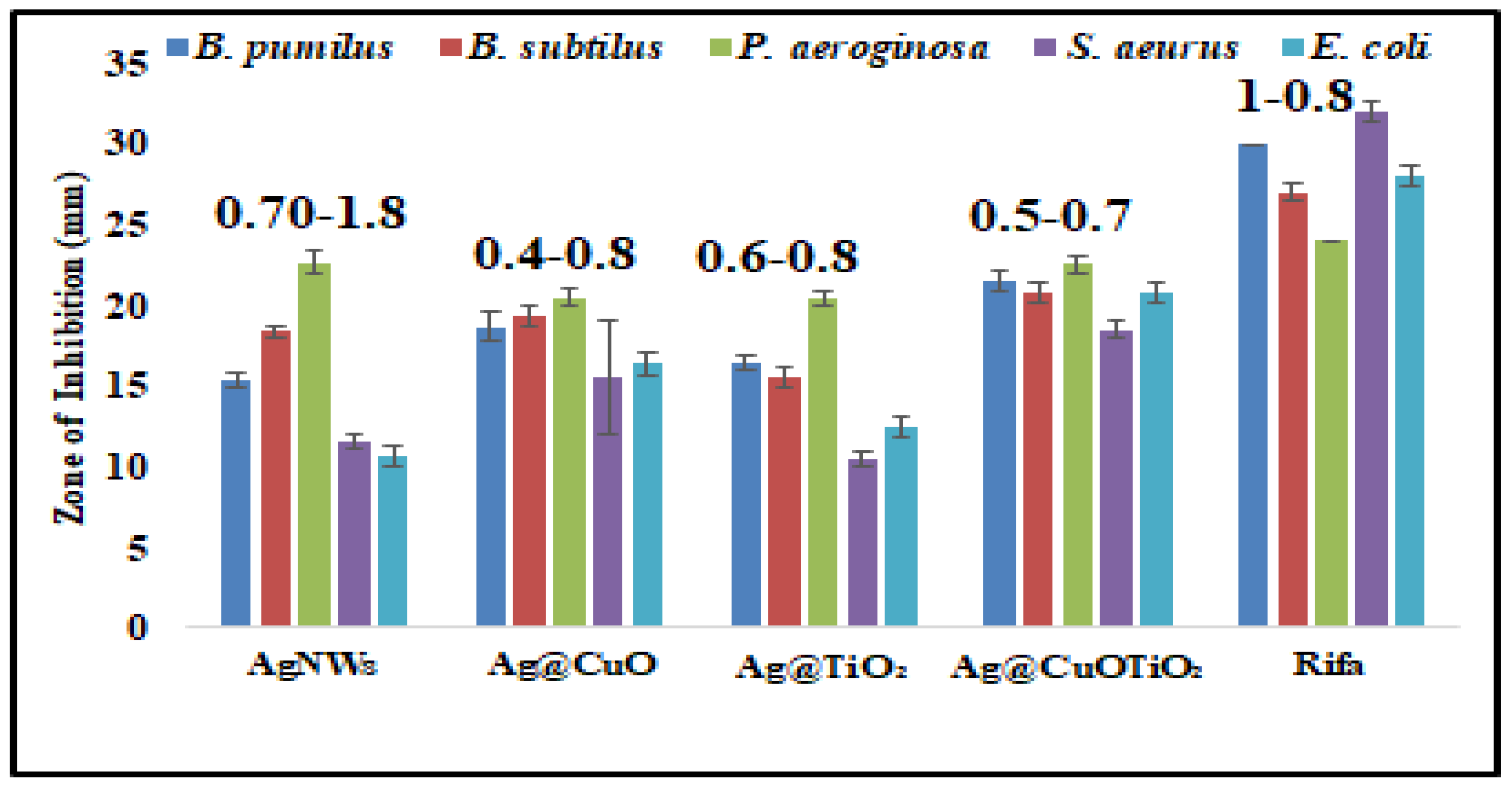
2.2. Antibacterial Activities

The antibacterial activity of the prepared samples including Ag NWs, Ag@CuO, Ag@TiO2, and Ag@CuO/TiO2 nanohybrids was tested toward Gram-positive bacteria (Staphylococcus aureus, Bacillus subtilis, and Bacillus pumilus) and Gram-negative bacteria (Pseudomonas aeruginosa and Escherichia coli), as shown in Figure 9. “Rifaximin” was used as a standard antibiotic in this evaluation. The zone of inhibition surrounding the well in the Petri dish was utilized to analyze the inhibitory power of the sample in millimeters. Figure 9 provides a detailed representation of the zone of inhibition using a bar graph. The bar graph indicates a general trend where each binary nanodesign of the Ag NWs exhibited higher inhibition zones than the pristine Ag NWs against all of the selected bacteria strains. Moreover, the ternary hybridized design Ag@CuO/TiO2 displayed the highest zones of inhibition: 24.8 ± 1.0, 23 ± 0.6, 24.4 ± 0.7, 19.4 ± 0.4, and 22.6 ± 0.6 for B. subtilis, B. pumilus, S. aureus, P. aeruginosa, and E. coli, respectively. The hybridized sample seemed to be closer to the antimicrobial activities of the standard used, where the values were mostly above 80%. The antimicrobial mechanism involves the interaction of Ag ions with bacteria and leads to the inhibition of cell growth and division. Therefore, the Ag NWs release silver ions, which adhere to the cell wall and cytoplasmic membrane, increasing their permeability [51]. Thus, cell rupture resulted, which led to cell death. When the Ag NWs were coated with CuO, TiO2, and their mixed form, they greatly increased the production of reactive oxygen species (ROS), which mainly react with proteins and DNA and also affect the ribosome [52]. Moreover, the presence of a higher number of surface defects in the binary and ternary Ag nanodesigns (Ag@CuO, Ag@TiO2, and Ag@CuO/TiO2) in comparison to that in the pristine Ag NWs was attributed to the high surface reactivity against bacterial cells, and hence responsible for better antibacterial activity against the tested pathogens. The metal oxide makes an ultra-thin amorphous sheath that can strongly interact with the pathogens. Moreover, these materials have also been reported as an antimicrobial; as Ag was the core material, their activity increased significantly.
The study found that the prepared samples were more effective against Gram-positive bacteria (such as S. aureus, B. subtilis, and B. pumilus) than against Gram-negative bacteria (like P. aeruginosa and E. coli). This is because of the differences in the composition and structure of their cell walls. Gram-positive bacteria have single-layer cell walls with low lipid content, while Gram-negative bacteria have three layers in their cell walls. Additionally, Gram-positive bacterial strains have single-layer cytoplasmic membranes, while Gram-negative bacterial strains have two membranes—an outer membrane and a cytoplasmic membrane [53]. The study also shows that the antibacterial properties of the fabricated nanohybrid materials could help disinfect and prevent the spread of pandemics caused by germs along with denaturing carcinogenic dyes.

3. Materials and Methods

3.1. Materials and Chemicals

Ethylene glycol (EG) 99% (Dae-Jung, Gyeonggi-do, Republic of Korea), silver nitrate (Merck, Rahway, NJ, USA), sodium chloride (Sigma Aldrich, Darmstadt, Germany), and the poly(vinyl pyrrolidone) (PVP) MW (90 k) product from Dae-Jung were used for the preparation of Ag linear structures. The encapsulating precursors (i.e., copper Cu(CH3COO)2.2H2O and titanium isopropoxide (Merck) were used as received. Analytical grade solvents like acetone and ethanol were from Sigma-Aldrich. Carcinogenic dyes used in the experiments like TB, SB, and MO were used as conventional dyes to evaluate the catalytic activities of the synthesized materials. The microbial strains were taken from Biotech Lab, MUST, Mirpur, Pakistan.

3.2. Preparation and Coating of Ag NWs

The polyol reduction approach was used to prepare the Ag NWs and their coated form with CuO, TiO2, and the hybrid of both (CuO/TiO2). The synthesis involved EG as a solvent and reductant and PVP as the capping agent. It is widely accepted method for high yield and monodispersity of the metallic outcome [54]. Herein, 10 mL of EG was preheated at 170 °C for an hour while continuously stirred. Approximately 2 μL of 0.01 M NaCl solution was added to slow the growth and increase the longitudinal dimension, followed by the dropwise addition of 5 mL solutions of 0.1 M AgNO3 and PVP in continuously stirred EG at 170 °C. This reaction was continued for 1 h, then the mixture was cooled, centrifuged, and washed with a mixture of acetone and ethanol. To prepare the coating of CuO, the first step was repeated at the same temperature for 40 min, followed by an addition of 2 mL of 0.01 M Cu (CH3COO)2.2H2O solution. This reaction was further heated for two hours. Similarly, the surface coating of Ag NWs with TiO2 was achieved by adding 4 µL of titanium isopropoxide with a molar concentration of 0.01 M. The product was then cooled, washed, and collected for characterization and application by centrifugation at 4000 rpm. Finally, the hybrid coating of CuO/TiO2 was similarly prepared by adding the precursors of CuO and TiO2 in growing Ag NWs and the reaction proceeded for a further two hours. In the end, the mixture was washed and collected as described above. All of the samples were wrapped in Whatman paper and dried. Consequently, the Ag NWs, Ag@CuO, Ag@TiO2, and Ag@CuO/TiO2 nanocables were prepared using the simple polyol method.

3.3. Catalytic Activities

A UV–Vis spectrophotometer was used to test the effectiveness of the Ag@CuO-, Ag@TiO2-, and Ag@CuO/TiO2 nanocable-based catalysts against dyes such as TB, SB, and MO with and without the addition of H2O2. In this experiment, 2 mg of catalyst was added to 50 mL of each dye solution, where each dye was 2.5 mg. The whole system was placed in a chamber adjusted with a tungsten bulb. The degradation reaction was observed in absorbance with the UV–Vis spectrophotometer. Additionally, 1 mL of H2O2 was added to the catalyst and dye solution to evaluate the effects of the degrading agent, and the reaction was closely and similarly monitored. Each dye was subjected to sole catalyst degradation and a catalyst with H2O2.

3.4. Antibacterial Activities

The antibacterial activities of the Ag NWs, Ag@CuO, Ag@TiO2, and Ag@CuO/TiO2 nanocables were tested using the Gram-positive bacteria Staphylococcus aureus, Bacillus subtilis, and Bacillus pumilus as well as the Gram-negative bacteria Pseudomonas aeruginosa and Escherichia coli. The common antibiotic “rifaximin” was used as the standard in the assessment. The well method was employed to measure the zones of inhibition. The testing environment was prepared by taking 50 mL of broth media, and 10 mL was pipetted into each of five separate Falcon tubes. Then, the bacteria were added, and the tubes were incubated for 24 h at 37 °C. After incubation, the bacterial culture appeared turbid, indicating that the bacteria were growing and multiplying. Wells were created using a cork borer after transferring agar material and streaking the bacterial culture into Petri plates. Subsequently, 15 microliters of each prepared sample was added to each well using laminar flow, and all Petri plates were covered. The Petri dishes containing the bacteria and each sample were incubated at 37 °C for another 24 h. The zones of inhibition were then detected and assessed using a traditional scale after one day.

3.5. Characterization

An XRD D8 ADVANCE BRUKER (Karlsruhe, Germany) was employed to analyze the crystal structures and phase growth of Ag, Ag@CuO, Ag@TiO2, and the hybrid of CuO and TiO2 around Ag, all being linear structures. The field emission scanning electron microscope (SEM) TSCAN Mira 3 (Kohoutovice, Czech Republic), coupled with EDX, proved helpful in observing the morphology and surface topography of all of the synthesized materials and also diagnosed the appearance of a respective ultra-thin coating around the Ag NWs. The coupled EDX determined the elemental nature of each sample. The coating thickness was explored using a JEOL 2100F FEG-TEM (Tokyo, Japan). The elemental composition was revealed through a Scientia-Omicron XPS instrument equipped with a micro-focused monochromatic Al K-alpha X-ray source with an operating energy of 15 KeV. UV–Vis absorption spectra were observed on a SHIMADZU UV 1800 Spectrophotometer (Kyoto, Japan) to analyze the SPR trend of the core material and the cast effect of the coating on this innate property. The instrument was further applied to study the catalytic activities (i.e., degradation of selected carcinogenic dyes). A SHIMADZU RF-6000 Spectro fluorophotometer measured the photoluminescence property of the coating materials and their hybridization. The electrochemical activities of the prepared samples were obtained using cyclic voltammetry (CHI 600E, CH Instruments, Dallas, TX, USA) with a scan rate of 0.1 V/s. In the end, a simple scale was used to measure the zone of inhibition to analyze the antimicrobial activities.

4. Conclusions

The polyol method under a single pot proved successful in designing the anisotropic structure of Ag and co-axial coating by metal oxides in the pure and hybridized form. The current work involved the synthesis of Ag NWs, binary core-shell nanocables of Ag@CuO and Ag@TiO2. Similarly, a ternary nanohybrid Ag@CuO/TiO2 with a hybridized coating was produced using polyol methodology. The presence of the metal oxide’s precursor remarkably reduced the diameter of the NWs and was highly manifested in the case of TiO2. An ultra-thin covering of CuO was found to be quite smooth, but in each case, TiO2 roughness arose due to the grain boundaries. An XPS study was carried out to explore the surface coating. The ultra-thin layer’s nature of CuO, TiO2, and their combined form was further observed through the decreased intensity of the metal SPR. PL also confirmed the hybridization of metal oxides around Ag, which possessed many discrete electronic energy levels from 400 nm to 750 nm to harvest light for photovoltaic applications and can trap the radicals for catalysis. The hybrid coating demonstrated high electrochemical activities as displayed by CV, making it suitable for activating the substrate and reagents and degrading the dyes. The highest photocatalytic activities were found for the Ag@CuO/TiO2 nanodesign for the TB, SB, and MO textile dyes, where 2 mg of catalyst degraded 2.5 g dyes by 1 mL H2O2. Similarly, the synthesized Ag core@shell nanocables exhibited antibacterial activities, particularly against Gram-negative bacterial strains. However, the ternary nanohybrid design was the most effective, with inhibition zone values in the range of 35–54 mm. Therefore, these nanostructures could be reliable candidates for applications in photovoltaic cells, anti-pollutants, and anti-microbial processes.

Author Contributions

J.A.: Methodology, Writing—Original Draft, Resources. Z.A.: Conceptualization, Methodology, Writing—original draft, Software, Project administration, Investigation, Resources. A.I.: Conceptualization, Visualization, Writing—review and editing, Formal analysis, Data curation. A.N.: Formal Analysis, Supervision, Visualization, Writing—review and editing. M.A.: Formal analysis, Methodology, Visualization, Writing—review and editing. R.S.: Conceptualization, Data curation, Writing—original draft, Investigation. M.R.: Formal analysis, Data curation, Visualization, Funding acquisition, Writing—review and editing; B.B.K.A.: Formal analysis, Resources, Validation, Writing—review and editing. Y.A.B.J.: Funding acquisition, Project administration, Formal analysis, Writing—review and editing. All authors have read and agreed to the published version of the manuscript.

Funding

This research is supported by the Researchers Supporting Project Number (RSP2025R457), King Saud University, Riyadh, Saudi Arabia.

Data Availability Statement

All of the data from this study are contained in this manuscript. More data related to this study can be accessed upon reasonable request to the corresponding author at  zahoor.chem@must.edu.pk.

Acknowledgments

The authors would like to extend their sincere appreciation to the Researchers Supporting Project Number (RSP2025R457), King Saud University, Riyadh, Saudi Arabia. All authors would like to extend their thanks to MG Shazly for the technical support provided for this research work.

Conflicts of Interest

The authors declare no conflicts of interest.

Correction Statement

This article has been republished with a minor correction to the Funding statement. This change does not affect the scientific content of the article.

References

  1. Wang, H.; Wu, C.; Huang, Y.; Sun, F.; Lin, N.; Soomro, A.M.; Zhong, Z.; Yang, X.; Chen, X.; Kang, J.; et al. One-pot synthesis of superfine core–shell Cu@ metal nanowires for highly tenacious transparent LED dimmer. ACS Appl. Mater. Interfaces 2016, 8, 28709–28717. [Google Scholar] [CrossRef] [PubMed]
  2. Zhu, Y.; Kim, S.; Ma, X.; Byrley, P.; Yu, N.; Liu, Q.; Sun, X.; Xu, D.; Peng, S.; Hartel, M.C.; et al. Ultrathin-shell epitaxial Ag@ Au core-shell nanowires for high-performance and chemically-stable electronic, optical, and mechanical devices. Nano Res. 2021, 14, 4294–4303. [Google Scholar] [CrossRef]
  3. Kang, H.; Kim, J.S.; Choi, S.-R.; Kim, Y.-H.; Kim, D.H.; Kim, J.-G.; Lee, T.-W.; Cho, J.H. Electroplated core–shell nanowire network electrodes for highly efficient organic light-emitting diodes. Nano Converg. 2022, 9, 1–8. [Google Scholar] [CrossRef] [PubMed]
  4. Wu, Z.-P.; Shan, S.; Zang, S.-Q.; Zhong, C.-J. Dynamic core–shell and alloy structures of multimetallic nanomaterials and their catalytic synergies. Acc. Chem. Res. 2020, 53, 2913–2924. [Google Scholar] [CrossRef]
  5. Yao, M.; Wang, B.; Sun, B.; Luo, L.; Chen, Y.; Wang, J.; Wang, N.; Komarneni, S.; Niu, X.; Hu, W. Rational design of self-supported Cu@ WC core-shell mesoporous nanowires for pH-universal hydrogen evolution reaction. Appl. Catal. B Environ. 2021, 280, 119451. [Google Scholar] [CrossRef]
  6. Liu, Y.; Tang, Z. Multifunctional nanoparticle@ MOF core–shell nanostructures. Adv. Mater. 2013, 25, 5819–5825. [Google Scholar] [CrossRef]
  7. Arabzadeh, A.; Salimi, A. One dimensional CdS nanowire@ TiO2 nanoparticles core-shell as high performance photocatalyst for fast degradation of dye pollutants under visible and sunlight irradiation. J. Colloid Interface Sci. 2016, 479, 43–54. [Google Scholar] [CrossRef]
  8. Choubrac, L.; Bär, M.; Kozina, X.; Félix, R.; Wilks, R.G.; Brammertz, G.; Levcenko, S.; Arzel, L.; Barreau, N.; Harel, S.; et al. Sn substitution by Ge: Strategies to overcome the open-circuit voltage deficit of kesterite solar cells. ACS Appl. Energy Mater. 2020, 3, 5830–5839. [Google Scholar] [CrossRef]
  9. Shin, J.C.; Kim, K.H.; Yu, K.J.; Hu, H.; Yin, L.; Ning, C.-Z.; Rogers, J.A.; Zuo, J.-M.; Li, X. InxGa1-x As nanowires on silicon: One-dimensional heterogeneous epitaxy, bandgap engineering, and photovoltaics. Nano Lett. 2011, 11, 4831–4838. [Google Scholar] [CrossRef]
  10. Han, L.; Zhan, W.; Liang, X.; Zhang, W.; Huang, R.; Chen, R.; Ni, H. In-situ generation Cu2O/CuO core-shell heterostructure based on copper oxide nanowires with enhanced visible-light photocatalytic antibacterial activity. Ceram. Int. 2022, 48, 22018–22030. [Google Scholar] [CrossRef]
  11. El-Toni, A.M.; Habila, M.A.; Labis, J.P.; Alothman, Z.A.; Alhoshan, M.; Elzatahry, A.A.; Zhang, F. Design, synthesis and applications of core–shell, hollow core, and nanorattle multifunctional nanostructures. Nanoscale 2016, 8, 2510–2531. [Google Scholar] [CrossRef] [PubMed]
  12. Zhang, D.; Wada, H. Laser ablation in liquids for nanomaterial synthesis and applications. In Handbook of Laser Micro- and Nano-Engineering; Springer: Berlin/Heidelberg, Germany, 2020; pp. 1–35. [Google Scholar]
  13. Rai, P. Plasmonic noble metal@ metal oxide core–shell nanoparticles for dye-sensitized solar cell applications. Sustain. Energy Fuels 2019, 3, 63–91. [Google Scholar] [CrossRef]
  14. Fiévet, F.; Ammar-Merah, S.; Brayner, R.; Chau, F.; Giraud, M.; Mammeri, F.; Peron, J.; Piquemal, J.-Y.; Sicard, L.; Viau, G. The polyol process: A unique method for easy access to metal nanoparticles with tailored sizes, shapes and compositions. Chem. Soc. Rev. 2018, 47, 5187–5233. [Google Scholar] [CrossRef]
  15. Li, G.; Tang, Z. Noble metal nanoparticle@ metal oxide core/yolk–shell nanostructures as catalysts: Recent progress and perspective. Nanoscale 2014, 6, 3995–4011. [Google Scholar] [CrossRef]
  16. Mondal, K.; Sharma, A. Recent advances in the synthesis and application of photocatalytic metal–metal oxide core–shell nanoparticles for environmental remediation and their recycling process. RSC Adv. 2016, 6, 83589–83612. [Google Scholar] [CrossRef]
  17. Baranowska-Korczyc, A.; Mackiewicz, E.; Ranoszek-Soliwoda, K.; Grobelny, J.; Celichowski, G. Core/shell Ag/SnO2 nanowires for visible light photocatalysis. Catalysts 2021, 12, 30. [Google Scholar] [CrossRef]
  18. Yadav, A.K.; Jain, C.; Malik, D. Toxic characterization of textile dyes and effluents in relation to human health hazards. J. Sustain. Environ. Res. 2014, 3, 95–102. [Google Scholar]
  19. Al-Tohamy, R.; Ali, S.S.; Li, F.; Okasha, K.M.; Mahmoud, Y.A.-G.; Elsamahy, T.; Jiao, H.; Fu, Y.; Sun, J. A critical review on the treatment of dye-containing wastewater: Ecotoxicological and health concerns of textile dyes and possible remediation approaches for environmental safety. Ecotoxicol. Environ. Saf. 2022, 231, 113160. [Google Scholar] [CrossRef]
  20. Senapati, S.; Srivastava, S.K.; Singh, S.B.; Mishra, H.N. Magnetic Ni/Ag core–shell nanostructure from prickly Ni nanowire precursor and its catalytic and antibacterial activity. J. Mater. Chem. 2012, 22, 6899–6906. [Google Scholar] [CrossRef]
  21. Sundaram, M.; Kalpana, S.; Sivaganesan, V.; Nandhakumar, E. Studies on the catalytic activity of CuO/TiO2/ZnO ternary nanocomposites prepared via one step hydrothermal green approach. Mater. Res. Express 2019, 6, 125043. [Google Scholar]
  22. Indumathi, T.; Suriyaprakash, J.; Alarfaj, A.A.; Hirad, A.H.; Jaganathan, R.; Mathanmohun, M. Synergistic effects of CuO/TiO2-chitosan-farnesol nanocomposites: Synthesis, characterization, antimicrobial, and anticancer activities on melanoma cells SK-MEL-3. J. Basic Microbiol. 2024, 64, 2300505. [Google Scholar] [CrossRef] [PubMed]
  23. Kerli, S.; Kavgacı, M.; Soğuksu, A.K.; Avar, B. Photocatalytic degradation of methylene blue, rhodamine-B, and malachite green by Ag@ ZnO/TiO2. Braz. J. Phys. 2022, 52, 22. [Google Scholar] [CrossRef]
  24. Idrees, S.; Ahmad, Z.; Akhtar, T.; Choudhary, M.A.; Rafiq, M.A.; Mehmood, A. Facile chemical strategy to synthesize Ag@ polypyrrole microarrays and investigating its anisotropic effect on polymer conductivity. Mater. Sci.-Pol. 2019, 37, 563–569. [Google Scholar] [CrossRef]
  25. Yang, X.; Fu, H.; Zhang, L.; An, X.; Xiong, S.; Jiang, X.; Yu, A. Enhanced gas sensing performance based on the fabrication of polycrystalline Ag@ TiO2 core-shell nanowires. Sens. Actuators B Chem. 2019, 286, 483–492. [Google Scholar] [CrossRef]
  26. Sharif, S.; Ahmad, Z.; Hoskins, C.; Choudhary, M.A.; Mehmood, A. Ag nanostructure morphologies and physicochemical properties dictated by the polyols used in the synthesis. J. Nano Res. 2022, 76, 93–106. [Google Scholar] [CrossRef]
  27. Yamashita, M. Next generation multifunctional nano-science of advanced metal complexes with quantum effect and nonlinearity. Bull. Chem. Soc. Jpn. 2021, 94, 209–264. [Google Scholar] [CrossRef]
  28. Ma, H.R.; Chen, X.Y.; Li, J.W.; Chang, C.T.; Wang, G.; Li, H.; Wang, X.M.; Li, R.W. Fe-based amorphous coating with high corrosion and wear resistance. Surf. Eng. 2017, 33, 56–62. [Google Scholar] [CrossRef]
  29. Gawande, M.B.; Goswami, A.; Asefa, T.; Guo, H.; Biradar, A.V.; Peng, D.-L.; Zboril, R.; Varma, R.S. Core–shell nanoparticles: Synthesis and applications in catalysis and electrocatalysis. Chem. Soc. Rev. 2015, 44, 7540–7590. [Google Scholar] [CrossRef]
  30. Prieto, P.; Nistor, V.; Nouneh, K.; Oyama, M.; Abd-Lefdil, M.; Díaz, R. XPS study of silver, nickel and bimetallic silver–nickel nanoparticles prepared by seed-mediated growth. Appl. Surf. Sci. 2012, 258, 8807–8813. [Google Scholar] [CrossRef]
  31. Chi, M.; Sun, X.; Lozano-Blanco, G.; Tatarchuk, B.J. XPS and FTIR investigations of the transient photocatalytic decomposition of surface carbon contaminants from anatase TiO2 in UHV starved water/oxygen environments. Appl. Surf. Sci. 2021, 570, 151147. [Google Scholar] [CrossRef]
  32. Ivanova, T.; Maslakov, K.; Sidorov, A.; Kiskin, M.; Linko, R.; Savilov, S.; Lunin, V.; Eremenko, I. XPS detection of unusual Cu (II) to Cu (I) transition on the surface of complexes with redox-active ligands. J. Electron Spectrosc. Relat. Phenom. 2020, 238, 146878. [Google Scholar] [CrossRef]
  33. Zahoor, A.; Qiu, T.; Zhang, J.; Li, X. Synthesis and characterization of Ag@ polycarbazole nanoparticles and their novel optical behavior. J. Mater. Sci. 2009, 44, 6054–6059. [Google Scholar] [CrossRef]
  34. Sharif, S.; Ahmed, T.; Ahmad, Z.; Choudhary, M.A.; Arshad, M. Synthesis and characterization of Ag@ Ni co-axial nanocables and their fluorescent and catalytic properties. Sci. Eng. Compos. Mater. 2024, 31, 20240010. [Google Scholar] [CrossRef]
  35. Arya, S.; Singh, A.; Kour, R. Comparative study of CuO, CuO@ Ag and CuO@ Ag: La nanoparticles for their photosensing properties. Mater. Res. Express 2019, 6, 116313. [Google Scholar] [CrossRef]
  36. Adekoya, J.; Dare, E.; Mesubi, M.; Nejo, A.A.; Swart, H.; Revaprasadu, N. Synthesis of polyol based Ag/Pd nanocomposites for applications in catalysis. Results Phys. 2014, 4, 12–19. [Google Scholar] [CrossRef]
  37. Blancon, J.; Nie, W.; Neukirch, A.J.; Gupta, G.; Tretiak, S.; Cognet, L.; Mohite, A.D.; Crochet, J.J. The effects of electronic impurities and electron–hole recombination dynamics on large-grain organic–inorganic perovskite photovoltaic efficiencies. Adv. Funct. Mater. 2016, 26, 4283–4292. [Google Scholar] [CrossRef]
  38. Hodges, B.C.; Cates, E.L.; Kim, J.-H. Challenges and prospects of advanced oxidation water treatment processes using catalytic nanomaterials. Nat. Nanotechnol. 2018, 13, 642–650. [Google Scholar] [CrossRef]
  39. Mondal, K.; Sharma, A. Photocatalytic oxidation of pollutant dyes in wastewater by TiO2 and ZnO nano-materials—A mini-review. Nanosci. Technol. Mank. 2014, 36–72. [Google Scholar]
  40. Ahmad, Z.; Maqsood, M.; Mehmood, M.; Ahmad, M.J.; Choudhary, M.A. Synthesis and characterization of pure and nano-Ag impregnated chitosan beads and determination of catalytic activities of nano-Ag. Bull. Chem. React. Eng. Catal. 2017, 12, 127–135. [Google Scholar] [CrossRef]
  41. Hu, H.; Liu, P.; Cao, S.; You, L.; Zhang, N.; Xi, J.; Guo, S.; Zhou, K. Single Metal Atoms Anchored on N-Doped Holey Graphene as Efficient Dual-Active-Component Catalysts for Nitroarene Reduction. Adv. Funct. Mater. 2024, 34, 2307162. [Google Scholar] [CrossRef]
  42. Abdelrahman, E.A.; Hegazey, R.; El-Azabawy, R.E. Efficient removal of methylene blue dye from aqueous media using Fe/Si, Cr/Si, Ni/Si, and Zn/Si amorphous novel adsorbents. J. Mater. Res. Technol. 2019, 8, 5301–5313. [Google Scholar] [CrossRef]
  43. Mantasha, I.; Hussain, S.; Ahmad, M.; Shahid, M. Two dimensional (2D) molecular frameworks for rapid and selective adsorption of hazardous aromatic dyes from aqueous phase. Sep. Purif. Technol. 2020, 238, 116413. [Google Scholar] [CrossRef]
  44. Bathla, A.; Rather, R.A.; Poonia, T.; Pal, B. Morphology dependent photocatalytic activity of CuO/CuO–TiO2 nanocatalyst for degradation of methyl orange under sunlight. J. Nanosci. Nanotechnol. 2020, 20, 3123–3130. [Google Scholar] [CrossRef] [PubMed]
  45. Kapoor, A.; Rajput, J.K. Solar light photocatalytic activity of CuO/TiO2 mixed oxide derived from conjugated azomethine metal complex for degradation of food colorants. J. Mol. Liq. 2022, 366, 120280. [Google Scholar] [CrossRef]
  46. Zuorro, A.; Lavecchia, R. Evaluation of UV/H2O2 advanced oxidation process (AOP) for the degradation of diazo dye Reactive Green 19 in aqueous solution. Desalin. Water Treat. 2014, 52, 1571–1577. [Google Scholar] [CrossRef]
  47. Ren, H.; Wang, S.; Labidi, A.; Pan, B.; Luo, J.; Wang, C. Nanoengineering of ultrathin N-CQDs/Bi2WO6 S-scheme heterojunction for enhanced photodegradation of antibiotics as emerging contaminants: Mechanism insight and toxicity assessment. Sep. Purif. Technol. 2025, 362, 131717. [Google Scholar] [CrossRef]
  48. Kochuveedu, S.T.; Jang, Y.H.; Kim, D.H. A study on the mechanism for the interaction of light with noble metal-metal oxide semiconductor nanostructures for various photophysical applications. Chem. Soc. Rev. 2013, 42, 8467–8493. [Google Scholar] [CrossRef]
  49. Sin, J.-C.; Lam, S.-M.; Zeng, H.; Lin, H.; Li, H.; Huang, L.; Liaw, S.-J.; Mohamed, A.R.; Lim, J.-W. Integrating BaFe2O4 nanoparticles onto N-doped Bi2WO6 microspheres for eminent visible light-driven photocatalytic performance towards aquaculture contaminants and pathogens. Colloids Surf. A Physicochem. Eng. Asp. 2024, 692, 133905. [Google Scholar] [CrossRef]
  50. Rawat, D.; Mishra, V.; Sharma, R.S. Detoxification of azo dyes in the context of environmental processes. Chemosphere 2016, 155, 591–605. [Google Scholar] [CrossRef]
  51. Rtimi, S.; Dionysiou, D.D.; Pillai, S.C.; Kiwi, J. Advances in catalytic/photocatalytic bacterial inactivation by nano Ag and Cu coated surfaces and medical devices. Appl. Catal. B Environ. 2019, 240, 291–318. [Google Scholar] [CrossRef]
  52. Vaishampayan, A.; Grohmann, E. Antimicrobials functioning through ros-mediated mechanisms: Current insights. Microorganisms 2021, 10, 61. [Google Scholar] [CrossRef] [PubMed]
  53. Vaiwala, R.; Sharma, P.; Ayappa, K.G. Differentiating interactions of antimicrobials with Gram-negative and Gram-positive bacterial cell walls using molecular dynamics simulations. Biointerphases 2022, 17, 061008. [Google Scholar] [CrossRef] [PubMed]
  54. Hemmati, S.; Harris, M.T.; Barkey, D.P. Polyol silver nanowire synthesis and the outlook for a green process. J. Nanomater. 2020, 2020, 9341983. [Google Scholar] [CrossRef]
Figure 1. XRD spectra of (a) Ag NWs, (b) Ag@CuO, (c) Ag@TiO2, and (d) Ag@CuO/TiO2 core@hybridized shell.
Catalysts 15 00283 g001
Figure 2. (A) SEM images with TEM inset of (a) Ag NWs, (b) Ag@CuO, (c) Ag@TiO2, and (d) Ag@CuO/TiO2 hybrid coating. (B) EDX patterns of (a) Ag NWs, (b) Ag@CuO, (c) Ag@TiO2, and (d) Ag@CuO/TiO2 nanocable with a hybridized shell.
Catalysts 15 00283 g002
Figure 3. XPS spectra of the Ag@CuO/TiO2 nanohybrid. (a) Full survey scan of Ag@CuO/TiO2, (b) HR spectra of Ag 3d, (c) HR spectra of Ti 2p, (d) HR spectra of Cu 2p3/2, (e) HR spectra of O 1s, and (f) HR spectra of C 1s.
Catalysts 15 00283 g003aCatalysts 15 00283 g003b
Figure 4. (A) UV–Vis spectra of Ag NWs, Ag@CuO, Ag@TiO2, and Ag@CuO/TiO2 nanocables. (B) PL spectra of (a) Ag NWs, (b) Ag@CuO, (c) Ag@TiO2, and (d) Ag@CuO/TiO2 nanocables with a hybridized shell.
Catalysts 15 00283 g004
Figure 5. Cyclic voltammogram of (a) Ag NWs, (b) Ag@CuO, (c) Ag@TiO2, and (d) Ag@CuO/TiO2.
Catalysts 15 00283 g005
Figure 6. Dye degradation of TB, SB, and MO without H2O2 by (ac) Ag@CuO, (df) Ag@TiO2, and (gi) Ag@CuO/TiO2 linear nanomaterials.
Catalysts 15 00283 g006
Figure 7. Dye degradation of TB, SB, and MO in the presence of H2O2 by (ac) Ag@CuO, (df) Ag@TiO2, and (gi) Ag@CuO/TiO2 NHs.
Catalysts 15 00283 g007
Figure 8. (A) % Dye degradation vs. time plots of TB, SB, and MO in the absence of H2O2. (B) Dye degradation of TB, SB, and MO in the presence of H2O2 using Ag@CuO, Ag@TiO2, and Ag@CuO/TiO2 NHs.
Catalysts 15 00283 g008
Figure 9. Comparative bar graph for the zone of inhibition of Ag NWs, Ag@CuO, Ag@TiO2 and Ag@CuO/TiO2 NHs with P. aeruginosa, E. coli, B. pumilus, S. aureus, and B. subtilis.
Catalysts 15 00283 g009
Disclaimer/Publisher’s Note: The statements, opinions and data contained in all publications are solely those of the individual author(s) and contributor(s) and not of MDPI and/or the editor(s). MDPI and/or the editor(s) disclaim responsibility for any injury to people or property resulting from any ideas, methods, instructions or products referred to in the content.

