Article

Detection of Dopamine Using Hybrid Materials Based on NiO/ZnO for Electrochemical Sensor Applications

1
Institute of Chemistry, University of Sindh, Jamshoro 76080, Pakistan
2
Institute of Chemistry, Shah Abdul Latif University Khairpur Mirs, Sindh 66111, Pakistan
3
Department of Mathematics and Sciences, College of Humanities and Sciences, Ajman University, Ajman P.O. Box 346, United Arab Emirates
4
Biomolecular Science, Earth and Life Science, Amsterdam University, 1081 HV Amsterdam, The Netherlands
5
Physics Department, Faculty of Science, Taibah University, Al-Madaina Al Munawarah 42353, Saudi Arabia
*
Authors to whom correspondence should be addressed.
Catalysts 2025, 15(2), 116; https://doi.org/10.3390/catal15020116
Submission received: 17 December 2024 / Revised: 7 January 2025 / Accepted: 21 January 2025 / Published: 24 January 2025
(This article belongs to the Section Electrocatalysis)

Abstract

:
Dopamine is a neurotransmitter which is classified as a catecholamine. It is also one of the main metabolites produced by some tumor types (such as paragangliomas and neoblastomas). As such, determining and monitoring the level of dopamine is of the utmost importance, ideally using analytical techniques that are sensitive, simple, and low in cost. Due to this, we have developed a non-enzymatic dopamine sensor that is highly sensitive, selective, and rapidly detects the presence of dopamine in the body. A hybrid material fabricated with NiO and ZnO, based on date fruit extract, was synthesized by hydrothermal methods and using NiO as a precursor material. This paper discusses the role of date fruit extracts in improving NiO’s catalytic performance with reference to ZnO and the role that they play in this process. An X-ray powder diffraction study, a scanning electron microscope study, and a Fourier transform infrared spectroscopy study were performed in order to investigate the structure of the samples. It was found that, in the composite NiO/ZnO, NiO exhibited a cubic phase and ZnO exhibited a hexagonal phase, both of which exhibited well-oriented aggregated cluster shapes in the composite. A hybrid material containing NiO and ZnO has been found to be highly electro-catalytically active in the advanced oxidation of dopamine in a phosphate buffer solution at a pH of 7.3. It has been found that this can be accomplished without the use of enzymes, and the range of oxidation used here was between 0.01 mM and 4 mM. The detection limit of non-enzymatic sensors is estimated to be 0.036 μM. Several properties of the non-enzymatic sensor presented here have been demonstrated, including its repeatability, selectivity, and reproducibility. A test was conducted on Sample 2 for the detection of banana peel and wheat grass, and the results were highly encouraging and indicated that biomass waste may be useful for the manufacture of medicines to treat chronic diseases. It is thought that date fruit extracts would prove to be valuable resources for the development of next-generation electrode materials for use in clinical settings, for energy conversion, and for energy storage.

1. Introduction

The neurotransmitter dopamine (DA) is widely recognized as one of the brain’s most important neurotransmitters [1]. Aside from its ability to function in both classes, dopamine is integral to the health and function of hormones, cardiovascular systems, kidneys, and the central nervous system [2,3]. It is possible that there could be a link between a high level of dopamine and cardiotoxicity as a result of excessive dopamine levels. This could lead to drug abuse in the future, which could have a negative impact on society [4], rapid heartbeat, heart failure, and hypertension. As opposed to this, low levels of dopamine are associated with depression [5], Parkinson’s disease [6], Alzheimer’s disease [7], schizophrenia [8,9], learning disabilities, and emotional disturbances. In the central nervous system, this neurotransmitter regulates a variety of functions, including memory, endocrine function, motor control, cognition, and learning. In this regard, it is of utmost importance to accurately measure the concentration of DA in physiological fluids as well as changes in that concentration over time. In recent decades, electrochemical (EC) sensors [10,11], optically based assays [12,13], capillary electrophoresis [14,15], mass spectrometry, and other techniques have all been developed for detecting DA. The advantages of EC sensors include their rapid response time, straightforward operation, and low cost. Dopamine levels in human bodily fluids are typically 100 to 1000 times lower than those in ascorbic acid (AA), the primary interfering substance when measuring dopamine levels using an electrochemical cell (EC) [16,17,18,19]. Furthermore, electrochemical sensors are frequently considered to perform better than other existing approaches due to their lower cost, faster response time, and higher sensitivity [20,21]. Due to their unique characteristics such as high stability, selectivity, and affordability, non-enzymatic sensing techniques have become more popular among researchers over the past decade. In order to achieve this, essential materials must be synthesized such that they can detect analytes without the use of enzymes under the necessary physiological conditions [22]. DA molecules are electroactive and therefore can be easily examined using EC methods [23,24,25]. In addition, EC methods have many limitations, particularly in the detection of DA in the presence of ascorbic acid, glucose, uric acid, and other derivatives of catecholamines. Furthermore, carbon supports, metal, and metal oxide decorations have been extensively reported to enhance sensing properties [23,26,27]. An electrochemical sensor with improved selectivity and electrocatalytic activity has been developed by anchoring metal oxide nanoparticles onto carbon surfaces. Nickel (Ni) and Ni-based nanomaterials are among the most desirable candidates among metals and metal oxides for electrocatalytic applications due to their considerable stability, abundance, low cost, low toxicity, and high catalytic activity [28,29]. ZnO is also a class of nanomaterials that have been used for enhancing electrochemical activity during sensing processes. Because of their numerous active sites and exceptional lattice conductivity, transition metals are highly electrocatalytic for the oxidation of hydroxyl (-OH) functional groups [30,31,32,33]. As a result of the simple, inexpensive, and scalable aspects of using biomass waste without using large amounts of harmful chemicals, environmentally friendly methods of utilizing biomass waste have garnered a great deal of attention in recent years. Consequently, different biomass wastes have been used in the synthesis of hybrid materials based on NiO/ZnO [34,35,36].
The aim of this research was to develop new green capping, stabilizing, and reducing agents derived from biomasses such as rotten dates. Many natural compounds can be used to produce nanostructured materials with improved performance in an environmentally friendly manner. Dates exhibit a natural chemistry of surface-modifying agents, such as carbohydrates and antioxidants. Among the bioactive compounds found in date fruit are phenolic acids, anthocyanins, carotenoids, flavonoids, sterols, and procyanidins. The free phenolic acids in date fruit include protocatechuic acid, vanilic acid, syringic acid, and ferulic acid, as well as bound phenolic acids such as gallic acid, caffeic acid, p-coumaric acid, and o-coumaric acid. Thus, these acids can be used to modify the morphology of hybrid materials as well as adjust their surface properties. In the hybrid materials growth process, antioxidant chemistry can provide multiple nucleation sites for the development of well-defined morphologies of hybrid materials that have potential functionalities. Hence, we have developed an ecofriendly and green method for preparing functional hybrid materials based on nickel-zinc oxide (NiO/ZnO) that can be applied to electrochemical sensing. To achieve this objective, we modified the structure and adjusted the surface properties of NiO/ZnO, which is derived from the promising properties of date fruit extract as potential catalysts to use in the electrochemical detection of dopamine. Importantly, variations of the content of co-catalysts like NiO during the development of hybrid systems have not been studied with reference to enhancing the electrocatalytic properties of ZnO based materials toward the fabrication of enzyme-free sensors.

2. Results and Discussion

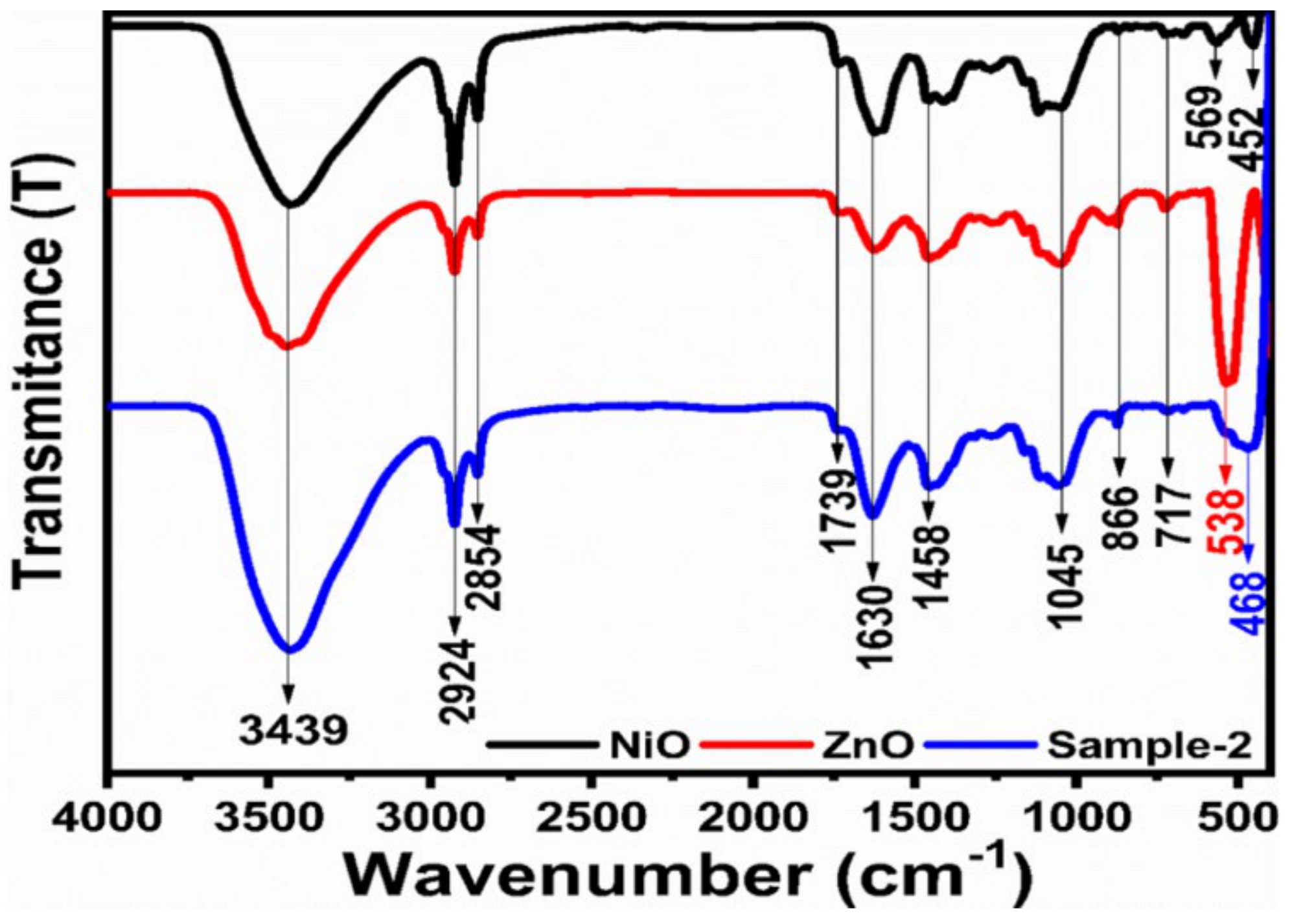
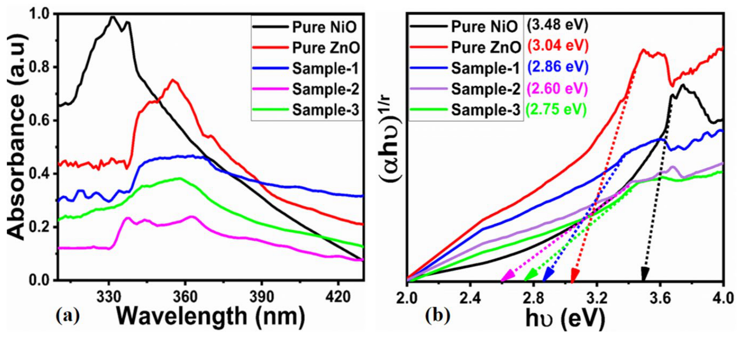
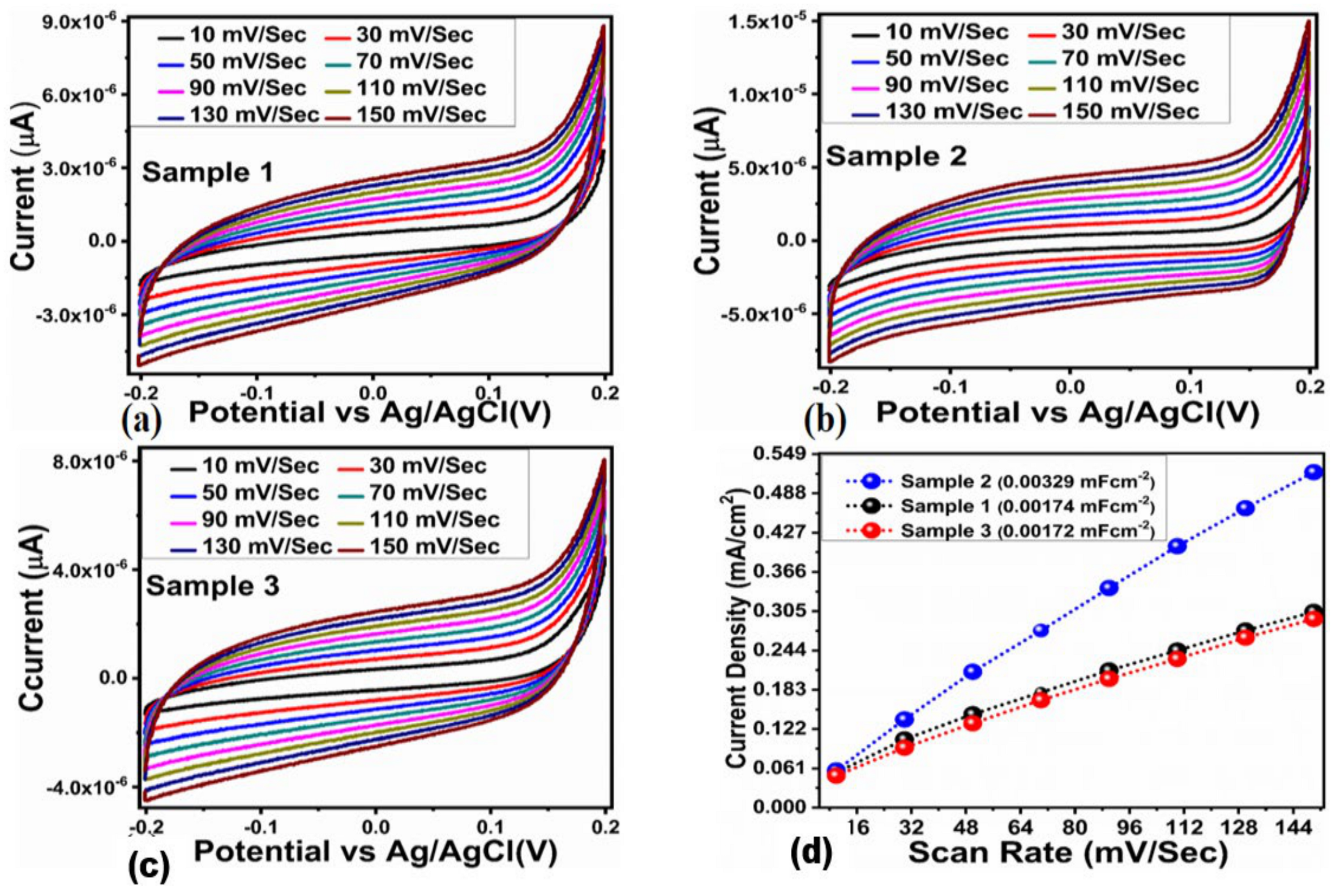
2.1. Crystal Quality, Morphology and Chemical Bonding Investigation NiO/ZnO Composites

Figure 1 illustrates an X-ray diffraction analysis of hybrid materials based on NiO/ZnO to assess their crystallinity. It can be concluded from the diffraction peak patterns of NiO (JCPDS card No: 01-089-7130) and ZnO (JCPDS card No: 01-079-0208) that NiO has a cubic structure and ZnO has a hexagonal structure. Based on NiO/ZnO, all hybrid materials presented the same phase information, which is consistent with previous studies [34,37,38]. In each hybrid material, NiO and ZnO reflections were observed, indicating the development of hetero-junction nanohybrids. Furthermore, the absence of additional reflections confirmed the purity of the hybrid materials based on NiO/ZnO. In addition, the XRD reflections were intense and sharp, indicating that the hybrid materials based on NiO/ZnO were highly crystalline.
A study of hybrid materials consisting of NiO and ZnO was conducted using FTIR to investigate their chemical bonding characteristics, as shown in Figure 2. It was determined that the stretching vibration for the O-H bond was associated with 3439 cm−1. Due to the adsorbed water molecules, the absorption bands at 1630 cm−1 are attributed to the hydroxyl group. The C-H stretching mode was linked to the band observed at 2924 cm−1 [35]. Band peaks at 2924 cm−1 and 2854 cm−1 indicate C-H vibration bands for CH2 [36]. Typical metal oxide peaks can be found at 538 cm−1 and 468 cm−1. The absorption band at 1458 cm−1 indicates the presence of carbonates. Observed peaks at 866 cm−1 and 717 cm−1 are associated with the in-plane bending mode and the out-of-plane bending mode of carbonates, respectively. NiO/ZnO was successfully obtained due to the presence of metal oxide stretching bands, especially for ZnO and NiO. This information confirms the presence of NiO and ZnO nanostructures in the hybrid system. There is, however, a slight fluctuation in the IR bands due to phytochemicals present in date fruit juice. The optical study was performed for the illustration of an optical band gap of as-synthesized materials, as shown in Figure 3. The typical UV-visible absorbance spectra are shown in Figure 3a for the pure ZnO, NiO, and their respective hybrid systems. It could be seen that with the addition of date fruit juice, the absorbance spectra were noticed with significant variation in the absorption, as shown in Figure 3a. The estimated Tauc’s plots are shown in Figure 3b for different as-synthesized materials. The optical band gap values were calculated by using a linear region of pure ZnO, NiO, and their composites with the use of 10 mL, 15 mL, and 20 mL of date fruit juice having values of 3.04 eV, 3.48 eV, 2.86 eV, 2.60 eV, and 2.75 eV respectively. It could be observed that the hybrid materials had relatively reduced optical band gap values due to the fact that the band gap is highly dependent on the shape, size, and defects in the structure of the prepared material. An SEM study was also conducted to investigate shape modifications in pure ZnO, NiO, and their hybrid systems. The SEM images were taken at (2 μm), as shown in Figure 4.
Based on Figure 4a, pure NiO exhibits a nanoparticle-like shape with a size of 100 to 200 nm. As shown in Figure 4b, pure ZnO displayed typical nanorod morphology under the same zinc precursor conditions as reported previously. Nanorods typically measured a few microns in length and had an average diameter of 200–300 nanometers. As shown in Figure 4c–e, hybrid materials were synthesized by using 0.3, 0.6, and 0.9 g of NiO for the deposition of ZnO under date fruit juice as a supplementary agent for the provision of reducing, capping, and stabilizing agents during the hydrothermal process with typical morphologies. With the use of date fruit juice, the surface texture of hybrid materials was significantly modified, indicating a role for phytochemicals in tailoring the surface properties of nanostructures. According to Figure 4c, Sample 1 displayed an aggregated structure and a non-uniform size distribution of nanoparticles. Using SEM analysis, we have determined that the shape orientation of Sample 2 is relatively well-defined, with a good homogeneity of particles and a well-defined surface texture. This indicates that Sample 2 can be perfectly tuned by incorporating date fruit juice, as shown in Figure 4d. A higher amount of NiO (0.9 g) was added to the deposition of ZnO under the influence of date fruit juice. As a result, the structure of hybrid materials was considerably aggregated and larger clusters formed, confirming that the surface of the material was significantly altered by date fruit phytochemicals, as illustrated in Figure 4e. The SEM analysis revealed that the use of different amounts of NiO under the same conditions of zinc precursors and date fruit juice has a significant impact on the production of well-defined hybrid materials with uniform size distributions and surface textures, as illustrated in Figure 4c–e.

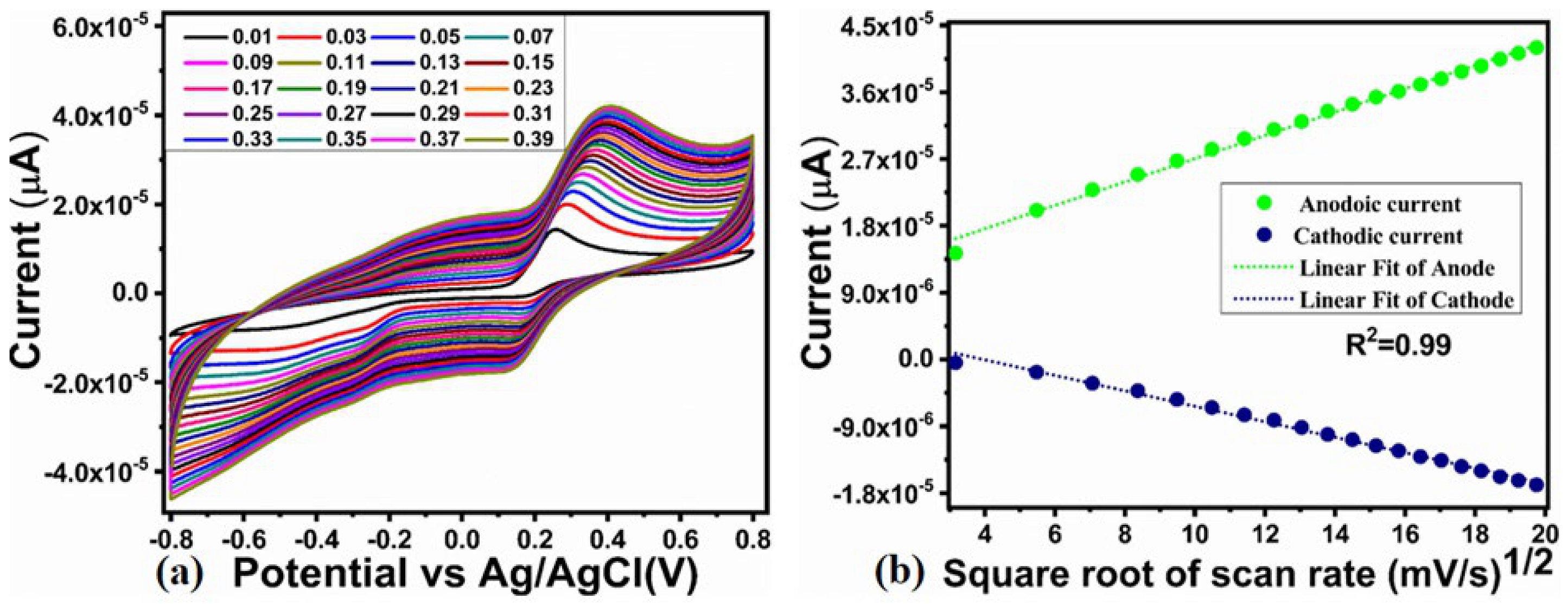
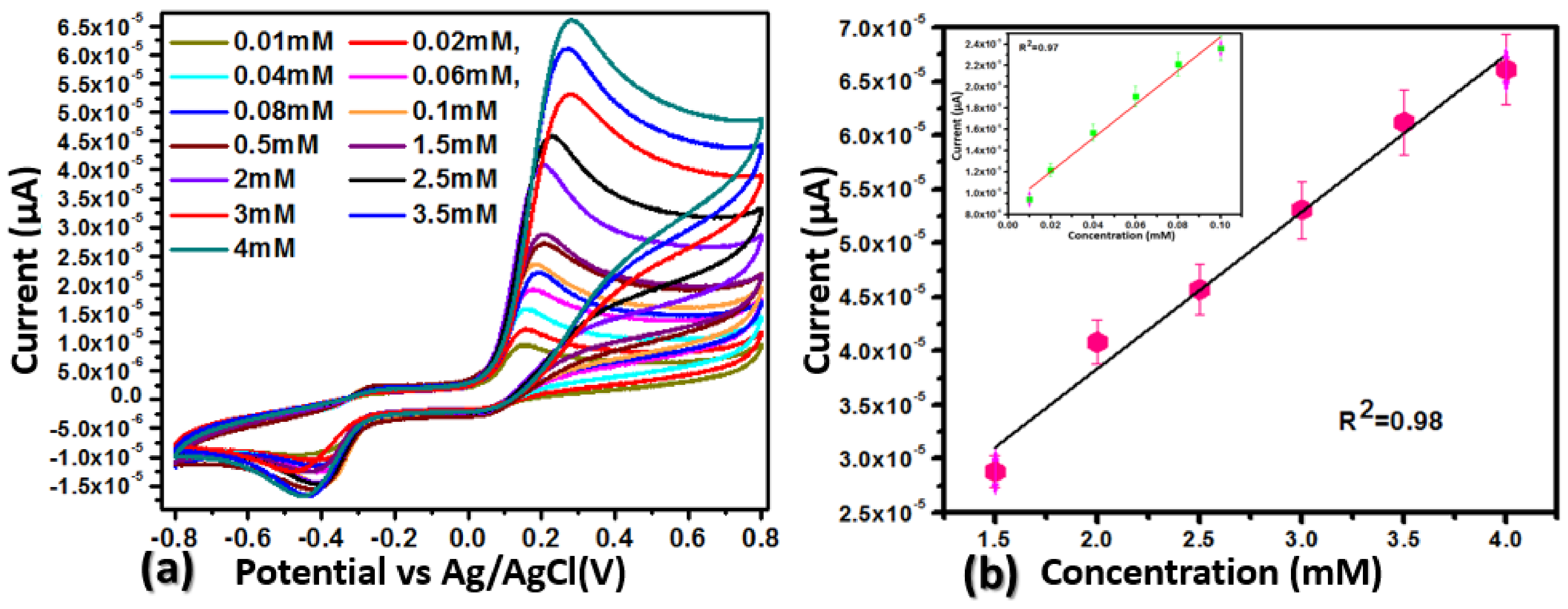
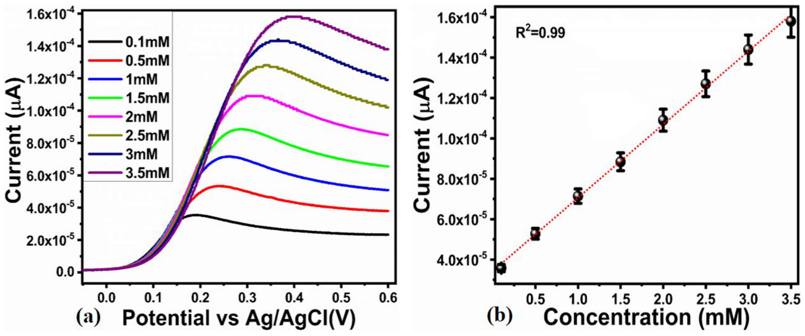
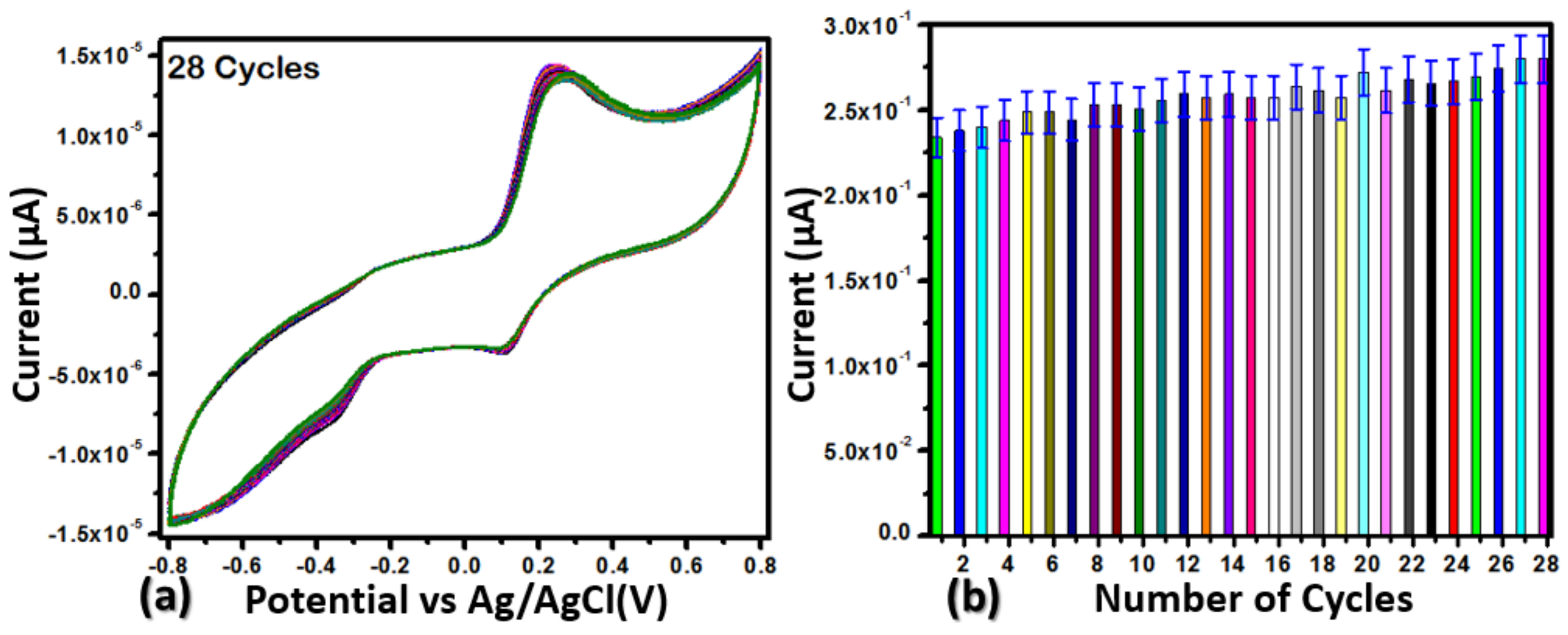
2.2. Enzyme-Free Dopamine Sensor Hybrid Materials Based on NiO/ZnO

We used a phosphate buffer at a pH of 7.3 as a supporting electrolyte and performed CV at a scan rate of 0.05 V/s. Using this method, the electrochemical signal of NiO/ZnO based hybrid materials was evaluated for the detection of dopamine. Hybrid systems consisting of 0.3, 0.6, and 0.9 g of NiO labeled as Samples 1, 2, and 3 were compared to pristine NiO and ZnO for a performance evaluation during non-enzymatic dopamine detection. Figure 5a illustrates that the bare glassy carbon electrode (BGCE) exhibits negligible redox signals in the electrolyte and analyte. Further, the pristine NiO and ZnO nanomaterials and their respective hybrid materials, did not exhibit any redox behavior in the electrolyte. However, pristine ZnO did not show any redox behavior in the 0.1 mM dopamine, whereas pristine NiO gave off an oxidation signal. Sample 2 showed a stronger oxidation signal for dopamine, indicating its excellent electrocatalytic properties, as shown in Figure 5b. Because of their capping, reducing, and stabilizing properties, the reducing agents in date fruit juice are highly active in transforming and modifying the surface properties of nanostructures. Sample 2 contained 0.6 g of NiO and a date fruit volume 15 mL as an optimum volume. Further increases in the volume of date fruit juice could reduce the catalytic sites and limit the activity of the hybrid system, hence 20 mL of date fruit juice showed relatively poor performance. Furthermore, the surface aspects of hybrid systems were significantly modified, as witnessed by the CV curves during the evaluation of the samples’ electrocatalytic properties. The composite system facilitates the electro-transfer rate via synergetic effects through the favorable interfacial chemistry. The composite carried the multiple catalytic sites’ contribution from both ZnO and NiO. Hence, improved electrocatalytic performance was noticed for the composite system.
As shown in Figure 6a, the effect of the scan rate on cathodic and anodic peak currents was investigated using CV and Sample 2 of the NiO/ZnO based hybrid materials. As the scan rate increased, both anodic and cathodic peak currents increased reversibly, demonstrating the enhanced electrochemical properties of the modified GCE. As shown in Figure 6b, the anodic and cathodic peak currents and the square root of the scan rate are fitted linearly. CV analyses of NiO/ZnO based hybrid materials exhibited dopamine-responsive diffusion-controlled kinetics.
To demonstrate the applicability of NiO/ZnO based on a hybrid material (Sample 2) for the sensitive detection of dopamine, a concentration window ranging from 0.01 mM to 4 mM was created and examined in (PBs) of pH 7.3 with CV curves at 50 mV/s. Based on the CV curves, the oxidation peak current increases with increasing dopamine levels, as shown in Figure 7a. As demonstrated in Figure 7b, the NiO/ZnO oxidation peak current vs. dopamine concentration was plotted linearly. A linear range of 0.01 mM to 4 mM concentrations was found to be appropriate in this study. Based on the hybrid material (Sample 2), the low limit of detection and the limit of quantification were determined to be 0.036 µM and 0.091 µM, respectively. By modulating the surface properties of the hybrid material (Sample 2), reducing, capping, and stabilizing agents enhanced the performance of hybrid systems dynamically. In this study, we have demonstrated that the use of a certain amount of date fruit juice can enable the creation of a surface with high active sites, rapid charge transfer, and good stability of the electrode material for a wide range of electrochemical applications.
Furthermore, Figure 8a shows a calibration plot for a dopamine detection using a NiO/ZnO based hybrid material (Sample 2) and linear seep voltammetry (LSV) at a scan rate of 50 mV/s. In addition to the linear increase in anodic peak current, the addition of more dopamine was clearly observed. As shown in Figure 8b, a linear relationship was established between anodic peak current and various dopamine concentrations. The results showed a well-known range of linearity for the NiO/ZnO based hybrid materials (Sample 2) with dopamine concentrations between 0.1 mM and 3.5 mM. The regression coefficient (R2 0.99) confirmed that the modified electrode exhibited excellent analytical performance. Figure 7 deals with the CV, and it has been imperatively seen that the CV is a slightly slow technique and involves a reversible process. Also, the concentration of dopamine close to the electrode surface is of a dynamic nature, as is the electrolyte. As a result, it could cause variations in the current at two consecutive dopamine concentrations. Comparing the CV curve current response with the LSV curve responses shown in Figure 7 and Figure 8a, the probability of variation in the dopamine concentration and the insertion of electrode area in the analyte solution could cause the difference between the current responses of the two different electrochemical modes.
The aim was to evaluate the dopamine selectivity of the hybrid material (Sample 2) based on NiO and ZnO, as shown in Figure 9a. An interference study was conducted using 0.1 mM dopamine and a similar concentration of potential interference species. The CV curves recorded at 50 mV/s revealed changes in peak currents associated with dopamine drug peak currents. As shown in Supplementary Table S1, which shows the recorded changes in the electrical signal, (G) glucose, (U)urea, (AA) ascorbic acid, and (UA) uric acid had little to no effect on the peak current. Similarly, the peak current of each interfering agent was compared with the dopamine electrical signal via bar graph representation, as shown in Figure 10b. It was performed in one container for the solution; all of the interfering agents were added sequentially, as shown in Figure 9b. It was seen clearly that compared to (D), the dopamine peak current, the peak current produced by the other interfering substances was very much lower and at different oxidation potentials, indicating that Sample 2 selectively quantified the dopamine.
However, the selectivity of an electrocatalytic material depends on the synthesis method, size, shape, and surface properties. The proposed material is based on the hybrid system of these two materials and has been treated with date fruit juice enriched with reducing agents. Hence, the size, shape and surface properties were altered in such a way that the presented material was mainly responding to glucose. This was the main aim of the present study: to produce a highly selective non-enzymatic sensor using the unique reducing agent chemistry of date fruit juice.
To determine the reproducibility of a non-enzymatic sensor, the CV curves of Sample 2 at 50 mV/s and 0.1 mM dopamine concentration are shown in Figure 10a, which depicts the inter-electrode response. It was observed that the electrochemical behavior of each electrode was almost the same, with negligible variations in the peak current, peak potential, and the area of the peak, demonstrating that Sample 2 displays consistent electrochemical activity, which further validates the accurate medication of the electrode with Sample 2. In Figure 10b, the bar graph illustrates the variations of the peak current of each inter-electrode response. As seen in the figure, the variation in the peak of each electrode modified with Sample 2 is less than 3%, indicating that the electrode is accurate and precise for measuring dopamine. Additionally, 28 repeatable CV curves were performed at 50 mV/s in 0.1 mM dopamine. The results are shown in Figure 11a. Once again, Sample 2’s performance confirms that it fully covers the electrode with a high degree of compatibility. It is evident that the date fruit juice is acting as a bio-mimic and that carbon was inserted during thermal annealing, which led to the uniform distribution of material during the preparation of the catalyst ink. As shown in Figure 11b, the peak current of each CV curve is plotted as a bar graph to provide a better understanding of the variation in current. The peak current variation was found to be negligible, demonstrating the excellent compatibility of Sample 2, which is responsible for its high stability.
An evaluation of electrochemical active surface area (ECSA) values was conducted to understand the reason for the enhanced electrochemical performance of Sample 2 in the oxidation of dopamine. Figure 12a–c illustrates the CV curves for Sample 1, Sample 2, and Sample 3 of the hybrid materials as measured at various scan rates in a non-Faradic region. In the given potential window range, all of the CV curves are well-shaped into non-Faradic regions.
In order to determine the electrochemical active surface area, the difference between the anodic and cathodic sides of the electrode was divided by two. As shown in Figure 12d, we plotted the current density versus the scan rate, with slopes corresponding to ECSA values. It was evident that Sample 2 exhibited an extensive active surface area. This may have contributed to its enhanced efficiency through the exposure of abundant active surface sites during dopamine oxidation. Plotting the anodic and cathodic side currents against the various scan rates and determining the slope from the linear result is the standard method for computing the ECSA. The CDL values for Samples 1, 2, and 3 were 0.00174 mF/cm2, 0.00329 mF/cm2, and 0.00172 mF/cm2, respectively, as illustrated in Figure 12d. Sample 2’s results show a higher CLD value, indicating the large surface area of this sample, and consequently it shows a well resolved oxidation peak with an increased peak current compared to other series of samples. This investigation proved that Sample 2 was rich in surface active sites for the advantageous oxidation of dopamine. As a result, it has been demonstrated to be a useful and efficient electrocatalytic material for dopamine oxidation in a phosphate buffer at a pH of 7.3. Moreover, this study used the variation of NiO content to deposit the ZnO, and we aimed to study the effect of NiO content on the electrocatalytic properties of ZnO under the unique environment of date fruit phytochemistry.
A real application of Sample 2 for the determination of dopamine was performed on wheat grass and banana peels. The selection of these samples can be considered a potential candidate for the development of medicines for treating several diseases, as described in the introduction. The real samples were chipped into small pieces and then boiled at 70 °C for 2 h in a phosphate buffer solution at a pH of 7.3. Extracts were then collected and directly used for the quantification of dopamine using Sample 2. The results of this study are presented in Supplementary Tables S2 and S3. Supplementary Tables S2 and S3 show that the relative value of dopamine concentration increased with increasing extract value for each real sample. The performance of as-prepared Sample 2 toward dopamine was compared with recently developed dopamine sensors as given in Table 1. Sample 2 is relatively simple, low cost, exhibited a wide linear range, can be scaled up, and is ecofriendly. The superiority of the proposed material in terms of its green nature, scalability, low cost, and facile and efficient response can be seen in Table 1. The green synthesis method for NiO/ZnO is indeed ecologically valuable, as it typically uses environmentally benign materials and processes, reducing the use of toxic chemicals and minimizing waste. The use of costly reducing agents could be minimized by the use of the presented approach of using date fruit juice, which would impact the cost of fabrication. Depositing higher amounts of material on the electrode’s surface and drying it for a longer time could result in low limits of detection. The presented study offered the opportunity to explore more ZnO based electrocatalysts by tailoring the amounts of co-catalysts for biomedical applications.

3. Used Materials and Methods

3.1. Chemical Reagents

The following items were purchased from Sigma Aldrich, Karachi, Sindh Pakistan: sodium dihydrogen phosphate, sodium chloride, disodium hydrogen phosphate, potassium chloride, ammonia (33%), nickel chloride hexahydrate, zinc chloride dihydrate, ammonia (33%), sodium dihydrogen phosphate, sodium chloride, potassium chloride, Nafion (5%), dopamine chloride, uric acid, ascorbic acid, and urea. All glassware was cleaned and rinsed with deionized water prior to experimentation and dried at room temperature following washing and rinsing. The desired solutions were prepared in deionized water (DI). A phosphate buffer solution at a pH of 7.3 was prepared using 10 mM Na2HPO4, 10 mM KH2PO4, 1 mM KCl, 1 mM NaCl, 0.1 M NaOH, and 0.1 M HCl. The dopamine (0.05 M) solution was prepared in a phosphate buffer solution at a pH of 7.3. Electrochemical measurements were conducted in this solution.

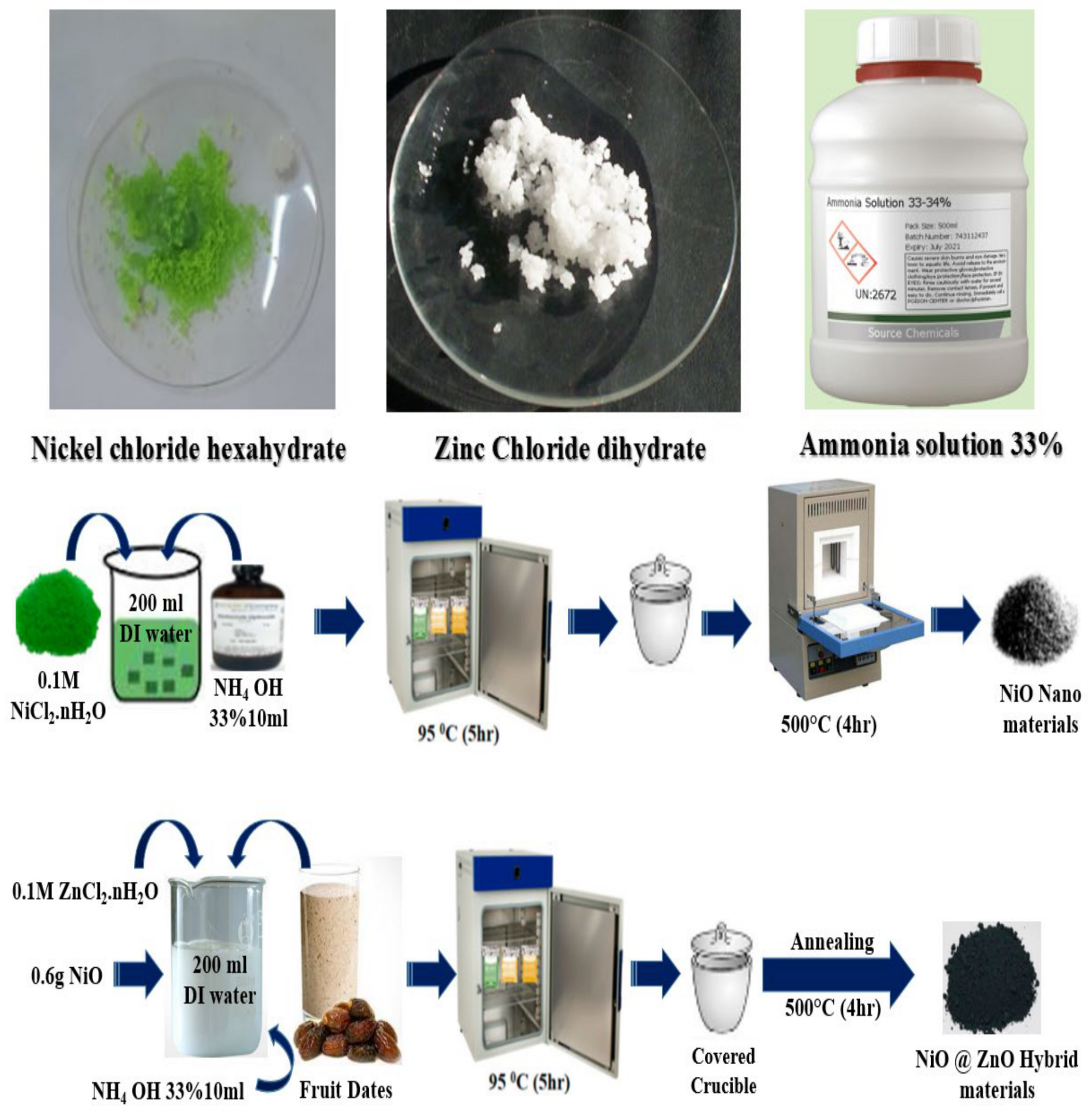
3.2. Hydrothermal Preparation of NiO/ZnO Using Date Fruit Juice as Natural Reducing and Surface Modifying Gent

The NiO/ZnO hybrid materials were prepared by hydrothermal synthesis, followed by thermal annealing in air. A NiO/ZnO hybrid material was synthesized by dissolving 0.1 M nickel chloride hexahydrate in 200 mL of deionized water and 10 mL of 33% aqueous NH3 solution. The growth solution beaker was covered with an aluminum sheet. The color of the nickel chloride hexahydrate could be readily observed. Afterwards, a five-hour hydrothermal process was conducted at 95 °C. An extraction of nickel hydroxide was performed using common laboratory filters, washed several times with DI water, and then allowed to dry overnight at room temperature. A china clay crucible was used to transfer the nickel hydroxide material. A thermal annealing procedure at 500 °C for four hours in air was used to convert the hydroxide phase into the metal oxide phase (NiO). A blackish gray color was obtained as a result of the process. Sample 1 consisted of pristine NiO and Sample 2 consisted of pristine ZnO, which was produced using 0.1 M zinc chloride dihydrate and 10 mL of 33% aqueous ammonia. Using reducing agents derived from rotten date fruit juice, one can change the surface properties and electrochemical charge transport properties by providing an environment that is more favorable for electrochemical reactions at the interface. The next step was to prepare three separate beakers containing 0.3 g, 0.6 g, and 0.9 g of NiO, respectively, and to add them to a solution of zinc chloride dihydrate containing 0.1 M and 33% NH3. The zinc chloride dihydrate solution was treated with three volumes of date juice (10 mL, 15 mL, and 20 mL). The three beakers were prepared using the same hydrothermal process and calcination conditions. As a result, a hybrid system that consists of NiO/ZnO was obtained. Hybrid systems have also been used to detect electrochemical dopamine and to conduct physical investigations.
Powder X-ray diffraction was used to evaluate the crystal quality of the hybrid NiO/ZnO systems under experimental conditions of 45 kV and 45 bad current. Cu-Kα was used as the radiation source (λ = 1.5418 Å) and was produced at 45 kV and 45 mA. The morphology of the samples was studied using scanning electron microscopy (SEM) at a voltage of up to 10 kV. The chemical vibrations of the NiO/ZnO were examined using a Shimadzu IR Affinity 1S series spectrometer (Shimadzu, Kyoto, Japan) with a frequency range of 400–4000 cm−1.

3.3. Electrochemical Measurements for the Non-Enzymatic Sensor for Dopamine

In order to prepare catalytic ink with a NiO/ZnO composite for surface modification of glassy carbon electrodes (GCEs), 10 mg of the hybrid material was dissolved in 3 mL of (DI) water and 30 μL of 5% Nafion. The mixture was then treated with an ultrasonic bath for ten minutes, resulting in a homogeneous catalyst ink. A glassy carbon electrode (GCE) was scrubbed with silicon paper and polished with 0.5 µM alumina paste before being cleaned with deionized water. Using a micropipette and the drop cast method, the glassy carbon electrode, which was designated as the (MGCE), was coated with a 10 µL layer of NiO/ZnO based hybrid materials. The electrochemical tests were conducted with a three electrode cell design, utilizing silver–silver chloride as the reference electrode, platinum wire as the counter electrode, and glassy carbon as the working electrode. PBs of pH 7.3 were used as the supporting electrolyte. To prepare the (0.05 M) dopamine stock solution, a phosphate buffer solution of pH 7.3 electrolyte solution was prepared prior to the electrochemical measurements. Various competing interfering agents were used in the selectivity experiment during the dopamine sensing process, at 0.1 mM concentrations in phosphate buffer pH 7.3. A variety of electrochemical methods were employed, such as cyclic voltammetry (CV) and linear sweep voltammetry (LSV).
A schematic illustration of the synthesis process of NiO/ZnO composites using various volumes of date fruit juice can be found in Scheme 1.

4. Conclusions

Hydrothermal processing of date fruit juice was used to design several NiO/ZnO based hybrid materials. A variety of analytical techniques were employed to study the samples’ structure, shape, and crystalline properties. Observations of Sample 2 of the NiO/ZnO hybrid material, prepared with 0.6 g of date fruit juice deposition, indicated that the distribution of the material was well organized, oriented in shape, and uniform in size. During the electrocatalytic oxidation of dopamine, this material demonstrated excellent electrocatalytic properties. In Sample 2, the dopamine activity ranged between 0.01 mM and 4 mM, indicating that the electrochemical properties are fully activated in phosphate buffer solutions at a pH of 7.3. Furthermore, Sample 2 was found to be highly selective, stable, reproducible, and sensitive when determining dopamine concentrations in a variety of real samples. According to the results obtained from Sample 2, it is evident that new hybrid materials can be developed by incorporating a certain amount of catalytic material into the development of functional composites in a fruit juice environment for applications such as biomedical, energy conversion, and storage systems.

Supplementary Materials

The following supporting information can be downloaded at: https://www.mdpi.com/article/10.3390/catal15020116/s1. Table S1: Interference study of Sample 2 for dopamine detection in phosphate buffer solution of pH using different interfering agents with same concentrations; Table S2: Real Sample Banana Peels; Table S3: Real Sample Wheat Grass.

Author Contributions

Conceptualization, I.N., A.T. and A.B.M.; methodology, L.S.; software, R.M.I.; validation, I.N., A.T. and R.M.I.; formal analysis, I.N., A.T., A.B.M., L.S. and R.M.I.; investigation, L.S.; resources, A.T.; data curation, A.B.M. and Z.H.I.; writing—original draft preparation, Z.H.I.; writing—review and editing, E.D.; visualization, I.N.; supervision, E.D.; project administration, Z.H.I.; funding acquisition, E.D. All authors have read and agreed to the published version of the manuscript.

Funding

The authors would like to gratefully acknowledge the Higher Education Commission Pakistan for partial support under the project NRPU/8350/8330. We also extend our sincere appreciation to the Researchers Supporting Project Number (RSP2024R79) at King Saud University, Riyadh, Saudi Arabia. Authors would also like to acknowledge partial funding of the Ajman University, Grant ID: DRG ref. 2024-IRG-HBS-01.

Data Availability Statement

Data sets generated during the current study are available from the corresponding author on reasonable request.

Conflicts of Interest

The authors declare no conflicts of interest.

References

  1. Eddin, F.B.K.; Fen, Y.W. Recent Advances in Electrochemical and Optical Sensing of Dopamine. Sensors 2020, 20, 1039. [Google Scholar] [CrossRef] [PubMed]
  2. Feng, P.; Chen, Y.; Zhang, L.; Qian, C.-G.; Xiao, X.; Han, X.; Shen, Q.-D. Near-Infrared Fluorescent Nanoprobes for Revealing the Role of Dopamine in Drug Addiction. ACS Appl. Mater. Interfaces 2018, 10, 4359–4368. [Google Scholar] [CrossRef] [PubMed]
  3. Whitton, A.E.; Reinen, J.M.; Slifstein, M.; Ang, Y.-S.; McGrath, P.J.; Iosifescu, D.V.; Abi-Dargham, A.; Pizzagalli, D.A.; Schneier, F.R. Baseline reward processing and ventrostriatal dopamine function are associated with pramipexole response in depression. Brain 2020, 143, 701–710. [Google Scholar] [CrossRef]
  4. Napier, T.C.; Kirby, A.; Persons, A.L. The role of dopamine pharmacotherapy and addiction-like behaviors in Parkinson’s disease. Prog. Neuro-Psychopharmacol. Biol. Psychiatry 2020, 102, 109942. [Google Scholar] [CrossRef]
  5. Katthagen, T.; Kaminski, J.; Heinz, A.; Buchert, R.; Schlagenhauf, F. Striatal Dopamine and Reward Prediction Error Signaling in Unmedicated Schizophrenia Patients. Schizophr. Bull. 2020, 46, 1535–1546. [Google Scholar] [CrossRef] [PubMed]
  6. Pan, X.; Kaminga, A.C.; Wen, S.W.; Wu, X.; Acheampong, K.; Liu, A. Dopamine and Dopamine Receptors in Alzheimer’s Disease: A Systematic Review and Network Meta-Analysis. Front. Aging Neurosci. 2019, 11, 175. [Google Scholar] [CrossRef]
  7. Vinay, M.M.; Arthoba Nayaka, Y. Iron oxide (Fe2O3) nanoparticles modified carbon paste electrode as an advanced material for electrochemical investigation of paracetamol and dopamine. J. Sci. Adv. Mater. Devices 2019, 4, 442–450. [Google Scholar] [CrossRef]
  8. Wen, M.; Xing, Y.; Liu, G.; Hou, S.; Hou, S. Electrochemical sensor based on Ti3C2 membrane doped with UIO-66-NH2 for dopamine. Microchim. Acta 2022, 189, 141. [Google Scholar] [CrossRef] [PubMed]
  9. Gaidukevic, J.; Aukstakojyte, R.; Barkauskas, J.; Niaura, G.; Murauskas, T.; Pauliukaite, R. A novel electrochemical sensor based on thermally reduced graphene oxide for the sensitive determination of dopamine. Appl. Surf. Sci. 2022, 592, 153257. [Google Scholar] [CrossRef]
  10. Rostami, S.; Mehdinia, A.; Jabbari, A. Intrinsic peroxidase-like activity of graphene nanoribbons for label-free colorimetric detection of dopamine. Mater. Sci. Eng. C 2020, 114, 111034. [Google Scholar] [CrossRef] [PubMed]
  11. Rasheed, P.A.; Lee, J.-S. Recent advances in optical detection of dopamine using nanomaterials. Microchim. Acta 2017, 184, 1239–1266. [Google Scholar] [CrossRef]
  12. Roychoudhury, A.; Francis, K.A.; Patel, J.; Jha, S.K.; Basu, S. A decoupler-free simple paper microchip capillary electrophoresis device for simultaneous detection of dopamine, epinephrine and serotonin. RSC Adv. 2020, 10, 25487–25495. [Google Scholar] [CrossRef] [PubMed]
  13. Iftikhar, I.; El-Nour, K.M.A.; Brajter-Toth, A. Detection of transient dopamine antioxidant radicals using electrochemistry in electrospray ionization mass spectrometry. Electrochim. Acta 2017, 249, 145–154. [Google Scholar] [CrossRef]
  14. Promsuwan, K.; Soleh, A.; Saisahas, K.; Saichanapan, J.; Kanatharana, P.; Thavarungkul, P.; Guo, C.; Li, C.M.; Limbut, W. Discrimination of dopamine by an electrode modified with negatively charged manganese dioxide nanoparticles decorated on a poly(3,4 ethylenedioxythiophene)/reduced graphene oxide composite. J. Colloid Interface Sci. 2021, 597, 314–324. [Google Scholar] [CrossRef] [PubMed]
  15. Chen, X.; Li, D.; Ma, W.; Yang, T.; Zhang, Y.; Zhang, D. Preparation of a glassy carbon electrode modified with reduced graphene oxide and overoxidized electropolymerized polypyrrole, and its application to the determination of dopamine in the presence of ascorbic acid and uric acid. Mikrochim. Acta 2019, 186, 407. [Google Scholar] [CrossRef]
  16. Rashed, M.A.; Ahmed, J.; Faisal, M.; Alsareii, S.A.; Jalalah, M.; Tirth, V.; Harraz, F.A. Surface modification of CuO nanoparticles with conducting polythiophene as a non-enzymatic amperometric sensor for sensitive and selective determination of hydrogen peroxide. Surf. Interfaces 2022, 31, 101998. [Google Scholar] [CrossRef]
  17. Ahmed, J.; Faisal, M.; Harraz, F.A.; Jalalah, M.; Alsareii, S.A. Porous silicon-mesoporous carbon nanocomposite based electrochemical sensor for sensitive and selective detection of ascorbic acid in real samples. J. Taiwan Inst. Chem. Eng. 2021, 125, 360–371. [Google Scholar] [CrossRef]
  18. Ahmed, J.; Rashed, M.A.; Faisal, M.; Harraz, F.A.; Jalalah, M.; Alsareii, S.A. Novel SWCNTs-mesoporous silicon nanocomposite as efficient non-enzymatic glucose biosensor. Appl. Surf. Sci. 2021, 552, 149477. [Google Scholar] [CrossRef]
  19. Ahmed, J.; Faisal, M.; Harraz, F.A.; Jalalah, M.; Alsareii, S.A. Development of an amperometric biosensor for dopamine using novel mesoporous silicon nanoparticles fabricated via a facile stain etching approach. Phys. E Low-Dimens. Syst. Nanostruct. 2022, 135, 114952. [Google Scholar] [CrossRef]
  20. Sarri, D.; Lombardo, S.; Lisci, R.; De Pascale, V.; Vieri, M. AgroBot Smash a Robotic Platform for the Sustainable Precision Agriculture. In Innovative Biosystems Engineering for Sustainable Agriculture, Forestry and Food Production; Springer: Cham, Switzerland, 2020; pp. 793–801. [Google Scholar]
  21. Deng, H.-H.; Hong, G.-L.; Lin, F.-L.; Liu, A.-L.; Xia, X.-H.; Chen, W. Colorimetric detection of urea, urease, and urease inhibitor based on the peroxidase-like activity of gold nanoparticles. Anal. Chim. Acta 2016, 915, 74–80. [Google Scholar] [CrossRef]
  22. Ju, J.; Chen, W. In Situ Growth of Surfactant-Free Gold Nanoparticles on Nitrogen-Doped Graphene Quantum Dots for Electrochemical Detection of Hydrogen Peroxide in Biological Environments. Anal. Chem. 2015, 87, 1903–1910. [Google Scholar] [CrossRef] [PubMed]
  23. Zhang, Y.; Ji, Y.; Wang, Z.; Liu, S.; Zhang, T. Electrodeposition synthesis of reduced graphene oxide–carbon nanotube hybrids on indium tin oxide electrode for simultaneous electrochemical detection of ascorbic acid, dopamine and uric acid. RSC Adv. 2015, 5, 106307–106314. [Google Scholar] [CrossRef]
  24. Zhou, J.; Chen, M.; Diao, G. Calix[4,6,8]arenesulfonates Functionalized Reduced Graphene Oxide with High Supramolecular Recognition Capability: Fabrication and Application for Enhanced Host–Guest Electrochemical Recognition. ACS Appl. Mater. Interfaces 2013, 5, 828–836. [Google Scholar] [CrossRef]
  25. Sun, J.; Li, L.; Zhang, X.; Liu, D.; Lv, S.; Zhu, D.; Wu, T.; You, T. Simultaneous determination of ascorbic acid, dopamine and uric acid at a nitrogen-doped carbon nanofiber modified electrode. RSC Adv. 2015, 5, 11925–11932. [Google Scholar] [CrossRef]
  26. Sun, C.-L.; Lee, H.-H.; Yang, J.-M.; Wu, C.-C. The simultaneous electrochemical detection of ascorbic acid, dopamine, and uric acid using graphene/size-selected Pt nanocomposites. Biosens. Bioelectron. 2011, 26, 3450–3455. [Google Scholar] [CrossRef]
  27. Ping, J.; Wu, J.; Wang, Y.; Ying, Y. Simultaneous determination of ascorbic acid, dopamine and uric acid using high-performance screen-printed graphene electrode. Biosens. Bioelectron. 2012, 34, 70–76. [Google Scholar] [CrossRef] [PubMed]
  28. Amin, S.; Tahira, A.; Solangi, A.; Beni, V.; Morante, J.R.; Liu, X.; Falhman, M.; Mazzaro, R.; Ibupoto, Z.H.; Vomiero, A. A practical non-enzymatic urea sensor based on NiCo2O4 nanoneedles. RSC Adv. 2019, 9, 14443–14451. [Google Scholar] [CrossRef]
  29. Nguyen, N.S.; Das, G.; Yoon, H.H. Nickel/cobalt oxide-decorated 3D graphene nanocomposite electrode for enhanced electrochemical detection of urea. Biosens. Bioelectron. 2016, 77, 372–377. [Google Scholar] [CrossRef] [PubMed]
  30. Shetti, N.P.; Malode, S.J.; Nayak, D.S.; Bagihalli, G.B.; Kalanur, S.S.; Malladi, R.S.; Reddy, C.V.; Aminabhavi, T.M.; Reddy, K.R. Fabrication of ZnO nanoparticles modified sensor for electrochemical oxidation of methdilazine. Appl. Surf. Sci. 2019, 496, 143656. [Google Scholar] [CrossRef]
  31. Shetti, N.P.; Bukkitgar, S.D.; Reddy, K.R.; Reddy, C.V.; Aminabhavi, T.M. ZnO-based nanostructured electrodes for electrochemical sensors and biosensors in biomedical applications. Biosens. Bioelectron. 2019, 141, 111417. [Google Scholar] [CrossRef] [PubMed]
  32. Beitollahi, H.; Tajik, S.; Garkani Nejad, F.; Safaei, M. Recent advances in ZnO nanostructure-based electrochemical sensors and biosensors. J. Mater. Chem. B 2020, 8, 5826–5844. [Google Scholar] [CrossRef]
  33. Wang, H.-H.; Chen, X.-J.; Li, W.-T.; Zhou, W.-H.; Guo, X.-C.; Kang, W.-Y.; Kou, D.-X.; Zhou, Z.-J.; Meng, Y.-N.; Tian, Q.-W.; et al. ZnO nanotubes supported molecularly imprinted polymers arrays as sensing materials for electrochemical detection of dopamine. Talanta 2018, 176, 573–581. [Google Scholar] [CrossRef] [PubMed]
  34. Weldekirstos, H.D.; Habtewold, B.; Kabtamu, D.M. Surfactant-Assisted Synthesis of NiO-ZnO and NiO-CuO Nanocomposites for Enhanced Photocatalytic Degradation of Methylene Blue Under UV Light Irradiation. Front. Mater. 2022, 9, 832439. [Google Scholar] [CrossRef]
  35. Budhiraja, N.; Sharma, A.; Dahiya, S.; Parmar, R.; Vidyadharan, V. Synthesis and Optical Characteristics of Silver Nanoparticles on Different Substrates. Int. Lett. Chem. Phys. Astron. 2013, 19, 80–88. [Google Scholar] [CrossRef]
  36. Yao, A.; Yang, H.; Wang, J.-Q.; Xu, W.; Huo, J.; Li, R.-W.; Qiu, H.; Chen, M. Flexible supercapacitor electrodes fabricated by dealloying nanocrystallized Al-Ni-Co-Y-Cu metallic glasses. J. Alloys Compd. 2019, 772, 164–172. [Google Scholar] [CrossRef]
  37. Goharshadi, E.K.; Ding, Y.; Jorabchi, M.N.; Nancarrow, P. Ultrasound-assisted green synthesis of nanocrystalline ZnO in the ionic liquid [hmim][NTf2]. Ultrason. Sonochem. 2009, 16, 120–123. [Google Scholar] [CrossRef] [PubMed]
  38. Hu, Q.; Li, W.; Abouelamaiem, D.I.; Xu, C.; Jiang, H.; Han, W.; He, G. Hollow Cu-doped NiO microspheres as anode materials with enhanced lithium storage performance. RSC Adv. 2019, 9, 20963–20967. [Google Scholar] [CrossRef]
  39. Wei, S.; Xiao, H.; Gu, M.; Chen, Z.; Cao, L. Ultrasensitive label-free electrochemical immunosensor based on core-shell Au@PtNPs functionalized rGO-TEPA/PB nanocomposite for HBsAg detection. J. Electroanal. Chem. 2021, 890, 115216. [Google Scholar] [CrossRef]
  40. Fayemi, O.E.; Adekunle, A.S.; Kumara Swamy, B.E.; Ebenso, E.E. Electrochemical sensor for the detection of dopamine in real samples using polyaniline/NiO, ZnO, and Fe3O4 nanocomposites on glassy carbon electrode. J. Electroanal. Chem. 2018, 818, 236–249. [Google Scholar] [CrossRef]
  41. Ni, M.; Chen, J.; Wang, C.; Wang, Y.; Huang, L.; Xiong, W.; Zhao, P.; Xie, Y.; Fei, J. A high-sensitive dopamine electrochemical sensor based on multilayer Ti3C2 MXene, graphitized multi-walled carbon nanotubes and ZnO nanospheres. Microchem. J. 2022, 178, 107410. [Google Scholar] [CrossRef]
  42. Zhang, W.; Sharma, G.; Kumar, A.; Shekh, M.I.; Stadler, F.J. Fabrication and characterization of Ni/Ag/Zn trimetal oxide nanocomposites and its application in dopamine sensing. Mater. Today Commun. 2021, 29, 102726. [Google Scholar] [CrossRef]
  43. Li, F.; Ni, B.; Zheng, Y.; Huang, Y.; Li, G. A simple and efficient voltammetric sensor for dopamine determination based on ZnO nanorods/electro-reduced graphene oxide composite. Surf. Interfaces 2021, 26, 101375. [Google Scholar] [CrossRef]
  44. Masood, T.; Asad, M.; Riaz, S.; Akhtar, N.; Hayat, A.; Shenashen, M.A.; Rahman, M.M. Non-enzymatic electrochemical sensing of dopamine from COVID-19 quarantine person. Mater. Chem. Phys. 2022, 289, 126451. [Google Scholar] [CrossRef]
  45. Ahmed, J.; Faisal, M.; Alsareii, S.A.; Jalalah, M.; Harraz, F.A. A novel gold-decorated porous silicon-poly(3-hexylthiophene) ternary nanocomposite as a highly sensitive and selective non-enzymatic dopamine electrochemical sensor. J. Alloys Compd. 2023, 931, 167403. [Google Scholar] [CrossRef]
  46. Chen, P.; Wang, H.; Li, H.; Niu, B.; Guo, H.; Chen, Z. A high-activity Fe-based MOFs fabricated through ultrasound strategy for electrochemical sensor of heavy metal ions and dopamine. J. Electroanal. Chem. 2024, 957, 118129. [Google Scholar] [CrossRef]
  47. Chen, C.; Ren, J.; Zhao, P.; Zhang, J.; Hu, Y.; Fei, J. A novel dopamine electrochemical sensor based on a β-cyclodextrin/Ni-MOF/glassy carbon electrode. Microchem. J. 2023, 194, 109328. [Google Scholar] [CrossRef]
Figure 1. Powder XRD reflection of pure ZnO (blue in color),pure NiO (red in color), and its composites with 0.3 g sample 1 (brown in color), 0.6 g sample 2 (green in color) and 0.9 g sample 3 (pink in color) of NiO using 10, 15, and 20 mL of date fruit juice respectively.
Catalysts 15 00116 g001
Figure 2. FTIR spectra of pure ZnO, NiO, and its composites with 0.3 g, 0.6 g, and 0.9 g of NiO using 10, 15, and 20 mL of date fruit juice, respectively.
Catalysts 15 00116 g002
Figure 3. (a) UV-visible absorption spectra of spectra of pure ZnO, NiO, and its composites with 0.3 g, 0.6 g, and 0.9 g of NiO using 10, 15, and 20 mL of date fruit juice, respectively. (b) Tauc’s plots.
Catalysts 15 00116 g003
Figure 4. (a,b) SEM images of pure NiO and pure ZnO, (c) SEM images of 0.3 g of NiO using 10 mL of date fruit juice for deposition of ZnO (Sample 1), (d) 0.6 g of NiO using 15 mL of date fruit juice for deposition of ZnO (Sample 2), (e) 0.9 g of NiO using 20 mL of date fruit juice for deposition of ZnO (Sample 3).
Catalysts 15 00116 g004
Figure 5. (a) BGCE’s CV curves and a modified glassy carbon electrode (MGCE) with two samples of pristine 1, 2, and 3 with NiO/ZnO formed on materials at 50 mV/s in the presence of (0.1 mM) dopamine and a phosphate buffer at a pH of 7.3, (b) Cyclic Voltagram of Samples 1, 2 and 3 formed on NiO/ZnO hybrid materials, at 0.05 V/s in the presence of (0.1 mM) dopamine prepared in a phosphate buffer at a pH of 7.3.
Catalysts 15 00116 g005
Figure 6. (a) CV curves at 0.05 V/s in phosphate buffer solution of pH 7.3 using pure ZnO, NiO, and their composites with 0.3 g, 0.6 g, and 0.9 g of NiO using 10, 15, and 20 mL of date fruit juice, respectively, (b) their corresponding CV responses under the same conditions with the use of 0.1 mM of dopamine.
Catalysts 15 00116 g006
Figure 7. (a) Curves of CV for Sample 2 NiO/ZnO based on hybrid materials, at 0.05 V/s various dopamine concentrations were prepared in PBs at a pH 7.3. (b) Anodic peak current plotted linearly versus different dopamine concentrations.
Catalysts 15 00116 g007
Figure 8. (a) The LSV of hybrid materials hinges on the NiO/ZnO of Sample 2, made at 0.6 V using different dopamine concentrations in PBS at pH 7.3. (b) Current was plotted linearly against various dopamine concentrations.
Catalysts 15 00116 g008
Figure 9. (a) CV curves at 0.05 V/s using hybrid material NiO/ZnO (Sample 2) under the influence of common interfering substances during dopamine sensing using the same concentration for each of them, (b) Peak current variation through bar graph of each CV curve measured for different interfering agents and dopamine using Sample 2 in PBS buffer pH 7.3 utilising same concentrations of all.
Catalysts 15 00116 g009
Figure 10. (a) CV curves at 0.05 V/s using five independent hybrid materials of NiO/ZnO (Sample 2) in 0.1 mM dopamine for the expression of reproducibility. (b) Peak current variation through bar graph of each electrode.
Catalysts 15 00116 g010
Figure 11. (a) Repeatable 28 CV curves at 0.05 V/s using of same modified hybrid material NiO/ZnO (Sample 2) in 0.1 mM dopamine for the expression of stability. (b) Peak current variation of 28 repeatable CV cycles through bar graph of each electrode.
Catalysts 15 00116 g011
Figure 12. (ac) CV curves at different scan rates using hybrid materials of NiO/ZnO (Sample 1, Sample 2, and Sample 3) in 0.1 mM dopamine for the expression of non-Faradic region for the estimation of ECSA values. (d) Linear plot for the estimation of ECSA of Sample 1, Sample 2, and Sample 3.
Catalysts 15 00116 g012
Scheme 1. Synthesis of hybrid materials based on NiO/ZnO by hydrothermal method following by annealing.
Catalysts 15 00116 sch001
Table 1. Performance evaluation comparative analysis of NiO/ZnO with recently published dopamine biosensors/sensors.
Electrode MaterialLinear Range (mM)Limit of Detection
(LOD μM)
Sensing MaterialMethod of DetectionReference
(ZnO-rGO-AuNPs/SPE)0.5 μM to 100 μM 0.294 μMDopamineNon-Enzymatic[39]
GCE/PANI-NiO, GCE/PANI-ZnO, and GCE/PANI-Fe3O4 sensors2.0 × 10−5 to 2.4 × 10⁻⁶ M0.153 × 10−7, 0.166 × 10⁻⁷, and 0.176 × 10⁻⁷ MDopamineNon-Enzymatic [40]
Ti3C2/G-MWCNTs/ZnO/GCE0.01–30 μM3.3 nMDopamineNon-Enzymatic [41]
Ni/Ag/Zn0.96 μA/μM cm20.3 μMDopamineNon-Enzymatic[42]
ZnONRs/ERGO/GCE0.01 to 6.0 μM and 6.0 to 80 μM3.6 nMDopamineNon-Enzymatic[43]
ZIF-67/PEDOT15–240 μM0.04 μMDopamineNon-Enzymatic[44]
Au/PSi-P3HT1.0–460 μM∼0.63 μMDopamineNon-Enzymatic[45]
NH2-MIL-101(Fe)/CPE0.3 to 450 μM0.025 μMDopamineNon-Enzymatic[46]
Ni–MOF0.7–310.2 μM0.227 μMDopamine [47]
NiO/ZnO/GCE0.01–4 mM0.03 μMDopamineNon-EnzymaticThis work
Disclaimer/Publisher’s Note: The statements, opinions and data contained in all publications are solely those of the individual author(s) and contributor(s) and not of MDPI and/or the editor(s). MDPI and/or the editor(s) disclaim responsibility for any injury to people or property resulting from any ideas, methods, instructions or products referred to in the content.

