Article

Facile Preparation of High-Performance Non-Enzymatic Glucose Sensors Based on Au/CuO Nanocomposites

1
School of Chemistry and Materials Science, Hubei Provincial Engineering Research Center of Key Technologies in Modern Paper and Hygiene Products Manufacturing, Hubei Engineering University, Xiaogan 432000, China
2
School of Physics and Electronic Information Engineering, Ningxia Normal University, Guyuan 756099, China
*
Authors to whom correspondence should be addressed.
Catalysts 2025, 15(11), 1020; https://doi.org/10.3390/catal15111020
Submission received: 8 October 2025 / Revised: 25 October 2025 / Accepted: 28 October 2025 / Published: 30 October 2025
(This article belongs to the Special Issue Heterogeneous Catalysis in China: New Horizons and Recent Advances)

Abstract

Non-enzymatic glucose sensing has attracted considerable interest as a promising alternative to enzyme-based sensors, addressing limitations such as poor stability and high cost. To overcome the challenges of expensive noble metals and the inherent issues of pure copper oxide (CuO), including low conductivity and aggregation tendency, this study developed a composite sensing material based on two-dimensional CuO nanosheets decorated with gold nanoparticles (Au NPs). A series of Au/CuO nanocomposites with varying Au loadings were synthesized through a combined hydrothermal and in situ reduction approach. Systematic electrochemical characterization revealed that the composite with 7.41 wt% Au loading exhibited optimal sensing performance, achieving sensitivities of 394.29 and 257.14 μA·mM−1·cm−2 across dual linear ranges of 5–3550 μM and 4550–11,550 μM, respectively, with a detection limit of 10 μM and a rapid response time of 3 s. The sensor also demonstrated selectivity against common interferents as well as long-term stability. This work highlights the importance of precise noble metal loading control in optimizing sensor performance and offers a feasible material design strategy for developing high-performance non-enzymatic glucose sensors.

1. Introduction

Glucose detection is crucial for biomedical monitoring and food quality control [1,2]. In clinical practice, blood glucose and urinary glucose levels provide crucial insights for diabetes management, while in the food industry, excessive glucose in beverages may lead to health risks including obesity [3,4,5]. Various analytical techniques have been developed for glucose determination, such as spectroscopy, chromatography, and electrochemical methods [6,7,8,9]. Although chromatographic and spectroscopic methods achieve high accuracy, they require sophisticated instrumentation and time-consuming procedures, limiting their use in point-of-care settings like home monitoring. In comparison, electrochemical sensing platforms offer superior practicality through operational simplicity, rapid response, high sensitivity, and cost-effectiveness. Enzyme-based electrochemical sensors utilizing glucose oxidase demonstrate excellent selectivity for glucose detection [10,11,12]. However, their susceptibility to denaturation under environmental fluctuations, coupled with high cost and limited stability, hinders widespread implementation [13,14,15]. These limitations have accelerated the development of non-enzymatic alternatives. Non-enzymatic glucose sensors employing metallic, metal oxide, or carbon-based electrocatalysts have attracted growing interest due to their enhanced durability, lower cost, and compatibility with complex biological media like urine, presenting a promising approach for practical glucose monitoring applications [16,17,18,19,20].
Noble metal nanoparticles, such as Au, Pt, and Pd, exhibit excellent electrocatalytic activity for glucose oxidation and are consequently widely used in non-enzymatic glucose sensing [21,22,23]. However, their practical application is significantly constrained by high cost, scarcity, susceptibility to interference from substances like uric acid and ascorbic acid in urine, and deactivation by reaction intermediates, leading to unsatisfactory stability [24,25]. Transition metal oxide copper oxide (CuO) has emerged as a highly promising alternative due to its abundant reserves, low cost, and the unique redox behavior of the Cu2+/Cu3+ pair, which aligns well with the kinetics of glucose oxidation, enabling efficient catalytic conversion [26,27,28]. Recent research has further enhanced the sensing performance of CuO-based materials by constructing specific morphologies such as nanoflowers and hollow structures [28,29,30]. Among various nanostructures, two-dimensional (2D) CuO nanosheets offer distinct advantages compared to other 2D materials like graphene oxide and MXene [31,32]. They not only share the common benefits of high specific surface area for abundant active sites and a planar structure conducive to mass transport and electron transfer but also possess intrinsic Cu2+/Cu3+ redox couples that directly participate in glucose catalysis, eliminating the need for additional catalytic components and thus demonstrating superior application potential [31,32,33]. Despite these advantages, pristine 2D CuO nanosheets suffer from inherent limitations, including poor electrical conductivity and a tendency to aggregate via van der Waals forces. This aggregation reduces accessible active sites and hinders electron transfer, ultimately resulting in sluggish sensor response. Furthermore, most reported Au/CuO composites utilize non-2D CuO nanostructures (e.g., nanoparticles, nanorods) as supports, which inherently limits active site exposure and mass transfer efficiency [34,35,36,37]. More critically, systematic optimization of the Au loading and in-depth investigation of the interfacial synergistic catalytic mechanism between Au and CuO are often lacking, making precise performance modulation challenging.
Herein, a series of Au/CuO nanocomposites were constructed using 2D CuO nanosheets as a support to overcome their inherent limitations in sensing applications and to correlate the Au loading with the catalytic performance. Through a two-step synthesis method involving hydrothermal growth and in situ reduction, we fabricated nanocomposites with precise Au loadings and systematically investigated the dependence of their microstructure and electrocatalytic behavior on the loading level, which enabled the interfacial synergy. This work thus provides a theoretical and practical foundation for developing advanced non-enzymatic glucose sensors and broadens the applications of 2D transition metal oxides in electrochemical sensing.

2. Results and Discussion

2.1. Morphological and Structural Characterization

As illustrated in Figure 1, the Au/CuO nanocomposites were fabricated through a rationally designed stepwise strategy. Initially, two-dimensional CuO nanosheets were synthesized via a facile, template-free hydrothermal reaction between NaOH and CuCl2. This two-dimensional architecture was intentionally constructed as a functional support, offering a high specific surface area and abundant surface anchoring sites, thereby providing an ideal substrate for the subsequent uniform loading of metal nanoparticles. Subsequently, Au nanoparticles were uniformly deposited onto the CuO nanosheets through an in situ reduction process using ascorbic acid as a mild reducing agent. During this process, the AuCl4 precursors were first adsorbed onto the nanosheet surfaces, followed by a controlled reduction step. This mechanism is crucial for the formation of fine, highly dispersed Au nanoparticles, effectively preventing particle agglomeration and enhancing the metal-support interaction at the Au-CuO interface, thereby laying the foundation for enhanced catalytic performance.
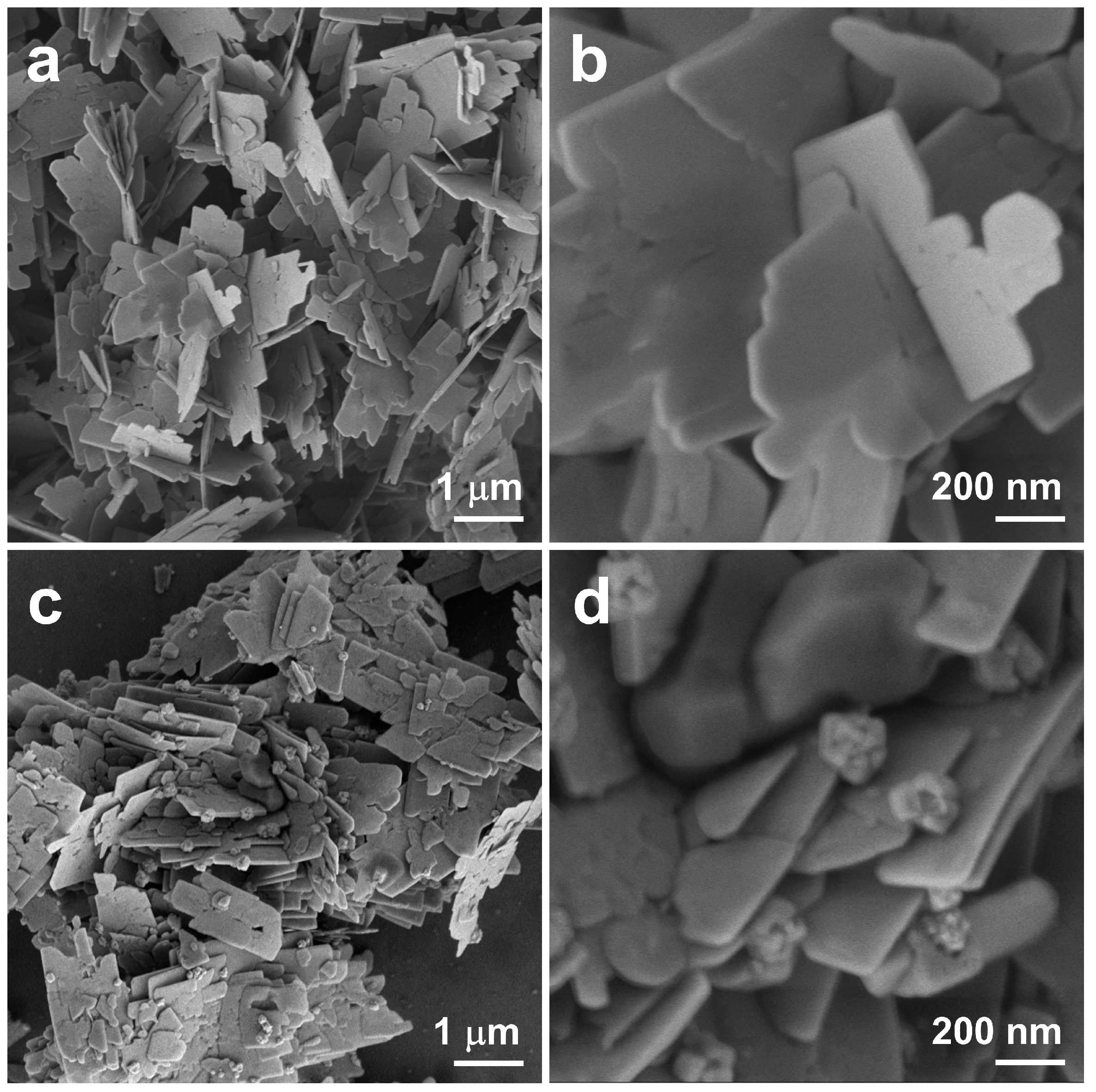
To systematically elucidate the structural evolution from the pristine support to the hybrid catalyst, morphological changes were first investigated. Figure 2a,b displays scanning electron microscopy (SEM) images of the pristine CuO nanosheets synthesized via a hydrothermal method, showing their characteristic two-dimensional sheet-like morphology with smooth surfaces and lateral dimensions of several micrometers. After the in situ deposition of Au nanoparticles, the resulting nanocomposite (Figure 2c,d) maintained the integrated two-dimensional framework, confirming that the reduction process preserved the structural integrity of the CuO support under sufficiently mild conditions.
The Au loadings of all samples were precisely determined by inductively coupled plasma optical emission spectrometry (ICP-OES), with measured values of 3.86 wt%, 7.41 wt%, 11.86 wt%, and 15.71 wt% (see Table S1). With increasing Au loading, SEM images (Figure 2 and Figure S1) clearly demonstrate an evolutionary trend. At the lower loading of 3.86 wt%, Au nanoparticles were sparsely distributed on the support surface. When the loading increased to 7.41 wt%, the nanoparticles exhibited a uniform and high-density distribution; However, further increases to 11.86 wt% and 15.71 wt% resulted in noticeable nanoparticle agglomeration. This systematic morphological evolution indicates that the 7.41 wt% loading achieves optimal dispersion of Au nanoparticles, while higher loadings lead to significant aggregation. Consequently, this optimal sample is expected to maximize the number of accessible active sites while maintaining structural stability, thereby providing a solid foundation for enhanced catalytic performance.
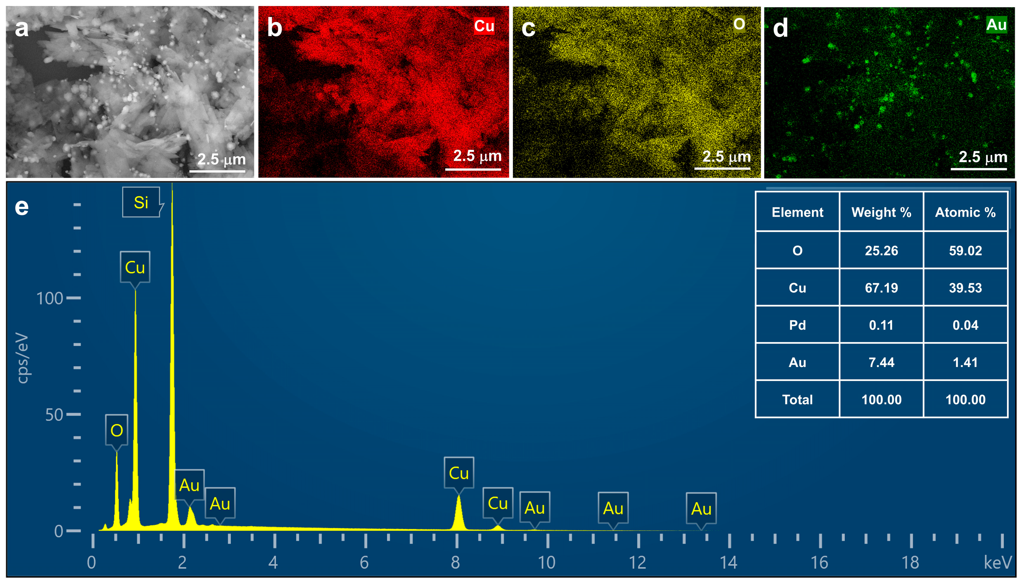
Energy-dispersive X-ray spectroscopy (EDS) elemental mapping was employed to verify the successful loading and distribution of Au. As shown in Figure 3, a representative nanosheet of the 7.41 wt% Au/CuO nanocomposites (Figure 3a) exhibits homogeneous spatial distribution of Cu, O, and Au (Figure 3b,d), with the Au signal entirely overlapping the support. The semi-quantitative EDS analysis determined a Au content of 7.44 wt% (Figure 3e), closely matching the bulk ICP-OES value (7.41 wt%). It is noted that the strong peaks located at 1.73 keV originate from the silicon substrate, while the agreement in Au content not only confirms the loading efficiency but directly evidences the uniform deposition of Au nanoparticles without severe agglomeration.
The nano-scale morphology and structure of the 7.41% Au/CuO nanocomposites were further elucidated by transmission electron microscopy (TEM). Low-magnification images (Figure 4a–c) confirm the uniform dispersion of Au nanoparticles on the CuO nanosheets. A more detailed analysis of the particle size distribution (Figure S2) indicates an average Au nanoparticle diameter of 71.79 ± 9.06 nm. Higher-resolution imaging (Figure 4d) resolves clear lattice fringes with a spacing of 0.235 nm, corresponding to the (111) plane of face-centered cubic Au. The observed intimate contact between the crystalline Au nanoparticles and the CuO support establishes a structural foundation for enhanced interfacial electron transfer, which is crucial for boosting catalytic performance.
The crystalline phases and structural characteristics of pure CuO nanosheets and the Au/CuO nanocomposites were analyzed by X-ray diffraction (XRD), as shown in Figure 5. All samples exhibit characteristic diffraction peaks corresponding to monoclinic CuO (JCPDS No. 48-1548). The two most intense reflections, located at 2θ = 35.5° and 38.7°, are indexed to the (11 1 ¯ ) and (111) planes, respectively. Additional identifiable CuO peaks are observed at approximately 2θ = 48.7° and 61.5°, collectively confirming the high crystallinity and structural integrity of the CuO support. As the Au loading increases from 3.86 wt% to 15.71 wt%, additional diffraction peaks gradually emerge at 2θ = 38.2°, 44.4°, 64.6°, and 77.6°, which correspond to the (111), (200), (220), and (311) planes of face-centered cubic Au (JCPDS No. 98-000-0230), confirming the successful formation of metallic Au nanoparticles. Notably, the intensity of the Au (111) peak increases with higher Au content, while its full width at half maximum decreases, indicating an increase in the average crystallite size of Au. This trend is consistent with the agglomeration behavior observed by SEM. Throughout the series, the positions of the CuO diffraction peaks remain unchanged, suggesting that Au deposition does not induce detectable lattice strain in the CuO framework. A weak diffraction signal is observed near 2θ = 35.5° in samples with Au loadings ≥ 7.41 wt%, which may be attributed to trace metallic Cu. Given its low intensity, this impurity is not expected to significantly influence the overall catalytic performance of the composites.

2.2. Electrochemical Behaviors of the Au/CuO Nanocomposites Modified Electrode

The electrocatalytic activity of the modified electrodes toward glucose oxidation was evaluated by cyclic voltammetry (CV) using a conventional three-electrode system. The working electrodes investigated were a bare glassy carbon electrode (GCE), CuO nanosheets modified electrode, and the Au/CuO nanocomposites (with varying Au loadings) modified electrodes. Prior to modification, each bare GCE underwent essential electrochemical calibration in a standard Fe(CN)63−/4− solution to confirm a properly pretreated and active surface state. This validation step ensured that the observed electrocatalytic responses originated specifically from the modified nanomaterials rather than from the base electrode. The complete electrode fabrication protocol, including polishing, cleaning, calibration, and modification steps, is explicitly described in Section 3.5.
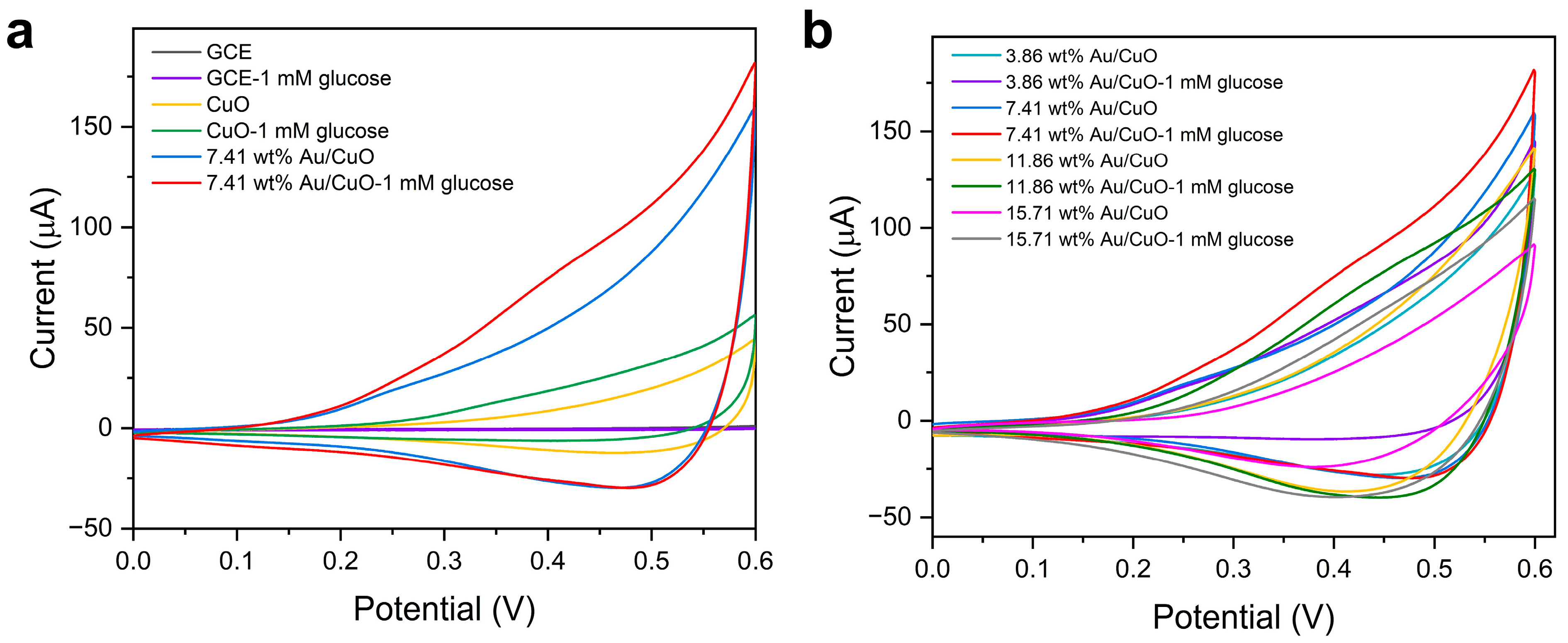
Figure 6 presents the CV curves of the different working electrodes, recorded in 0.1 M NaOH solution with and without 1 mM glucose at a scan rate of 0.05 V/s, to evaluate their non-enzymatic glucose sensing performance. The optimal potential window for these measurements was determined through preliminary tests. Initial CV scans over a broader range of 0–1.0 V (Figure S3) revealed that the glucose oxidation current primarily occurred between 0 and 0.7 V. However, at potentials approaching 0.7 V, the onset of side reactions such as oxygen evolution became noticeable. To establish a conservative potential window that completely avoids any interference and ensures a highly specific and stable electrocatalytic response, the range was optimized to 0–0.6 V for all subsequent analyses. Unless otherwise specified, all CV measurements in this study were conducted at 0.05 V/s within this optimized potential range. As illustrated in Figure 6a, the bare GCE shows negligible electrochemical response to glucose, which is confirmed by the overlapping CV curves obtained in the absence and presence of 1 mM glucose (Figure S4). In contrast, CuO nanosheets modified electrode and 7.41 wt% Au/CuO nanocomposites modified electrode exhibit distinct electrochemical responses in the glucose-containing electrolyte. Although no obvious redox peaks are observed, the current variation characteristics clearly reflect the electrocatalytic activity toward glucose. The 7.41 wt% Au/CuO nanocomposites modified electrode demonstrates a significantly enhanced current variation compared to the bare GCE and CuO nanosheets modified electrode, indicating that the Au/CuO nanocomposites synergistically promote electron transfer during glucose oxidation. To further optimize the Au loading amount, Figure 6b compares the CV behaviors of Au/CuO nanocomposites modified electrode with different Au contents (3.86 wt%, 7.41 wt%, 11.86 wt%, and 15.71 wt%). The 7.41 wt% Au/CuO nanocomposites modified electrode exhibits the largest current change amplitude, while electrodes with either lower or higher Au loadings show decreased current variations due to insufficient active sites or nanoparticle aggregation, respectively. These results collectively demonstrate that the 7.41 wt% Au/CuO nanocomposites modified electrode possesses optimal electrocatalytic activity for glucose oxidation, making it a promising electrode material for non-enzymatic glucose sensing applications.
To further elucidate the sensing performance and electrocatalytic mechanism of the optimized 7.41 wt% Au/CuO nanocomposites modified GCE, systematic characterizations were performed, and the results are illustrated in Figure 7. Figure 7a depicts the CV responses of the 7.41 wt% Au/CuO nanocomposites modified GCE in 0.1 M NaOH with varying glucose concentrations (0–10 mM), recorded at a scan rate of 0.05 V/s. Within this concentration range, the catalytic current increases progressively with rising glucose concentration, and no further increment is observed at 10 mM. This behavior is attributed to the finite active sites on the electrode surface, where high glucose concentrations fully occupy available catalytic sites, thereby restricting additional current enhancement. Analysis of the CV curves revealed that the oxidation current at 0.5 V provided a well-balanced response, avoiding noise from higher potentials and weak signals from lower potentials. Consistent with the general behavior of glucose oxidation reactions, higher potentials often introduce significant noise, whereas lower potentials yield insufficient signal intensity [38,39]. Therefore, 0.5 V was selected as the optimal working potential for subsequent chronoamperometric detection. Figure 7b presents the CV curves of the 7.41 wt% Au/CuO/GCE in 0.1 M NaOH containing 1 mM glucose at different scan rates (0.001–0.05 V/s). Well-defined oxidation peaks are observed at low scan rates (0.001–0.005 V/s), whereas peak definition diminishes at higher scan rates (0.01–0.05 V/s). This phenomenon is ascribed to reduced interaction time between glucose molecules and the electrode’s active sites at elevated scan rates, which leads to incomplete redox reactions and compromised peak resolution. The corresponding relationship of oxidative currents at 0.5 V and the square root of scan rate (v1/2) at the rates ranging from 0.001 to 0.05 V/s is a good linear relationship (I = 674.63 v1/2 − 13.07, R2 = 0.993), shown in Figure 7c, revealing that the glucose oxidation reaction on Au/CuO nanocomposites is a diffusion-controlled process in an electrolyte containing 1 mM glucose. These findings, including concentration-dependent current saturation, scan rate-dependent peak behavior and diffusion-controlled kinetics, collectively validate the feasibility of the 7.41 wt% Au/CuO/GCE for non-enzymatic glucose sensing applications.
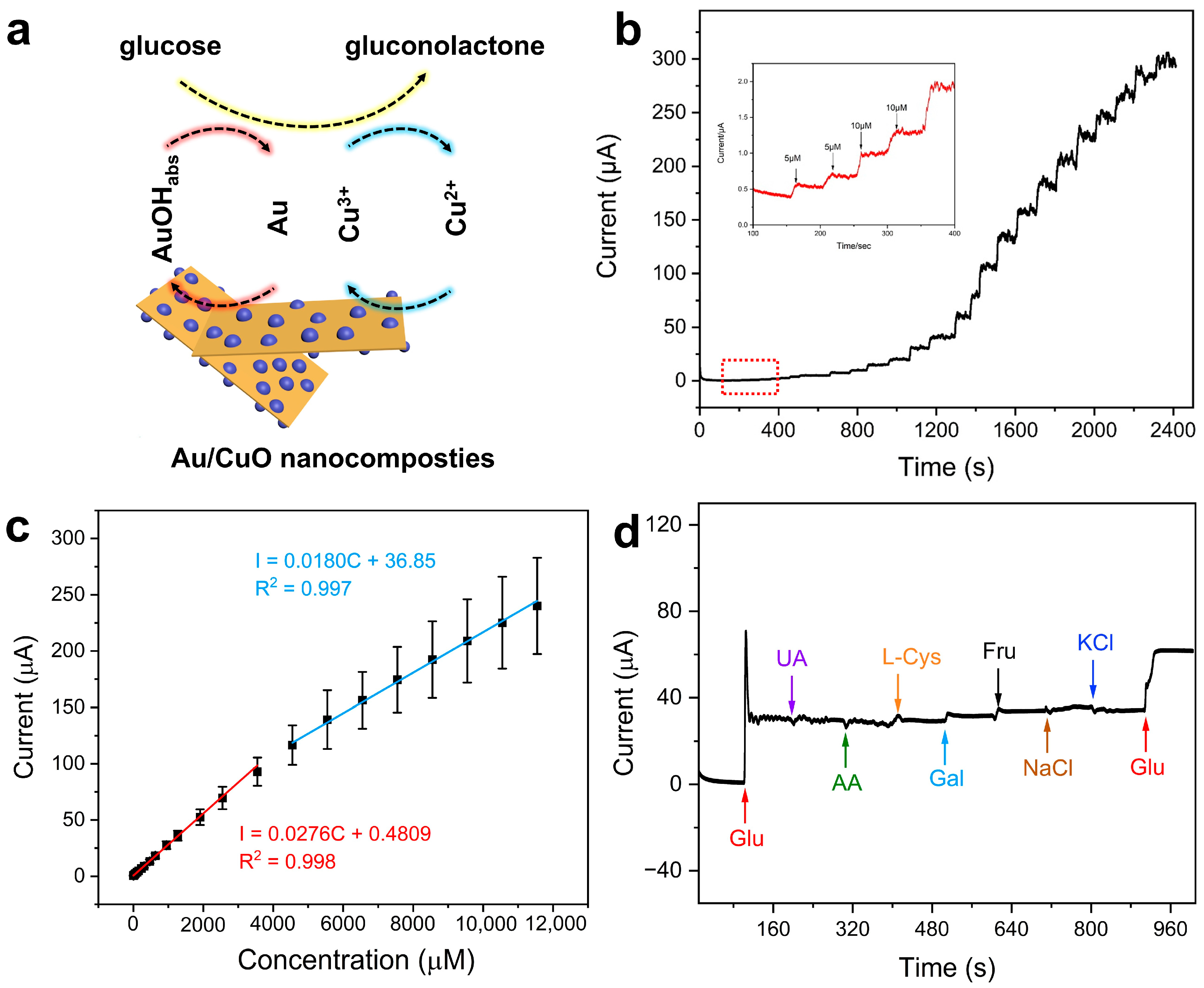
Based on the electrochemical characterization and optimized potential, Figure 8 systematically evaluates the glucose-sensing performance of the 7.41 wt% Au/CuO nanocomposites modified electrode. A comparative analysis with the CuO nanosheets modified electrode (Figure S5) highlights the beneficial effect of Au loading. As illustrated in the proposed mechanism (Figure 8a), Au promotes the formation of adsorbed AuOH (AuOHads), while Cu2+ in CuO is oxidized to Cu3+ [34,36]. These two active species work synergistically to catalyze the oxidation of glucose to gluconolactone, completing the catalytic cycle through the subsequent reduction in the active sites. Amperometric i-t responses were recorded at 0.5 V in stirred 0.1 M NaOH. Both the 7.41 wt% Au/CuO nanocomposites modified electrode (Figure 8b) and the CuO nanosheets modified electrode (Figure S5a) showed rapid, stable, and stepwise current increases upon successive glucose additions. For the 7.41 wt% Au/CuO nanocomposites modified electrode (Figure 8c), the current-concentration relationship showed two linear ranges: from 5 μM to 3550 μM (I = 0.0276C + 0.4809, R2 = 0.998) with a sensitivity of 394.29 μA·mM−1·cm−2, and from 4550 μM to 11,550 μM (I = 0.0180C + 36.85, R2 = 0.995) with a sensitivity of 257.14 μA·mM−1·cm−2. The CuO nanosheets modified electrode (Figure S5b) also exhibited two linear ranges: from 5 μM to 3550 μM (I = 0.0193C + 0.4809, R2 = 0.998) with a sensitivity of 257.71 μA·mM−1·cm−2, and from 4550 μM to 1255 μM (I = 0.0096C + 37.82, R2 = 0.998) with a sensitivity of 137.14 μA·mM−1·cm−2. The bilinear behavior observed in both electrodes is a common feature of nanomaterial-based sensors and it likely arises from variations in reaction kinetics and mass transport across different concentration regimes.
The detection limits (LOD) were calculated using the formula LOD = 3σ/S, where σ is the standard deviation of the blank response and S is the sensitivity (slope of the calibration curve). The calculated LOD values were 10 μM for the 7.41 wt% Au/CuO nanocomposites modified electrode and 27 μM for the CuO nanosheets modified electrode, demonstrating a discernible advantage in detection capability for the composite material. Additionally, the 7.41 wt% Au/CuO nanocomposites modified electrode had a fast response time of 3 s (Figure S6). Selectivity was assessed through successive additions where 1 mM glucose was first injected and 0.1 mM physiological interferents (L-cysteine (L-Cys), ascorbic acid (AA), uric acid (UA), galactose (Gal), fructose (Fru), NaCl, and KCl) were added at 100 s intervals, with a second injection of 1 mM glucose after all interferents had been introduced (Figure 8d). Among the organic interferents, cysteine, galactose, and fructose induce slightly larger responses than uric acid and ascorbic acid, but all are negligible compared to the pronounced current increases from both glucose injections. Specifically, the response signals of these organic interferents (0.1 mM) account for only ~5–7% of the glucose response (1 mM), confirming the relative selectivity of the sensor towards glucose in the presence of these species. Notably, NaCl and KCl cause a slight current decrease yet do not affect the response to glucose, further demonstrating its practical selectivity. While the CuO nanosheets modified electrode demonstrates a broader linear range, the 7.41 wt% Au/CuO nanocomposites modified electrode achieves higher sensitivity and a lower detection limit and these advantages are directly attributed to the synergistic interaction between Au and CuO. This synergy enhances electron transfer efficiency and catalytic activity thereby highlighting the critical role of Au loading in optimizing key performance metrics for practical glucose sensing applications.
The practical applicability of an electrode depends not only on its sensing performance and reaction mechanism but also critically on its reproducibility and long-term stability. Accordingly, the long-term stability was systematically evaluated in Figure 9, while the reproducibility was assessed in Figure S7. The long-term stability was monitored over four weeks by measuring the current response in 0.1 M NaOH containing 1 mM glucose at weekly intervals (Figure 9). The 7.41 wt% Au/CuO nanocomposites modified electrode retained over 90% of its initial current response after this period. This outstanding stability can be attributed to the robust structure of the Au/CuO composite and the strong interaction between the Au nanoparticles and the CuO support, which effectively suppresses nanoparticle aggregation and material degradation. Furthermore, the reproducibility was investigated as shown in Figure S7. The amperometric response of a single electrode to six successive additions of 0.5 mM glucose in 0.1 M NaOH (Figure S7a) showed good consistency, with a relative standard deviation (RSD) of 12.7%, indicating stable electrocatalytic activity over repeated measurements. To assess the electrode-to-electrode reproducibility, five independently fabricated electrodes were tested under identical conditions (Figure S7b). The high degree of overlap in their response curves, coupled with an RSD of 14.9%, confirms the excellent controllability of the electrode preparation process. Collectively, these results demonstrate the excellent long-term stability and favorable reproducibility of the modified electrode, significantly enhancing its potential for practical application in non-enzymatic glucose sensing.
To evaluate the quantitative accuracy of the sensor in a simplified biological medium, spike-and-recovery experiments were conducted in 1000-fold diluted human urine samples. The urine samples (first-morning void) were obtained from healthy adult volunteers under fasting conditions with informed consent, to minimize dietary influence on urine composition. Specifically, 20 μL of raw urine was added to 20 mL of stirred 0.1 M NaOH without further pretreatment to mimic the matrix conditions of real biological samples. For spiking experiments, a glucose stock solution was added to the above urine containing electrolyte system, and the final concentration of glucose in the 20 mL 0.1 M NaOH solution was 40 μM. Each spiked sample was measured three times independently. The current responses were recorded via amperometric i-t measurements, and the glucose concentrations were calculated using the established calibration curve (I = 0.0276C + 0.4809, R2 = 0.998). As shown in Table 1, the sensor exhibited excellent accuracy in the complex urine matrix with recoveries ranging from 105.9% to 108.5% and good repeatability (RSD < 10%). These results demonstrate the feasibility of quantitative analysis in a bio-fluid context. Given that the urine samples were from healthy volunteers and subsequently diluted 1000-fold, the endogenous glucose concentration was negligible compared to the spiked concentration (40 μM).
Furthermore, a systematic comparison with previously reported non-enzymatic glucose sensors, as summarized in Table 2, demonstrates that the 7.41 wt% Au/CuO nanocomposites modified electrode developed in this work exhibits a competitive overall performance profile. It operates at a moderate detection potential of 0.5 V, comparable to that of many high-performance sensing materials, thereby contributing to reduced interference from easily oxidizable species. The electrode displays a distinctive dual-linear response that effectively spans the physiologically relevant range of urine glucose, from trace concentrations found under normal conditions to elevated pathological levels up to 10 mM. Moreover, the measured sensitivities of 394.29 and 257.14 μA·mM−1·cm−2 in the respective linear regions surpass those reported for numerous pure CuO, pure Au, and even selected Au/CuO composite based sensors. While the achieved detection limit of 10 μM is not the lowest documented in literature, it is entirely sufficient for the practical detection of pathological glucosuria, which typically requires a threshold sensitivity of 1 mM.

3. Materials and Methods

3.1. Materials

Copper(II) chloride dihydrate (CuCl2·2H2O), chloroauric acid (HAuCl4), uric acid (C5H4N4O3), potassium chloride (KCl), L-cysteine (C3H7NO2S) were all acquired from Beijing Leyan Technology Co., Ltd. (Beijing, China). Sodium hydroxide (NaOH), sodium chloride (NaCl) and ethanol (C2H6O) were purchased from Sinopharm Group Chemical Reagent Co., Ltd. (Shanghai, China). ascorbic acid (C6H8O6) and glucose (C6H12O6) were purchased from Anhui Zesheng Technology Co., Ltd. (Anhui, China). potassium ferricyanide (K3[Fe(CN)6]), potassium hexacyanoferrate (II) (K4[Fe(CN)6]) and were obtained from Shanghai Maclin Biochemical Technology Co., Ltd. (Shanghai, China). The ultrapure water (≥18.25 MΩ cm−1) was utilized for the preparation of all solutions.

3.2. Hydrothermal Synthesis CuO Nanosheets

A 4.5 M NaOH solution was prepared by dissolving 2.16 g of NaOH in 12 mL of ultrapure water. Separately, a 0.5 M CuCl2 solution was obtained by dissolving 0.0800 g of CuCl2 in 4.0 mL of ultrapure water. Under magnetic stirring at 500 rpm, the CuCl2 solution was added dropwise into the NaOH solution. The resulting mixture was stirred continuously for 30 min, forming a blue suspension. This suspension was then transferred into a stainless-steel autoclave and maintained at 100 °C for 12 h. After the reaction vessel had cooled to room temperature, the black solid product was collected by centrifugation, washed three times with ultrapure water and once with absolute ethanol, and finally dried at 60 °C for 6 h in a vacuum oven to yield gray-black copper oxide nanosheets.

3.3. Synthesis Au/CuO Nanocomposites

First, 0.1 M HAuCl4 solution and 0.25 M ascorbic acid (AA) solutions were prepared. Then, 100 mg of CuO nanosheets were ultrasonically dispersed in 10.0 mL of ultrapure water to form a homogeneous dispersion. A specified volume of the 0.1 M HAuCl4 solution was introduced under vigorous stirring, followed by brief ultrasonication. During continuous stirring, a corresponding volume of the AA solution was added dropwise at a fixed HAuCl4 to AA molar ratio of 1:5, and the reaction proceeded for 5 min. The resulting products were collected by centrifugation, sequentially washed with deionized water and absolute ethanol, and vacuum-dried at 60 °C to obtain a series of Au/CuO nanocomposites with different Au loadings.

3.4. Materials Characterization

The crystal structures of the as-prepared CuO nanosheets and Au/CuO nanocomposites were characterized by X-ray diffraction (XRD, Bruker, Karlsruhe, Germany) using Cu Kα radiation. Data were collected over the 2° range of 20° to 100° at a scanning rate of 5° min−1. The morphology and microstructure of the samples were examined using field-emission scanning electron microscopy (FE-SEM, ZEISS, Oberkochen, Germany) and transmission electron microscopy (TEM, JEOL, Tokyo, Japan). Elemental composition and distribution were analyzed by energy-dispersive X-ray spectroscopy (EDS) and elemental mapping performed on the FE-SEM system. The actual elemental concentrations were quantified by ICP-OES, Agilent Technologies, California, USA on an Agilent 5110 instrument.

3.5. Electrochemical Measurement

The 7.41 wt% Au/CuO nanocomposites modified GCE was prepared via a simple drop-casting method. A bare GCE (dia. 3 mm) was first polished sequentially with 0.3 and 0.05 μm α-alumina slurry on a microcloth. After each polishing step, the GCE was ultrasonically cleaned in ethanol and ultrapure water (each for 2–3 min) and dried under a nitrogen stream. Prior to modification, each polished bare GCE was individually calibrated electrochemically to confirm a clean and active surface. The calibration was performed in a standard solution containing 1 mM K3[Fe(CN)6], 1 mM K4[Fe(CN)6], and 0.1 M KCl using cyclic voltammetry from −0.2 to 0.8 V at a scan rate of 0.05 V·s−1. An electrode was approved for modification only if the peak-to-peak separation (ΔEp;) for the [Fe(CN)6]3−/4− redox couple was 80 mV or less. For comparison, the CuO nanosheets modified GCE and Au/CuO nanocomposites with different Au loadings were fabricated following this identical procedure, with each underlying bare GCE undergoing the same electrochemical validation. A 10 μL volume of a homogeneous Au/CuO suspension (10 mg·mL−1 in ultrapure water) was drop-cast onto the pre-validated GCE surface and dried under an infrared lamp. Subsequently, 10 μL of a 0.5 wt% Nafion solution was applied as a protective coating and allowed to dry completely in air at room temperature.
The electrochemical tests of the as-prepared samples were evaluated by using an electrochemical workstation (CHI 760E, Chenhua, Shanghai, China). The system comprised a bare or modified GCE as the working electrode, an Ag/AgCl electrode (saturated KCl) as the reference electrode, and a platinum mesh (1 × 1 cm) as the counter electrode. All potentials are reported versus the Ag/AgCl (saturated KCl) reference. CV was conducted at a scan rate of 0.05 V·s−1 over a potential window of 0 to 0.6 V in 0.1 M NaOH solution with varying glucose concentrations. With constant stirring at 200 rpm to provide convective transport, the amperometric i-t curves were recorded in 20 mL of 0.1 M NaOH at an applied potential of 0.5 V. The selectivity was evaluated by challenging the electrode with 0.1 mM solutions of common physiological interferents, including L-cysteine (L-Cys), ascorbic acid (AA), uric acid (UA), galactose (Gal), fructose (Fru), NaCl, and KCl. All experiments were conducted at room temperature.

4. Conclusions

In summary, this work successfully constructs a high-performance non-enzymatic glucose sensor using CuO nanosheets-supported Au nanoparticles, with systematic optimization confirming that the 7.41 wt% Au/CuO nanocomposites deliver exceptional sensing properties, including a wide dual-linear detection range (5–3550 μM, 4550–11,550 μM), high sensitivity 43 (394.29, 257.14 μA·mM−1·cm−2), a low detection limit (10 μM), and fast response time (3 s). In addition, the sensor’s high accuracy in the analysis of diluted human urine and robust long-term stability underscore the promise for its practical application. Beyond presenting a superior sensor, this study provides fundamental insights into the role of metal oxide support and noble metal loading in electrocatalytic glucose oxidation, offering a valuable design strategy for future development of transition metal oxide-based sensing platforms.

Supplementary Materials

The following supporting information can be downloaded at: https://www.mdpi.com/article/10.3390/catal15111020/s1, Figure S1: SEM images of (a) the 3.86 wt% Au/CuO nanocomposites; (b) the 11.86 wt% Au/CuO nanocomposites; (c) the 15.71 wt% Au/CuO nanocomposites; Table S1: Actual gold loading in the Au/CuO nanocomposites determined by ICP-OES; Figure S2: Distribution of Au NP diameter in the 7.41 wt% Au/CuO nanocomposites; Figure S3: CV curves of (a) bare GCE, (b) 7.41 wt% Au/CuO electrode, and (c) their overlaid comparison in 0.1 M NaOH with 1 mM glucose, measured from 0 to 1.0 V at a scan rate of 0.05 V/s; Figure S4: CV curves of the bare GCE in 0.1 M NaOH with and without 1 mM glucose, measured from 0 to 0.6 V at a scan rate of 0.05 V/s; Figure S5: (a) Amperometric response of the CuO nanosheets modified electrode to successive glucose additions in 0.1 M NaOH, recorded at 0.5 V. (b) Corresponding calibration curve of current vs. glucose concentration; Figure S6: Response time of the 7.41 wt% Au/CuO nanocomposites; Figure S7: (a) Chronoamperometric response of one Au/CuO nanocomposites electrode to six sequential injections of 0.5 mM glucose in 0.1 M NaOH. (b) Chronoamperometric responses of five independently fabricated Au/CuO nanocomposites electrode to a single injection of 0.5 mM glucose in 0.1 M NaOH.

Author Contributions

Conceptualization, L.M.; methodology, L.M. and L.Z.; investigation, T.W., H.M., Y.Y. and Z.L.; resources, W.L., B.L. and L.Z.; data curation, H.M. and L.M.; writing—original draft preparation, L.M.; writing—review and editing, L.M. and L.Z.; visualization, L.M., T.W. and H.M.; supervision, S.K. and L.Z.; project administration, W.L. and B.L.; funding acquisition, L.M., S.K., W.L. and L.Z. All authors have read and agreed to the published version of the manuscript.

Funding

This research was funded by Project of Science and Technology Research of Hubei Provincial Department of Education (Q20242705, D20232701, Q20232704), Hubei Provincial Natural Science Foundation of China (2024DJC032, 2025AFB889, 2023AFA108), Ningxia Hui Autonomous Region Natural Science Foundation of China (2024AAC03316).

Data Availability Statement

The data are contained within the article and Supplementary Materials.

Conflicts of Interest

The authors declare no conflicts of interest.

References

  1. Bolla, A.S.; Priefer, R. Blood glucose monitoring-an overview of current and future non-invasive devices. Diabetes Metab. Synd. 2020, 14, 739–751. [Google Scholar] [CrossRef]
  2. Cheng, S.; Zhang, M.; Cong, X.; Li, J.; Shi, Q.; Min, J.Z. Sweeteners in diabetes and blood glucose management: Advances, challenges, and trends in the food industry. Food Rev. Int. 2025, 1–26. [Google Scholar] [CrossRef]
  3. Wang, L.; Chang, S.J.; Chen, C.J.; Liu, J.T. Metal-organic frameworks for electrochemical glucose sensors: Progress and challenges. cardiovascular disease, and mortality in diabetes: Epidemiology, pathogenesis, and management. Coord. Chem. Rev. 2025, 543, 216907. [Google Scholar]
  4. Li, S.; Zhang, H.; Zhu, M.; Kuang, Z.; Li, X.; Xu, F.; Miao, S.; Zhang, Z.; Lou, X.; Li, H.; et al. Electrochemical biosensors for whole blood analysis: Recent progress, challenges, and future perspectives. Chem. Rev. 2023, 123, 7953–8039. [Google Scholar] [CrossRef]
  5. Kumar, A.; Castro, M.; Feller, J. Review on sensor array-based analytical technologies for quality control of food and beverages. Sensors 2023, 23, 4017. [Google Scholar] [CrossRef]
  6. Sun, X. Glucose detection through surface-enhanced Raman spectroscopy: A review. Anal. Chim. Acta 2022, 1206, 339226. [Google Scholar] [CrossRef] [PubMed]
  7. Halko, R.; Pavelek, D.; Kaykhaii, M. High performance liquid chromatography-fourier transform infrared spectroscopy coupling: A comprehensive review. Crit. Rev. Anal. Chem. 2024, 1–12. [Google Scholar] [CrossRef] [PubMed]
  8. Saha, T.; Caño, R.D.; Mahato, K.; Paz, E.D.; Chen, C.; Ding, S.; Yin, L.; Wang, J. Wearable electrochemical glucose sensors in diabetes management: A comprehensive review. Chem. Rev. 2023, 123, 7854–7889. [Google Scholar] [CrossRef]
  9. Teymourian, H.; Barfidokht, A.; Wang, J. Electrochemical glucose sensors in diabetes management: An updated review (2010–2020). Chem. Soc. Rev. 2020, 49, 7671–7709. [Google Scholar] [CrossRef]
  10. Reyes-De-Corcuera, J.I.; Olstad, H.E.; García-Torres, R. Stability and stabilization of enzyme biosensors: The key to successful application and commercialization. Annu. Rev. Food Sci. Technol. 2018, 9, 293–322. [Google Scholar] [CrossRef] [PubMed]
  11. Lee, H.; Hong, Y.J.; Baik, S.; Hyeon, T.; Kim, D.H. Enzyme-based glucose sensor: From invasive to wearable device. Adv. Healthc. Mater. 2018, 9, 293–322. [Google Scholar] [CrossRef]
  12. Anusha, J.R.; Kim, H.J.; Fleming, A.T.; Das, S.J.; Yu, K.H.; Kim, B.C.; Raj, C.J. Simple fabrication of ZnO/Pt/chitosan electrode for enzymatic glucose biosensor. Sens. Actuators B Chem. 2014, 202, 827–833. [Google Scholar] [CrossRef]
  13. Hassan, M.H.; Vyas, C.; Grieve, B.; Bartolo, P. Recent advances in enzymatic and non-enzymatic electrochemical glucose sensing. Sensors 2021, 21, 4672. [Google Scholar] [CrossRef]
  14. Nor, N.M.; Ridhuan, N.S.; Razak, K.A. Progress of enzymatic and non-enzymatic electrochemical glucose biosensor based on nanomaterial-modified electrode. Biosensors 2022, 12, 1136. [Google Scholar]
  15. Singh, K.; Maurya, K.K.; Malviya, M.; Cheng, S.; Zhang, M.; Cong, X.; Li, J.; Shi, Q.; Min, J.Z. Review of Electrochemical Sensors and Biosensors Based on First-Row Transition Metals, Their Oxides, and Noble Metals Nanoparticles. J. Anal. Test. 2024, 8, 143–159. [Google Scholar] [CrossRef]
  16. Wei, M.; Qiao, Y.; Zhao, H.; Liang, J.; Li, T.; Luo, Y.; Lu, S.; Shi, X.; Lu, W.; Sun, X. Electrochemical non-enzymatic glucose sensors: Recent progress and perspectives. Chem. Commun. 2020, 56, 14553–14569. [Google Scholar] [CrossRef] [PubMed]
  17. Wang, G.; He, X.; Wang, L.; Gu, A.; Huang, Y.; Fang, B.; Geng, B.; Zhang, X. Non-enzymatic electrochemical sensing of glucose. Microchim. Acta 2013, 180, 161–186. [Google Scholar] [CrossRef]
  18. He, C.; Asif, M.; Liu, Q.; Xiao, F.; Liu, H.; Xia, B.Y. Noble metal construction for electrochemical nonenzymatic glucose detection. Adv. Mater. Technol. 2023, 8, 2200272. [Google Scholar] [CrossRef]
  19. Li, D.; Yang, M.; Li, W.; Wang, A.; Wang, X.; Hong, L.; Dong, L.; Xu, H.; Wang, G. WS2/CuO-based non-enzymatic sensor for the detection of glucose in sweat. Anal. Chim. Acta 2025, 1378, 344696. [Google Scholar] [CrossRef]
  20. Li, Z.; Zeng, W.; Li, Y. Recent progress in MOF-based electrochemical sensors for non-enzymatic glucose detection. Molecules 2023, 28, 4891. [Google Scholar] [CrossRef]
  21. Shen, L.; Liang, Z.; Chen, Z.; Wu, C.; Hu, X.; Zhang, J.; Jiang, Q.; Wang, Y. Reusable electrochemical non-enzymatic glucose sensors based on Au-inlaid nanocages. Nano Res. 2022, 15, 6490–6499. [Google Scholar]
  22. Chang, G.; Shu, H.; Huang, Q.; Oyama, M.; Ji, K.; Liu, X.; He, Y. Synthesis of highly dispersed Pt nanoclusters anchored graphene composites and their application for non-enzymatic glucose sensing. Electrochim. Acta 2015, 157, 149–157. [Google Scholar]
  23. Ye, J.S.; Chen, C.W.; Lee, C.L. Pd nanocube as non-enzymatic glucose sensor. Sens. Actuators B Chem. 2015, 208, 569–574. [Google Scholar] [CrossRef]
  24. Tee, S.Y.; Teng, C.P.; Ye, E. Metal nanostructures for non-enzymatic glucose sensing. Biomater. Adv. 2017, 70, 1018–1030. [Google Scholar]
  25. Tang, J.; Tang, D. Non-enzymatic electrochemical immunoassay using noble metal nanoparticles: A review. Microchim. Acta 2015, 182, 2077–2089. [Google Scholar] [CrossRef]
  26. Kalhoro, K.A.; Anwar, M.; Zhang, C.; Khan, A.; Wu, D.; Rehman, A.U.; Shokouhimehr, M.; Liu, Z. Recent Trends and Prospective Developments in Metal Oxide Composites-Based Electrochemical Nonenzymatic Glucose Sensors. Talanta 2025, 295, 128366. [Google Scholar] [CrossRef]
  27. Yang, J.; Yin, J.; Xu, L. Electrochemical non-enzymatic glucose sensors based on CuO nanostructures. J. Alloys Compd. 2025, 1010, 177796. [Google Scholar] [CrossRef]
  28. Ashok, A.; Kumar, A.; Tarlochan, F. Highly efficient nonenzymatic glucose sensors based on CuO nanoparticles. Appl. Surf. Sci. 2019, 481, 712–722. [Google Scholar] [CrossRef]
  29. Li, K.; Fan, G.; Yang, L.; Li, F. Novel ultrasensitive non-enzymatic glucose sensors based on controlled flower-like CuO hierarchical films. Sens. Actuators B Chem. 2014, 199, 175–182. [Google Scholar]
  30. Haghparas, Z.; Kordrostami, Z.; Sorouri, M.; Rajabzadeh, M.; Khalifeh, R.; Cheng, S.; Zhang, M.; Cong, X.; Li, J.; Shi, Q.; et al. Fabrication of non-enzymatic electrochemical glucose sensor based on nano-copper oxide micro hollow-spheres. Biotechnol. Bioprocess Eng. 2020, 25, 528–535. [Google Scholar]
  31. Wang, Q.; Cui, X.; Chen, J.; Zheng, X.; Liu, C.; Xue, T.; Wang, H.; Jin, Z.; Qiao, L.; Zheng, W. Well-dispersed palladium nanoparticles on graphene oxide as a non-enzymatic glucose sensor. RSC Adv. 2012, 2, 6245–6249. [Google Scholar]
  32. Gopal, T.S.; Jeong, S.K.; Alrebdi, T.A.; Pandiaraj, S.; Alodhayb, A.; Muthuramamoorthy, M.; Grace, A.N. MXene-based composite electrodes for efficient electrochemical sensing of glucose by non-enzymatic method. Mater. Today Chem. 2022, 24, 100891. [Google Scholar] [CrossRef]
  33. Dai, S.; Wang, Y.; Xiao, L.; Hao, G.; Hu, Y.; Zhang, G.; Jiang, W. 2D/2D/2D CuO-MXene-OCN heterojunction with enhanced photocatalytic removal of pharmaceuticals and personal care products: Characterization, efficiency and mechanism. J. Alloys Compd. 2022, 919, 165873. [Google Scholar] [CrossRef]
  34. Wang, S.Z.; Zheng, M.; Zhang, X.; Zhuo, M.; Zhou, M.; Su, Y.; Zheng, M.; Yuan, G.; Wang, Z. Flowerlike CuO/Au nanoparticle heterostructures for nonenzymatic glucose detection. ACS Appl. Nano Mater. 2021, 4, 5808–5815. [Google Scholar] [CrossRef]
  35. Felix, S.; Grace, A.N.; Jayavel, R. Sensitive electrochemical detection of glucose based on Au-CuO nanocomposites. J. Phys. Chem. Solids 2018, 122, 255–260. [Google Scholar] [CrossRef]
  36. Chakraborty, P.; Dhar, S.; Debnath, K.; Majumder, T.; Mondal, S.P. Non-enzymatic and non-invasive glucose detection using Au nanoparticle decorated CuO nanorods. Sens. Actuators B Chem. 2019, 283, 776–785. [Google Scholar] [CrossRef]
  37. Tang, Y.; Liu, Q.; Yang, X.; Wei, M.; Zhang, M.; Mishra, A.K.; Jarwal, D.K.; Mukherjee, B.; Kumar, A.; Ratan, S.; et al. Copper oxide coated gold Nanorods like a film: A facile route to nanocomposites for electrochemical application. J. Electroanal. Chem. 2017, 806, 8–14. [Google Scholar] [CrossRef]
  38. Yang, H.; Wang, S.; Wang, X.; Zhang, P.; Yan, C.; Luo, Y.; Chen, L.; Li, M.; Fan, F.; Zhou, Z.; et al. Grain boundary enriched CuO nanobundle for efficient non-invasive glucose sensors/fuel cells. J. Colloid Interface Sci. 2022, 609, 139–148. [Google Scholar] [CrossRef]
  39. Pu, F.; Miao, H.; Lu, W.; Zhang, X.; Yang, Z.; Kong, C. High-performance non-enzymatic glucose sensor based on flower-like Cu2O-Cu-Au ternary nanocomposites. Appl. Surf. Sci. 2022, 581, 152389. [Google Scholar] [CrossRef]
  40. Chang, H.W.; Chen, S.C.; Chen, P.W.; Liu, F.J.; Tsai, Y.C. Constructing morphologically tunable copper oxide-based nanomaterials on Cu wire with/without the deposition of manganese oxide as bifunctional materials for glucose sensing and supercapacitors. Int. J. Mol. Sci. 2022, 23, 3299. [Google Scholar] [CrossRef]
  41. Mamleyev, E.R.; Weidler, P.G.; Nefedov, A.; Szabó, D.V.; Islam, M.; Mager, D.; Korvink, J.G. Nano-and microstructured copper/copper oxide composites on laser-induced carbon for enzyme-free glucose sensors. ACS Appl. Nano Mater. 2021, 4, 13747–13760. [Google Scholar] [CrossRef]
  42. Li, S.; Xia, H.; Liu, Y.; Cao, C.; Li, S.; Wang, X.; Tian, N.; Liu, L.; Lu, P.; Quan, C.; et al. Room-temperature and gram-scale constructed Cu@CuO with promoted kinetics for glucose electrooxidation in the Faraday process. Sci. China Mater. 2023, 66, 4396–4402. [Google Scholar] [CrossRef]
  43. Zhou, Z.; Zhu, Z.; Cui, F.; Shao, J.; Zhou, H.S. CuO/Cu composite nanospheres on a TiO2 nanotube array for amperometric sensing of glucose. Microchim. Acta 2020, 187, 123. [Google Scholar] [CrossRef]
  44. Sun, S.; Shi, N.; Liao, X.; Zhang, B.; Yin, G.; Huang, Z.; Chen, X.; Ximing Pu, X. Facile synthesis of CuO/Ni(OH)2 on carbon cloth for non-enzymatic glucose sensing. Appl. Surf. Sci. 2020, 529, 147067. [Google Scholar] [CrossRef]
  45. Chang, G.; Shu, H.; Ji, K.; Oyama, M.; Liu, X.; He, Y.; Cheng, S.; Zhang, M.; Cong, X.; Li, J.; et al. Gold nanoparticles directly modified glassy carbon electrode for non-enzymatic detection of glucose. Appl. Surf. Sci. 2014, 288, 524–529. [Google Scholar] [CrossRef]
  46. Mishra, A.K.; Mukherjee, B.; Kumar, A.; Jarwal, D.K.; Ratan, S.; Kumar, C.; Satyabrata Jit, S.; Cheng, S.; Zhang, M.; Cong, X.; et al. Superficial fabrication of gold nanoparticles modified CuO nanowires electrode for non-enzymatic glucose detection. RSC Adv. 2019, 9, 1772–1781. [Google Scholar] [CrossRef]
  47. Fang, Q.; Wang, H.; Wei, X.; Tang, Y.; Luo, X.; Xu, W.; Hu, L.; Gu, W.; Zhu, C. Cu aerogels with sustainable Cu (I)/Cu (II) redox cycles for sensitive nonenzymatic glucose sensing. Adv. Healthc. Mater. 2023, 12, 2301073. [Google Scholar] [CrossRef] [PubMed]
  48. Dayakar, T.; Rao, K.V.; Bikshalu, K.K.; Malapati, V.; Sadasivuni, K.K. Non-enzymatic sensing of glucose using screen-printed electrode modified with novel synthesized CeO2@CuO core shell nanostructure. Biosens. Bioelectron. 2018, 111, 166–173. [Google Scholar]
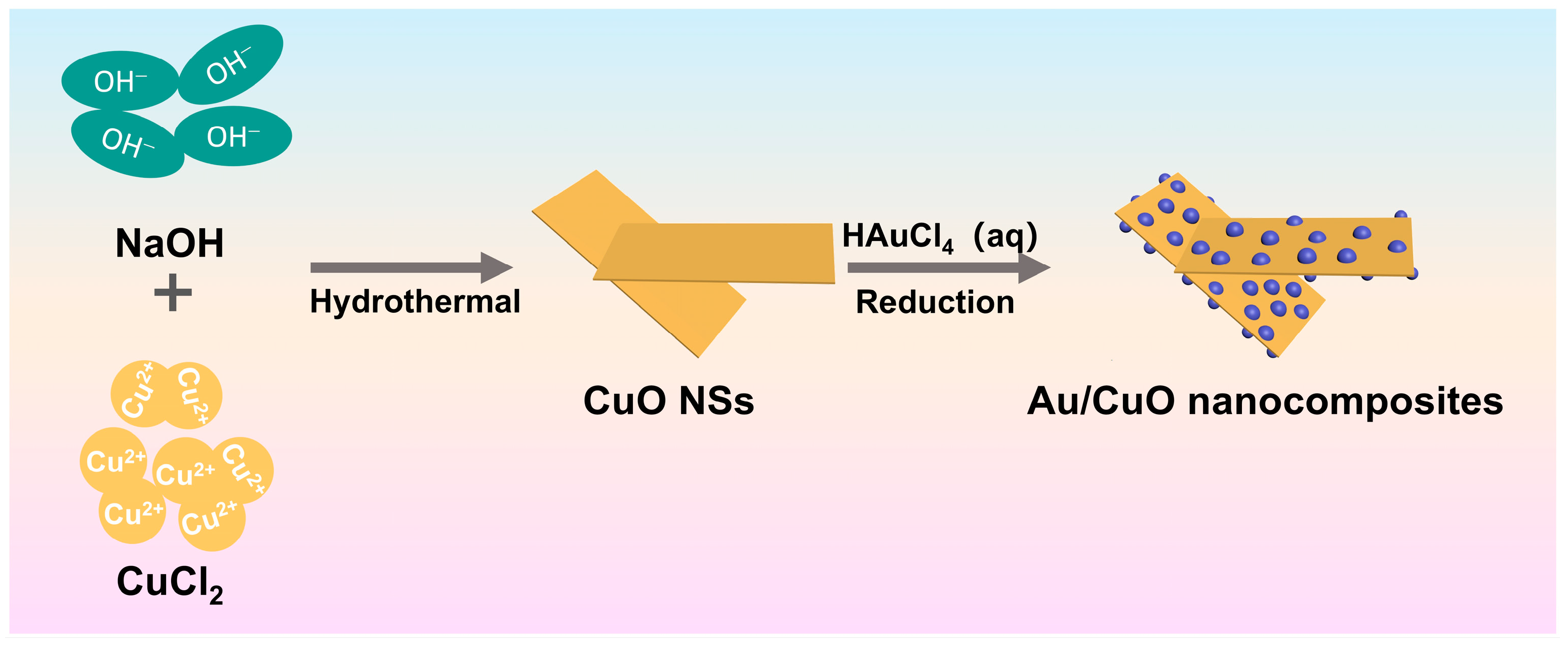
Figure 1. Schematic illustration of the Au/CuO nanocomposites.
Catalysts 15 01020 g001
Figure 2. SEM images of (a,b) pure CuO nanosheets and (c,d) the 7.41 wt% Au/CuO nanocomposites at different magnifications.
Catalysts 15 01020 g002
Figure 3. SEM images of (a) the 7.41 wt% Au/CuO nanocomposites and the corresponding EDS elemental mapping of (b) Cu, (c) O and (d) Au, respectively. (e) EDS spectra of the 7.41 wt% Au/CuO nanocomposites.
Catalysts 15 01020 g003
Figure 4. (ac) TEM and (d) HRTEM images of the 7.41 wt% Au/CuO nanocomposites.
Catalysts 15 01020 g004
Figure 5. XRD patterns for pure CuO nanosheets and the Au/CuO nanocomposites.
Catalysts 15 01020 g005
Figure 6. CV curves of modified electrodes for non-enzymatic glucose sensing evaluation. (a) CV curves of bare GCE, CuO nanosheets modified electrode, and 7.41 wt% Au/CuO nanocomposites modified electrode; (b) CV curves of Au/CuO nanocomposites modified electrode with Au loadings of 3.86 wt%, 7.41 wt%, 11.86 wt%, and 15.71 wt%. All measurements: 0.05 V/s scan rate in 0.1 M NaOH (with/without 1 mM glucose).
Catalysts 15 01020 g006
Figure 7. (a) CV curves of the 7.41 wt% Au/CuO electrode at 0.05 V/s in 0.1 M NaOH with different glucose concentrations; (b) CV curves of the 7.41 wt% Au/CuO nanocomposites modified electrode in 0.1 M NaOH containing 1 mM glucose at scan rates ranging from 0.001 to 0.05 V/s. (c) Oxidative current at 0.5 V vs. the square root of the scan rate.
Catalysts 15 01020 g007
Figure 8. (a) Schematic illustration of the glucose sensing mechanism for 7.41 wt% Au/CuO nanocomposites; (b) Amperometric responses of the 7.41 wt% Au/CuO nanocomposites modified electrode with successive additions of glucose into 0.1 M NaOH at 0.5 V; (c) the calibration curve of the modified electrode with current against glucose concentration; (d) Amperometric response of 7.41 wt% Au/CuO nanocomposites modified electrode in glucose (1 mM) and different interferences (0.1 mM) in 0.1 M NaOH at 0.5 V.
Catalysts 15 01020 g008
Figure 9. Long-term stability of 7.41 wt% Au/CuO nanocomposites electrode at 0.1 M NaOH by adding 1 mM glucose every week for 4 weeks.
Catalysts 15 01020 g009
Table 1. Determination results of glucose in real human urine samples (n = 3) on Au/CuO/GCE.
SampleAdded (μM)Detected (μM)Recovery (%)RSD (%) (n = 3)
Urine 140.0043.40108.54.2
Urine 240.0042.36105.98.5
Urine 340.0043.12107.86.5
Table 2. Comparison of the electrocatalytic performance of non-enzymatic glucose sensors.
ElectrodeDetection Potential
(V)
Sensitivity
(μA·mM−1·cm−2)
Linear Range
(μM)
LOD
(μM)
Reference
CuO nanosheets0.45233120–300020[40]
CuO urchins0.53201–33007.56[41]
CuO/Au0.54245510–12,0000.53[34]
Cu@CuO0.550.031.3–25001.3[42]
CuO/Cu0.65234200–90,00019[43]
CuO/Ni(OH)20.55598.650–85000.31[44]
Au nanoparticles-87.5100–25,00050[45]
Au decorated CuO NR0.5520095–13250.17[36]
Au NP modified CuO NWs0.64398.80.5–59000.5[46]
Cu aerogels0.5714.31–10000.48[47]
Au@CuO/nafion0.671520–200018[48]
Nafion/Au-CuO0.63126.765–6501.4[35]
Au/CuO0.5393.925–22505This
work
269.873550–11,550
Disclaimer/Publisher’s Note: The statements, opinions and data contained in all publications are solely those of the individual author(s) and contributor(s) and not of MDPI and/or the editor(s). MDPI and/or the editor(s) disclaim responsibility for any injury to people or property resulting from any ideas, methods, instructions or products referred to in the content.