
Biocatalysis as a Green Approach for Synthesis of Iron Nanoparticles—Batch and Microflow Process Comparison


Abstract
1. Introduction—The Definition of Nanotechnology
2. General Approaches to Nanomaterial Production
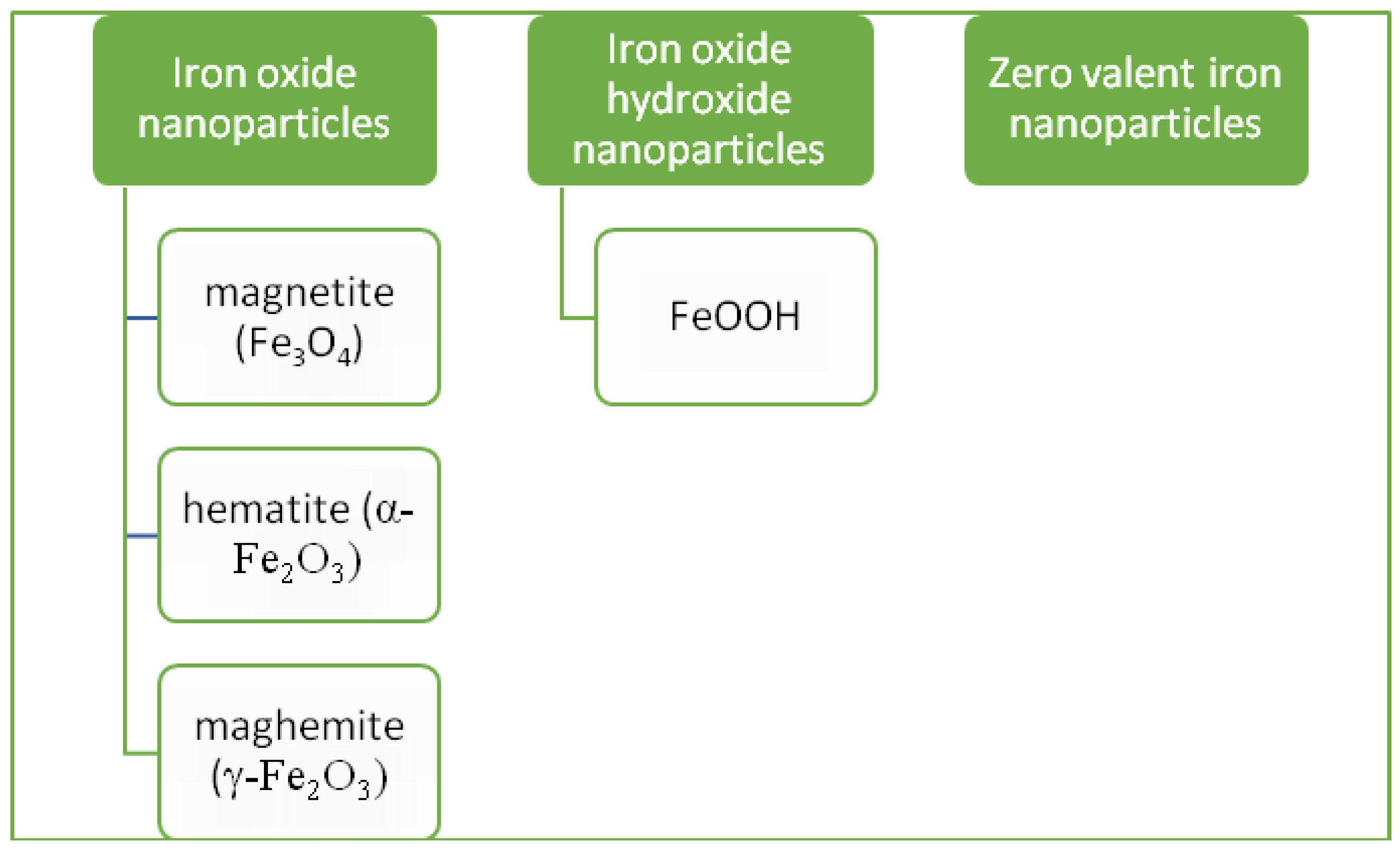
3. Iron Nanoparticles
4. Green Synthesis of Iron Nanoparticles
5. Batch (“One-Pot”) Biosynthesis of Iron Nanoparticles
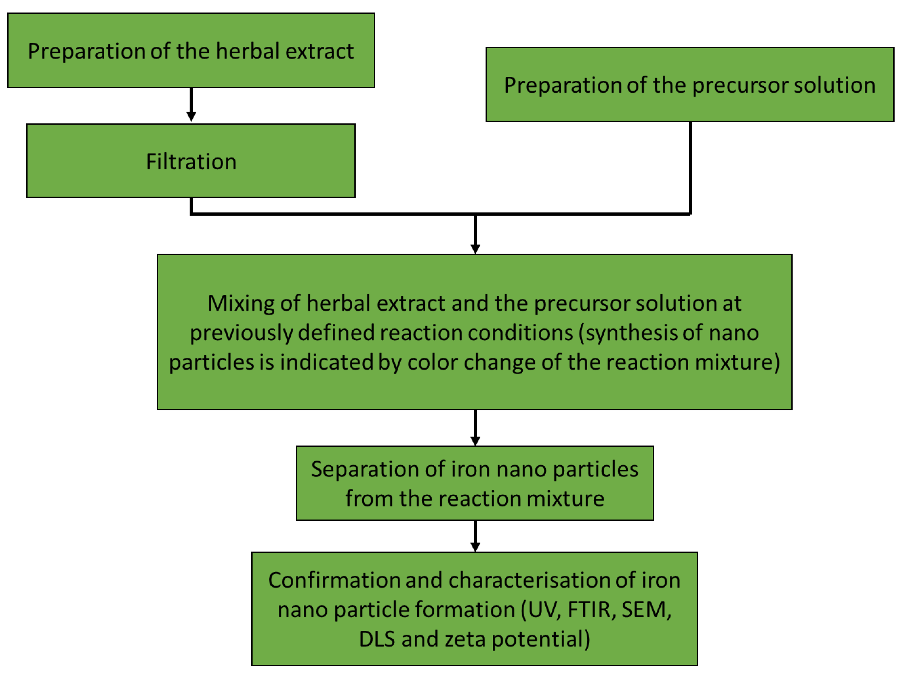
5.1. Batch Synthesis Using Herbal Extracts
5.2. Batch Synthesis Using Algae and Seaweed
5.3. Batch Synthesis Using Bacteria and Fungi
6. Continuous Synthesis Using Microfluidic Systems
6.1. Strategies for Iron Nanoparticle Synthesis Using Microfluidic Devices

| Nanoparticle | Microfluidic Device Geometry | Process Efficiency | Reference |
|---|---|---|---|
| Iron oxide core chitosan nanoparticles | 3D printed microchannel with T-shape inlet | With 20–120 mL/h flow rates and 0.06–0.03% concentrations at pH 4.5 for chitosan-tripolyphosphat, nanoparticles of diameter 190 ± 15 nm were obtained | [156] |
| Iron oxide nanoparticles | (Poly)tetrafluoro-ethylene (PTFE) tube microreactor with coaxial flows | Magnetic and stable colloidal iron oxide particles with a size less than 7 nm have been prepared | [157] |
| Superparamagnetic iron oxide nanoparticles | The experimental setup comprises two microreactors made of stainless steel lined up in succession. The reactor volume, which was 160 μL, was contained within microchannels that were 370 μm wide and 150 μm deep. | Residence time of 19.2 s resulted in a particle size distribution of 3.9 ± 0.9 nm | [158] |
6.2. Green Synthesis of Iron Nanoparticles Using Microfluidic Devices, Future Perspective and Research
- (1)
- Waste reduction
- (2)
- Atom economy during synthesis
- (3)
- Less dangerous chemical synthesis
- (4)
- Design of environmentally friendly chemicals
- (5)
- Use of safer solvents
- (6)
- Energy efficiency
- (7)
- Use of chemicals made from renewable sources
- (8)
- Reduced use of chemical derivatives
- (9)
- Reduced use of catalysts
- (10)
- Use of degradable chemicals
- (11)
- Real-time monitoring of pollution
- (12)
- Safe chemical procedures.
7. Conclusions
Author Contributions
Funding
Data Availability Statement
Conflicts of Interest
References
- Som, C.; Berges, M.; Chaudhry, Q.; Dusinska, M.; Fernandes, T.F.; Olsen, S.I.; Nowack, B. The Importance of Life Cycle Concepts for the Development of Safe Nanoproducts. Toxicology 2010, 269, 160–169. [Google Scholar] [CrossRef] [PubMed]
- Cushen, M.; Kerry, J.; Morris, M.; Cruz-Romero, M.; Cummins, E. Nanotechnologies in the Food Industry—Recent Developments, Risks and Regulation. Trends Food Sci. Technol. 2012, 24, 30–46. [Google Scholar] [CrossRef]
- Koivisto, A.J.; Aromaa, M.; Mäkelä, J.M.; Pasanen, P.; Hussein, T.; Hämeri, K. Concept to Estimate Regional Inhalation Dose of Industrially Synthesized Nanoparticles. ACS Nano 2012, 6, 1195–1203. [Google Scholar] [CrossRef] [PubMed]
- Mäkelä, J.M.; Keskinen, H.; Forsblom, T.; Keskinen, J. Generation of Metal and Metal Oxide Nanoparticles by Liquid Flame Spray Process. J. Mater. Sci. 2004, 39, 2783–2788. [Google Scholar] [CrossRef]
- Savolainen, K.; Pylkkänen, L.; Norppa, H.; Falck, G.; Lindberg, H.; Tuomi, T.; Vippola, M.; Alenius, H.; Hämeri, K.; Koivisto, J.; et al. Nanotechnologies, Engineered Nanomaterials and Occupational Health and Safety—A Review. Saf. Sci. 2010, 48, 957–963. [Google Scholar] [CrossRef]
- Vanlalveni, C.; Lallianrawna, S.; Biswas, A.; Selvaraj, M.; Changmai, B.; Rokhum, S.L. Green Synthesis of Silver Nanoparticles Using Plant Extracts and Their Antimicrobial Activities: A Review of Recent Literature. RSC Adv. 2021, 11, 2804–2837. [Google Scholar] [CrossRef]
- Iravani, S.; Korbekandi, H.; Mirmohammadi, S.V.; Zolfaghari, B. Synthesis of Silver Nanoparticles: Chemical, Physical and Biological Methods. Res. Pharm. Sci. 2014, 9, 385–406. [Google Scholar]
- Marquis, G.; Ramasamy, B.; Banwarilal, S.; Munusamy, A.P. Evaluation of Antibacterial Activity of Plant Mediated CaO Nanoparticles Using Cissus quadrangularis Extract. J. Photochem. Photobiol. B Biol. 2016, 155, 28–33. [Google Scholar] [CrossRef]
- Kaur, M.; Chopra, D.S. Green Synthesis of Iron Nanoparticles for Biomedical Applications. Glob. J. Nanomed. 2018, 4, 68–77. [Google Scholar] [CrossRef]
- Akintelu, S.A.; Oyebamiji, A.K.; Olugbeko, S.C.; Folorunso, A.S. Green Synthesis of Iron Oxide Nanoparticles for Biomedical Application and Environmental Remediation: A Review. Eclet. Quim. 2021, 46, 17–37. [Google Scholar] [CrossRef]
- Cao, Y.; Dhahad, H.A.; El-Shorbagy, M.A.; Alijani, H.Q.; Zakeri, M.; Heydari, A.; Bahonar, E.; Slouf, M.; Khatami, M.; Naderifar, M.; et al. Green Synthesis of Bimetallic ZnO–CuO Nanoparticles and Their Cytotoxicity Properties. Sci. Rep. 2021, 11, 23479. [Google Scholar] [CrossRef] [PubMed]
- Bharti, K.; Lone, S.A.; Singh, A.; Nathani, S.; Roy, P.; Sadhu, K.K. Green Synthesis of Luminescent Gold-Zinc Oxide Nanocomposites: Cell Imaging and Visible Light–Induced Dye Degradation. Front. Chem. 2021, 9, 105. [Google Scholar] [CrossRef]
- Spanos, A.; Athanasiou, K.; Ioannou, A.; Fotopoulos, V.; Krasia-Christoforou, T. Functionalized Magnetic Nanomaterials in Agricultural Applications. Nanomaterials 2021, 11, 3106. [Google Scholar] [CrossRef]
- Cao, M.; Li, Z.; Wang, J.; Ge, W.; Yue, T.; Li, R.; Colvin, V.L.; Yu, W.W. Food Related Applications of Magnetic Iron Oxide Nanoparticles: Enzyme Immobilization, Protein Purification, and Food Analysis. Trends Food Sci. Technol. 2012, 27, 47–56. [Google Scholar] [CrossRef]
- Attia, N.F.; El-Monaem, E.M.A.; El-Aqapa, H.G.; Elashery, S.E.A.; Eltaweil, A.S.; El Kady, M.; Khalifa, S.A.M.; Hawash, H.B.; El-Seedi, H.R. Iron Oxide Nanoparticles and Their Pharmaceutical Applications. Appl. Surf. Sci. Adv. 2022, 11, 100284. [Google Scholar] [CrossRef]
- Ebrahiminezhad, A.; Zare-Hoseinabadi, A.; Sarmah, A.K.; Taghizadeh, S.; Ghasemi, Y.; Berenjian, A. Plant-Mediated Synthesis and Applications of Iron Nanoparticles. Mol. Biotechnol. 2018, 60, 154–168. [Google Scholar] [CrossRef]
- Mittal, A.K.; Chisti, Y.; Banerjee, U.C. Synthesis of Metallic Nanoparticles Using Plant Extracts. Biotechnol. Adv. 2013, 31, 346–356. [Google Scholar] [CrossRef] [PubMed]
- Zhu, K.X.; Huang, S.; Peng, W.; Qian, H.F.; Zhou, H.M. Effect of Ultrafine Grinding on Hydration and Antioxidant Properties of Wheat Bran Dietary Fiber. Food Res. Int. 2010, 43, 943–948. [Google Scholar] [CrossRef]
- Shibata, T. Method for Producing Green Tea in Microfine Powder. U.S. Patent No 6,416,803, 7 July 2002. [Google Scholar]
- Brody, A.L.; Bugusu, B.; Han, J.H.; Sand, C.K.; McHugh, T.H. Innovative Food Packaging Solutions. J. Food Sci. 2008, 73, R107–R116. [Google Scholar] [CrossRef] [PubMed]
- Jadoun, S.; Arif, R.; Jangid, N.K.; Meena, R.K. Green Synthesis of Nanoparticles Using Plant Extracts: A Review. Environ. Chem. Lett. 2021, 19, 355–374. [Google Scholar] [CrossRef]
- Asghar, M.A.; Zahir, E.; Asghar, M.A.; Iqbal, J.; Rehman, A.A. Facile, One-Pot Biosynthesis and Characterization of Iron, Copper and Silver Nanoparticles Using Syzygium cumini Leaf Extract: As an Effective Antimicrobial and Aflatoxin B1 Adsorption Agents. PLoS ONE 2020, 15, e0234964. [Google Scholar] [CrossRef] [PubMed]
- Machado, S.; Pinto, S.L.; Grosso, J.P.; Nouws, H.P.A.; Albergaria, J.T.; Delerue-Matos, C. Green Production of Zero-Valent Iron Nanoparticles Using Tree Leaf Extracts. Sci. Total Environ. 2013, 445–446, 1–8. [Google Scholar] [CrossRef] [PubMed]
- Sozer, N.; Kokini, J.L. Nanotechnology and Its Applications in the Food Sector. Trends Biotechnol. 2009, 27, 82–89. [Google Scholar] [CrossRef]
- Herlekar, M.; Barve, S.; Kumar, R. Plant-Mediated Green Synthesis of Iron Nanoparticles. J. Nanoparticles 2014, 2014, 1–9. [Google Scholar] [CrossRef]
- Chauhan, S.; Upadhyay, L.S.B. Biosynthesis of Iron Oxide Nanoparticles Using Plant Derivatives of Lawsonia inermis (Henna) and Its Surface Modification for Biomedical Application. Nanotechnol. Environ. Eng. 2019, 4, 1–10. [Google Scholar] [CrossRef]
- Batool, F.; Iqbal, M.S.; Khan, S.U.D.; Khan, J.; Ahmed, B.; Qadir, M.I. Biologically Synthesized Iron Nanoparticles (FeNPs) from Phoenix dactylifera Have Anti-Bacterial Activities. Sci. Rep. 2021, 11, 22132. [Google Scholar] [CrossRef]
- Huber, D.L. Synthesis, Properties, and Applications of Iron Nanoparticles. Small 2005, 1, 482–501. [Google Scholar] [CrossRef]
- Mahanty, S.; Bakshi, M.; Ghosh, S.; Gaine, T.; Chatterjee, S.; Bhattacharyya, S.; Das, S.; Das, P.; Chaudhuri, P. Mycosynthesis of Iron Oxide Nanoparticles Using Manglicolous Fungi Isolated from Indian Sundarbans and Its Application for the Treatment of Chromium Containing Solution: Synthesis, Adsorption Isotherm, Kinetics and Thermodynamics Study. Environ. Nanotechnol. Monit. Manag. 2019, 12, 100276. [Google Scholar] [CrossRef]
- Ash, A.; Revati, K.; Pandey, B.D. Microbial Synthesis of Iron-Based Nanomaterials—A Review. Bull. Mater. Sci. 2011, 34, 191–198. [Google Scholar] [CrossRef]
- Nadeem, M.; Khan, R.; Shah, N.; Bangash, I.R.; Abbasi, B.H.; Hano, C.; Liu, C.; Ullah, S.; Hashmi, S.S.; Nadhman, A.; et al. A Review of Microbial Mediated Iron Nanoparticles (IONPs) and Its Biomedical Applications. Nanomaterials 2022, 12, 130. [Google Scholar] [CrossRef]
- Kianpour, S.; Ebrahiminezhad, A.; Deyhimi, M.; Negahdaripour, M.; Raee, M.J.; Mohkam, M.; Rezaee, H.; Irajie, C.; Berenjian, A.; Ghasemi, Y. Structural Characterization of Polysaccharide-Coated Iron Oxide Nanoparticles Produced by Staphylococcus warneri, Isolated from a Thermal Spring. J. Basic Microbiol. 2019, 59, 569–578. [Google Scholar] [CrossRef] [PubMed]
- Sundaram, P.A.; Augustine, R.; Kannan, M. Extracellular Biosynthesis of Iron Oxide Nanoparticles by Bacillus Subtilis Strains Isolated from Rhizosphere soil. Biotechnol. Bioprocess Eng. 2012, 17, 835–840. [Google Scholar] [CrossRef]
- Arias, S.L.; Shetty, A.R.; Senpan, A.; Echeverry-Rendón, M.; Reece, L.M.; Allain, J.P. Fabrication of a Functionalized Magnetic Bacterial Nanocellulose with Iron Oxide Nanoparticles. J. Vis. Exp. 2016, 2016, e52951. [Google Scholar] [CrossRef]
- Fani, M.; Ghandehari, F.; Rezaee, M. Biosynthesis of Iron Oxide Nanoparticles by Cytoplasmic Extract of Bacteria Lactobacillus fermentum. J. Med. Chem. Sci. 2018, 1, 28–30. [Google Scholar]
- Kaur, K.; Sidhu, A.K. Green Synthesis: An Eco-Friendly Route for the Synthesis of Iron Oxide Nanoparticles. Front. Nanotechnol. 2021, 3, 47. [Google Scholar]
- Mohamed, Y.M.; Azzam, A.M.; Amin, B.H.; Safwat, N.A. Mycosynthesis of Iron Nanoparticles by Alternaria alternata and Its Antibacterial Activity. Afr. J. Biotechnol. 2015, 14, 1234–1241. [Google Scholar] [CrossRef]
- Sidkey, N. Biosynthesis, Characterization and Antimicrobial Activity of Iron Oxide Nanoparticles Synthesized by Fungi. Al-Azhar J. Pharm. Sci. 2020, 62, 164–179. [Google Scholar] [CrossRef]
- Baskar, G.; Chandhuru, J.; Praveen, A.S.; Sheraz Fahad, K. Anticancer Activity of Iron Oxide Nanobiocomposite of Fungal Asparaginase. Int. J. Mod. Sci. Technol. 2017, 2, 98–104. [Google Scholar]
- Baccile, N.; Noiville, R.; Stievano, L.; Bogaert, I. Van Sophorolipids-Functionalized Iron Oxide Nanoparticles. Phys. Chem. Chem. Phys. 2013, 15, 1606–1620. [Google Scholar] [CrossRef]
- Mathur, P.; Saini, S.; Paul, E.; Sharma, C.; Mehtani, P. Endophytic Fungi Mediated Synthesis of Iron Nanoparticles: Characterization and Application in Methylene Blue Decolorization. Curr. Res. Green Sustain. Chem. 2021, 4, 100053. [Google Scholar] [CrossRef]
- Adeleye, T.M.; Kareem, S.O.; Kekere-Ekun, A.A. Optimization Studies on Biosynthesis of Iron Nanoparticles Using Rhizopus stolonifer. In Proceedings of the IOP Conference Series: Materials Science and Engineering, Ulaanbaatar, Mongolia, 10–13 September 2020; Volume 805. [Google Scholar]
- Arya, A.; Chundawat, T.S. Metal Nanoparticles from Algae: A Green Approach for the Synthesis, Characterization and Their Biological Activity. Nanosci. Nanotechnol.-Asia 2018, 10, 185–202. [Google Scholar] [CrossRef]
- LewisOscar, F.; Vismaya, S.; Arunkumar, M.; Thajuddin, N.; Dhanasekaran, D.; Nithya, C. Algal Nanoparticles: Synthesis and Biotechnological Potentials. In Algae—Organisms for Imminent Biotechnology; Books on Demand GmbH: Hamburg, Germany, 2016. [Google Scholar]
- Taghizadeh, S.; Taherpoor, A.; Ebrahiminezhad, A. Review Paper: Algae and Microalgae Mediated Synthesis of Iron Nanoparticles and Their Applications. JAMSAT 2020, 5, 1–11. [Google Scholar]
- Win, T.T.; Khan, S.; Bo, B.; Zada, S.; Fu, P.C. Green Synthesis and Characterization of Fe3O4 Nanoparticles Using Chlorella-K01 Extract for Potential Enhancement of Plant Growth Stimulating and Antifungal Activity. Sci. Rep. 2021, 11, 21996. [Google Scholar] [CrossRef] [PubMed]
- El-Kassas, H.Y.; Aly-Eldeen, M.A.; Gharib, S.M. Green Synthesis of Iron Oxide (Fe3O4) Nanoparticles Using Two Selected Brown Seaweeds: Characterization and Application for Lead Bioremediation. Acta Oceanol. Sin. 2016, 35, 89–98. [Google Scholar] [CrossRef]
- Salem, D.M.S.A.; Ismail, M.M.; Tadros, H.R.Z. Evaluation of the Antibiofilm Activity of Three Seaweed Species and Their Biosynthesized Iron Oxide Nanoparticles (Fe3O4-NPs). Egypt. J. Aquat. Res. 2020, 46, 333–339. [Google Scholar] [CrossRef]
- Mukherjee, A.; Sarkar, D.; Sasmal, S. A Review of Green Synthesis of Metal Nanoparticles Using Algae. Front. Microbiol. 2021, 12, 693899. [Google Scholar] [CrossRef] [PubMed]
- Puthukkara, A.R.P.; Jose, S.T.; Lal, D.S. Plant Mediated Synthesis of Zero Valent Iron Nanoparticles and Its Application in Water Treatment. J. Environ. Chem. Eng. 2021, 9, 104569. [Google Scholar] [CrossRef]
- AL-Husseini, A.H.; Sih, B.T.; Al-Araji, A.M. Green Synthesis of Iron Oxide Nanoparticles (Fe2O3) Using Saffron Extract. Proc. J. Phys. Conf. Ser. 2021, 2114, 012082. [Google Scholar] [CrossRef]
- Groiss, S.; Selvaraj, R.; Varadavenkatesan, T.; Vinayagam, R. Structural Characterization, Antibacterial and Catalytic Effect of Iron Oxide Nanoparticles Synthesised Using the Leaf Extract of Cynometra ramiflora. J. Mol. Struct. 2017, 1128, 572–578. [Google Scholar] [CrossRef]
- Jagtap, U.R.; Bapat, V.A. Green synthesis of silver nanoparticles using Artocarpus heterophyllus Lam. seed extract and its antibacterial activity. Ind. Crops Prod. 2013, 46, 132–137. [Google Scholar] [CrossRef]
- Kanagasubbulakshmi, S.; Kadirvelu, K. Green Synthesis of Iron Oxide Nanoparticles Using Lagenaria siceraria and Evaluation of Its Antimicrobial Activity. Def. Life Sci. J. 2017, 2, 422. [Google Scholar] [CrossRef]
- Kiwumulo, H.F.; Muwonge, H.; Ibingira, C.; Lubwama, M.; Kirabira, J.B.; Ssekitoleko, R.T. Green Synthesis and Characterization of Iron-Oxide Nanoparticles Using Moringa oleifera: A Potential Protocol for Use in Low and Middle Income Countries. BMC Res. Notes 2022, 15, 149. [Google Scholar] [CrossRef] [PubMed]
- Gottimukkala, K. Green Synthesis of Iron Nanoparticles Using Green Tea Leaves Extract. J. Nanomedine. Biother. Discov. 2017, 7, 1000151. [Google Scholar] [CrossRef]
- Mahdavi, M.; Namvar, F.; Ahmad, M.B.; Mohamad, R. Green Biosynthesis and Characterization of Magnetic Iron Oxide (Fe3O4) Nanoparticles Using Seaweed (Sargassum muticum) Aqueous Extract. Molecules 2013, 18, 5954–5964. [Google Scholar] [CrossRef]
- Mirza, A.U.; Kareem, A.; Nami, S.A.A.; Khan, M.S.; Rehman, S.; Bhat, S.A.; Mohammad, A.; Nishat, N. Biogenic Synthesis of Iron Oxide Nanoparticles Using Agrewia optiva and Prunus persica Phyto Species: Characterization, Antibacterial and Antioxidant Activity. J. Photochem. Photobiol. B Biol. 2018, 185, 262–274. [Google Scholar] [CrossRef]
- Shahwan, T.; Abu Sirriah, S.; Nairat, M.; Boyaci, E.; Eroĝlu, A.E.; Scott, T.B.; Hallam, K.R. Green Synthesis of Iron Nanoparticles and Their Application as a Fenton-like Catalyst for the Degradation of Aqueous Cationic and Anionic Dyes. Chem. Eng. J. 2011, 172, 258–266. [Google Scholar] [CrossRef]
- Thacker, H.H.; Ram, V.R.; Dave, P.N. Plant Mediated Synthesis of Iron Nanoparticles and Their Applications: A Review. Prog. Chem. Biochem. Res. 2019, 2, 84–91. [Google Scholar] [CrossRef]
- Majumdar, M.; Khan, S.A.; Nandi, N.B.; Roy, S.; Panja, A.S.; Roy, D.N.; Misra, T.K. Green Synthesis of Iron Nanoparticles for Investigation of Biofilm Inhibition Property. ChemistrySelect 2020, 5, 13575–13583. [Google Scholar] [CrossRef]
- Karunakaran, S.; Ramanujam, S.; Gurunathan, B. Green Synthesised Iron and Iron-Based Nanoparticle in Environmental and Biomedical Application: A Review. IET Nanobiotechnol. 2018, 12, 1003–1008. [Google Scholar] [CrossRef] [PubMed]
- Huang, L.; Weng, X.; Chen, Z.; Megharaj, M.; Naidu, R. Green Synthesis of Iron Nanoparticles by Various Tea Extracts: Comparative Study of the Reactivity. Spectrochim. Acta. A Mol. Biomol. Spectrosc. 2014, 130, 295–301. [Google Scholar] [CrossRef]
- Harshiny, M.; Iswarya, C.N.; Matheswaran, M. Biogenic Synthesis of Iron Nanoparticles Using Amaranthus dubius Leaf Extract as a Reducing Agent. Powder Technol. 2015, 286, 744–749. [Google Scholar] [CrossRef]
- Martínez-Cabanas, M.; López-García, M.; Barriada, J.L.; Herrero, R.; Sastre de Vicente, M.E. Green Synthesis of Iron Oxide Nanoparticles. Development of Magnetic Hybrid Materials for Efficient As(V) Removal. Chem. Eng. J. 2016, 301, 83–91. [Google Scholar] [CrossRef]
- Lenders, J.J.M.; Mirabello, G.; Sommerdijk, N.A.J.M. Bioinspired Magnetite Synthesis via Solid Precursor Phases. Chem. Sci. 2016, 7, 5624–5634. [Google Scholar] [CrossRef]
- Woźnica, A.; Dzirba, J.; Mańka, D.; Łabuzek, S. Effects of Electron Transport Inhibitors on Iron Reduction in Aeromonas hydrophila Strain KB1. Anaerobe 2003, 9, 125–130. [Google Scholar] [CrossRef] [PubMed]
- Huang, L.; Luo, F.; Chen, Z.; Megharaj, M.; Naidu, R. Green Synthesized Conditions Impacting on the Reactivity of Fe NPs for the Degradation of Malachite Green. Spectrochim. Acta Part A Mol. Biomol. Spectrosc. 2015, 137, 154–159. [Google Scholar] [CrossRef]
- Nadagouda, M.N.; Castle, A.B.; Murdock, R.C.; Hussain, S.M.; Varma, R.S. In Vitro Biocompatibility of Nanoscale Zerovalent Iron Particles (NZVI) Synthesized Using Tea Polyphenols. Green Chem. 2010, 12, 114–122. [Google Scholar] [CrossRef]
- Saranya, S.; Vijayarani, K.; Pavithra, S. Green Synthesis of Iron Nanoparticles Using Aqueous Extract of Musa ornata Flower Sheath against Pathogenic Bacteria. Indian J. Pharm. Sci. 2017, 79, 688–694. [Google Scholar] [CrossRef]
- Yehia, R.S.; Ali, A.M. Biosynthesis and Characterization of Iron Nanoparticles Produced by Thymus vulgaris L. And Their Antimicrobial Activity. Acta Bot. Croat. 2020, 77, 114–120. [Google Scholar] [CrossRef]
- Ehrampoush, M.H.; Miria, M.; Salmani, M.H.; Mahvi, A.H. Cadmium Removal from Aqueous Solution by Green Synthesis Iron Oxide Nanoparticles with Tangerine Peel Extract. J. Environ. Health Sci. Eng. 2015, 13, 84. [Google Scholar] [CrossRef] [PubMed]
- Muthukumar, H.; Matheswaran, M. Amaranthus spinosus Leaf Extract Mediated FeO Nanoparticles: Physicochemical Traits, Photocatalytic and Antioxidant Activity. ACS Sustain. Chem. Eng. 2015, 3, 3149–3156. [Google Scholar] [CrossRef]
- Patra, J.K.; Baek, K.H. Green Biosynthesis of Magnetic Iron Oxide (Fe3O4) Nanoparticles Using the Aqueous Extracts of Food Processing Wastes under Photo-Catalyzed Condition and Investigation of Their Antimicrobial and Antioxidant Activity. J. Photochem. Photobiol. B Biol. 2017, 173, 291–300. [Google Scholar] [CrossRef] [PubMed]
- Rajendran, K.; Sen, S. Optimization of Process Parameters for the Rapid Biosynthesis of Hematite Nanoparticles. J. Photochem. Photobiol. B Biol. 2016, 159, 82–87. [Google Scholar] [CrossRef] [PubMed]
- Bibi, I.; Nazar, N.; Ata, S.; Sultan, M.; Ali, A.; Abbas, A.; Jilani, K.; Kamal, S.; Sarim, F.M.; Khan, M.I.; et al. Green Synthesis of Iron Oxide Nanoparticles Using Pomegranate Seeds Extract and Photocatalytic Activity Evaluation for the Degradation of Textile Dye. J. Mater. Res. Technol. 2019, 8, 6115–6124. [Google Scholar] [CrossRef]
- Khan, S.; Bibi, G.; Dilbar, S.; Iqbal, A.; Ahmad, M.; Ali, A.; Ullah, Z.; Jaremko, M.; Iqbal, J.; Ali, M.; et al. Biosynthesis and Characterization of Iron Oxide Nanoparticles from Mentha spicata and Screening Its Combating Potential against Phytophthora Infestans. Front. Plant Sci. 2022, 13, 1001499. [Google Scholar] [CrossRef]
- Sayed, F.N.; Polshettiwar, V. Facile and Sustainable Synthesis of Shaped Iron Oxide Nanoparticles: Effect of Iron Precursor Salts on the Shapes of Iron Oxides. Sci. Rep. 2015, 5, 9733. [Google Scholar] [CrossRef]
- Verma, A.; Mehata, M.S. Controllable Synthesis of Silver Nanoparticles Using Neem Leaves and Their Antimicrobial Activity. J. Radiat. Res. Appl. Sci. 2016, 9, 109–115. [Google Scholar] [CrossRef]
- Madivoli, E.S.; Kareru, P.G.; Maina, E.G.; Nyabola, A.O.; Wanakai, S.I.; Nyang’au, J.O. Biosynthesis of Iron Nanoparticles Using Ageratum conyzoides Extracts, Their Antimicrobial and Photocatalytic Activity. SN Appl. Sci. 2019, 1, 500. [Google Scholar] [CrossRef]
- Lakshmnarayanan, S.; Shereen, M.F.; Niraimathi, K.L.; Brindha, P.; Arumugam, A. One-Pot Green Synthesis of Iron Oxide Nanoparticles from Bauhinia tomentosa: Characterization and Application towards Synthesis of 1, 3 Diolein. Sci. Rep. 2021, 11, 8643. [Google Scholar] [CrossRef]
- Bhuiyan, M.S.H.; Miah, M.Y.; Paul, S.C.; Aka, T.D.; Saha, O.; Rahaman, M.M.; Sharif, M.J.I.; Habiba, O.; Ashaduzzaman, M. Green Synthesis of Iron Oxide Nanoparticle Using Carica papaya Leaf Extract: Application for Photocatalytic Degradation of Remazol Yellow RR Dye and Antibacterial Activity. Heliyon 2020, 6, e04603. [Google Scholar] [CrossRef]
- Devi, H.S.; Boda, M.A.; Shah, M.A.; Parveen, S.; Wani, A.H. Green Synthesis of Iron Oxide Nanoparticles Using Platanus orientalis Leaf Extract for Antifungal Activity. Green Process. Synth. 2019, 8, 38–45. [Google Scholar] [CrossRef]
- Buarki, F.; AbuHassan, H.; Al Hannan, F.; Henari, F.Z. Green Synthesis of Iron Oxide Nanoparticles Using Hibiscus rosa Sinensis Flowers and Their Antibacterial Activity. J. Nanotechnol. 2022, 2022, 5474645. [Google Scholar] [CrossRef]
- Jain, R.; Mendiratta, S.; Kumar, L.; Srivastava, A. Green Synthesis of Iron Nanoparticles Using Artocarpus heterophyllus Peel Extract and Their Application as a Heterogeneous Fenton-like Catalyst for the Degradation of Fuchsin Basic Dye. Curr. Res. Green Sustain. Chem. 2021, 4, 100086. [Google Scholar] [CrossRef]
- Kamath, V.; Chandra, P.; Jeppu, G.P. Comparative Study of Using Five Different Leaf Extracts in the Green Synthesis of Iron Oxide Nanoparticles for Removal of Arsenic from Water. Int. J. Phytoremed. 2020, 22, 1278–1294. [Google Scholar] [CrossRef]
- Nahari, M.H.; Al Ali, A.; Asiri, A.; Mahnashi, M.H.; Shaikh, I.A.; Shettar, A.K.; Hoskeri, J. Green Synthesis and Characterization of Iron Nanoparticles Synthesized from Aqueous Leaf Extract of Vitex leucoxylon and Its Biomedical Applications. Nanomaterials 2022, 12, 2404. [Google Scholar] [CrossRef]
- Truskewycz, A.; Shukla, R.; Ball, A.S. Phytofabrication of Iron Nanoparticles for Hexavalent Chromium Remediation. ACS Omega 2018, 3, 10781–10790. [Google Scholar] [CrossRef]
- Jafer Aziz, W.; Abd Urabe, A. Chemical Preparation of Iron Oxide Nanoparticles Using Plants Extracts in Antibacterial Application. Int. J. Bioorganic Chem. 2019, 4, 1–6. [Google Scholar] [CrossRef]
- Üstün, E.; Önbaş, S.C.; Kübra Çelik, S.; Çol Ayvaz, M.; Şahin, N. Green Synthesis of Iron Oxide Nanoparticles by Using Ficus carica Leaf Extract and Its Antioxidant Activity. Biointerface Res. Appl. Chem. 2022, 12, 2108–2116. [Google Scholar] [CrossRef]
- Syarifah, S.; Imawan, C.; Handayani, W.; Djuhana, D. Biosynthesis of Ferric Oxide Nanoparticles Using Pometia pinnata J.R. Frost. & G.Forst. Leaves Water Extract. Proc. AIP Conf. Proc. 2018, 2023, 20054. [Google Scholar] [CrossRef]
- Yusefi, M.; Shameli, K.; Yee, O.S.; Teow, S.Y.; Hedayatnasab, Z.; Jahangirian, H.; Webster, T.J.; Kuča, K. Green Synthesis of Fe3O4 Nanoparticles Stabilized by a Garcinia mangostana Fruit Peel Extract for Hyperthermia and Anticancer Activities. Int. J. Nanomed. 2021, 16, 2515–2532. [Google Scholar] [CrossRef]
- Prabhu, N.N.; Kowshik, M. Green Route Synthesis and Preliminary Characterization of Iron Oxide Nanoparticles Using Leaf Extract of Ocimum tenuiflorum. Adv. Mater. Sci. Eng. 2019, 3. [Google Scholar] [CrossRef]
- Pallela, P.N.V.K.; Ummey, S.; Ruddaraju, L.K.; Gadi, S.; Cherukuri, C.S.L.; Barla, S.; Pammi, S.V.N. Antibacterial Efficacy of Green Synthesized α-Fe2O3 Nanoparticles Using Sida cordifolia Plant Extract. Heliyon 2019, 5, e02765. [Google Scholar] [CrossRef]
- Parajuli, K.; Sah, A.K.; Paudyal, H.; Parajuli, K.; Sah, A.K.; Paudyal, H. Green Synthesis of Magnetite Nanoparticles Using Aqueous Leaves Extracts of Azadirachta indica and Its Application for the Removal of As(V) from Water. Green Sustain. Chem. 2020, 10, 117–132. [Google Scholar] [CrossRef]
- Bensy, A.D.V.; Christobel, G.J.; Muthusamy, K.; Alfarhan, A.; Anantharaman, P. Green Synthesis of Iron Nanoparticles from Ulva lactuca and Bactericidal Activity against Enteropathogens. J. King Saud Univ. Sci. 2022, 34, 101888. [Google Scholar] [CrossRef]
- Salem, D.M.S.A.; Ismail, M.M.; Aly-Eldeen, M.A. Biogenic Synthesis and Antimicrobial Potency of Iron Oxide (Fe3O4) Nanoparticles Using Algae Harvested from the Mediterranean Sea, Egypt. Egypt. J. Aquat. Res. 2019, 45, 197–204. [Google Scholar] [CrossRef]
- Shalaby, S.M.; Madkour, F.F.; El-Kassas, H.Y.; Mohamed, A.A.; Elgarahy, A.M. Green Synthesis of Recyclable Iron Oxide Nanoparticles Using Spirulina platensis Microalgae for Adsorptive Removal of Cationic and Anionic Dyes. Environ. Sci. Pollut. Res. 2021, 28, 65549–65572. [Google Scholar] [CrossRef]
- El-Sheekh, M.M.; El-Kassas, H.Y.; Shams El-Din, N.G.; Eissa, D.I.; El-Sherbiny, B.A. Green Synthesis, Characterization Applications of Iron Oxide Nanoparticles for Antialgal and Wastewater Bioremediation Using Three Brown Algae. Int. J. Phytoremed. 2021, 23, 1538–1552. [Google Scholar] [CrossRef]
- Ghanbariasad, A.; Taghizadeh, S.M.; Show, P.L.; Nomanbhay, S.; Berenjian, A.; Ghasemi, Y.; Ebrahiminezhad, A. Controlled Synthesis of Iron Oxyhydroxide (FeOOH) Nanoparticles Using Secretory Compounds from Chlorella vulgaris Microalgae. Bioengineered 2019, 10, 390–396. [Google Scholar] [CrossRef]
- Ercan, G.; Uzunoğlu, D.; Ergüt, M.; Özer, A. Biosynthesis and Characterization of Iron Oxide Nanoparticles from Enteromorpha spp. Extract: Determination of Adsorbent Properties for Copper (II) Ions. Int. Adv. Res. Eng. J. 2019, 3, 65–74. [Google Scholar]
- Chandran, M.; Yuvaraj, D.; Christudhas, L.; Kv, R. Bio-Synthesis of Iron Nanoparticles Using the Brown Seaweed, Dictyota dicotoma. Biotechnol. 2016, 12, 5–9. [Google Scholar]
- Lahiri, D.; Nag, M.; Sheikh, H.I.; Sarkar, T.; Edinur, H.A.; Pati, S.; Ray, R.R. Microbiologically-Synthesized Nanoparticles and Their Role in Silencing the Biofilm Signaling Cascade. Front. Microbiol. 2021, 12, 180. [Google Scholar] [CrossRef]
- Ghani, S.; Rafiee, B.; Sadeghi, D.; Ahsani, M. Biosynthesis of Iron Nano-Particles by Bacillus megaterium and Its Anti-Bacterial Properties. J. Babol. Univ. Med. Sci. 2017, 19, 13–19. [Google Scholar] [CrossRef]
- Al-Maliki, Q.A.; Taj-Aldeen, W.R. Antibacterial and Antibiofilm Activity of Bacteria Mediated Synthesized Fe3O4 Nanoparticles Using Bacillus coagulans. J. Nanostruct. 2021, 11, 782–789. [Google Scholar] [CrossRef]
- Khan, A.A.; Khan, S.; Khan, S.; Rentschler, S.; Laufer, S.; Deigner, H.P. Biosynthesis of Iron Oxide Magnetic Nanoparticles Using Clinically Isolated Pseudomonas aeruginosa. Sci. Rep. 2021, 11, 20503. [Google Scholar] [CrossRef]
- Bharde, A.; Wani, A.; Shouche, Y.; Joy, P.A.; Prasad, B.L.V.; Sastry, M. Bacterial Aerobic Synthesis of Nanocrystalline Magnetite. J. Am. Chem. Soc. 2005, 127, 9326–9327. [Google Scholar] [CrossRef]
- Obayemi, J.D.; Dozie-Nwachukwu, S.; Danyuo, Y.; Odusanya, O.S.; Anuku, N.; Malatesta, K.; Soboyejo, W.O. Biosynthesis and the Conjugation of Magnetite Nanoparticles with Luteinizing Hormone Releasing Hormone (LHRH). Mater. Sci. Eng. C 2015, 46, 482–496. [Google Scholar] [CrossRef]
- Rajeswaran, S.; Somasundaram Thirugnanasambandan, S.; Dewangan, N.K.; Moorthy, R.K.; Kandasamy, S.; Vilwanathan, R. Multifarious Pharmacological Applications of Green Routed Eco-Friendly Iron Nanoparticles Synthesized by Streptomyces sp. (SRT12). Biol. Trace Elem. Res. 2020, 194, 273–283. [Google Scholar] [CrossRef]
- Byrne, J.M.; Telling, N.D.; Coker, V.S.; Pattrick, R.A.D.; Van Der Laan, G.; Arenholz, E.; Tuna, F.; Lloyd, J.R. Control of Nanoparticle Size, Reactivity and Magnetic Properties during the Bioproduction ofmagnetite by Geobacter sulfurreducens. Nanotechnology 2011, 22, 455709. [Google Scholar] [CrossRef]
- Zaki, S.A.; Eltarahony, M.M.; Abd-El-Haleem, D.A. Disinfection of Water and Wastewater by Biosynthesized Magnetite and Zerovalent Iron Nanoparticles via NAP-NAR Enzymes of Proteus mirabilis 10B. Environ. Sci. Pollut. Res. 2019, 26, 23661–23678. [Google Scholar] [CrossRef]
- Desai, M.P.; Pawar, K.D. Immobilization of Cellulase on Iron Tolerant Pseudomonas stutzeri Biosynthesized Photocatalytically Active Magnetic Nanoparticles for Increased Thermal Stability. Mater. Sci. Eng. C 2020, 106, 110169. [Google Scholar] [CrossRef]
- Ali, A.T.; Elewa, N.; Elbostany, N.A.; Shetaia, Y.M.; Swilm, M. Antimicrobial Potentiality of Green Synthesized Iron Oxide Nanoparticles by Penicillium roqueforti. Egy. J. Pure Appl. Sci 2021, 59, 29–43. [Google Scholar] [CrossRef]
- Asha Ranjani, V.; Tulja Rani, G.; Sowjanya, M.; Preethi, M.; Srinivas, M.; Nikhil, M. Yeast Mediated Synthesis of Iron Oxide Nano Particles: Its Characterization and Evaluation of Antibacterial Activity. Int. Res. J. Pharm. Med. Sci. 2022, 5, 12–16. [Google Scholar]
- Abdeen, M.; Sabry, S.; Ghozlan, H.; El-Gendy, A.A.; Carpenter, E.E. Microbial-Physical Synthesis of Fe and Fe3O4 Magnetic Nanoparticles Using Aspergillus niger YESM1 and Supercritical Condition of Ethanol. J. Nanomater. 2016, 2016, 9174891. [Google Scholar] [CrossRef]
- Mahanty, S.; Bakshi, M.; Ghosh, S.; Chatterjee, S.; Bhattacharyya, S.; Das, P.; Das, S.; Chaudhuri, P. Green Synthesis of Iron Oxide Nanoparticles Mediated by Filamentous Fungi Isolated from Sundarban Mangrove Ecosystem, India. Bionanoscience 2019, 9, 637–651. [Google Scholar] [CrossRef]
- Tarafdar, J.C.; Raliya, R. Rapid, Low-Cost, and Ecofriendly Approach for Iron Nanoparticle Synthesis Using Aspergillus oryzae TFR9. J. Nanopart. 2013, 2013, 141274. [Google Scholar] [CrossRef]
- Manikandan, G.; Ramasubbu, R. Biosynthesis of Iron Nanoparticles from Pleurotus Florida and Its Antimicrobial Activity against Selected Human Pathogens. Indian J. Pharm. Sci. 2021, 83, 45–51. [Google Scholar] [CrossRef]
- Vainshtein, M.; Belova, N.; Kulakovskaya, T.; Suzina, N.; Sorokin, V. Synthesis of Magneto-Sensitive Iron-Containing Nanoparticles by Yeasts. J. Ind. Microbiol. Biotechnol. 2014, 41, 657–663. [Google Scholar] [CrossRef]
- Han, Z.; Jiang, X. Microfluidic Synthesis of Functional Nanoparticles. Nanotechnol. Microfluid. 2020. [Google Scholar] [CrossRef]
- Ma, J.; Lee, S.M.Y.; Yi, C.; Li, C.W. Controllable Synthesis of Functional Nanoparticles by Microfluidic Platforms for Biomedical Applications-a Review. Lab Chip 2017, 17, 209–226. [Google Scholar] [CrossRef]
- Frey, N.; Sönmez, U.M.; Minden, J.; LeDuc, P. Microfluidics for Understanding Model Organisms. Nat. Commun. 2022, 13, 3195. [Google Scholar] [CrossRef]
- Nielsen, J.B.; Hanson, R.L.; Almughamsi, H.M.; Pang, C.; Fish, T.R.; Woolley, A.T. Microfluidics: Innovations in Materials and Their Fabrication and Functionalization. Anal. Chem. 2020, 92, 150. [Google Scholar] [CrossRef]
- Chen, Z.; Lv, Z.; Zhang, Z.; Weitz, D.A.; Zhang, H.; Zhang, Y.; Cui, W. Advanced Microfluidic Devices for Fabricating Multi-Structural Hydrogel Microsphere. Exploration 2021, 1, 20210036. [Google Scholar] [CrossRef]
- Fong, E.J.; Huang, C.; Hamilton, J.; Benett, W.J.; Bora, M.; Burklund, A.; Metz, T.R.; Shusteff, M. A Microfluidic Platform for Precision Small-Volume Sample Processing and Its Use to Size Separate Biological Particles with an Acoustic Microdevice. J. Vis. Exp. 2015, 2015, 53051. [Google Scholar] [CrossRef] [PubMed]
- Streets, A.M.; Huang, Y. Chip in a Lab: Microfluidics for next Generation Life Science Research. Biomicrofluidics 2013, 7, 1–23. [Google Scholar] [CrossRef] [PubMed]
- Damiati, S.; Kompella, U.B.; Damiati, S.A.; Kodzius, R. Microfluidic Devices for Drug Delivery Systems and Drug Screening. Genes 2018, 9, 103. [Google Scholar] [CrossRef] [PubMed]
- Zhu, Y.; Fang, Q. Analytical Detection Techniques for Droplet Microfluidics-A Review. Anal. Chim. Acta 2013, 787, 24–35. [Google Scholar] [CrossRef]
- Tsui, J.H.; Lee, W.; Pun, S.H.; Kim, J.; Kim, D.H. Microfluidics-Assisted in Vitro Drug Screening and Carrier Production. Adv. Drug Deliv. Rev. 2013, 65, 1575–1588. [Google Scholar] [CrossRef]
- Thomas, A.; Tan, J.; Liu, Y. Characterization of Nanoparticle Delivery in Microcirculation Using a Microfluidic Device. Microvasc. Res. 2014, 94, 17–27. [Google Scholar] [CrossRef]
- Tammaro, O.; Costagliola di Polidoro, A.; Romano, E.; Netti, P.A.; Torino, E. A Microfluidic Platform to Design Multimodal PEG—Crosslinked Hyaluronic Acid Nanoparticles (PEG-CHANPs) for Diagnostic Applications. Sci. Rep. 2020, 10, 6028. [Google Scholar] [CrossRef]
- Ahn, J.; Ko, J.; Lee, S.; Yu, J.; Kim, Y.; Li Jeon, N.; Woodruff, G.W. Microfluidics in Nanoparticle Drug Delivery; From Synthesis to Pre-Clinical Screening. Adv. Drug Deliv. Rev. 2018, 128, 29–53. [Google Scholar] [CrossRef]
- Jurinjak Tušek, A.; Šalić, A.; Valinger, D.; Jurina, T.; Benković, M.; Kljusurić, J.G.; Zelić, B. The Power of Microsystem Technology in the Food Industry—Going Small Makes It Better. Innov. Food Sci. Emerg. Technol. 2021, 68, 102613. [Google Scholar] [CrossRef]
- Miličević, N.; Panić, M.; Valinger, D.; Cvjetko Bubalo, M.; Benković, M.; Jurina, T.; Gajdoš Kljusurić, J.; Radojčić Redovniković, I.; Jurinjak Tušek, A. Development of Continuously Operated Aqueous Two-Phase Microextraction Process Using Natural Deep Eutectic Solvents. Sep. Purif. Technol. 2020, 244, 116746. [Google Scholar] [CrossRef]
- Bačić, M.; Ljubić, A.; Gojun, M.; Šalić, A.; Tušek, A.J.; Zelić, B. Continuous Integrated Process of Biodiesel Production and Purification—The End of the Conventional Two-Stage Batch Process? Energies 2021, 14, 403. [Google Scholar] [CrossRef]
- James, M.; Revia, R.A.; Stephen, Z.; Zhang, M. Microfluidic Synthesis of Iron Oxide Nanoparticles. Nanomaterials 2020, 10, 2113. [Google Scholar] [CrossRef]
- Bennett, J.A.; Campbell, Z.S.; Abolhasani, M. Role of Continuous Flow Processes in Green Manufacturing of Pharmaceuticals and Specialty Chemicals. Curr. Opin. Chem. Eng. 2019, 26, 9–19. [Google Scholar] [CrossRef]
- Wang, X.; Liu, J.; Wang, P.; deMello, A.; Feng, L.; Zhu, X.; Wen, W.; Kodzius, R.; Gong, X. Synthesis of Biomaterials Utilizing Microfluidic Technology. Genes 2018, 9, 283. [Google Scholar] [CrossRef]
- Ma, J.; Wang, Y.; Liu, J. Biomaterials Meet Microfluidics: From Synthesis Technologies to Biological Applications. Micromachines 2017, 8, 255. [Google Scholar] [CrossRef]
- Niculescu, A.G.; Chircov, C.; Bîrcă, A.C.; Grumezescu, A.M. Nanomaterials Synthesis through Microfluidic Methods: An Updated Overview. Nanomaterials 2021, 11, 864. [Google Scholar] [CrossRef]
- Abiev, R.S.; Almjasheva, O.V.; Popkov, V.I.; Proskurina, O.V. Microreactor Synthesis of Nanosized Particles: The Role of Micromixing, Aggregation, and Separation Processes in Heterogeneous Nucleation. Chem. Eng. Res. Des. 2022, 178, 73–94. [Google Scholar] [CrossRef]
- Yao, C.; Liu, Y.; Zhao, S.; Dong, Z.; Chen, G. Bubble/Droplet Formation and Mass Transfer during Gas–Liquid–Liquid Segmented Flow with Soluble Gas in a Microchannel. AIChE J. 2017, 63, 1727–1739. [Google Scholar] [CrossRef]
- Yao, C.; Liu, Y.; Xu, C.; Zhao, S.; Chen, G. Formation of Liquid–Liquid Slug Flow in a Microfluidic T-Junction: Effects of Fluid Properties and Leakage Flow. AIChE J. 2018, 64, 346–357. [Google Scholar] [CrossRef]
- Dang, M.; Yue, J.; Chen, G.; Yuan, Q. Formation Characteristics of Taylor Bubbles in a Microchannel with a Converging Shape Mixing Junction. Chem. Eng. J. 2013, 223, 99–109. [Google Scholar] [CrossRef]
- Zong, J.; Yue, J. Gas–Liquid Slug Flow Studies in Microreactors: Effect of Nanoparticle Addition on Flow Pattern and Pressure Drop. Front. Chem. Eng. 2022, 3, 788241. [Google Scholar] [CrossRef]
- Tušek, A.J.; Anić, I.; Kurtanjek, Ž.; Zelić, B. Mass Transfer Coefficient of Slug Flow for Organic Solvent-Aqueous System in a Microreactor. Korean J. Chem. Eng. 2015, 32, 1037–1045. [Google Scholar] [CrossRef]
- Etminan, A.; Muzychka, Y.S.; Pope, K. Liquid Film Thickness of Two-Phase Slug Flows in Capillary Microchannels: A Review Paper. Can. J. Chem. Eng. 2022, 100, 325–348. [Google Scholar] [CrossRef]
- Wu, Z.; Cao, Z.; Sunden, B. Flow Patterns and Slug Scaling of Liquid-Liquid Flow in Square Microchannels. Int. J. Multiph. Flow 2019, 112, 27–39. [Google Scholar] [CrossRef]
- Zhang, Q.; Liu, H.; Zhao, S.; Yao, C.; Chen, G. Hydrodynamics and Mass Transfer Characteristics of Liquid–Liquid Slug Flow in Microchannels: The Effects of Temperature, Fluid Properties and Channel Size. Chem. Eng. J. 2019, 358, 794–805. [Google Scholar] [CrossRef]
- Hakke, V.; Sonawane, S.; Anandan, S.; Sonawane, S.; Ashokkumar, M. Process Intensification Approach Using Microreactors for Synthesizing Nanomaterials—A Critical Review. Nanomaterials 2021, 11, 98. [Google Scholar] [CrossRef]
- Suryawanshi, P.L.; Sonawane, S.H.; Bhanvase, B.A.; Ashokkumar, M.; Pimplapure, M.S.; Gogate, P.R. Synthesis of Iron Oxide Nanoparticles in a Continuous Flow Spiral Microreactor and Corning® Advanced FlowTM Reactor. Green Process. Synth. 2018, 7, 1–11. [Google Scholar] [CrossRef]
- Göpfert, L.; Schoenen, M.; Reisen, O.; Buhl, E.M.; Mues, B.; Schmitz-Rode, T.; Slabu, I. Enabling Continuous Flow Manufacturing of Magnetic Nanoparticles with a Millifluidic System. J. Magn. Magn. Mater. 2022, 563, 169985. [Google Scholar] [CrossRef]
- Ahrberg, C.D.; Choi, J.W.; Chung, B.G. Droplet-Based Synthesis of Homogeneous Magnetic Iron Oxide Nanoparticles. Beilstein J. Nanotechnol 2018, 9, 2413–2420. [Google Scholar] [CrossRef]
- Panariello, L.; Wu, G.; Besenhard, M.O.; Loizou, K.; Storozhuk, L.; Thanh, N.T.K.; Gavriilidis, A. A Modular Millifluidic Platform for the Synthesis of Iron Oxide Nanoparticles with Control over Dissolved Gas and Flow Configuration. Materials 2020, 13, 1019. [Google Scholar] [CrossRef] [PubMed]
- Larrea, A.; Sebastian, V.; Ibarra, A.; Arruebo, M.; Santamaria, J. Gas Slug Microfluidics: A Unique Tool for Ultrafast, Highly Controlled Growth of Iron Oxide Nanostructures. Chem. Mater. 2015, 27, 4254–4260. [Google Scholar] [CrossRef] [PubMed]
- Aşık, M.D.; Kaplan, M.; Çetin, B.; Sağlam, N. Synthesis of Iron Oxide Core Chitosan Nanoparticles in a 3D Printed Microfluidic Device. J. Nanopart. Res. 2021, 23, 62. [Google Scholar] [CrossRef]
- Abou Hassan, A.; Sandre, O.; Cabuil, V.; Tabeling, P. Synthesis of Iron Oxide Nanoparticles in a Microfluidic Device: Preliminary Results in a Coaxial Flow Millichannel. Chem. Commun. 2008, 178, 1783–1785. [Google Scholar] [CrossRef] [PubMed]
- Uson, L.; Arruebo, M.; Sebastian, V.; Santamaria, J. Single Phase Microreactor for the Continuous, High-Temperature Synthesis of <4 nm Superparamagnetic Iron Oxide Nanoparticles. Chem. Eng. J. 2018, 340, 66–72. [Google Scholar] [CrossRef]
- Anastas, P.T.; Warner, J.C. Green Chemistry: Theory and Practice; Oxford University Press: New York, NY, USA, 1998. [Google Scholar]
- Ma, H.; Jin, N.; Zhang, P.; Zhou, Y.; Zhao, Y.; Zhang, X.; Lü, H.; Liu, J. Dynamic Characterization of Nanoparticles Production in a Droplet-Based Continuous Flow Microreactor. Chem. Eng. Res. Des. 2019, 144, 247–257. [Google Scholar] [CrossRef]
- García-Merino, B.; Bringas, E.; Ortiz, I. Synthesis and Applications of Surface-Modified Magnetic Nanoparticles: Progress and Future Prospects. Rev. Chem. Eng. 2022, 38, 821–842. [Google Scholar] [CrossRef]


| Plant | Precursor | Synthesis Conditions | Iron Nanoparticle Properties | Reference |
|---|---|---|---|---|
| Ageratum conyzoides (whole plant extract) | FeCl3·6H2O (0.1 M) solution | Room temperature, extract:precursor ratio: 1:1, t = 3 min | Cubic crystals, average diameter 85.98 nm | [80] |
| Bauhinia tomentosa (leave extract) | FeCl3 (0.01 M) solution | Room temperature, extract:precursor ratio: 1:1, continuous stirring until visible color change | Crystalline particles, average diameter 70 nm | [81] |
| Carica papaya (leaves extract) | FeCl3·6H2O (0.1 M) solution | Room temperature, extract:precursor ratio: 1:1, t = 30 min, continuous stirring | Irregular, non-uniform crystalline particles, visible formation of agglomerates, average diameter 21.59 nm | [82] |
| Platanus orientalis (leaves extract) | Fe(NO3)3·9H2O salt (99.8 %) | 1 g of Fe(NO3)3·9H2O salt added to 10 mL leaf extract, t = 1 h, T = 25 °C, continuous stirring | Spherical shape with an average diameter of 38 nm | [83] |
| Hibiscus rosa sinensis (flowers extract) | FeCl2·4H2O (1 mM) solution | Precursor:extract ratio: 1:1, 1:2 and 1:3, t = 20 s microwave radiation | Crystalline nanoparticles of nearly spherical shape, polydisperse, average diameter 51 nm | [84] |
| Artocarpus heterophyllus (peel extract) | 0.1 M FeCl2 solution | Precursor:extract ratio 2:3, room temperature, pH = 6 | Spherical particles, irregular surface, agglomerated, average diameter 33 nm | [85] |
| Punica granatum (seeds extract) | FeCl3 (1 M) solution | Extract:precursor ratio 12:1, 70 °C for 15 min, continuous stirring | Aggregates, maximum distribution in the range of 28.4–66.2 nm | [76] |
| Crocus sativus (whole plant extract) | FeCl3 (1 M) solution | Extract:precursor ratio 1:1, 60 °C for 30 min, continuous stirring | Nanoparticle structures with average grain sizes of 24.27–46.27 nm | [51] |
| Quercus virginiana, Eucalyptus globulus (leaves extract) | FeCl3 solution (0.1 M) | Extract:precursor in a 2:1 ratio, room temperature, continuous stirring at 300 rpm | Spherical, particle diameter around 10–100 nm, amorphous structure | [86] |
| Thymus vulgaris L. (leaves extract) | FeCl3·6H2O (0.1 M) | extract:precursor ratio: 1:1, 5 min, room temperature | Spherical particles, bimodal distribution with peaks at 2 nm and 40 nm | [71] |
| Vitex leucoxylon (leaves extract) | FeSO4 solution (0.05 mM) | extract:precursor ratio: 1:10, room temperature, incubated in the dark until a visible color change occurred | Spherical, with a diameter ranging from 45 nm to 100 nm based on SEM images | [87] |
| Lawsonia inermis (parts of the plant not identified, extract) | FeSO4·7H2O (0.02 M) solution | extract:precursor ratio: 1:1, 60 °C, pH = 11, 30 min | Spherical, average size 150–200 nm | [26] |
| Syzygium cumini (leaves extract) | FeCl3 solution (0.01 M) | Precursor: extract ratios 1:1, 4:1 and 9:1, continuous stirring, room temperature | Spherical, average size 55–65 nm (SEM) | [22] |
| Pittosporum undulatum, Melia azedarach, Schinus molle and Syzygium paniculatum (var. australe) (leaves extract) | 0.1 M FeCl3·6H2O solution | 0.1 M FeCl3·6H2O solution was slowly introduced using a peristaltic pump with a flow rate of 2 mL/min, ensuring a 2:1 extract:precursor ratio, continuous mixing, room temperature | Irregular particles, chain-like structures, size dependant on the extract used (min. 5–10 nm formed using Pittosporum undulatum extract, max. >100 nm for Schinus mole) | [88] |
| Apium graveolens (stalks extract) and Camellia sinensis (leaves extract) | Fe(NO3)2·6H2O, 0.02 M solution | 75 °C initial temperature, reduced to 60 °C and incubated for 60 min | Irregular, cubic and hexagonal shapes of various sizes, visible aggregates, smooth surface, diameter raged from 30.52 to 95.14 nm | [89] |
| Ficus carica (leaves extract) | 0.01M FeCl3·6H2O solution | 40 mL of the extract added to 100 mL of the precursor solution, pH = 11, continuous stirring at 70 °C for 1h | Crystalline multiform particles, aggregates visible, diameter 43–57 nm | [90] |
| Pometia pinnata (leaves extract) | FeCl3·6H2O (0.01 M) solution | Extract:precursor ratio 1:1, 500 rpm, 25 °C, 2h, with (pH = 11) and without pH adjustment | Polydisperse amorphous magnetic particles, size 10–20 nm | [91] |
| Camellia sinensis (leaves extract) | FeCl3 0.01M solution | Extract:precursor ratio 1:1, room temperature | Particles of maximum diameter of 116 nm | [56] |
| Garcinia mangostana (fruit peel extract) | FeCl3·6H2O, 97% and FeCl2·4H2O (0.99 g) at a molar ratio of 2:1 | pH = 11, 30 min, continuous stirring, different extract concentrations (1, 2, 5 and 10 % w/w) | Irregularly shaped magnetic particles, mean size of 13.42 ± 1.58 nm | [92] |
| Ocimum tenuiflorum (leaves extract) | FeCl2·4H2O (1 mol) FeCl3·6H2O (2 mol) in 100 mL distilled water (solution) | 100 mL of precursor solution with 5 mL of extract heated at 80 °C with continuous stirring until color change | Nanospheres (3–5 nm), aggregates, and nanoelipsoidal particles with a diameter of 100–200 nm | [93] |
| Sidacordifolia (whole plant extract) | 0.07 M Fe(NO3)2 solution | 5 mL of the extract was added to 10 mL of the precursor solution, heated at 60 °C for 5 min under continuous stirring | Spherical nano clusters, hematite of average diameter 16 nm | [94] |
| Azadirachta indica (leaves extract) | FeCl3∙6H2O, FeSO4∙7H2O (aqueous mixture of ferric (Fe3+) and ferrous ions (Fe2+) at a 2:1 molar ratio) | pH 10–11, 80 °C for 1 h with constant stirring | Spherical, uniform and particles smaller than 100 nm, aggregates visible | [95] |
| Mentha spicata (leaves extract) | FeCl3, 0.4 M solution | Extract:precursor ratio 1:1, 100 °C, 30 min | Circular and rod-shaped nanoparticles with an average diameter 21–82 nm | [77] |
| Citrus macroptera juice extract | FeCl3 0.14 M solution | Not specified | Spherical particles with diameter of 12 nm | [61] |
| Algae | Precursor | Synthesis Conditions | Iron Nanoparticle Properties | Reference |
|---|---|---|---|---|
| Ulva lactuca extract | 0.1 M FeCl3 solution | Extract:precursor ratio 1:1; 30 °C, 2 h, continuous stirring | Uniform, spherical, diameter 20–40 nm | [96] |
| Colpomenia sinuosa and Pterocladia capillacea extracts | 0.1 M FeCl3 solution | Extract:precursor ratio 1:1; 800 rpm, room temperature for 1h (C. sinuosa) and 2 h (P. capillacea) | Nanospheres, diameter 16.85–22.47 nm | [97] |
| Chlorella-K01 extract | 0.1 M FeCl2·4H2O solution | Precursor:algal extract ratio 2:3, 6–70 °C, different pH values (6, 8, 10 and 12) | Spherical Fe3O4 nanoparticles, diameter in the range of approximately 50 to 100 nm | [46] |
| Padina pavonica Thivy and Sargassum acinarium Setchell 1933 extracts | FeCl3 0.1 M solution | Extract:precursor ratio 1:1, 60 min, room temperature, continuous stirring | Spherical Fe3O4 nanoparticles in the range of sizes 10 to 19.5 nm (P. pavonica) and 21.6 to 27.4 nm for S. acinarium | [47] |
| Spirulina platensis extracts | FeCl3·6H2O from 0.1 to 0.6 M | Extract:precursor ratio 1:1, immediate color change, stabilization for another 2 h at room temperature | Agglomerated non- uniform magnetic particles, diameter <10 nm | [98] |
| Petalonia fascia, Colpomenia sinuosa extracts | FeCl3.6H2O (0.1 M) solution | Extract:precursor ratio 1:1, immediate color change, incubation for 72 h at room temperature | - spherical shape and size ranged from 6.54 to 13.46 nm with an average 9.42 ± 2.84 nm (P. fascia) - cubic shape of the Fe3O4–NPs synthesized using C. sinuosa with a size range 10.56 to 19.91 nm and an average 17.78 ± 3.63 nm | [99] |
| Sargassum muticum extract | 0.1 M FeCl3 solution | Extract:precursor ratio 1:1, immediate color change, the mixture was stirred for 60 min and then allowed to stand at room temperature for another 30 min | Cubic particles with mean diameter of 18 ± 4 nm | [57] |
| Chlorella vulgaris extract | FeCl3.6H2O | 15 min, continuous stirring, room temperature (controlled synthesis using algal extract and uncontrolled synthesis without algal extract) | Large aggregates of ultrafine nanoparticles obtained by uncontrolled synthesis, discrete spherical nanoparticles ranging from 8 to 17 nm for controlled synthesis in the presence of C. vulgaris secretory carbohydrates contained in the extract | [100] |
| Enteromorpha spp. extract | 0.1 M FeCl3 solution | Extract was added drop by drop manually into 0.1 M FeCl3 solution in a 1:2 volume ratio at room temperature, immediate color change | Spherical, porous agglomerates visible, mean diameter 78.83 nm | [101] |
| Dictyota dicotoma extract | FeCl3 (2% solution) and FeSO4 (1% solution) | Iron nanoparticles were prepared by adding ferric chloride (2%), ferrous sulphate (1%) solution, to the extracts (10 mL) and precipitated with 2 mL sodium hydroxide (0.1 M), pH 7–10, room temperature, 1 h, continuous stirring | Cubic nanoparticles, size range about 40 nm to 50 nm | [102] |
| Moringa oleifera extract | 0.6 M FeCl3 solution | 80 mL of iron(III)chloride solution was mixed with 20 mL of the extract, 60 °C, 4h | Granular, homogenous, spherical-shaped structure with an average diameter of approximately 16 nm | [55] |
| Microorganism | Precursor | Synthesis Conditions | Iron Nanoparticle Properties | Reference |
|---|---|---|---|---|
| Bacteria | ||||
| Bacillus megaterium cell culture | 0.1 M Fe(NO3)2 solution | Bacterial suspensions combined with precursor solution at 1:1 ratio, room temperature, 20 min | Cubic shape, 40–60 nm | [104] |
| Bacillus coagulans (cell-free broth supernatant) | iron salt precursor Fe3+ (FeCl3∙6H2O) and Fe2+ (FeCl2·4H2O) at a 2:1 M ratio | Cell-free broth supernatant was added to the precursor solution, 35 °C, 30 min, continuous stirring, pH = 11 | Irregular cubic shaped particles, diameter 4–33 nm | [105] |
| Pseudomonas aeruginosa (cell culture) | ferric quinate (FQ) solution | Intracellular synthesis, the isolates were cultured in a simple 9 K medium to isolate magnetotactic bacteria, magnetic properties confirmed by a magnet, magnetosomes extracted by sonication | Metallic structures, size 35–45 nm | [106] |
| Actinobacter spp. bacetrial biomass broth | K3Fe(CN)6/K4Fe(CN)6 mixture (2:1 molar ratio) | Bacterial biomass was cultured in a Luria broth, sterilized and, after 24 h, aqueous K3Fe(CN)6/K4Fe(CN)6 mixture (2:1 molar ratio) was added, 3 days, 150 rpm, 28 °C | After 24 h, quasi-spherical nanoparticles of 10−40 nm were obtained, after 48 h uniform cubic particles of 50−150 nm | [107] |
| Magnetospirillum magneticum culture broth | FeCl3 | 29 °C, 24 h, 141 rpm, reaction mixture comprised of MTB culture and ferric chloride at different pH values (4–12) | Cuboidal and rectangular prisms, nearly spherical faceted nanoparticles, size 18–52 nm, depending on the pH of the mixture | [108] |
| Streptomyces sp. (SRT12) cell-free broth supernatant | FeCl2·4H2O, FeCl3·6H2O | Cell-free supernatant was mixed with the precursor solution, 120 min, room temperature | Quasi-spherical, granular, crystalline and smooth cubical surfaced clusters with the size range from 65.0 to 86.7 nm | [109] |
| Geobacter sulfurreducens bacterial suspensions | FeO(OH), 50 mM | Cell cultures in deionized water containing an electron donor (sodium acetate 20 mM), an electron acceptor (Fe(III)-oxyhydroxide, 50 mM) and a sodium bicarbonate buffer (30 mM), 30 °C, 1 week incubation in the dark | Spherical and square shapes, particle size during synthesis can be controlled in the range 10–50 nm | [110] |
| Proteus mirabilis 10B bacterial suspensions | 7 mM of Fe (NO3)3·9H2O | Proteus mirabilis 10B strain (108 CFU/mL) was inoculated to the optimized medium supplemented with 7 mM of Fe (NO3)3·9H2O; reaction was monitored based on color change of the reaction mixture | Uniform, small, monodispersed and spherical nanoparticles without distinct aggregation with a diameter of 1.44 to 1.92 nm; spider-web-like shape size 11.7 to 60.8 nm | [111] |
| Pseudomonas stutzeri bacterial suspension | Ferric quinate (FQ) solution: 10 mM FQ solution was prepared by dissolving 0.27 g of ferric chloride and 0.19 g of quinic acid in 100 mL H2O | Bacterial culture was inoculated in 100 mL nutrient broth supplemented with 0.5 mM FQ. Of the two sets, one was incubated as static culture and other was agitated at 120 rpm at 37 °C for 72 h | Spherical in shape with rough surface. The mean diameter of IOMNPs clusters was estimated to be below 100 nm (SEM); spherical morphology with size in the range of 10–20 nm (TEM) | [112] |
| Fungi, yeasts | ||||
| Pennicillium roqeforti fungal mycelia suspension | FeCl3∙6H2O, ferric chloride hexahydrate (0.001 M) and FeCl2·4H2O (0.001 mM) in the ratio 2:1 | 10 g of wet biomass was added to 90 mL mixture of salt solution and was incubated for 24 h at 28 °C in a dark-shaking incubator at 150 rpm | Non-spherical, coated, diameter 5–16 nm | [113] |
| Saccharomyces cerevisiae yeast culture suspended in a growth medium | FeCl3∙6H2O (0.001 M), FeSO4∙7H2O (0.1 M) | Freshly prepared yeast culture was added to the mixture of precursor solution, incubated in a rotary shaker at 30 °C at 120 rpm for 2–3 days | Spherical Fe2O3 particles, diameter 70–100 nm | [114] |
| Alternaria alternata (Mili-Q water cell-free filtrate) | Fe(NO3)2, 1 mM solution | Cell-free filtrate mixed with 1 mM Fe(NO3)2 incubated at 28 °C in dark with shaking (150 rpm) for 72 h | Cubic particles, diameter 5.4–12.1 nm | [37] |
| Penicillium oxalicum (Mili-Q water cell-free filtrate) | FeSO4, 100 mM solution | Fungal mycelia filtrate was mixed with precursor solution (1:1), overnight, continuous stirring, 30 °C | Spherical shape, diameter 140 nm, agglomerates visible | [41] |
| Aspergillus niger homogenized mycelia cells solution | FeSO4 and FeCl3 solutions (2000 ppm) | Precursor/fungus solutions were incubated for 6 days at room temperature | Spherical particles synthesized intra- and extracellularly, Fe particles of 18 nm, Fe3O4 of 50 nm | [115] |
| Trichoderma asperellum, Phialemoniopsis ocularis, and Fusarium incarnatum fungal cell broth filtrate | FeCl3 and FeCl2 salt solution (2:1 mM final concentration) | Fungal cell filtrate mixed with FeCl3 and FeCl2 salt solution, 5 min, 30 °C, continuous stirring | Spherical with average particle size ranging between 25 ± 3.94 nm for T. asperellum, 13.13 ± 4.32 nm for P. ocularis and 30.56 ± 8.68 nm for F. incarnatum | [116] |
| Aspergillus oryzae TFR9 Mili-Q water cell-free filtrate | FeCl3, 0.001 M solution | Cell-free filtrate and precursor solution were mixed and kept on rotary shaker at 28 °C at 150 rpm for 12 h | Spherical crystalline particles with diameters 10 nm and 24.6 nm | [117] |
| Pleurotus florida water extract | FeCl3, 1 M solution | Mushroom extract:ferric chloride solution ratio 1:1, temperature 50–60°, stirred continuously | Spherical, roughly with 100 nm diameter | [118] |
| Cryptococcus humicola 9–6 cells suspended in a growth medium | Fe2SO4·7H2O, 0.05 M solution added to the growth medium | C. humicola nutrient media were enriched with iron, incubated at 22–25 °C, mixed every 3 days | Spherical nanoparticles present in the cell and adhered to the cell wall, diameter 8–9 nm | [119] |
| Rhizopus stolonifera (Mili-Q water cell-free filtrate) | FeCl3, 1 mM solution | Cell filtrate(50 mL) was mixed with 50 mL of 1 M FeCl3 solution, agitated in the orbital shaker for 72 h at room temperature | Diameter and morphology not determined in the study | [42] |
Disclaimer/Publisher’s Note: The statements, opinions and data contained in all publications are solely those of the individual author(s) and contributor(s) and not of MDPI and/or the editor(s). MDPI and/or the editor(s) disclaim responsibility for any injury to people or property resulting from any ideas, methods, instructions or products referred to in the content. |
© 2023 by the authors. Licensee MDPI, Basel, Switzerland. This article is an open access article distributed under the terms and conditions of the Creative Commons Attribution (CC BY) license (https://creativecommons.org/licenses/by/4.0/).
Share and Cite
Benković, M.; Valinger, D.; Jurina, T.; Gajdoš Kljusurić, J.; Jurinjak Tušek, A. Biocatalysis as a Green Approach for Synthesis of Iron Nanoparticles—Batch and Microflow Process Comparison. Catalysts 2023, 13, 112. https://doi.org/10.3390/catal13010112
Benković M, Valinger D, Jurina T, Gajdoš Kljusurić J, Jurinjak Tušek A. Biocatalysis as a Green Approach for Synthesis of Iron Nanoparticles—Batch and Microflow Process Comparison. Catalysts. 2023; 13(1):112. https://doi.org/10.3390/catal13010112
Chicago/Turabian StyleBenković, Maja, Davor Valinger, Tamara Jurina, Jasenka Gajdoš Kljusurić, and Ana Jurinjak Tušek. 2023. "Biocatalysis as a Green Approach for Synthesis of Iron Nanoparticles—Batch and Microflow Process Comparison" Catalysts 13, no. 1: 112. https://doi.org/10.3390/catal13010112
APA StyleBenković, M., Valinger, D., Jurina, T., Gajdoš Kljusurić, J., & Jurinjak Tušek, A. (2023). Biocatalysis as a Green Approach for Synthesis of Iron Nanoparticles—Batch and Microflow Process Comparison. Catalysts, 13(1), 112. https://doi.org/10.3390/catal13010112