Article

Protein Engineering of Pasteurella multocida α2,3-Sialyltransferase with Reduced α2,3-Sialidase Activity and Application in Synthesis of 3′-Sialyllactose

1
Key Laboratory of Food Nutrition and Safety, Ministry of Education, Tianjin University of Science and Technology, Tianjin 300457, China
2
National Key Laboratory of Biochemical Engineering, National Engineering Research Center for Biotechnology (Beijing), Institute of Process Engineering, Chinese Academy of Sciences, Beijing 100190, China
3
School of Chemistry and Chemical Engineering, University of Jinan, Jinan 250022, China
*
Authors to whom correspondence should be addressed.
These authors contributed equally to this work.
Catalysts 2022, 12(6), 579; https://doi.org/10.3390/catal12060579
Submission received: 9 April 2022 / Revised: 18 May 2022 / Accepted: 21 May 2022 / Published: 25 May 2022
(This article belongs to the Special Issue Advances in Biocatalysis and Enzyme Engineering)

Abstract

:
Sialyltransferases are key enzymes for the production of sialosides. The versatility of Pasteurella multocida α2,3-sialyltransferase 1 (PmST1) causes difficulties in the efficient synthesis of α2,3-linked sialylatetd compounds, especial its α2,3-sialidase activity. In the current study, the α2,3-sialidase activity of PmST1 was further reduced by rational design-based protein engineering. Three double mutants PMG1 (M144D/R313Y), PMG2 (M144D/R313H) and PMG3 (M144D/R313N) were designed and constructed using M144D as the template and kinetically investigated. In comparison with M144D, the α2,3-sialyltransferase activity of PMG2 was enhanced by 1.4-fold, while its α2,3-sialidase activity was reduced by 4-fold. Two PMG2-based triple mutants PMG2-1 (M144D/R313H/T265S) and PMG2-2 (M144D/R313H/E271F) were then designed, generated and characterized. Compared with PMG2, triple mutants showed slightly improved α2,3-sialyltransferase activity, but their α2,3-sialidase activities were increased by 2.1–2.9 fold. In summary, PMG2 was used for preparative-scale production of 3′-SL (3′-sialyllactose) with a yield of >95%. These new PmST1 mutants could be potentially utilized for efficient synthesis of α2,3-linked sialosides. This work provides a guide to designing and constructing efficient sialyltransferases.

1. Introduction

Sialylated oligosaccharides play important roles in many physiological and pathological processes, including intercellular adhesion, signaling, microbial attachment, etc., and most sialic acid-related biological processes require specific sialic acid forms, glycosidic linkage and defined underlying glycan chains [1]. Noticeably, sialic acid (SA) is generally located at the nonreducing ends of the sugar chains [2], and this outmost position and ubiquitous distribution enable sialylated glycans to be involved in numerous cellular processes. Particularly, sialyllactose (SL) moieties are found in gangliosides in neuron and brain tissues, which modulate cell signal transduction events such as brain development and memory formation [3]. SL is also one of the major components of human milk oligosaccharides (HMOs), consisting of an SA bound to lactose at the 3 or 6 position, and can promote probiotic proliferation and optimized metabolism in the brain, facilitate liver and muscle development in infants, induce differentiation such as in human intestinal epithelial cells [4] and function as a potent inhibitor of bacterial or viral adhesion to the epithelial surface in the initial stages of the infection process, among other functions. [5,6]. 3′-SL (3′-sialyllactose) and 6′-SL (6′-sialyllactose), containing Neu5Ac(α2,3)Gal and Neu5Ac(α2,6)Gal, are differentially recognized by avian and human influenza viruses, respectively [7], through interaction with viral hemagglutinins. Synthetic SLs can be used for various applications, such as functional food ingredients, nutraceutical medicine and virus adsorbent materials, so that their efficient preparative-scale production is urgently needed.
Due to the hindered tertiary anomeric center and the lack of a neighboring participating group in sialic acids, chemical sialylation is considered one of the most difficult glycosylation reactions [8]. Enzymatic sialylation is preferred for synthesis of sialylated compounds. Sialyltransferases (STs) are the key enzymes that catalyze the transfer of a sialic acid residue from cytidine 5′-monophosphate-sialic acid (CMP-Sia) to an acceptor to produce sialosides [9,10,11,12,13]. STs can catalyzed synthesis of α2,3/2,6/2,8-linked sialylated compounds and are currently divided into five different GT (glycosyltransferase) families according to the CAZy (Carbohydrate-Active enZYmes) database (http://www.cazy.org/, accessed on 26 March 2022): GT29, GT38, GT42, GT52 and GT8. The GT29 family mainly contains eukaryotic and viral STs, whereas GT38, GT42, GT52 and GT80 mainly include bacterial STs. Bacterial STs have been extensively used in the synthesis of sialylated glycoconjugates [14,15]. The GT38 family includes PolySTs (Neus, Siad) from E. coli and N. meningitidis [16,17]. The GT42 family contains mono-functional α2,3-STs (Cst-I, Cst-II, Cst-III) and bifunctional α2,3/α2,8-STs (LiC3A, LiC3B) from C. jejuni and H. influenza [18,19]. The GT52 family includes mono-functional α2,3-STs and bifunctional α2,3-STs from Neisseria spp. (LST) [20,21]. The family GT80 includes α2,3-STs, α2,6-STs and bifunctional α2,3/α2,6-STs from Haemophilus ducreyi, Pasteurella multocida, Photobacterium spp. and Vibrio sp. [9,22,23,24].
One interesting feature is that some bacterial STs are multifunctional. For example, Pasteurella multocida α2,3-sialyltransferase 1 (PmST1), which has been widely used in enzymatic and chemoenzymatic synthesis of diverse α2-3-linked sialosides, is a highly promiscuous enzyme with major α2,3-sialyltransferase activity (optimal pH range of 7.5–9.0), weak α2,6-sialyltransferase activity (at pH 4.5–7.0), CMP-Neu5Ac hydrolase activity, α2,3-sialidase activity (optimal at pH 5.0–5.5) and α2,3-trans-sialidase activity (optimal at pH 5.5–6.5) [9,25]. The promiscuity of PmST1 caused great challenges in the synthesis of α2,3-linked sialosides. For instance, the α2,3-sialidase activity of PmST1 results in the hydrolysis of the formed product, which affects the yield of sialylated oligosaccharides and complicates the reaction. Thus, it is essential to control the amount of PmST1 used and the time of the PmST1-catalyzed reaction. If PmST1-catalyzed α2,3-sialylation takes longer, such as sialylation of glycoproteins and synthesis of sialyl Lewisx [SLex, Siaα2−3Galβ1−4(Fucα1−3) GlcNAcβOR] from Lewisx [Lex, Galβ1−4(Fucα1−3)GlcNAcβOR], PmST1 containing α2-3-sialidase activity would lead to a lower yield of the anticipated products and become a big issue [25]. Therefore, in addition to reaction engineering, it is of great significance to reduce the α2,3-sialidase activity of PmST1 through protein engineering.
In order to improve the yield of the α2,3-sialylation reaction, protein engineering of PmST1 was performed. In 2011, Chen et al. reported that the α2-3-sialidase activity of PmST1 was significantly decreased by structure-based site-directed mutagenesis [26]. A rationally designed double mutant E271F/R313Y showed 6333-fold reduced α2,3-sialidase activity without affecting its α2-3-sialyltransferase activity. Later, in 2012, and likewise based on a structure-based rational design, the same group constructed a single mutant (M144D) with decreased donor (CMP-SiaA) hydrolysis activity without influencing its α2-3-sialylation activity very much, and M144D also had drastically reduced α2-3-sialidase activity and was successfully used for synthesis of sialyl Lewisx antigens containing different sialic acid forms [25]. In 2014, PmST1 was engineered for enhancement of its α2-3-sialyltransferase activity by combing rational design with site-saturation mutagenesis by Kim et al. [27]. Putative functional residues involved in the substrate-binding pocket were selected by multiple sequence alignment and alanine scanning and subsequently subjected to site-saturation mutagenesis. Several single and double mutants, such as R313N, R313N/T265S and R313H/T265S, were screened with improved yield and productivity of 3′-SL.
In the current study, we found that the residual α2-3-sialidase activity of M144D of PmST1 still led to hydrolysis of the formed 3′-SL when the time of the M144D-catalyzed reaction was prolonged. Given the fact that M144D has been extensively used as an powerful biocatalyst for α2,3-sialylation, our observation prompted us to make new PmST1 mutants with further decreased α2-3-sialidase activity. Therefore, based on previous studies, we designed and constructed several double (the first generation) and triple (the second generation) mutants of PmST1 and characterized their α2-3-sialidase and α2-3-sialyltransferase activities in detail. Some interesting results were obtained.

2. Results and Discussion

2.1. α2,3-Sialidase Activity of PmST1 M144D Led to Low Yield of 3′-SL

Currently, M144D of PmST1 has been widely used for the production of α2,3-linked sialosides. However, when M144D-catalyzed synthesis of 3′-SL was followed by TLC (thin-layer chromatography) (Scheme 1), it was found that the concentration of 3′-SL first increased very quickly for 0–6 h, and then gradually decreased with the extension of reaction time at pH 8.5 (the optimum pH of α2,3-sialyltransferase). The results suggested that M144D still retained residual α2,3-sialidase activity when the time of the M144D-cataltzed reaction was prolonged (Figure 1A). Then, the α2,3-sialidase activity of M144D was analyzed at pH = 5.5 (the optimal pH for sialidase) using 3′-SL as the substrate, as shown in Scheme 2. Hydrolysis of 3′-SL was clearly observed, demonstrating the presence of the residual α2,3-activity of M144 (Figure 1B). Considering the low sensitivity of TLC analysis, the α2,3-sialyltransferase and α2,3-sialidase activities of M144D were further analyzed by following the changes of 3′-SL over time based on MS (mass spectrometry) analysis, which were consistent with TLC analysis (Figure 2A,B).

2.2. Design and Construction of M144D-Based First Generation of Mutants

Considering the fact that Asp141 of PmST1 is a possible general acid/base and H311 is a possible nucleophile, their mutants were not considered for investigation (Figure 3) [26]. According to the published results, it seems that mutants (R313Y, R313H and R313N) of hydrophilic amino acid residue Arg313, which is close to the lactose acceptor in the ternary structure of PmST1-CMP-3F(axial)Neu5Ac–lactose, showed reduced α2,3-sialidase activities (Table 1). Specifically, Arg313 was close to His311 (Figure 4), which has been demonstrated to be critical for the α2,3-sialidase activity but not as critical for the α2,3-sialyltransferase activity of PmST1, and the α2,3-sialyltransferase activity of R313Y was reduced by 40.6% and those of R313H and R313N were raised by 84.9–111.1% or 33.2%-55.3% (for different substrates) (Table 1) [26,27]. α2-3-Sialidase activities of E271F (Glu271 is also close to the lactose acceptor according to the crystal structure) and T265S were reduced significantly too, while the α2,3-sialyltransferase activity of E271F was not influenced too much and that of T265S was improved (Table 1). It was supposed that mutation(s) of Arg313 and/or Glu271 into hydrophobic residues might create a more hydrophobic environment to discourage water molecules entering the substrate-binding pocket and decrease the α2,3-sialidase activity without affecting the α2,3-sialyltransferase of PmST1 significantly [26]. In the case of T265S, the reoriented hydroxyl group generated by T265S mutation is likely to result in H-bonding (2.87 Å) to the N4 atom of the cytosine ring of CMP, whereas the distance between Oγ1 of Thr265 and CMP is 4.87 Å in the 2ILV structure of closed conformation. This interaction might help to stabilize the CMP in a more active conformation, which may contribute to slightly enhanced α2,3-sialyltransferase activity of T265S [27]. Based on the above analysis and previous studies, the first generation of M144D-based mutants was designed and constructed, including PMG1 (M144D/R313Y), PMG2 (M144D/R313H) and PMG3 (M144D/R313N) (Table 2).

2.3. Characterization of M144D-Based First Generation of Mutants

The protein expression levels of three double mutants PMG1, PMG2 and PMG3 were quite similar to those of M144D (data not shown), suggesting that additional mutations did not nearly affect PmST1 expression.
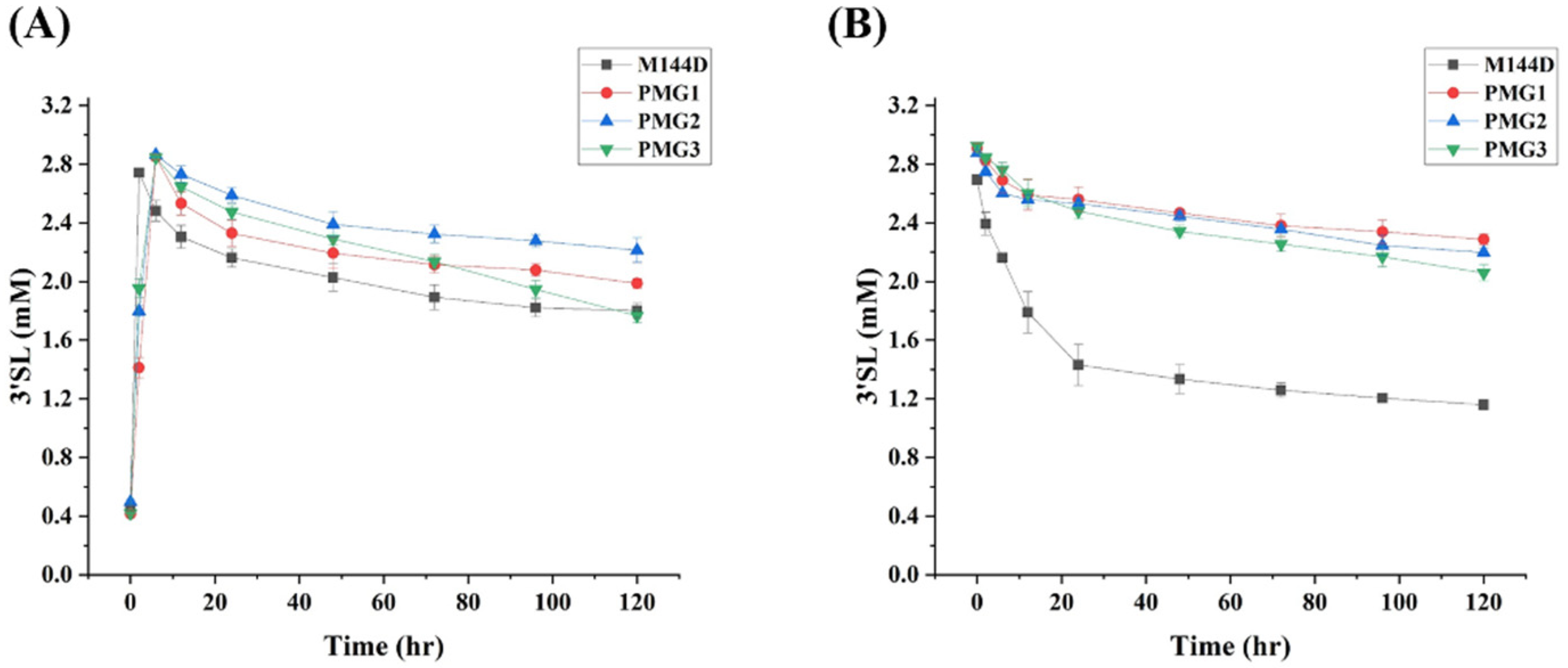
First, their α2,3-sialyltransferase and α2,3-sialidase activities were quickly analyzed by TLC. As shown in Figure S1 (in the Supplementary Materials), three double mutants, just like M144D, lactose was completely consumed very quickly, demonstrating that their α2,3-sialyltransferase activities were not influenced greatly. In particular, the formed 3′-SL product was not obviously hydrolyzed even after 120 h, indicating that residual α2,3-sialidase activity of M144D was further decreased (see Figure S1 in the Supplementary Materials). The significant reduction of the α2,3-sialidase activities of the three double mutants was further proved by incubating three double mutants with 3′-SL respectively at pH 5.5, and no obvious 3′-SL hydrolysis was observed (see Figure S2 in the Supplementary Materials). The α2,3-sialyltransferase and α2,3-sialidase activities of three double mutants were also characterized by following the changes of 3′-SL over time based using MS analysis (Figure 4). It seems that their α2,3-sialyltransferase activities were not impacted too much, but their α2,3-sialidase activities decreased drastically.
In order to fully understand their performance and the underlying mechanisms of mutations of PmST1 in relation to the changes in activities, the kinetic parameters of their α2,3-sialyltransferase activities were determined in detail by monitoring changes of 3′-SL over time based on MS analysis. As shown in Table 3, compared with M144D, the Km values of PMG1 and PMG2 towards lactose and CMP-Neu5Ac were almost not influenced, whereas those of PMG3 for lactose and CMP-Neu5Ac were reduced by 44.2% and 71.5%, respectively. These results demonstrated that the binding affinities of PMG3 for lactose and CMP-Neu5Ac were enhanced, and those of PMG1 and PMG2 towards lactose and CMP-Neu5A did not change. In addition, in comparison with M144D, the kcat value of PMG1 was affected slightly, while that of PMG2 was improved by 38.2 and that of PMG3 decreased by 48.8%. The kcat values of PMG1, PMG2 and PMG3 towards CMP-Neu5Ac were reduced by 23.7%, 28.2% and 66.4%, respectively, compared with that of M144D. Taken together, the α2,3-sialyltransferase catalytic efficiencies (kcat/Km) of PMG1 and PMG2 towards lactose were increased by 2.3% and 38.8%, respectively, while that of PMG3 was decreased by 8.5%. The α2,3-sialyltransferase catalytic efficiencies (kcat/Km) of PMG1 and PMG2 against CMP-Neu5Ac were reduced by 18.0% and 30.7%, respectively, whereas that of PMG3 was raised by 16.4%.
Their α2,3-sialidase activities were also kinetically characterized by using 3′-SL as the substrate. As shown in Table 4, the Km values of PMG1, PMG2 and PMG3 decreased by 14.6, 47.6% and 68.9%, respectively, compared with that of M144D, whereas their kcat values were reduced by 87.5%, 87% and 91%, respectively. Therefore, in comparison with M144D, their binding affinities towards 3′-SL were lower and their α2,3-sialidase activities drastically decreased. All in all, their α2,3-sialidase catalytic efficiencies (kcat/Km) were reduced by 85%, 75% and 79.8%, respectively.
Based on the above kinetic data, it appears that synergistic effects of α2,3-sialidase activities for three double mutants obviously occurred. Given the fact that the α2,3-sialyltransferase activity of M144D was lower than WT PmST1 [25], the additional mutations of R313Y and R313H contributed greatly to the improved α2,3-sialyltransferase activities of PMG1 and PMG2, consistent with the reported results for R313Y and R313H [27].

2.4. Design, Construction and Characterization of Second Generation of Mutants

According to the kinetic parameters of three double mutants, previous studies and comprehensive consideration, four triple mutants PMG2-1 (M144D/R313H/T265S), PMG2-2 (M144D/R313H/E271F), PMG3-1 (M144D/R313N/T265S) and PMG3-2 (M144D/R313N/E271F) were designed, constructed and characterized (Table 2).
First, α2,3-sialidase and α2,3-sialyltransferase activities of the four triple mutants were quickly analyzed by TLC (see Figures S3–S6 in the Supplementary Materials). It is clear that α2,3-sialidase activities for the four triple mutants were still not observed (see Figures S4 and S6 in the Supplementary Materials). However, it seems that their α2,3-sialyltransferase activities were affected to some extent, since residual lactose was still found for these four mutants even after 120 h (see Figures S3 and S5 in the Supplementary Materials). Therefore, two triple mutants PMG2-1 and PMG2-2 were selected for detailed kinetic characterization.
According to Table 3, it seems that α2,3-sialyltransferase activities of PMG2-1 and PMG2-2 towards lactose and CMP-Neu5Ac were reduced in comparison with those of both PMG2 and M144D. Unexpectedly, their α2,3-sialidase activities were not further lowered compared with that of PMG2, though they showed lower α2,3-sialidase activities than M144D. No obvious synergistic effects of α2,3-sialidase activities were observed for two triple mutants. It was presumed that triple mutations in PMG2-1, PMG2-2, PMG3-1 and PMG3-2 might lead to a big impact on the local environment of the substrate-binding pocket, and accordingly both α2,3-sialidase and α2,3-sialyltransferase activities were affected. A crystal structure for triple mutants might help to explain the results.

2.5. Preparative-Scale Synthesis of 3′-SL Using PMG2

Given the fact that triple mutants showed reduced α2,3-sialyltransferase activities and slightly increased α2,3-sialidase, they are not appropriate for preparative-scale synthesis of 3′-SL. Therefore, PMG2 was chosen for this.
A one-pot two-enzyme α2,3-sialylation system was adopted for introducing α2,3-linked sialic acid (N-acetylneuraminic acid, Neu5Ac) onto the galactose unit of lactose (Scheme 1), where Neu5Ac was converted into CMP-Neu5Ac in the presence of cytidine 5′-triphosphate (CTP) by a recombinant CMP-Neu5Ac synthetase from Neisseria meningitides (NmCSS), and CMP-Neu5Ac as a donor was transferred onto the lactose acceptor to form 3′-SL by an α2,3-sialyltransferase (PMG2), with a yield of around 95% after convenient purification by gel filtration. 3′-SL was characterized by MS and NMR, consistent with the reported analysis.

3. Materials and Methods

3.1. General Information

Unless otherwise stated, chemicals were purchased and used without further purification. Gel filtration chromatography was performed using a column (100 cm × 2.5 cm) packed with BioGel P-2 Fine resins (Bio-Rad, Hercules, CA, USA). 1H and 13C NMR spectra were recorded on a Bruker AVANCE-600 spectrometer at 25 °C. ESI (Electrospray ionization) mass spectra were obtained at the Institute of Process Engineering, Chinese Academy of Sciences.
Neisseria meningitides CMP-sialic acid synthetase (NmCSS) and the Pasteurella multocida α2,3-sialyltransferase 1 M144D mutant (PmST1 M144D) [25] were expressed in E. coli BL21 (DE3) and purified on nickel-chelating resin as reported in the literature. Codon-optimized genes encoding NmCSS and PmST1 M144D were cloned into pET-22b (with C-terminal His6 tag) and pET-23a (with C-terminal His6 tag), respectively. In order to eliminate the possibility of protein contamination, the nickel column was first eluted with the binding buffer containing 0.5 M imidazole before it was used for purifying a mutant.

3.2. Site-Directed Mutagenesis, Expression and Purification of PmST1 M144D Mutants

According to the sequence of PmST1 M144D, primers for site-directed mutations of Aug313 (R313Y, R313H and R313N), Glu271 and Thr265 were respectively designed, as shown in Table 5. All site-directed mutants were constructed according to the standard QuikChange Site-Directed Mutagenesis protocol. Mutants were expressed and purified as for the previously described wild-type PmST1 [9].

3.3. Analysis of α2,3-Sialyltransferase and α2,3-Sialidase Activity of PmST1 M144D Mutants by TLC

The reaction mixture (800 μL) containing Tris-HCl (100 mM, pH = 8.5), 20 mM MgCl2, 3 mM lactose, 3.6 mM Neu5Ac and 3.6 mM CTP was incubated with NmCSS (0.2 mg/mL) and a variant enzyme (0.25 mg/mL) at 37 °C for different periods. At different time points, 25 μL of the sample was transferred into an Eppendorf tube, and 25 μL cold ethanol was added. The mixture was kept at 4 °C for 30 min, then centrifuged at 12,000 rpm for 10 min and the precipitates were removed. The supernatant was analyzed by TLC (EtOAc:MeOH:H2O:HOAc = 4:2:1:0.2, v/v) and stained with 10% (v/v) H2SO4 in ethanol. The intensity of each spot on TLC plates was quantified using grayscale analysis in ImageJ software (NIH, Bethesda, MD, USA).

3.4. Kinetics of the α2,3-Sialyltransferase Activity of PmST1 M144D Mutants by LC-MS Analysis

The reaction mixture (25 μL) containing Tris-HCl (100 mM, pH = 8.5) and 20 mM MgCl2 was incubated at 37 °C for 10 min with an enzyme (5 μg/mL), a fixed concentration of CMP-Neu5Ac (6 mM) and different concentrations (0.5, 1, 2, 5, 10, 20, 30, 40 and 50 mM) of Lac. Alternatively, the reaction was performed at 37 °C for 5 min, with a fixed concentration of Lac (3 mM) and different concentrations of CMP-Neu5Ac (0.1, 0.5, 1, 2, 5, 10 and 15 mM).
All reactions were performed in triplicate. The reactions were stopped by adding 25 μL prechilled ethanol. The mixtures were incubated on ice for 30 min and centrifuged at 12,000 rpm for 10 min. The supernatants were collected in a sample bottle and analyzed by LC-MS on a 2.5 μm XBridge BEH Amide column (2.1 mm × 150 mm). The HPLC conditions are as follows: flow rate: 0.3 mL/min; detection duration: 30 min; A: H2O; B: acetonitrile; C: 100 mM ammonium formate (pH = 3.2). The C phase was fixed at 10%, and the B phase was decreased from 85% to 50% for 30 min and increased to 85% from 35 min to 35.5 min. 3′-SL was detected at m/z = 632.55. The average values were obtained from repeated measurement results, which were fitted to the Michaelis–Menten equation to obtain the apparent kinetic parameters.

3.5. Kinetics of the α2,3-Sialidase Activity of PmST1 M144D Mutants

The reaction mixture (25 μL) containing MES buffer (100 mM, pH = 5.5) and 20 mM MgCl2 was incubated at 37 °C for 10 min with an enzyme (1 mg/mL) and different concentrations (1, 2, 5, 10, 20, 40 and 50 mM) of 3′-SL. All reactions were performed in triplicate. Sample treatment after the reaction and analysis was carried out by LC-MS similarly as described above for the α2,3-sialyltransferase assays.

3.6. Preparative-scale Synthesis of 3′-SL Using PMG2

The reaction mixture (100 mL) of Tris-HCl (100 mM, pH = 8.5), 20 mM MgCl2, 0.2 mg/mL NmCSS, 0.25 mg/mL PMG2, 3 mM Lac, 3.6 mM Neu5Ac and 3.6 mM CTP was incubated at 37 °C for 6 h. The reactions were stopped by adding an equal volume of prechilled ethanol. The mixtures were incubated on ice for 30 min and centrifuged at 12,000 rpm for 20 min. 3-′SL was purified by silica gel column chromatography, which was eluted with an eluent (ethyl acetate:methanol:water = 7:2:1, v/v). Purified 3′-SL was analyzed by NMR and MS. 3′-SL (yield > 95%), white solid after lyophilization. 1H NMR 600 MHz (D2O) δ 5.14 (d, J = 3.6 Hz, 1H), 4.59 (d, J = 4.8 Hz, 1H), 4.45 (d, J = 8.4 Hz, 1H), 4.04 (m, 1H), 3.90–3.45 (m, 17H), 3.21 (t, J = 7.8 Hz, 1H), 2.69 (dd, J = 13.2 and 4.8 Hz, 1H)1.96 (s, 3H, Ac), 1.73 (t, J = 12.0 Hz, 1H); 13C NMR (600 MHz, D2O) δ 175.00, 173.80, 102.63, 99.79, 95.77, 91.82, 78.19, 75.16, 74.80, 74.32, 73.80, 72.87, 72.04, 71.76, 69.36, 68.34, 62.48, 61.03, 60.42, 59.80, 51.68, 39.64, 23.26. MS (ESI) m/z calcd for C23H40NO19 [M−H] 632.55, found 632.25.

4. Conclusions

In conclusion, based on previous studies, α2,3-sialidase activity of PmST1 was further decreased through a rational protein engineering strategy without greatly affecting α2,3-sialyltransferase activity. The new double mutant PMG2 (M144D/R313H) identified was successfully applied for synthesis of 3′-SL with high yield and efficiency and would be very useful for production of α2,3-linked sialosides.

Supplementary Materials

The following supporting information can be downloaded at: https://www.mdpi.com/article/10.3390/catal12060579/s1, Figure S1. Analysis of α2,3-sialyltransferase activities of PMG1, PMG2 and PMG3 by TLC. Figure S2. Analysis of α2,3-sialidase activities of PMG1, PMG2 and PMG3 by TLC. Figure S3. Analysis of α2,3-sialyltransferase activities of PMG2-1 and PMG2-2 by TLC. Figure S4. Analysis of α2,3-sialidase activities of PMG2-1 and PMG2-2 by TLC. Figure S5. Analysis of α2,3-sialyltransferase activities of PMG3-1 and PMG3-2 by TLC. Figure S6. Analysis of α2,3-sialidase activities of PMG3-1 and PMG3-2 by TLC. Figure S7. SDS-PAGE analysis of overexpressed and purified PmST1 M144D. Figure S8. SDS-PAGE analysis of overexpressed and purified PMG1. Figure S9. SDS-PAGE analysis of overexpressed and purified PMG2. Figure S10. SDS-PAGE analysis of overexpressed and purified PMG3. Figure S11. SDS-PAGE analysis of overexpressed and purified PMG2-1. Figure S12. SDS-PAGE analysis of overexpressed and purified PMG2-2.

Author Contributions

Conceptualization, J.L., Y.D., M.P. and Z.Z.; Methodology, validation, investigation, formal analysis, data curation, M.G., R.Y., S.J., J.H. and C.F.; Writing—original draft preparation, M.G., R.Y. and J.L.; Writing—review and editing, M.G., R.Y. and J.L.; Supervision, J.L., Y.D., M.P. and Z.Z.; Project administration, J.L.; Funding acquisition, Y.D. All authors have read and agreed to the published version of the manuscript.

Funding

This research was funded by National Natural Science Foundation of China (grant number 21877114).

Conflicts of Interest

The authors declare no conflict of interest.

References

  1. Varki, A. Sialic acids in human health and disease. Trends Mol. Med. 2008, 14, 351–360. [Google Scholar] [CrossRef] [PubMed] [Green Version]
  2. Varki, A. Glycan-based interactions involving vertebrate sialic-acid-recognizing proteins. Nature 2007, 446, 1023–1029. [Google Scholar] [CrossRef] [PubMed]
  3. Wang, B. Sialic Acid Is an Essential Nutrient for Brain Development and Cognition. Annu. Rev. Nutr. 2009, 29, 177–222. [Google Scholar] [CrossRef] [PubMed]
  4. Perdijk, O.; van Baarlen, P.; Fernandez-Gutierrez, M.M.; van den Brink, E.; Schuren, F.H.J.; Brugman, S.; Savelkoul, H.F.J.; Kleerebezem, M.; van Neerven, R.J.J. Sialyllactose and Galactooligosaccharides Promote Epithelial Barrier Functioning and Distinctly Modulate Microbiota Composition and Short Chain Fatty Acid Production In Vitro. Front. Immunol. 2019, 10, 94. [Google Scholar] [CrossRef] [PubMed] [Green Version]
  5. Moore, R.E.; Xu, L.Y.L.; Townsend, S.D. Prospecting Human Milk Oligosaccharides as a Defense Against Viral Infections. Acs Infect. Dis. 2021, 7, 254–263. [Google Scholar] [CrossRef]
  6. Kunz, C.; Rudloff, S.; Baier, W.; Klein, N.; Strobel, S. Oligosaccharides in human milk: Structural, functional, and metabolic aspects. Annu. Rev. Nutr. 2000, 20, 699–722. [Google Scholar] [CrossRef]
  7. Imai, M.; Kawaoka, Y. The role of receptor binding specificity in interspecies transmission of influenza viruses. Curr. Opin. Virol. 2012, 2, 160–167. [Google Scholar] [CrossRef] [Green Version]
  8. Izumi, M.; Shen, G.J.; Wacowich-Sgarbi, S.; Nakatani, T.; Plettenburg, O.; Wong, C.H. Microbial glycosyltransferases for carbohydrate synthesis: Alpha-2,3-sialyltransferase from Neisseria gonorrheae. J. Am. Chem. Soc. 2001, 123, 10909–10918. [Google Scholar] [CrossRef]
  9. Yu, H.; Chokhawala, H.; Karpel, R.; Yu, H.; Wu, B.Y.; Zhang, J.B.; Zhang, Y.X.; Jia, Q.; Chen, X. A multifunctional Pasteurella multocida sialyltransferase: A powerful tool for the synthesis of sialoside libraries. J. Am. Chem. Soc. 2005, 127, 17618–17619. [Google Scholar] [CrossRef]
  10. Chokhawala, H.A.; Huang, S.S.; Lau, K.; Yu, H.; Cheng, J.S.; Thon, V.; Hurtado-Ziola, N.; Guerrero, J.A.; Varki, A.; Chen, X. Combinatorial chemoenzymatic synthesis and high-through put screening of sialosides. Acs Chem. Biol. 2008, 3, 567–576. [Google Scholar] [CrossRef] [Green Version]
  11. Cao, H.Z.; Li, Y.H.; Lau, K.; Muthana, S.; Yu, H.; Cheng, J.S.; Chokhawala, H.A.; Sugiarto, G.; Zhang, L.; Chen, X. Sialidase substrate specificity studies using chemoenzymatically synthesized sialosides containing C5-modified sialic acids. Org. Biomol. Chem. 2009, 7, 5137–5145. [Google Scholar] [CrossRef] [PubMed]
  12. Drouillard, S.; Mine, T.; Kajiwara, H.; Yamamoto, T.; Samain, E. Efficient synthesis of 6′-sialyllactose, 6,6′-disialyllactose, and 6′-KDO-lactose by metabolically engineered E. coli expressing a multifunctional sialyltransferase from the Photobacterium sp JT-ISH-224. Carbohydr. Res. 2010, 345, 1394–1399. [Google Scholar] [CrossRef] [PubMed]
  13. Schelch, S.; Zhong, C.; Petschacher, B.; Nidetzky, B. Bacterial sialyltransferases and their use in biocatalytic cascades for sialo-oligosaccharide production. Biotechnol. Adv. 2020, 44, 107613. [Google Scholar] [CrossRef]
  14. Fierfort, N.; Samain, E. Genetic engineering of Escherichia coli for the economical production of sialylated oligosaccharides. J. Biotechnol. 2008, 134, 261–265. [Google Scholar] [CrossRef] [PubMed]
  15. Yu, H.; Yu, H.; Karpel, R.; Chen, X. Chemoenzymatic synthesis of CMP-sialic acid derivatives by a one-pot two-enzyme system: Comparison of substrate flexibility of three microbial CMP-sialic acid synthetases. Bioorg. Med. Chem. 2004, 12, 6427–6435. [Google Scholar] [CrossRef] [PubMed]
  16. Shen, G.J.; Datta, A.K.; Izumi, M.; Koeller, K.M.; Wong, C.H. Expression of alpha 2,8/2,9-polysialyltransferase from Escherichia coli K92—Characterization of the enzyme and its reaction products. J. Biol. Chem. 1999, 274, 35139–35146. [Google Scholar] [CrossRef] [Green Version]
  17. Willis, L.M.; Gilbert, M.; Karwaski, M.F.; Blanchard, M.C.; Wakarchuk, W.W. Characterization of the alpha-2,8-polysialyltransferase from Neisseria meningitidis with synthetic acceptors, and the development of a self-priming polysialyltransferase fusion enzyme. Glycobiology 2008, 18, 177–186. [Google Scholar] [CrossRef] [Green Version]
  18. Gilbert, M.; Brisson, J.R.; Karwaski, M.F.; Michniewicz, J.; Cunningham, A.M.; Wu, Y.Y.; Young, N.M.; Wakarchuk, W.W. Biosynthesis of ganglioside mimics in Campylobacter jejuni OH4384—Identification of the glycosyltransferase genes, enzymatic synthesis of model compounds, and characterization of nanomole amounts by 600-MHz H-1 and C-13 NMR analysis. J. Biol. Chem. 2000, 275, 3896–3906. [Google Scholar] [CrossRef] [Green Version]
  19. Fox, K.L.; Cox, A.D.; Gilbert, M.; Wakarchuk, W.W.; Li, J.J.; Makepeace, K.; Richards, J.C.; Moxon, E.R.; Hood, D.W. Identification of a bifunctional lipopolysaccharide sialyltransferase in Haemophilus influenzae—Incorporation of disialic acid. J. Biol. Chem. 2006, 281, 40024–40032. [Google Scholar] [CrossRef] [Green Version]
  20. Gilbert, M.; Watson, D.C.; Cunningham, A.M.; Jennings, M.P.; Young, N.M.; Wakarchuk, W.W. Cloning of the lipooligosaccharide alpha-2,3-sialyltransferase from the bacterial pathogens Neisseria meningitidis and Neisseria gonorrhoeae. J. Biol. Chem. 1996, 271, 28271–28276. [Google Scholar] [CrossRef] [Green Version]
  21. Kaniuk, N.A.; Monteiro, M.A.; Parker, C.T.; Whitfield, C. Molecular diversity of the genetic loci responsible for lipopolysaccharide core oligosaccharide assembly within the genus Salmonella. Mol. Microbiol. 2002, 46, 1305–1318. [Google Scholar] [CrossRef] [PubMed] [Green Version]
  22. Yamamoto, T.; Nakashizuka, M.; Terada, I. Cloning and expression of a marine bacterial beta-galactoside alpha 2,6-sialyltransferase gene from Photobacterium damsela JT0160. J. Biochem. Tokyo 1998, 123, 94–100. [Google Scholar] [CrossRef] [PubMed]
  23. Takakura, Y.; Tsukamoto, H.; Yamamoto, T. Molecular cloning, expression and properties of an alpha/beta-galactoside alpha 2,3-sialyltransferase from Vibrio sp JT-FAJ-16. J. Biochem. Tokyo 2007, 142, 403–412. [Google Scholar] [CrossRef]
  24. Tsukamoto, H.; Takakura, Y.; Yamamoto, T. Purification, cloning, and expression of an alpha/beta-galactoside alpha-2,3-sialyltransferase from a luminous marine bacterium, Photobacterium phosphoreum. J. Biol. Chem. 2007, 282, 29794–29802. [Google Scholar] [CrossRef] [PubMed] [Green Version]
  25. Sugiarto, G.; Lau, K.; Qu, J.Y.; Li, Y.H.; Lim, S.; Mu, S.; Ames, J.B.; Fisher, A.J.; Chen, X. A Sialyltransferase Mutant with Decreased Donor Hydrolysis and Reduced Sialidase Activities for Directly Sialylating Lewis(X). Acs Chem. Biol. 2012, 7, 1232–1240. [Google Scholar] [CrossRef] [PubMed] [Green Version]
  26. Sugiarto, G.; Lau, K.; Li, Y.H.; Khedri, Z.; Yu, H.; Le, D.T.; Chen, X. Decreasing the sialidase activity of multifunctional Pasteurella multocida alpha 2-3-sialyltransferase 1 (PmST1) by site-directed mutagenesis. Mol. Biosyst. 2011, 7, 3021–3027. [Google Scholar] [CrossRef]
  27. Choi, Y.H.; Kim, J.H.; Park, J.H.; Lee, N.; Kim, D.H.; Jang, K.S.; Park, I.H.; Kim, B.G. Protein engineering of alpha 2,3/2,6-sialyltransferase to improve the yield and productivity of in vitro sialyllactose synthesis. Glycobiology 2014, 24, 159–169. [Google Scholar] [CrossRef] [Green Version]
Scheme 1. One-pot two-enzyme synthesis of 3′-sialyllactose (3′-SL). NmCSS: Neisseria meningitidis CMP-sialic acid synthetase; PmST1: Pasteurella multocida α2,3-sialyltransferase.
Catalysts 12 00579 sch001
Scheme 2. Enzymatic hydrolysis of 3′-sialyllactose (3′-SL) by PmST1.
Catalysts 12 00579 sch002
Figure 1. Analysis of α2,3-sialyltransferase (A) and α2,3-sialidase (B) activities of M144D by TLC. Lane 1, Lac; Lane 2, 0 h; Lane 3, 2 h; Lane 4, 6 h; Lane 5, 10 h; Lane 6, 24 h; Lane 7, 48 h; Lane 8, 72 h; Lane 9, 96 h; Lane 10, 120 h; Lane 11, S’-SL.
Catalysts 12 00579 g001
Figure 2. Analysis of α2,3-sialyltransferase (A) and α2,3-sialidase (B) activities of M144D by MS. For the α2,3-sialyltransferase activity assay, the reaction mixture (800 μL) containing Tris-HCl (100 mM, pH = 8.5), 20 mM MgCl2, 3 mM lactose, 3.6 mM Neu5Ac and 3.6 mM CTP was incubated with NmCSS (0.2 mg/mL) and a variant enzyme (0.25 mg/mL) at 37 °C for different periods. For the α2,3-sialidase activity assay, the reaction mixture (800 μL) containing MES buffer (100 mM, pH = 5.5) and 20 mM MgCl2 was incubated with 3 mM 3′-SL, a variant enzyme (2.5 mg/mL), at 37 °C for different periods. At different time points, 25 μL of the sample was transferred into an Eppendorf tube, and 25 μL cold ethanol was added. The mixture was kept at 4 °C for 30 min, then centrifuged at 12,000 rpm for 10 min and the precipitates were removed. The supernatants were used for analysis. All reactions were done in triplicate.
Catalysts 12 00579 g002
Figure 3. Some of the residues involved in substrate binding. Met144, Arg313, Glu271 and Thr265 in magenta are yet to be investigated. The catalytic residues Asp141 and His311 are in yellow and red. Lactose is in cyan and CMP-Neu5Ac in green.
Catalysts 12 00579 g003
Figure 4. Comparison of α2,3-sialyltransferase (A) and α2,3-sialidase (B) activities of M144D, PMG1, PMG2 and PMG3. For the α2,3-sialyltransferase activity assay, the reaction mixture (800 μL) containing Tris-HCl (100 mM, pH = 8.5), 20 mM MgCl2, 3 mM lactose, 3.6 mM Neu5Ac and 3.6 mM CTP was incubated with NmCSS (0.2 mg/mL) and a variant enzyme (0.25 mg/mL) at 37 °C for different periods. For the α2,3-sialidase activity assay, the reaction mixture (800 μL) containing MES buffer (100 mM, pH = 5.5) and 20 mM MgCl2 was incubated with 3 mM 3′-SL, a variant enzyme (2.5 mg/mL), at 37 °C for different periods. At different time points, 25 μL of the sample was transferred into an Eppendorf tube, and 25 μL cold ethanol was added. The mixture was kept at 4 °C for 30 min, then centrifuged at 12,000 rpm for 10 min and the precipitates were removed. The supernatants were used for analysis. All reactions were done in triplicate.
Catalysts 12 00579 g004
Table 1. Summary of effects of different mutations on ST and sialidase activity in previous studies.
Variantsα2,3-Sialyltransferaseα2,3-Sialidase
M144D [25]a 94%↓99.98%
R313Y [27]b 25%↓83%
R313H [27]↑46%↓60%
R313N [27]↑131%↓33%
E271F [26]↑15%↓98%
T265S [27]↑16%Not determined
R313N/T265S [27]↑116%↓93%
R313H/T265S [27]↑137%↓90%
a↓: Reduced; b↑: Decreased.
Table 2. All variants and their corresponding mutations in the current study.
VariantsMutations
PGM1M144D/R313Y
PGM2M144D/R313H
PGM3M144D/R313N
PGM2-1M144D/R313H/T265S
PGM2-2M144D/R313H/E271F
PGM3-1M144D/R313N/T265S
PGM3-2M144D/R313N/E271F
Table 3. Apparent kinetics of the α2,3-sialyltransferase activities of M144D and mutants.
VariantsLacCMP-Neu5Ac
Km (mM)M144D19.7 ± 1.81.93 ± 0.17
PMG117.4 ± 1.51.60 ± 0.14
PMG219.6 ± 1.91.97 ± 0.19
PMG311.0 ± 0.90.55 ± 0.041
PMG2-19.7 ± 0.80.91 ± 0.079
PMG2-222.7 ± 2.10.80 ± 0.064
kcat (s−1)M144D25.4 ± 2.24.64 ± 0.039
PMG123.0 ± 1.73.54 ± 0.032
PMG235.1 ± 2.63.33 ± 0.028
PMG313.0 ± 1.11.56 ± 0.013
PMG2-18.97 ± 0.71.70 ± 0.013
PMG2-218.7 ± 1.51.75 ± 0.012
kcat/Km (s−1 mM−1)M144D1.29 ± 0.0892.44 ± 0.21
PMG11.32 ± 0.0832.20 ± 0.18
PMG21.79 ± 0.0121.69 ± 0.15
PMG31.18 ± 0.0112.84 ± 0.23
PMG2-10.93 ± 0.091.88 ± 0.17
PMG2-20.82 ± 0.0762.20 ± 0.19
Table 4. Apparent kinetics of the α2,3-sialidase activities of M144D and mutants.
Variants3′-SL
Km (mM)M144D16.4 ± 1.57
PMG114.0 ± 1.1
PMG28.6 ± 0.73
PMG35.1 ± 0.46
PMG2-126.0 ± 2.2
PMG2-232.8 ± 2.8
kcat (s−1)M144D2.0 ± 0.19
PMG10.25 ± 0.021
PMG20.26 ± 0.023
PMG30.18 ± 0.016
PMG2-12.25 ± 0.17
PMG2-22.05 ± 0.19
kcat/Km (s−1 mM−1)M144D0.120 ± 0.011
PMG10.018 ± -0.0014
PMG20.030 ± 0.0024
PMG30.035 ± 0.0029
PMG2-10.086 ± 0.0083
PMG2-20.063 ± 0.0057
Table 5. Primer sequences for different site-directed mutations.
GenePrimerSequence (5′→3′)
PmST1 M144DR313YFCAAGGGTCATCCGTACGGTGGCGAAATTAATG
R313YRCATTAATTTCGCCACCGTACGGATGACCCTTG
R313HFCAAGGGTCATCCGCACGGTGGCGAAATTAATG
R313HRCATTAATTTCGCCACCGTGCGGATGACCCTTG
R313NFCAAGGGTCATCCGAACGGTGGCGAAATTAATG
R313NRCATTAATTTCGCCACCGTTCGGATGACCCTTG
PMG2 (M144D/R313H)/PMG3 (M144D/R313N)T265S FGCAGGCAAAATTCATTTTTAGCGGCACCACCACC
T265S RGGTGGTGGTGCCGCTAAAAATGAATTTTGCCTGC
E271FFACCGGCACCACCACCTGGTTTGGCAATACCGATG
E271FRTTCGCGCACATCGGTATTGCCAAACCAGGTGGTGGTG
Publisher’s Note: MDPI stays neutral with regard to jurisdictional claims in published maps and institutional affiliations.