Article

In Situ Partial Sulfidation for Preparing Cu/Cu2−xS Core/Shell Nanorods with Enhanced Photocatalytic Degradation

1
Key Laboratory of Artificial Micro- and Nano-Structures of the Ministry of Education, Hubei Nuclear Solid Physics Key Laboratory, School of Physics and Technology, Wuhan University, Wuhan 430072, China
2
School of Education, Jiangxi Science and Technology Normal University, Nanchang 330000, China
*
Authors to whom correspondence should be addressed.
Catalysts 2022, 12(2), 147; https://doi.org/10.3390/catal12020147
Submission received: 26 December 2021 / Revised: 17 January 2022 / Accepted: 18 January 2022 / Published: 25 January 2022
(This article belongs to the Special Issue Plasmon-Assisted Photocatalysis in Hybrid Nanoparticles)

Abstract

:
Herein, we report an approach to prepare Cu/Cu2−xS core/shell nanorods by in situ sulfidation of copper nanorods. Firstly, copper nanorods with tunable longitudinal surface plasmon resonances were synthesized by a seed-mediated method using Au nanoparticles as seeds. A convenient in situ sulfidation method was then applied to convert the outermost layer of Cu nanorods into Cu2−xS, to increase their stability and surface activity in photocatalytic applications. The thickness of Cu2−xS layer can be adjusted by controlling the amount of S source. The Cu/Cu2−xS core/shell nanorods exhibits two characteristic surface plasmon resonances located in visible and near-infrared regions, respectively. The photocatalytic performances of Cu nanorods and their derivatives were evaluated by measuring the degradation rate of methyl orange dyes. Compared with Cu nanorods, the Cu/Cu2−xS core/shell nanorods demonstrate more than a 13.6-fold enhancement in the degradation rate at 40 min. This work suggests a new direction for constructing derivative nanostructures of copper nanorods and exploring their applications.

1. Introduction

In the past few decades, one-dimensional, metal nanomaterials with surface plasmon resonances (SPRs) have attracted intense attention because of their unique properties [1,2,3,4,5]. In particular, the preparation process of nanorods (NRs) with gold or silver as the main component is very well-developed. On this basis, the growth of semiconductor components on plasmonic NRs forms metal–semiconductor heterogeneous nanostructures with a fascinating interaction between the plasmon and semiconductor [6,7,8,9]. These nanostructured materials show promising potentials in the applications of energy devices, photocatalytic hydrogen production, second-harmonic generation enhancement and ultrafast, optical, nonlinear properties [9,10].
Compared with gold and silver, copper is an abundant and low-cost metal resource. Nanostructured copper and its derivative compounds have many applications in catalytic degradation, hydrogen production and surface-enhanced Raman scattering (SERS) [11,12]. Nanoscale copper has a strong tendency to oxidize [1], and its preparation process is very difficult. In recent years, with the attempts of many scholars, the synthesis of one-dimensional copper NRs made great progress. Precious metal nanoparticles (Au, Pt, Pd) were used as seeds, and bimetallic copper NRs were prepared by reacting different amine reducing agents at a high temperature under an argon atmosphere [2]. Wang and colleagues reported that single-crystal Au nanoparticles were used as seeds to induce the local epitaxial growth of copper shells in the water phase, and the prepared NRs have thick centers and thin ends due to anisotropic growth [5]. Luo and colleagues used Pd decahedrons as seeds to synthesize Cu NRs with a uniform diameter and strong stability in the water phase, and the Pd seeds are located at one end of the asymmetric Cu NRs [13]. Recently, Jeong et al. reported a seed-mediated method for synthesizing penta-twinned Cu NRs in the oil phase using Au nanocrystals as seeds [1]. The synthesized Cu NRs with a high purity have a uniform diameter and tunable aspect ratio in a wide range.
The shortcoming of oxidation also hinders the applications that use Cu nanocrystals. Especially in catalytic reactions, the stability of Cu nanocrystals is very poor. In order to address this issue, the preparation of Cu derivatives based on Cu nanocrystals made considerable progress [14,15,16]. Oxidation or sulfidation could produce a protective shell to improve the stability of Cu nanocrystals. The combination of Cu nanocrystals with newly formed derivatives, such as CuxO and CuxS, could achieve excellent catalytic performances. For instance, Xia and colleagues demonstrated a controlled surface oxidation of Cu nanowires, improving their catalytic selectivity and stability toward C2+ products in CO2 reduction [17]. A copper-based catalyst Cu2O-Cu, supported on carbon microspheres, exhibits Fenton-like, catalytic properties for organic dyes [18], providing a simple and cost-effective method for the catalytic degradation of organic dyes. Moreover, the catalytic performance of semiconductor nanomaterials can be further improved by doping copper nanocrystals and their derivatives. For example, doping copper or copper oxide into a zinc oxide nanostructure can delay the recombination of electrons and holes, broaden the absorption spectrum, promote certain specific reactions on the catalyst surface, and improve photocatalytic activity [19,20,21,22]. These copper compounds (copper oxide and copper sulfide) can absorb light energy under visible light illumination, causing holes (h+) in the valence band (VB) and electrons (e) in the conduction band (CB) for catalytic reactions [23,24,25]. It is attractive that copper has two oxide states of Cu(II) and Cu(I), and they combine with H2O2 in a Fenton-like process to generate hydroxyl radicals (·OHs) and other reactive groups [26,27,28,29,30], which assist the reaction [31,32,33].
Here, a seed-mediated method using Au nanoparticles as seeds was used to synthesize Cu NRs with tunable longitudinal SPRs (LSPRs) ranging from 740 nm to 1190 nm. Subsequently, Cu NRs were partially sulfurized by mixing with an ethanol solution of sodium hydrosulfide. The hetero-nanostructures were carefully characterized to reveal structural features. The photocatalytic performance of Cu/Cu2−xS NRs was evaluated via the degradation of methyl orange (MO). The results in this work provide new insight into exploring the preparation and application of Cu nanostructures and their derivatives.

2. Results and Discussion

The synthesis strategy of Cu/Cu2−xS NRs is schematically illustrated in Figure 1. Cu NRs were prepared by a seed-mediated growth method, using oleylamine as both a solvent and reducing agent. To protect Cu from oxidation, nitrogen gas blowing was maintained in the whole synthesis process. Heating a mixture solution of copper chloride and oleylamine to 180 °C reduced Cu2+ to Cu+, and the color of mixture solution was changed from blue to yellow in this process. Au seeds (Figure S1) were added to initiate the growth of Cu NRs, and Cu NRs with penta-twinned structure were obtained after 1 h reaction. After adding the gold seeds, the color of the solution changed from orange–red to red, then to black–red, and finally to brown–yellow within 10 min, indicating the morphology evolution of the Cu nanocrystals.
The length of Cu NRs can be controlled by the amount of Au seeds. Figure 2a illustrates the extinction spectra of Cu NRs with a varied amount of Au seeds. The prepared Cu NRs exhibit two extinction bands, corresponding to the transverse and longitudinal SPRs (TSPRs and LSPRs). When the amount of Au seeds is decreased, the LSPRs located in the near-infrared (NIR) region are red-shifted and can be tuned from 740 to 1190 nm. According to Jeong’s experimental results [1], the aspect ratio of Cu NRs is estimated to range from 2.5 to 5.6. As the length increases, the TSPR is slightly blue-shifted from 576 nm to 571 nm, and the intensity ratio of LSPR/TSPR is increased from 1.04 to 5.25. Two transmission electron microscopy (TEM) images of Cu NRs with LSPRs of 740 and 821 nm are shown in Figure 2b,c. The average length of these two samples is about 44.5 and 57.5 nm, respectively. We note that the Cu NRs exhibit a rough surface, which is caused by the deposition of Cu atoms and surface diffusion during the anisotropic growth of Cu NRs [1]. The rough surface of Cu NRs implies that they have abundant surface hotspots due to localized SPR field enhancements. Meanwhile, the rough structure also creates an active surface caused by the increased surface area and exposed high-index facets.
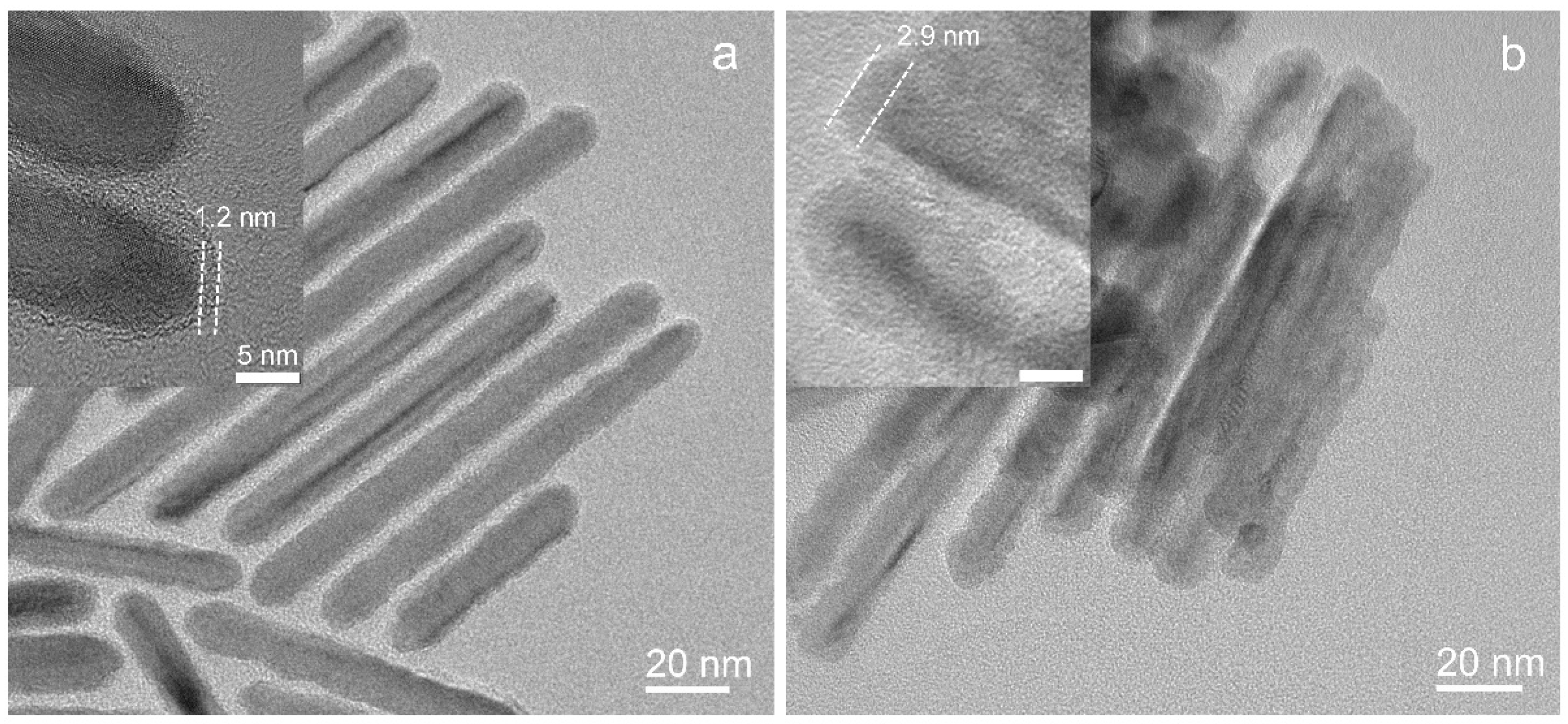
Compared with Au and Ag, Cu is an abundant and inexpensive metal. However, Cu nanocrystals are easily oxidized, especially in aqueous solutions, resulting in: (1) the controlled synthesis of Cu nanostructures remains a challenge, (2) the stability of Cu nanocrystals in catalytic applications is not good. Here, an in situ sulfidation method is applied to prepare a copper sulfide shell coating on Cu NRs. At room temperature, NaHS ethanol solution (1 mM), as a sulfur source, was added into Cu NRs dispersed in toluene. Figure 3 illustrates the extinction spectra of NRs with the addition of the sulfur source. The initial Cu NRs exhibit a LSPR band at 938 nm. When the sulfur source is increased from 50 to 120 μL, the LSPR is red-shifted to 1100 nm, 1140 nm, 1190 nm, with broadened width and decreased intensity, respectively. The TSPR band is also red-shifted from 572 nm to 595 nm. Figure 4 shows the TEM images of sulfurized Cu NRs with varied amounts of sulfur sources. A coating layer is clearly observed on the surface of the Cu NRs. The rough surface is kept after partial sulfidation. In fact, in situ sulfidation could further increase the roughness. The average layer thickness is about 1.2 nm in Figure 4a and 2.9 nm in Figure 4b, respectively. The results indicate the thickness of the sulfide layer increases with the increase in the S source. In experiments, the sulfidation affects the dispersibility of the Cu NRs in toluene, which is caused by the damage of surface capping agents during the sulfidation process. When the amount of sulfur source is further increased to a very high level, the dispersibility of sample becomes poor.
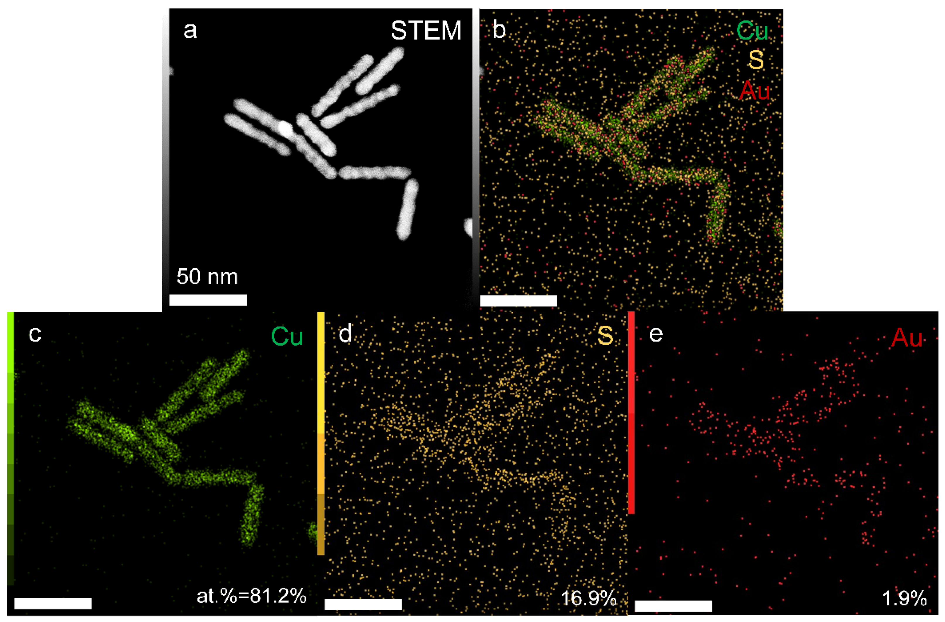
To reveal the composition of the coating layer on Cu NRs, we performed carefully structured characterizations of the sulfurized Cu NRs. Figure 5 shows the energy-dispersive X-ray spectroscopy (EDS) element mapping of sulfurized Cu NRs in scanning TEM (STEM). The feature of rough surface is clearly observed in the STEM image (Figure 5a). The merged image of element mapping shown in Figure 5B indicates the distribution of Cu, S, and Au along the NRs. The main component of these NRs is Cu, with an atomic percentage of 81.2%. A small amount of S distribution (16.9%) along the NRs is due to the partial sulfidation on the surface of Cu NRs. A trace of Au distribution (1.9%) along the NRs is caused by the atom diffusion during the synthesis of Cu NRs, which is consistent with previous reports [1].
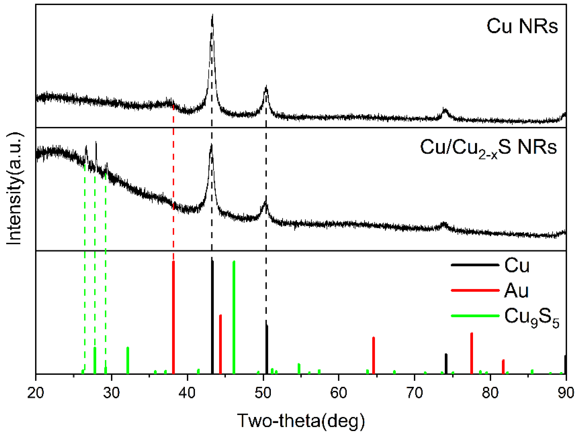
The X-ray diffraction (XRD) patterns of initial and sulfurized Cu NRs are shown in Figure 6. The diffractive peaks of initial Cu NRs at 43.3°, 50.5°, and 74.1° are consistent with the diffractions from the (111), (200) and (220) planes of face-centered cubic (fcc) Cu. These three peaks of sulfurized Cu NRs shifted by about 0.2° to the left, to 43.1°, 50.2°, 73.9°, respectively. For the sulfurized Cu NRs, a raised wide band below 35° indicates that an amorphous structure may be formed after sulfidation. Three special peaks at 26.6°, 27.9° and 29.4° are matched with the (101), (0015), and (107) diffractions of the hexagonal close-packed (hcp) Cu9S5 (Cu1.8S) phase. In addition, as can be seen in Figure S2, the three special peaks could correspond to the diffraction peaks at 26.5° (211), 26.6° (103), 27.9° (022) and 29.0° (113) of the orthorhombic Cu7S4 (Cu1.75S) phase, or at 26.5° (002) and 29.2° (101) of the hcp Cu2S phase. Therefore, the coating layer may be a variety of mixed phases of copper sulfide, and it is termed as Cu2−xS in this work. The formed Cu2−xS layer by partial sulfidation could protect the inside of the Cu from oxidation. Meanwhile, Cu2−xS is a p-doped semiconductor associated with copper deficiency, supporting tunable SPRs in the NIR spectral region [34,35], and has an active surface for catalytic applications [36].
The structural characterizations demonstrate that the sulfurized Cu NRs consist of a Cu core and a Cu2−xS shell. The formed Cu/Cu2−xS core/shell NRs integrate many advantages including: (1) the SPRs of Cu NRs have intense absorption and local field enhancements, (2) the rough surface is kept during the sulfidation, which provides large-area active surface of Cu2−xS, (3) the copper ions of Cu2−xS have both oxidizing and reducing properties, (4) Cu2−xS is a functional semiconductor possessing optical absorption and hole-induced SPRs. All of these properties indicate that the prepared Cu/Cu2−xS is a promising material for photocatalytic application.
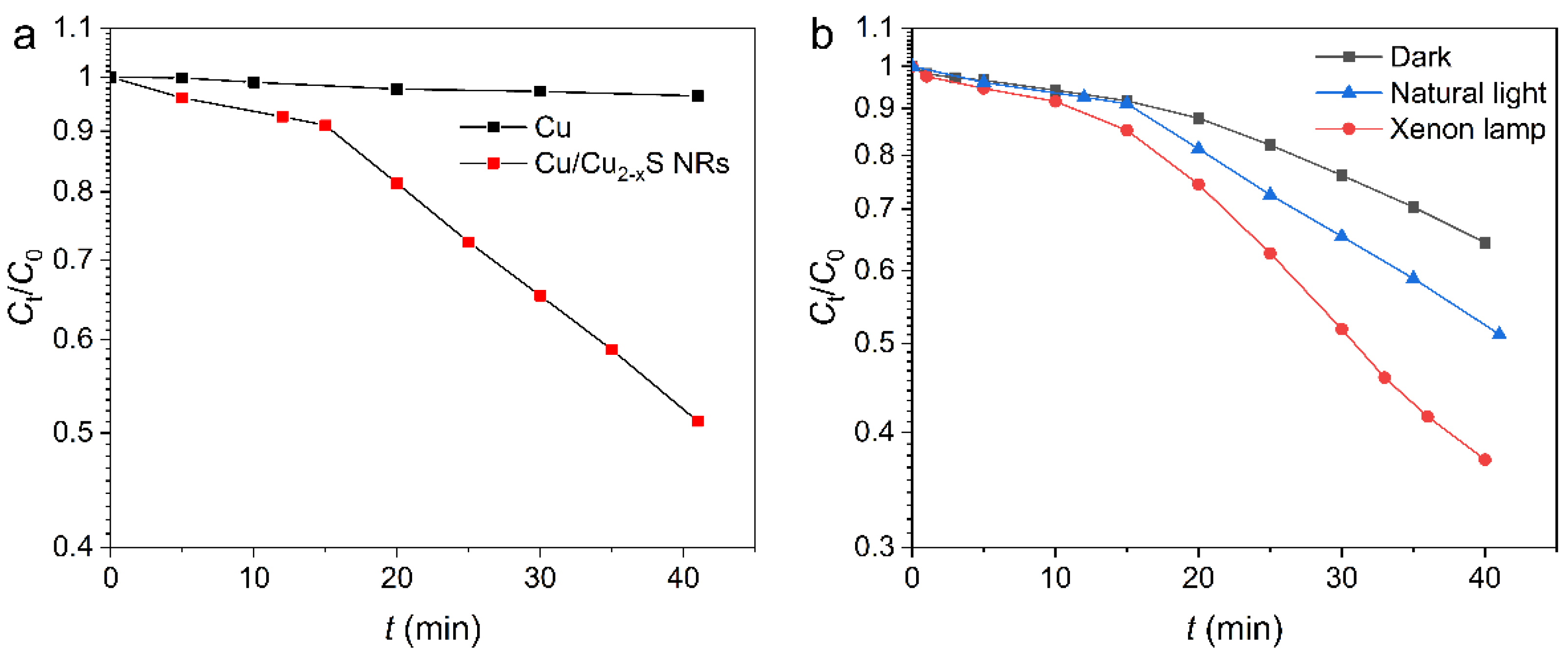
We performed the degradation of methyl orange (MO) to evaluate the photocatalytic performance of Cu/Cu2−xS core/shell NRs. Figure 7a compares the degradation of MO over Cu NRs and Cu/Cu2−xS NRs under natural light. The MO concentration is measured by the absorbance of MO at 464 nm (Figure S3). C0 and Ct are the concentrations of MO dyes initially and at time t (min), respectively. The degradation rate is calculated by (C0Ct)/C0. For the Cu NRs under natural light, only 3.6% MO dyes are degraded after 40 min. For the Cu/Cu2−xS NRs under natural light, the degradation rate is 48.9% after 40 min, which is 13.6-fold higher than that of the Cu NRs. Figure 7b illustrates the degradation performance of Cu/Cu2−xS NRs on MO dyes under the conditions of dark, natural light, and xenon lamp irradiation. Even under the dark condition, the sulfurized NRs exhibit a 35.7% degradation rate within 40 min. The dye degradation in the dark was reported previously for Cu2−xS nanocrystals [37], and this may be due to a self-cyclic redox reaction of multivalent copper [18,26], or a low-temperature, thermocatalytic reaction in the dark system [38]. On the other hand, natural light can improve the degradation rate to 51.5% at 40 min, and xenon lamp irradiation can further improve the degradation rate to 62.6% at 40 min. Under natural light and xenon lamp irradiation, the degradation rates of the Cu/Cu2−xS core/shell NRs at 40 min are 1.4 times and 1.8 times higher than those in darkness, respectively.
The logarithmic plots, as a function of time, indicate that there is a two-stage process during the photocatalytic degradation reaction by the Cu/Cu2−xS NRs. A pseudo-first-order equation ln(Ct/C0) = kt is used to fit the experimental data (Figure S4), where k is the reaction rate constant (min−1). There is only one k value of 9.4 × 10⁻⁴ min−1 for the Cu NRs under natural light, whereas for the sulfurized NRs, the k values of the two stages of the photocatalytic degradation process are 6.1 × 10⁻³ min−1 and 2.2 × 10⁻² min−1, respectively. In Figure S4b, the photocatalytic degradation reaction of Cu/Cu2−xS NRs under each condition has two stages. Under the dark condition, the k values are 5.8 × 10⁻³ min−1 and 1.6 × 10⁻² min−1, respectively. Under xenon lamp irradiation, the k values are 8.2 × 10⁻³ min−1 and 3.4 × 10⁻² min−1, respectively. The two stages are supposedly related to the dye adsorption process [11] and photocatalytic degradation reaction [39]. The dye adsorption process by catalysts can be demonstrated in the dark condition [40,41,42]. In this work, the kinetic process in the dark is clearly divided into two stages, indicating the coexistence of the adsorption process and degradation reaction under the dark condition.
We performed another photocatalytic reaction for methyl blue (MB) by Cu/Cu2−xS NRs. Figure S5 illustrates the degradation performance of Cu/Cu2−xS NRs on MB dyes under xenon lamp irradiation. The degradation is faster for MB molecules because the stability of MB dyes is poorer than that of MO molecules [18,42]. The results show that 33% of MB molecules with no catalysts are spontaneously degraded under xenon lamp irradiation after 20 min (Figure S5). On the contrary, no MO degradation is observed under xenon lamp irradiation after 40 min when the catalyst is not added (see Figure S6).
The CB and VB edge potentials of Cu2S are located at −4.1 and −5.5 eV, respectively, with a band gap of 1.4 eV (~886 nm) [37]. For the CuS, the CB and VB edge potentials are −2.2 and −4.45 eV, respectively, and the band gap is 2.25 eV (~551 nm) [37]. The optical responses of Cu2−xS are not observed in the extinction spectra because the SPRs of Cu NRs are still strong after sulfidation and the amount of Cu2−xS is small. Under light irradiation, the interaction between Cu NRs and the Cu2−xS layer leads to an increased amount of photoexcited charge carriers and the effective separation of electrons and holes [35,36]. The photoexcited electrons (e) and holes (h+) migrate to the surface of Cu/Cu2−xS NRs, generating active species (such as the hydroxyl radicals of ·OH and superoxide radicals of ·O2−) for a photocatalytic degradation reaction. To investigate the main active species generated during the decomposition process of MO, we used methanol and disodium ethylenediaminetetraacetate (EDTA) as scavengers to examine ·OHs and h+, respectively [40]. Figures S6 and S7 show the effect of two scavengers on the degradation rate of MO degradation by Cu/Cu2−xS NRs under xenon lamp irradiation. Compared with the scavenger-free sample, with a degradation rate of 89% at 40 min, the photocatalytic degradation rate decreases to 10% and 4% after the addition of methanol and EDTA, respectively. The result indicates that both ·OH and h+ are the major reactive species [40,41]. The MO molecules could be degraded into CO2, H2O, NO3, SO42−, and other incomplete products after the reaction with active radicals [18]. Bubbles can be observed during the degradation process under natural light (Figure S8), which is due to the product of CO2 from MO degradation.

3. Materials and Methods

3.1. Materials

Tetrachloroauric(III) acid tetrahydrate (HAuCl4·4H2O), copper(II) chloride dehydrate (CuCl2·2H2O, 99.999%), toluene (99.5%), acetone (99.5%), isopropanol (99.7%), ethanol absolute (99.7%), methanol, methyl orange (MO), methyl blue (MB), and ethylenediaminetetraacetate (EDTA, 99.5%) were purchased from Sinopharm Chemical Reagent limited corporation (Shanghai, China). Sodium hydrosulfide hydrate (68–72%), borane-tert-butylamine complex (TBAB, 97%), and oleylamine (80–90%) were purchased from Aladdin (Shanghai, China). Oleylamine (50%) was purchased from TCI Shanghai (Shanghai, China).

3.2. Synthesis of Au Seeds

Au seeds were synthesized by referring to the method of Jeong [1]. Oleylamine (10 mL, 50%) was loaded into a 100 mL three-necked flask with nitrogen flushing for 10 min at room temperature (25 °C). Then, 10 mL anhydrous toluene was added into the flask, followed by nitrogen purging for 10 min. Afterward, 100 mg of HAuCl4·4H2O was added into the flask with N2 purging for another 10 min. A solution mixture of 0.20 mmol (17 mg) of TBAB, 1 mL of oleylamine (50%) and 1 mL of anhydrous toluene was quickly injected into the mixture, followed by stirring for 1 h at 25 °C. After the reaction, the product was precipitated with 50 mL of acetone and centrifuged at 6000 rpm for 5 min. Finally, the precipitate was dispersed in 40 mL of toluene for storage. The average size of Au seeds with a deep-red color was 7.8 nm.

3.3. Synthesis of Cu NRs with Tunable Length

For preparing Cu NRs, 0.5 mmol (85 mg) of CuCl2∙2H2O and 10 mL of oleylamine (80–90%) were firstly mixed in a 50 mL three-necked flask at room temperature. Nitrogen blowing was applied and maintained until the end of reaction. The mixture was heated to 180 °C, and stirred within 20 min. The reaction solution was maintained at 180 °C for another 30 min. At room temperature, the copper dichloride was partially dissolved, and the solution turned blue. At about 160 °C, the solution turned from blue to green and quickly turned yellow, indicating the reduction of Cu(II) to Cu(I). After heating for 30 min, a certain amount of Au seeds was injected into the mixture at 180 °C. When the Au seeds with a deep-red color were added, the color of the solution changed rapidly from yellow to orange–red. The reaction was allowed to continue at 180 °C for one hour. After the reaction was completed, the three-necked flask was placed in cold water to cool down to room temperature. Then, 10 mL of isopropanol was added to precipitate the products. The sample was centrifuged at 3000 rpm for 3 min and the precipitates were washed with 5 mL of toluene. Finally, the precipitates were re-dispersed in toluene.

3.4. In Situ Sulfidation of Cu NRs

Sodium hydrosulfide ethanol solution (1 mM) was used as the sulfur source, and 2 mL of Cu NR toluene solution was uniformly mixed with different amounts of sulfur source (50, 80, 120 μL) for 5 min. After that, the mixed solution was centrifuged at 3000 rpm for 3 min to obtain a black precipitate. The precipitate was re-dispersed in 2 mL of toluene.

3.5. Characterization

Extinction spectra were measured by a Varian Cary 5000 UV-Vis-NIR (Agilent, Santa Clara, CA, USA) spectrophotometer. XRD patterns were recorded by Bruker D8 advanced XRD (Karlsruhe, Germany) under Cu-α irradiation (λ = 0.15418 nm). TEM analysis was performed by a JEOL JEM 2100 microscope (Tokyo, Japan). STEM imaging and EDS mapping were acquired on a JEOL JEM-ARM200F microscope (Tokyo, Japan) operated at 200 kV with a Schottky cold-field emission gun in Wuhan University.

3.6. Photocatalytic Degradation of Dye

A concentrated toluene solution of sulfurized Cu NRs was transferred to a 20 mL small beaker, which was then placed in a fume hood to air dry. After air drying, the solid was ultrasonically dispersed in 3 mL of water for subsequent degradation experiments. For photocatalytic degradation, 1.5 mL of 20 mg/L MO aqueous solution was mixed with 1.5 mL catalyst aqueous solution in a silica cuvette with optical path length of 1 cm. The sample was left undisturbed under different conditions (dark, natural light, xenon lamp irradiation). The xenon lamp irradiation was performed by placing the sample 10 cm away from a xenon lamp source (PLS-SXE300+) (Beijing, China). The irradiation power of xenon lamp was measured as 1.5 W. The concentration of MO dyes was measured every 5 min. The MO concentration is measured through directly measuring the absorbance of the sample at 464 nm by an UV-Vis-NIR spectrophotometer (Varian Cary 5000, Agilent, Santa Clara, CA, USA). The degradation test of MB dyes was carried out under xenon lamp irradiation. The concentration of 1.5 mL of MB was 40 mg/L. For the dye stability experiment, 1.5 mL of 20 mg/L MO solution and 1.5 mL of 40 mg/L MB solution were mixed with 1.5 mL of water, respectively. The samples were placed under xenon lamp irradiation and the absorption intensity was monitored.
EDTA and methanol were added in the photocatalytic degradation of MO to investigate the main active species, such as h+, ·OHs [40,41]. The experiments were carried out under the illumination of xenon lamp. Catalyst aqueous solution (1 mL) was added into 1 mL of 20 mg/L MO solution. Subsequently, 0.2 mL of water, methanol, or 0.05 M EDTA was added into the solution. Then, the absorption intensity of dyes was measured every 5 min.

4. Conclusions

In this work, Cu NRs with tunable LSPRs were successfully synthesized by the seed-mediated method, and the outermost layer of Cu NRs was transformed into the Cu2−xS phase by a convenient in situ sulfidation method. The thickness of the Cu2−xS layer could be adjusted by controlling the amount of S source. Compared with the pure Cu NRs, the Cu/Cu2−xS core/shell NRs integrate plasmonic enhancement, functional Cu2−xS material and a rough active surface, demonstrating an excellent photocatalytic dye degradation performance. Moreover, the coating of Cu2−xS improves the stability of Cu NRs in the photocatalytic reaction. The experimental results show that the degradation rate of Cu/Cu2−xS core/shell NRs at 40 min is 13.6 times higher than that of the pure Cu NRs, under natural light. The results in this work demonstrate a promising way to construct Cu nanostructures in optical and catalytic applications.

Supplementary Materials

The following supporting information can be downloaded at: https://www.mdpi.com/article/10.3390/catal12020147/s1, Figure S1: absorption spectrum and TEM image of Au seeds, Figure S2: XRD pattern analysis of Cu/Cu2−xS NRs, Figure S3: absorption spectra of MO photocatalytic degradation, Figure S4: curve fitting for the plot of ln(Ct/C0) vs. time, Figure S5: influence of radical scavengers on the MO degradation, Figure S6: sample photograph after MO degradation, Figure S7: absorption spectra of MO photocatalytic degradation with scavengers, Figure S8: photocatalytic degradation of MB dyes.

Author Contributions

Conceptualization, L.Z. and Q.-Q.W.; methodology and investigation, L.C. and Y.-T.Z.; writing—original draft preparation, L.C.; writing—review and editing, L.Z. and L.C.; supervision, L.Z. All authors have read and agreed to the published version of the manuscript.

Funding

This research was funded by the National Key R&D Program of China, grant number 2020YFA0211300 and 2017YFA0303402, and the National Natural Science Foundation of China, grant number 12174294, 11874293 and 12074296.

Data Availability Statement

The supplemental data are provided in Supplementary Materials. Data presented in this study are available on request from the corresponding author.

Acknowledgments

The authors would like to acknowledge the Center for Electron Microscopy at Wuhan University for their substantial supports to TEM work.

Conflicts of Interest

The authors declare no conflict of interest.

References

  1. Jeong, S.; Liu, Y.; Zhong, Y.; Zhan, X.; Li, Y.; Wang, Y.; Cha, P.M.; Chen, J.; Ye, X. Heterometallic seed-mediated growth of monodisperse colloidal copper nanorods with widely tunable plasmonic resonances. Nano Lett. 2020, 20, 7263–7271. [Google Scholar] [CrossRef] [PubMed]
  2. Chen, S.; Jenkins, S.V.; Tao, J.; Zhu, Y.; Chen, J. Anisotropic seeded growth of Cu–M (M = Au, Pt, or Pd) Bimetallic nanorods with tunable optical and catalytic properties. J. Phys. Chem. C 2013, 117, 8924–8932. [Google Scholar] [CrossRef]
  3. Cui, F.; Yu, Y.; Dou, L.; Sun, J.; Yang, Q.; Schildknecht, C.; Schierle-Arndt, K.; Yang, P. Synthesis of ultrathin copper nanowires using Tris(trimethylsilyl)silane for high-performance and low-haze transparent conductors. Nano Lett. 2015, 15, 7610–7615. [Google Scholar] [CrossRef] [PubMed]
  4. Huo, D.; Kim, M.J.; Lyu, Z.; Shi, Y.; Wiley, B.J.; Xia, Y. One-dimensional metal nanostructures: From colloidal syntheses to applications. Chem. Rev. 2019, 119, 8972–9073. [Google Scholar] [CrossRef]
  5. Wang, Z.; Chen, Z.; Zhang, H.; Zhang, Z.; Wu, H.; Jin, M.; Wu, C.; Yang, D.; Yin, Y. Lattice-mismatch-induced twinning for seeded growth of anisotropic nanostructures. ACS Nano 2015, 9, 3307–3313. [Google Scholar] [CrossRef] [PubMed] [Green Version]
  6. Liang, S.; Liu, X.L.; Yang, Y.Z.; Wang, Y.L.; Wang, J.H.; Yang, Z.J.; Wang, L.B.; Jia, S.F.; Yu, X.F.; Zhou, L.; et al. Symmetric and asymmetric Au-AgCdSe hybrid nanorods. Nano Lett. 2012, 12, 5281–5286. [Google Scholar] [CrossRef]
  7. Ma, L.; Liang, S.; Liu, X.L.; Yang, D.J.; Zhou, L.; Wang, Q.Q. Synthesis of dumbbell-like gold-metal sulfide core-shell nanorods with largely enhanced transverse plasmon resonance in visible region and efficiently improved photocatalytic activity. Adv. Funct. Mater. 2015, 25, 898–904. [Google Scholar] [CrossRef]
  8. Wang, P.F.; Chen, K.; Ma, S.; Wang, W.; Qiu, Y.H.; Ding, S.J.; Liang, S.; Wang, Q.Q. Asymmetric synthesis of Au-CdSe core-semishell nanorods for plasmon-enhanced visible-light-driven hydrogen evolution. Nanoscale 2020, 12, 687–694. [Google Scholar] [CrossRef]
  9. Wu, B.; Wang, P.F.; Qiu, Y.H.; Liang, S.; Wu, Z.Y.; Zhou, L.; Wang, Q.Q. Enhanced second-harmonic generation of asymmetric Au@CdSe heterorods. Sci. China Mater. 2020, 63, 1472–1479. [Google Scholar] [CrossRef] [Green Version]
  10. Zhong, Y.; Ma, S.; Chen, K.; Wang, P.F.; Qiu, Y.H.; Liang, S.; Zhou, L.; Chen, Y.; Wang, Q.Q. Controlled growth of plasmonic heterostructures and their applications. Sci. China Mater. 2020, 63, 1398–1417. [Google Scholar] [CrossRef] [Green Version]
  11. Jourshabani, M.; Shariatinia, Z.; Badiei, A. Sulfur-doped mesoporous carbon nitride decorated with Cu particles for efficient photocatalytic degradation under visible-light irradiation. J. Phys. Chem. C 2017, 121, 19239–19253. [Google Scholar] [CrossRef]
  12. Chen, L.; Hu, H.; Chen, Y.; Li, Y.; Gao, J.; Li, G. Sulfur precursor reactivity affecting the crystal phase and morphology of Cu2−xS nanoparticles. Chem. Eur. J. 2021, 27, 1057–1065. [Google Scholar] [CrossRef]
  13. Luo, M.; Ruditskiy, A.; Peng, H.C.; Tao, J.; Figueroa-Cosme, L.; He, Z.K.; Xia, Y.N. Penta-twinned copper nanorods: Facile synthesis via seed-mediated growth and their tunable plasmonic properties. Adv. Funct. Mater. 2016, 26, 1209–1216. [Google Scholar] [CrossRef]
  14. Fu, W.; Liu, L.; Yang, G.; Deng, L.; Zou, B.; Ruan, W.; Zhong, H. Oleylamine-assisted phase-selective synthesis of Cu2−xS nanocrystals and the mechanism of phase control. Part. Part. Syst. Char. 2015, 32, 907–914. [Google Scholar] [CrossRef]
  15. Hsu, S.W.; Ngo, C.; Bryks, W.; Tao, A.R. Shape focusing during the anisotropic growth of CuS triangular nanoprisms. Chem. Mater. 2015, 27, 4957–4963. [Google Scholar] [CrossRef]
  16. Sun, C.; Liu, M.; Zou, Y.; Wei, J.; Jiang, J. Synthesis of plasmonic Au–CuS hybrid nanocrystals for photothermal transduction and chemical transformations. RSC Adv. 2016, 6, 26374–26379. [Google Scholar] [CrossRef]
  17. Lyu, Z.; Zhu, S.; Xie, M.; Zhang, Y.; Chen, Z.; Chen, R.; Tian, M.; Chi, M.; Shao, M.; Xia, Y. Controlling the surface oxidation of cu nanowires improves their catalytic selectivity and stability toward C2+ Products in CO2 Reduction. Angew. Chem. Int. Ed. 2021, 60, 1909–1915. [Google Scholar] [CrossRef]
  18. Sun, B.; Li, H.; Li, X.; Liu, X.; Zhang, C.; Xu, H.; Zhao, X.S. Degradation of organic dyes over fenton-like Cu2O–Cu/C catalysts. Ind. Eng. Chem. Res. 2018, 57, 14011–14021. [Google Scholar] [CrossRef]
  19. Chakraborty, A.; Islam, D.A.; Acharya, H. One pot synthesis of ZnO-CuO nanocomposites for catalytic peroxidase like activity and dye degradation. Mater. Res. Bull. 2019, 120, 110592. [Google Scholar] [CrossRef]
  20. Jyothi, N.S.; Ravichandran, K. Optimum pH for effective dye degradation: Mo, Mn, Co and Cu doped ZnO photocatalysts in thin film form. Ceram. Int. 2020, 46, 23289–23292. [Google Scholar] [CrossRef]
  21. Kale, G.; Arbuj, S.; Kawade, U.; Kadam, S.; Nikam, L.; Kale, B. Paper templated synthesis of nanostructured Cu–ZnO and its enhanced photocatalytic activity under sunlight. J. Mater. Sci. Mater. Electron. 2019, 30, 7031–7042. [Google Scholar] [CrossRef]
  22. Vasanthi, D.S.; Ravichandran, K.; Kavitha, P.; Sriram, S.; Praseetha, P.K. Combined effect of Cu and N on bandgap modification of ZnO film towards effective visible light responsive photocatalytic dye degradation. Superlattices Microstruct. 2020, 145, 106637. [Google Scholar] [CrossRef]
  23. Hoseini, S.J.; Fath, R.H. Formation of nanoneedle Cu(0)/CuS nanohybrid thin film by the disproportionation of a copper(i) complex at an oil–water interface and its application for dye degradation. RSC Adv. 2016, 6, 76964–76971. [Google Scholar] [CrossRef]
  24. Wan, M.; Cui, S.; Wei, W.; Cui, S.; Chen, K.; Chen, W.; Mi, L. Bi-component synergic effect in lily-like CdS/Cu7S4 QDs for dye degradation. RSC Adv. 2019, 9, 2441–2450. [Google Scholar] [CrossRef] [Green Version]
  25. Kumar, P.S.; Lakshmi Prabavathi, S.; Indurani, P.; Karuthapandian, S.; Muthuraj, V. Light assisted synthesis of hierarchically structured Cu/CdS nanorods with superior photocatalytic activity, stability and photocatalytic mechanism. Sep. Purif. Technol. 2017, 172, 192–201. [Google Scholar] [CrossRef]
  26. Alhebshi, N.; Huang, H.; Ghandour, R.; Alghamdi, N.K.; Alharbi, O.; Aljurban, S.; He, J.H.; Al-Jawhari, H. Green synthesized CuxO@Cu nanocomposites on a Cu mesh with dual catalytic functions for dye degradation and hydrogen evaluation. J. Alloys Compd. 2020, 848, 156284. [Google Scholar] [CrossRef]
  27. Basu, M.; Sinha, A.K.; Pradhan, M.; Sarkar, S.; Negishi, Y.; Govind; Pal, T. Evolution of hierarchical hexagonal stacked plates of CuS from liquid−liquid interface and its photocatalytic application for oxidative degradation of different dyes under indoor lighting. Environ. Sci. Technol. 2010, 44, 6313–6318. [Google Scholar] [CrossRef]
  28. Jiang, Q.; Jiang, J.; Deng, R.; Xie, X.; Meng, J. Controllable preparation of CuO/Cu2O composite particles with enhanced photocatalytic performance. New J. Chem. 2020, 44, 6369–6374. [Google Scholar] [CrossRef]
  29. Periyayya, U.; Madhu, D.; Subramaniyam, K.; Son, H.; Lee, I.H. Enhanced cyclic performance initiated via an in situ transformation of Cu/CuO nanodisk to Cu/CuO/Cu2O nanosponge. Environ. Sci. Pollut. Res. 2021, 28, 6459–6469. [Google Scholar]
  30. Solomane, N.; Ajibade, P.A. Synthesis and crystal structure of bis(thiomorpholinyldithiocarbamato)Cu(II) complex and its use as precursor for CuS nanoparticles photocatalyst for the degradation of organic dyes. J. Sulphur. Chem. 2020, 42, 167–179. [Google Scholar] [CrossRef]
  31. Bhowmick, S.; Moi, C.T.; Kalita, N.; Sahu, A.; Suman, S.; Qureshi, M. Spontaneous Fenton-like dye degradation in clustered-petal di-manganese copper oxide by virtue of self-cyclic redox couple. J. Environ. Chem. Eng. 2021, 9, 106094. [Google Scholar] [CrossRef]
  32. Hou, Y.; Li, X.; Zou, X.; Quan, X.; Chen, G. Photoeletrocatalytic activity of a Cu2O-loaded self-organized highly oriented TiO2 nanotube array electrode for 4-chlorophenol degradation. Environ. Sci. Technol. 2009, 43, 858–863. [Google Scholar] [CrossRef]
  33. Rahmatolahzadeh, R.; Ebadi, M.; Motevalli, K. Preparation and characterization of Cu clusters and Cu–Ag alloy via galvanic replacement method for azo dyes degradation. J. Mater. Sci. Mater. Electron. 2017, 28, 6056–6063. [Google Scholar] [CrossRef]
  34. Li, Y.Y.; Pan, G.M.; Liu, Q.Y.; Ma, L.; Xie, Y.; Zhou, L.; Hao, Z.H.; Wang, Q.Q. Coupling resonances of surface plasmon in gold nanorod/copper chalcogenide core-shell nanostructures and their enhanced photothermal effect. ChemPhysChem 2018, 19, 1852–1858. [Google Scholar] [CrossRef]
  35. Ma, S.; Chen, K.; Qiu, Y.H.; Gong, L.L.; Pan, G.M.; Lin, Y.J.; Hao, Z.H.; Zhou, L.; Wang, Q.Q. Controlled growth of CdS-Cu2−xS lateral heteroshells on Au nanoparticles with improved photocatalytic activity and photothermal efficiency. J. Mater. Chem. A 2019, 7, 3408–3414. [Google Scholar] [CrossRef]
  36. Wang, W.; Ma, S.; Chen, K.; Wang, P.F.; Liu, Q.Y.; Zhou, L.; Wang, Q.Q. Controlled growth of Cu2−xS sheet-like nanoshells and Cu2−xS-CdS p-n junctions on Au nanorods with coupled plasmon resonances and enhanced photocatalytic activities. J. Mater. Chem. C 2020, 8, 3058–3068. [Google Scholar] [CrossRef]
  37. Kuo, K.Y.; Chen, S.H.; Hsiao, P.H.; Lee, J.T.; Chen, C.Y. Day-night active photocatalysts obtained through effective incorporation of Au@CuxS nanoparticles onto ZnO nanowalls. J. Hazard. Mater. 2022, 421, 126674. [Google Scholar] [CrossRef]
  38. Deng, S.Q.; Miao, Y.L.; Tan, Y.L.; Fang, H.N.; Li, Y.T.; Mo, X.J.; Cai, S.L.; Fan, J.; Zhang, W.G.; Zheng, S.R. An anionic nanotubular metal-organic framework for high-capacity dye adsorption and dye degradation in darkness. Inorg. Chem. 2019, 58, 13979–13987. [Google Scholar] [CrossRef]
  39. Nandi, P.; Das, D. ZnO-CuO heterostructure photocatalyst for efficient dye degradation. J. Phys. Chem. Solids 2020, 143, 109463. [Google Scholar] [CrossRef]
  40. Song, C.; Wang, X.; Zhang, J.; Chen, X.; Li, C. Enhanced performance of direct Z-scheme CuS-WO3 system towards photocatalytic decomposition of organic pollutants under visible light. Appl. Surf. Sci. 2017, 425, 788–795. [Google Scholar] [CrossRef]
  41. Li, L.; Yang, X.; Xie, C.; Wang, Y.; Wei, L.; Yang, J. Synthesis and photocatalytic mechanism of visible-light-driven Ag/AgBr/(I/S) composites for organic dyes degradation. Opt. Mater. 2022, 123, 111947. [Google Scholar] [CrossRef]
  42. Tavakoli Joorabi, F.; Kamali, M.; Sheibani, S. Effect of aqueous inorganic anions on the photocatalytic activity of CuO–Cu2O nanocomposite on MB and MO dyes degradation. Mater. Sci. Semicon. Proc. 2022, 139, 106335. [Google Scholar] [CrossRef]
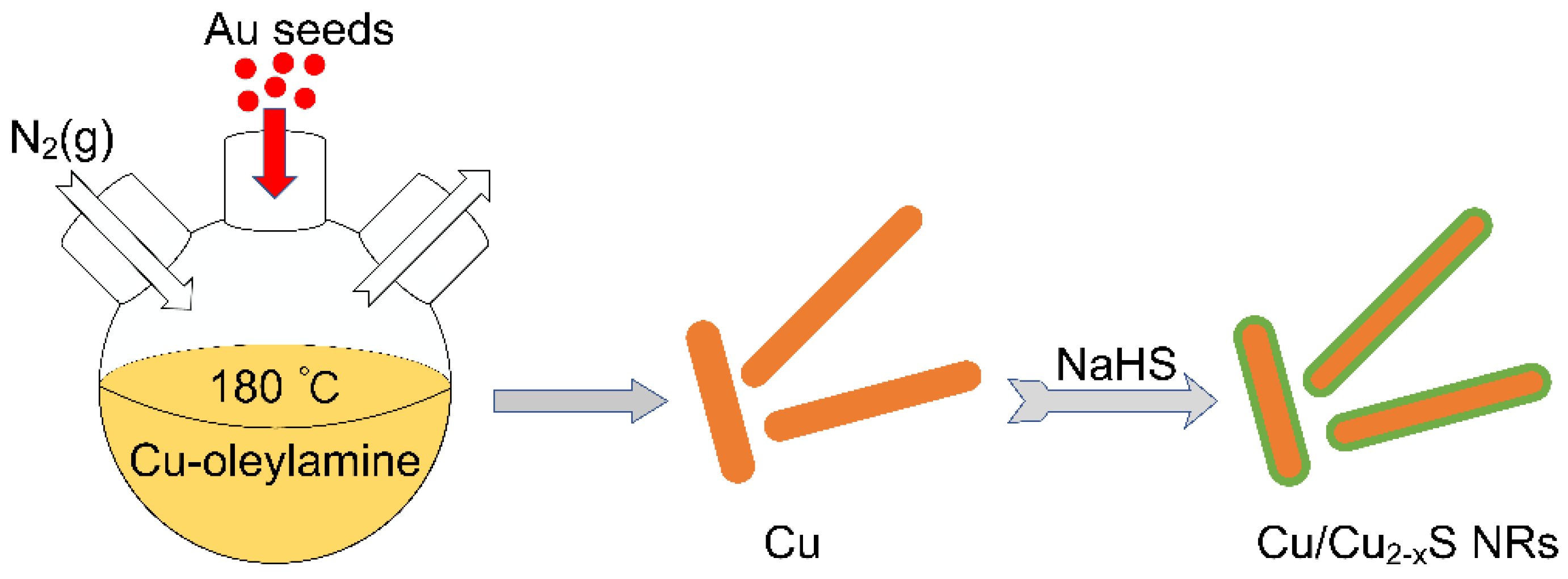
Figure 1. Scheme of seed-mediated growth of Cu NRs and subsequent sulfidation.
Catalysts 12 00147 g001
Figure 2. (a) Extinction spectra of Cu NRs with LSPRs located at 740 (purple), 821 (yellow), 888 (green), 960 (blue), 1054 (red) and 1190 nm (black). (b,c) TEM images of Cu NRs with LSPR of 740 nm (b) and 821 nm (c).
Catalysts 12 00147 g002
Figure 3. Extinction spectra of Cu NRs when adding different amounts of sulfur source (1 mM NaHS).
Catalysts 12 00147 g003
Figure 4. TEM images of Cu NRs with the addition of (a) 10 μL and (b) 60 μL of NaHS solution. The insets show the amplifying TEM images around the tip region of sulfurized Cu NRs.
Catalysts 12 00147 g004
Figure 5. STEM-EDS elemental analysis of the sulfurized Cu NRs: (a) STEM image, (b) merged element mapping, (ce) element distributions of Cu, S, and Au. The atomic percentage is also shown.
Catalysts 12 00147 g005
Figure 6. XRD patterns of Cu NRs before and after sulfidation. The bottom column diagram represents the standard diffraction pattern of Cu (#85-1326), Au (#04-0784), and Cu9S5 (#47-1748).
Catalysts 12 00147 g006
Figure 7. Logarithmic plots of Ct/C0 as a function of time for MO degradation by (a) Cu and Cu/Cu2−xS NRs under natural light, (b) Cu/Cu2−xS NRs under three irradiation conditions (dark, natural light and xenon lamp).
Catalysts 12 00147 g007
Publisher’s Note: MDPI stays neutral with regard to jurisdictional claims in published maps and institutional affiliations.