Article

Structural Studies of Bypass of Forespore Protein C from Bacillus Subtilis to Reveal Its Inhibitory Molecular Mechanism for SpoIVB

1
College of Chemistry, Fuzhou University, Fuzhou 350116, China
2
College of Biological Science and Engineering, Fuzhou University, Fuzhou 350116, China
3
Fujian Key Laboratory of Marine Enzyme Engineering, Fuzhou University, Fuzhou 350116, China
*
Authors to whom correspondence should be addressed.
Catalysts 2022, 12(12), 1530; https://doi.org/10.3390/catal12121530
Submission received: 15 October 2022 / Revised: 24 November 2022 / Accepted: 25 November 2022 / Published: 28 November 2022
(This article belongs to the Special Issue State-of-the-Art in Enzyme Inhibitors)

Abstract

:
Activation of pro-σK processing requires a signaling protease SpoIVB that is secreted from the forespore into the space between the two cells during sporulation in Bacillus subtilis. Bypass of forespore protein C (BofC) is an inhibitor preventing the autoproteolysis of SpoIVB, ensuring the factor σK operates regularly at the correct time during the sporulation. However, the regulatory mechanisms of BofC on pro-σK processing are still unclear, especially in the aspect of the interaction between BofC and SpoIVB. Herein, the recombinant BofC (rBofC) was expressed in the periplasm by the E. coli expression system, and crystal growth conditions were obtained and optimized. Further, the crystal structure of rBofC was determined by X-ray crystallography, which is nearly identical to the structures determined by NMR and predicted by AlphaFold. In addition, the modeled structure of the BofC–SpoIVB complex provides insights into the molecular mechanism by which domain 1 of BofC occupies the active site of the SpoIVB serine protease domain, leading to the inhibition of the catalytical activity of SpoIVB and prevention of the substrate of SpoIVB (SpoIVFA) from binding to the active site.

1. Introduction

Sporulation has been the subject of continuous microbiological investigation since the 19th century [1]. A series of proteins exhibit synergistic effects during sporulation when bacteria have to resist harsh environments [2]. During sporulation, the promoter specificities on RNA polymerase are mainly conferred by sigma factors, including σH, σE, σF, σG and σK [3,4]. Sigma factor σK, localized in the sporangium’s mother cell compartment, is proteolytically cleaved from the inactive precursor pro-σK [5,6]. The conversion of pro-σK is considered as a checkpoint of the developing sporangium, which coordinates gene expression between two chambers, the mother cell and the forespore [7].
The processing of pro-σK involves regulated intramembrane proteolysis (RIP) [8]. At least six proteins are involved in controlling the σK cleavage process, including SpoIVFA, SpoIVFB, BofA, SpoIVB, CtpB and bypass of forespore protein C (BofC) [9,10,11]. The former three proteins, SpoIVFA, SpoIVFB and BofA, formed a ternary complex and are situated at the outer forespore membrane (OFM) that surrounds the forespore [10,12,13], whereas SpoIVB is secreted from the forespore [3], and CtpB is produced from both the mother cell and the forespore [14]. SpoIVB and CtpB are secreted into the intermembrane space [6,15]. Firstly, SpoIVB and CtpB trigger pro-σK processing by sequentially cleaving the regulatory protein SpoIVFA. Additionally, CtpB serves as a substrate for SpoIVB and cleaves BofA to relieve the inhibition of SpoIVFB [11,16]. These modifications result in forming the active SpoIVFB for the proteolytic activation of pro-σK [16,17]. Lastly, BofC, expressed by forespores, acts negatively on SpoIVB to regulate intercompartmental signaling of pro-σK processing in the σK-checkpoint of B. subtilis [18,19].
bofC has been cloned and sequenced, which encodes a 19 kDa protein containing 170 amino acids with 30 amino acids as a signal peptide [18]. bofC transcription is controlled by both σF and σG and the σG-phase is negatively regulated by the putative DNA-binding protein, SpoVT [20]. It is speculated there is a direct protein–protein interaction between BofC and the unprocessed form of SpoIVB or other potentially active species, by which BofC can inhibit the autoproteolysis of SpoIVB and ensure no premature signal to process pro-σK [19]. BofC could maintain SpoIVB in a stable but inactive form leading to a delay in proteolytic cleavage of pro-σK. According to the three-dimensional structure determined by solution-state NMR, two domains make up the BofC monomer [21]. The N-terminal domain includes an α-helix packed onto one face of a four-stranded β-sheet. The C-terminal domain includes a three-stranded β-sheet and three α-helices in a novel domain topology [21]. Nevertheless, the crystal structure of BofC remains unknown, which would provide detailed structural information at high atomic resolution, especially in some hinge and flexible regions.
In this study, we used the prokaryotic expression system to express the recombinant BofC (rBofC) in the E. coli periplasm. Furthermore, we determined the crystal structure of rBofC and predicted the binding interaction within the BofC–SpoIVB complex, which provides a structural basis to study the molecular mechanisms of the regulation of BofC’s inhibitory activity to SpoIVB.

2. Results and Discussion

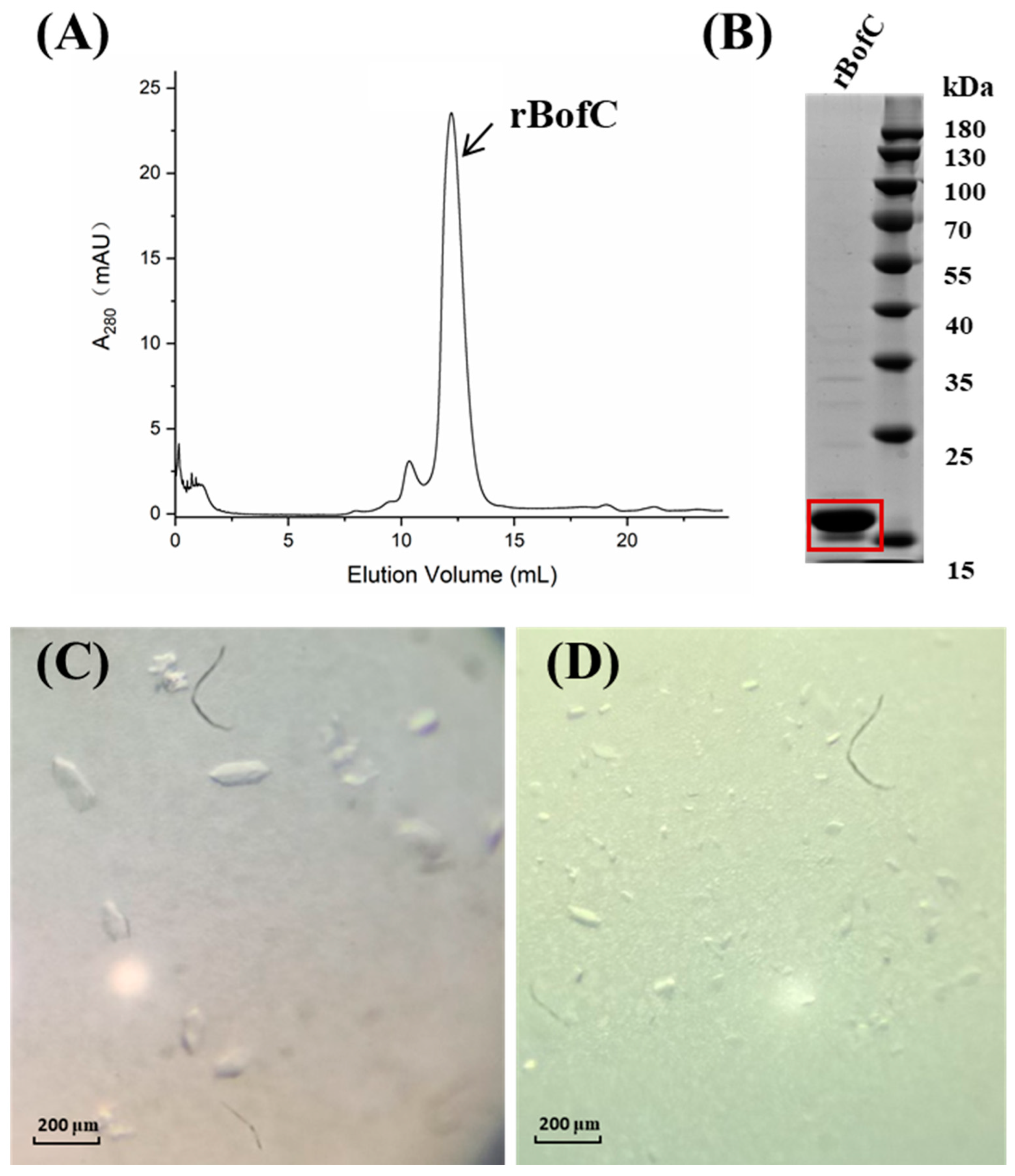
2.1. Expression, Purification and Crystallization of the Recombinant BofC (rBofC)

The cDNA of rBofC was amplified by PCR, and the agarose gel electrophoresis results showed that the molecular weight of the PCR product was consistent with the theoretical molecular weight of the bofC, indicating that the target fragment was successfully amplified (Figure S1A). The bofC was cloned into the pET-22b(+) vector by the LIC method to generate BofC (residue Met1-Gly170) with a Hexa-His tag at the C-terminus (Figure S1B,C). The sequencing result confirmed that the pET-22b(+)-bofC plasmid was successfully constructed.
rBofC was extracted from the periplasm by the osmotic shock method. The supernatants and the deposits of hypertonic solution and hypotonic solution were both checked by 15% SDS-PAGE analysis (Figure S1D). Subsequently, rBofC was successfully captured by Ni-affinity chromatography, and the elution containing the target protein rBofC was also analyzed by 15% SDS-PAGE analysis which showed that a single band of target protein BofC showed between 15 kDa and 20 kDa of marker bands (Figure S1E). To further purify the rBofC, the elution fractions were applied onto a gel-filtration column (superdex75 10/300 GL), and the elution volume of the rBofC was 12.2 mL (Figure 1A). The elution fractions of the rBofC were collected and analyzed by 15% SDS-PAGE analysis which showed that the rBofC had high purity (Figure 1B). Thereafter, the rBofC was concentrated at 9 mg/mL for the crystal growth screen.
Crystals appeared under several conditions over 3 days. The primary screened crystals were small; therefore, these crystallization conditions needed to be optimized. The optimization work was initially focused on expanding the two reagents (sodium acetate trihydrate and polyethylene glycol). After the optimization, single crystals suitable for the X-ray diffraction measurement were grown in two different conditions at room temperature (Figure 1C,D). The crystallization conditions are shown in Table S1.

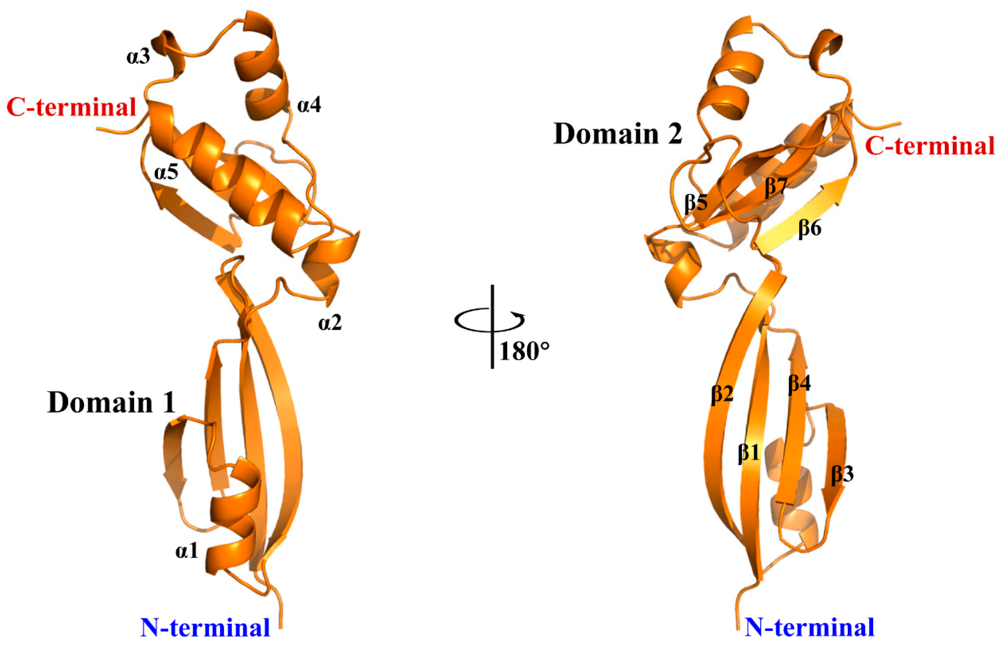
2.2. X-ray Crystallographic Studies of rBofC

The X-ray diffraction data were collected on a beamline 17UB at the Shanghai Synchrotron Radiation Facility (SSRF) at 100 K. All the frames were collected using an oscillation angle of 1 at a wavelength of 0.9792 Å. The structure of rBofC from B. subtilis was determined in the monoclinic space group P212121 with one monomer in the asymmetric unit at 1.98 Å (Table 1). In overall structure, 136 residues of rBofC (Glu35-Gly170) have been modeled into the electron density map, which has two sub-domains which include five α-helices (α1-α5) and seven β-strands (β1-β7) in all (Figure 2 and Table S2). In detail, domain 1 from the N-terminal of rBofC has four β-sheets and one α-helices, and the other domain (domain 2) has three β-strands and four α-helices.
The three-dimensional structure of BofC has been determined by the NMR method [21]. BofC-Xtal and BofC-NMR structures look highly similar, with a root mean square deviation (RMSD) of 2.63 Å on superposing all main-chain atoms of both structures, and they both have seven β-sheets and five α-helices (Figure 3A). However, there are some different conformations of domain 1 between BofC-Xtal and BofC-NMR structures (Figure 3B), which might be related to the physical basis for these differences by modeling protein cores as jammed packings of amino acid-shaped particles [22]. In detail, the α4 helix in the BofC-Xtal structure tilts about 12.61° compared to that in the BofC-NMR structure (Figure 3C). In addition, the α5 helix in the BofC-Xtal structure tilts about 14.91° compared to that in the BofC-NMR structure (Figure 3D), and these differences seen have to do with the greater dynamics of the solution structures [23]. Furthermore, the AlphaFold Protein Structure Database has released the prediction structure of BofC, which is also highly similar to the crystal structure of rBofC (RMSD = 1.74 Å, Figure S3).

2.3. Structural Prediction of BofC–SpoIVB Interactions

Although we have also expressed recombinant inactive SpoIVB (rSpoIVBS378A), we did not directly detect the interaction between rBofC and rSpoIVBS378A by size-exclusion chromatography (Figure S4). This result was verified in the previous report [21] which also showed that the way these two proteins would interact would be extremely difficult to mimic in vitro, which may be due to a lack of a suitable intercompartmental space environment or other components of the pro-σK processing complex.
Thus, to generate structures of complexes formed by the predicted SpoIVB and BofC, protein–protein docking simulations were performed using the Cluspro 2.0 server. Computational complex modeling of BofC–SpoIVB shows that domain 1 of BofC occupies the active site of the SpoIVB serine protease domain (Figure 4A). The prediction of binding affinity (ΔG) between BofC and SpoIVB is −8.6 kcal/mol, and the value of the dissociation constant (KD) is 5.1 × 10−7 M. The interface area is 958.7 A2, and 45 residues are involved in the interactions, including the formation of eight hydrogen bonds and five salt bridges analyzed by the PISA service [24] (Table S3 and Figure 4B). Thus, we speculated that the active site of SpoIVB was occupied by domain 1 of BofC, leading to the inhibition of the catalytical activity of SpoIVB and prevention of the substrate (SpoIVFA) of SpoIVB from binding to the active site; therefore, BofC plays a negative role in the regulation of intercompartmental signaling of pro-σK processing. Further experiments, such as NMR chemical shift perturbations [25], are useful to determine BofC and SpoIVB interaction that may be a transient process, and additionally, to identify specific proteins which assist in the formation of interactions between BofC and SpoIVB in vivo.

3. Materials and Methods

3.1. PCR Amplification of bofC Gene

The designation of primers was referred to as the full-length BofC sequence (residue Met1-Gly170) and the pET-22b(+) plasmid. The two restriction enzyme sites (NdeI and XhoI) were added to the forward primer and the reverse primer, respectively (Table S4). Two primers were synthesized by Fuzhou Sunya Biotechnology Co., LTD (Fuzhou, China). The PCR reaction was performed according to the protocol of the PrimeSTAR® HS DNA Polymerase (TaKaRa, Beijing, China). The reaction mixture volume was 50 μL, which consisted of 5 × PS buffer (10 μL), forward and reverse primers (10 μM and 1 μL each), DMSO (5 μL), dNTP mix (4 μL), PrimeSTAR (0.5 μL), template (0.5 μL) and double-distilled water (28 μL). After pre-denaturation at 98 °C, 30 cycles at 98 °C for 10 s, 66 °C for 15 s and 72 °C for 40 s were performed, and the temperature was decreased to 16 °C at the end. The amplified product was subjected to electrophoresis on a 1.0% agarose gel. The PCR product was recovered and purified by the glass milk method. The gel band of the PCR product was cut and dissolved in NaI (6 M, 800 μL) in a water bath (60 °C, 10 min). The glass milk (15 μL) was added to the solution [26]. Then the mixture was bathed in water (60 °C) for 10 min, and the mixture was gently flipped repeatedly every 2 min. Next, the mixture was centrifuged (6000× g, 30 s), and the supernatant was discarded. The new wash buffer (1 mL, 10 mM Tris-HCl pH 7.5, 1 mM EDTA, 0.1 M NaCl and 50% v/v ethyl alcohol) was gently mixed with the sedimented material and centrifuged (6000× g, 30 s), the supernatant was discarded and then this step was repeated once. The lid was removed to evaporate the ethyl alcohol, double-distilled water (50 μL) was mixed with the sedimented material, and finally, removal of the supernatant after the mixture was centrifuged (6000× g, 30 s).

3.2. Construction and Identification of rBofC Expression Plasmid

The expression vector pET-22b(+) (590 ng/μL) was digested using 0.5 μL NdeI (20 U/μL, New England Biolabs, Ipswich, MA, USA) and 0.5 μL XhoI (20 U/μL, New England Biolabs) restriction endonucleases at 37 °C overnight. The product was subjected to electrophoresis on a 1.0% agarose gel. The fragment (5379 bp) of pET-22b(+) was recovered by the glass milk method. Ligation independent cloning (LIC) was used to construct the expression plasmid [27]. In total, 6 μL of PCR product (100 ng/μL) was mixed with 2 μL of the fragment of pET-22b(+) (90 ng/μL), and the ExoIII buffer (1 μL) was added to the mixture and incubated on ice for 10 min. Subsequently, 1 μL of ExoIII (20 U/μL, TaKaRa, Beijing, China) was added and incubated on ice for 1 h. Thereafter, 1 μL of EDTA (0.5 M) was added, and the mixture was incubated at 65 °C for 10 min. The product was transformed into E. coli DH5α, and the cells were cultured overnight on LB agar plates containing 50 μg/mL of ampicillin. The single colony was chosen to be inoculated into an LB medium with 50 μg/mL of ampicillin. The plasmids were extracted from DH5α cultured by LB medium using a HiPure Plasmid Micro Kit (Shanghai HLingene Biological Technology Co., LTD, Shanghai, China), and the final expression vector used was confirmed by DNA sequencing.

3.3. Expression and Purification of the Recombinant BofC (rBofC)

The plasmid was transformed into E. coli BL21(DE3) and the single colony was chosen to inoculate LB medium containing 50 μg/mL of ampicillin. The LB culture was grown at 37 °C, 220 rpm/min for 12 h. In total, 10 mL of LB was inoculated into a 1 L LB medium containing 50 μg/mL of ampicillin. E. coli cells were grown at 37 °C, and then placed in a 220 rpm/min shaker incubator until OD600 up to 0.6. Then the temperature and revolving speed were decreased to 18 °C and 160 rpm/min, respectively. Next, the expression of recombinant protein was induced by isopropyl beta-D-thiogalactoside (IPTG) at the final concentration (1 mM) for 8 h. The culture was harvested and centrifuged under the condition of 4000 rpm/min and 4 °C for 20 min, and the supernatant was dropped. We used the osmotic shock method to extract the periplasmic protein BofC. BL21(DE3) cells were resuspended in 20 mL of hypertonic solution (100 mM Tris-HCl, 10 mM EDTA, 20% sucrose and pH 7.4) and incubated on ice for 30 min. After centrifugation (7000× g, 10 min and 4 °C), the supernatant was collected and the cells were resuspended in 20 mL of hypotonic solution (100 mM Tris-HCl, 10 mM EDTA and pH 7.4). After centrifugation (16000× g, 10 min and 4 °C), the supernatant was collected.
Both the hypertonic and hypotonic supernatant was filtered through 0.45 μm filter membranes and exchanged into an equilibrium buffer (20 mM Tris-HCl, 200 mM NaCl, 5% glycerin and pH 7.4), and then rBofC were captured by Ni Sepharose Excel resin (GE Healthcare Life Sciences, Marlborough, MA, USA). After washing with 100 mL of wash buffer (20 mM Tris-HCl, 200 mM NaCl, 50 mM imidazole, 5% glycerin and pH 7.4), the target protein rBofC was eluted with 10 mL of elution buffer (20 mM Tris-HCl, 200 mM NaCl, 300 mM imidazole, 5% glycerin, and pH 7.4). The pooled rBofC fractions were concentrated and further purified using size-exclusion chromatography (Superdex75 10/300 GL, GE, Marlborough, MA, USA) with elution buffer consisting of 20 mM Tris-HCl, 200 mM NaCl, 5% glycerin and pH 7.4. rBofC protein was concentrated at 9 mg/mL for crystal-growth experiments. All fractions were analyzed on 12% polyacrylamide gels, and proteins were stained with Coomassie Brilliant Blue R-250.

3.4. Crystallization of rBofC

We used a Phoenix crystallization robot (Art Robbins Instruments, Sunnyvale, CA, USA) and commercial screen kits (Qiagen, Hilden, Germany; XtalQuest, Beijing China and Hampton Research, Aliso Viejo, CA, USA) to screen the crystallization conditions of rBofC by the sitting-drop vapor-diffusion method. Reproducible crystals were finally obtained in two conditions at 25 °C after 3 days: 0.1 M sodium acetate trihydrate pH 4.5, 30% w/v PEG1500 and 0.1 M sodium acetate trihydrate pH 4.5, 30% w/v PEG5000 MME; the detailed information is shown in Table S1. Crystals were picked up and stored in crystallization solutions containing 25% v/v glycerin. Then, these crystals were cooled in liquid nitrogen for X-ray data collection.

3.5. Data Collection and Processing

Data collection was performed under cryogenic conditions at 100 K on beamline BL17U at Shanghai Synchrotron Radiation Facility (SSRF, Shanghai, China). A total of 720 images were collected at a crystal-to-detector distance of 300 mm with 1 s exposure time for every 1° oscillation frame. The data were processed using the HKL2000 package [28]. Data collection and processing statistics are summarized in Table 1.

3.6. Phasing and Refinement

The structure of BofC was solved by the molecular replacement method using the MolRep program [29] which gave very strong and unambiguous solutions. The molecular replacement model was subjected to iterative refinement and manual model rebuilding using Refmac [30] and Coot [31], alternately, giving a final R factor and Rfree factor of 0.234 and 0.269, respectively, at a resolution range of 50.0–2.0 Å (Table 1). The structure was validated with PROCHECK [32]. None of these residues was in the disallowed region of the Ramachandran plot. The coordinate of the final model was deposited in PDB with an access code of 7XT1. The final results were analyzed and visualized by PyMol [33].

3.7. Structural Prediction of BofC–SpoIVB Interactions

The crystal structure of rBofC determined from this study was used for the structural prediction of the BofC–SpoIVB interactions. Although the high-resolution 3D structure of apo–SpoIVB has not been reported, the 3D structure of apo–SpoIVB can be retrieved from the AlphaFold Protein Structure Database (https://www.alphafold.ebi.ac.uk/, accessed on 15 May 2022) [34]. The molecular docking simulation was performed in the Cluspro 2.0 server (https://cluspro.org/, accessed on 29 August 2022) [35] to predict interactions between the BofC and SpoIVB. The predicted values for binding affinity (ΔG, kcal/mol) and dissociation constant (KD) at 25.0 °C were obtained from the Prodigy website (https://wenmr.science.uu.nl/prodigy/, accessed on 30 August 2022) [36,37]. PDBsum was used to visualize the interaction network, including salt bridges, hydrogen interactions and non-bonded contacts.

4. Conclusions

In this study, BofC from B. subtilis was cloned and expressed in a prokaryotic expression system. The crystal structure of BofC was determined, which indicated the presence of one BofC molecule in the asymmetric unit, and is highly comparable to the structures determined by NMR and predicted by AlphaFold. Moreover, the computational structure of the BofC–SpoIVB complex reveals that domain 1 of BofC occupies the active site of the SpoIVB serine protease domain, leading to inhibiting the catalytical activity of SpoIVB and preventing the substrate of SpoIVB (SpoIVFA) from binding to the active site. Thus, this study provides a structural basis for exploring how BofC inhibits the autoproteolysis of SpoIVB to regulate the proteolytic processing of pro-σK.

Supplementary Materials

The following supporting information can be downloaded at: https://www.mdpi.com/article/10.3390/catal12121530/s1, Figure S1: construction of rBofC expression plasmid, extraction of the periplasmic rBofC by osmotic shock method and purification of rBofC by the Ni NTA affinity chromatography. Figure S2: sequence alignment of BofC-Xtal with the sequence of BofC-NMR in structures, and secondary structural elements are also indicated. Figure S3: comparison of BofC structures determined by the X-ray crystallography (BofC-Xtal, colored in orange) and the AlphaFold prediction (BofC-AF, colored in cyan). Figure S4: size-exclusion chromatography analysis of the interaction between rBofC and rSpoIVBS378A. Table S1: crystallization conditions of recombinant BofC. Table S2: protein sequences in different secondary structures in BofC-Xray. Table S3: eight hydrogen bonds and five salt bridges are involved in the interactions between BofC and SpoIVB. Table S4: construction of pET-22b(+)-BofC plasmid and expression of rBofC.

Author Contributions

L.J. and M.H. designed the study. X.Z., G.S. and L.J. performed research. C.Y. and L.J. analyzed the data. X.Z. and L.J. wrote the initial version of the manuscript. All authors revised the manuscript. All authors have read and agreed to the published version of the manuscript.

Funding

This research was funded by the National Natural Science Foundation of China (No. 82070142 and 22077016), the Key Project of Science and Technology Innovation of Fujian Province (2021G02004), the National Key R&D Program of China (2017YFE0103200) and the Natural Science Foundation of Fujian Province (2018J01729).

Data Availability Statement

The coordinate of BofC was deposited in the Protein Data Bank (PDB code 7XT1).

Conflicts of Interest

The authors declare no conflict of interest.

References

  1. Koch, R. Die Ätiologie der Milzbrand-Krankheit: Begründet auf die Entwicklungsgeschichte des Bacillus Anthracis; Springer: Berlin/Heidelberg, Germany, 1876. [Google Scholar]
  2. Tu, Z.; Dekker, H.L.; Roseboom, W.; Swarge, B.N.; Setlow, P.; Brul, S.; Kramer, G. High Resolution Analysis of Proteome Dynamics during Bacillus subtilis Sporulation. Int. J. Mol. Sci. 2021, 22, 9345. [Google Scholar] [CrossRef]
  3. Errington, J. Bacillus subtilis sporulation: Regulation of gene expression and control of morphogenesis. Microbiol. Rev. 1993, 57, 1–33. [Google Scholar] [CrossRef]
  4. Higgins, D.; Dworkin, J. Recent progress in Bacillus subtilis sporulation. FEMS Microbiol. Rev. 2012, 36, 131–148. [Google Scholar] [CrossRef] [Green Version]
  5. Kroos, L.; Kunkel, B.; Losick, R. Switch Protein Alters Specificity of RNA Polymerase Containing a Compartment-Specific Sigma Factor. Science 1989, 243, 526–529. [Google Scholar] [CrossRef]
  6. Sun, G.; Yang, M.; Jiang, L.; Huang, M. Regulation of pro-sigma(K) activation: A key checkpoint in Bacillus subtilis sporulation. Environ. Microbiol. 2021, 23, 2366–2373. [Google Scholar] [CrossRef]
  7. Cutting, S.; Oke, V.; Driks, A.; Losick, R.; Lu, S.; Kroos, L. A forespore checkpoint for mother cell gene expression during development in B. subtilis. Cell 1990, 62, 239–250. [Google Scholar] [CrossRef]
  8. Konovalova, A.; Søgaard-Andersen, L.; Kroos, L. Regulated proteolysis in bacterial development. FEMS Microbiol. Rev. 2014, 38, 493–522. [Google Scholar] [CrossRef] [Green Version]
  9. Cutting, S.; Roels, S.; Losick, R. Sporulation operon spoIVF and the characterization of mutations that uncouple mother-cell from forespore gene expression in Bacillus subtilis. J. Mol. Biol. 1991, 221, 1237–1256. [Google Scholar] [CrossRef]
  10. Ricca, E.; Cutting, S.; Losick, R. Characterization of bofA, a gene involved in intercompartmental regulation of pro-sigma K processing during sporulation in Bacillus subtilis. J. Bacteriol. 1992, 174, 3177–3184. [Google Scholar] [CrossRef] [Green Version]
  11. Zhou, R.; Kroos, L. Serine proteases from two cell types target different components of a complex that governs regulated intramembrane proteolysis of pro-σK during Bacillus subtilis development. Mol. Microbiol. 2005, 58, 835–846. [Google Scholar] [CrossRef]
  12. Resnekov, O.; Alper, S.; Losick, R. Subcellular localization of proteins governing the proteolytic activation of a developmental transcription factor in Bacillus subtilis. Genes Cells 1996, 1, 529–542. [Google Scholar] [CrossRef] [PubMed]
  13. Rudner, D.Z.; Losick, R. A sporulation membrane protein tethers the pro-ςK processing enzyme to its inhibitor and dictates its subcellular localization. Genes Dev. 2002, 16, 1007–1018. [Google Scholar] [CrossRef] [PubMed] [Green Version]
  14. Campo, N.; Rudner, D.Z. SpoIVB and CtpB are both forespore signals in the activation of the sporulation transcription factor σK in Bacillus subtilis. J. Bacteriol. 2007, 189, 6021–6027. [Google Scholar] [CrossRef] [PubMed] [Green Version]
  15. Kaiser, D.; Losick, R. How and why bacteria talk to each other. Cell 1993, 73, 873–885. [Google Scholar] [CrossRef] [PubMed]
  16. Campo, N.; Rudner, D.Z. A branched pathway governing the activation of a developmental transcription factor by regulated intramembrane proteolysis. Mol. Cell 2006, 23, 25–35. [Google Scholar] [CrossRef]
  17. Ramirez-Guadiana, F.H.; Rodrigues, C.D.A.; Marquis, K.A.; Campo, N.; Barajas-Ornelas, R.D.C.; Brock, K.; Marks, D.S.; Kruse, A.C.; Rudner, D.Z. Evidence that regulation of intramembrane proteolysis is mediated by substrate gating during sporulation in Bacillus subtilis. PLoS Genet. 2018, 14, e1007753. [Google Scholar] [CrossRef] [Green Version]
  18. Gomez, M.; Cutting, S.M. bofC encodes a putative forespore regulator of the Bacillus subtilis sigma(K) checkpoint. Microbiology 1997, 143, 157–170. [Google Scholar] [CrossRef] [Green Version]
  19. Wakeley, P.; Hoa, N.G.; Cutting, S. BofC negatively regulates SpolVB-mediated signalling in the Bacillus subtilis sigma(K)-checkpoint. Mol. Microbiol. 2000, 36, 1415–1424. [Google Scholar] [CrossRef] [PubMed]
  20. Bagyan, I.; Hobot, J.; Cutting, S. A compartmentalized regulator of developmental gene expression in Bacillus subtilis. J. Bacteriol. 1996, 178, 4500–4507. [Google Scholar] [CrossRef] [Green Version]
  21. Patterson, H.M.; Brannigan, J.A.; Cutting, S.M.; Wilson, K.S.; Wilkinson, A.J.; AB, E.; Diercks, T.; de Jong, R.N.; Truffault, V.; Folkers, G.E.; et al. The structure of bypass of forespore C, an intercompartmental signaling factor during sporulation in Bacillus. J. Biol. Chem. 2005, 280, 36214–36220. [Google Scholar] [CrossRef]
  22. Mei, Z.; Treado, J.D.; Grigas, A.T.; Levine, Z.A.; Regan, L.; O’Hern, C.S. Analyses of protein cores reveal fundamental differences between solution and crystal structures. Proteins 2020, 88, 1154–1161. [Google Scholar] [CrossRef] [Green Version]
  23. Garbuzynskiy, S.O.; Melnik, B.S.; Lobanov, M.Y.; Finkelstein, A.V.; Galzitskaya, O.V. Comparison of X-ray and NMR structures: Is there a systematic difference in residue contacts between X-ray- and NMR-resolved protein structures? Proteins 2005, 60, 139–147. [Google Scholar] [CrossRef] [PubMed]
  24. Krissinel, E.; Henrick, K. Inference of macromolecular assemblies from crystalline state. J. Mol. Biol. 2007, 372, 774–797. [Google Scholar] [CrossRef] [PubMed]
  25. Krishnarjuna, B.; Sugiki, T.; Morales, R.A.V.; Seow, J.; Fujiwara, T.; Wilde, K.L.; Norton, R.S.; MacRaild, C.A. Transient antibody-antigen interactions mediate the strain-specific recognition of a conserved malaria epitope. Commun. Biol. 2018, 1, 58. [Google Scholar] [CrossRef] [Green Version]
  26. Oliveira, L.F.V.d.; Wallau, G.d.L.; Loreto, E.L.S. Isolation of high quality DNA: A protocol combining rennet and glass milk. Electron. J. Biotechnol. 2009, 12, 1–6. [Google Scholar] [CrossRef]
  27. Li, M.Z.; Elledge, S.J. Harnessing homologous recombination in vitro to generate recombinant DNA via SLIC. Nat. Methods 2007, 4, 251–256. [Google Scholar] [CrossRef] [PubMed]
  28. Otwinowski, Z.; Minor, W. Processing of X-ray diffraction data collected in oscillation mode. Methods Enzym. 1997, 276, 307–326. [Google Scholar]
  29. Vagin, A.; Teplyakov, A. MOLREP: An Automated Program for Molecular Replacement. J. Appl. Crystallogr. 1997, 30, 1022–1025. [Google Scholar] [CrossRef]
  30. Murshudov, G.N.; Vagin, A.A.; Dodson, E.J. Refinement of Macromolecular Structures by the Maximum-Likelihood Method. Acta Crystallogr. 1997, 53, 240–255. [Google Scholar] [CrossRef] [PubMed]
  31. Emsley, P.; Cowtan, K. Coot: Model-building tools for molecular graphics. Acta Crystallogr. Sect. D 2004, 60, 2126–2132. [Google Scholar] [CrossRef] [Green Version]
  32. Laskowski, R.A.; MacArthur, M.W.; Moss, D.S.; Thornton, J.M. PROCHECK: A program to check the stereochemical quality of protein structures. J. Appl. Crystallogr. 1993, 26, 283–291. [Google Scholar] [CrossRef]
  33. Delano, W.L. The PyMOL Molecular Graphics System; De-Lano Scientific: San Carlos, CA, USA, 2002. [Google Scholar]
  34. Jumper, J.; Evans, R.; Pritzel, A.; Green, T.; Figurnov, M.; Ronneberger, O.; Tunyasuvunakool, K.; Bates, R.; Zidek, A.; Potapenko, A.; et al. Highly accurate protein structure prediction with AlphaFold. Nature 2021, 596, 583–589. [Google Scholar] [CrossRef] [PubMed]
  35. Desta, I.T.; Porter, K.A.; Xia, B.; Kozakov, D.; Vajda, S. Performance and Its Limits in Rigid Body Protein-Protein Docking. Structure 2020, 28, 1071–1081.e3. [Google Scholar] [CrossRef]
  36. Vangone, A.; Bonvin, A.M. Contacts-based prediction of binding affinity in protein-protein complexes. Elife 2015, 4, e07454. [Google Scholar] [CrossRef] [PubMed]
  37. Xue, L.C.; Rodrigues, J.P.; Kastritis, P.L.; Bonvin, A.M.; Vangone, A. PRODIGY: A web server for predicting the binding affinity of protein-protein complexes. Bioinformatics 2016, 32, 3676–3678. [Google Scholar] [CrossRef] [PubMed] [Green Version]
Figure 1. Purification and crystallization of rBofC. (A) Size-exclusion chromatography (superdex75 10/300 GL) profile of rBofC protein where 200 μL rBofC (0.5 mg/mL) was injected in the superdex75 10/300 GL column, and the elution volume of rBofC was 12.2 mL. (B) 15% SDS-PAGE analysis of the purified rBofC by superdex75 10/300 GL. B: fractions of peak BofC; L: Ladder. (C) Crystals of rBofC in 0.1 M sodium acetate trihydrate pH4.5 and 30% w/v polyethylene glycol 1500. (D) Crystals of rBofC in 0.1 M sodium acetate trihydrate pH4.5 and 30%w/v polyethylene glycol monomethyl ether 5000. Scale bars are 200 μm.
Catalysts 12 01530 g001
Figure 2. Two views of a simulated representation of rBofC. There are two domains including seven β-strands (β1–β7) and five α-helices (α1–α5).
Catalysts 12 01530 g002
Figure 3. Comparison of BofC structures determined by the X-ray crystallography (BofC-Xtal, colored in orange) and NMR (BofC-NMR, colored in lightblue). (A) Overall, BofC-Xtal and BofC-NMR have similar structures (RMSD = 2.63 Å). (B) Two views of representation of domain 1 in BofC; there are some different conformations of α4 and α5 in domain 1. (C) The α4 helix in the BofC-Xtal structure tilts about 12.61° compared to that in the BofC-NMR structure. (D) The α5 helix in the BofC-Xtal structure tilts about 14.91° compared to that in the BofC-NMR structure.
Catalysts 12 01530 g003
Figure 4. The docking result of BofC to SpoIVB. (A) Domain 1 of BofC participates in binding to the serine protease domain of SpoIVB, leading to BofC occupying the active site of SpoIVB that is normally occupied by its substrate or inhibitor. The structure of BofC is shown as orange and SpoIVB is shown in dark green. (B) The detailed interface between BofC and SpoIVB, and key residues involved in hydrogen bonds and salt bridges are shown in the stick model.
Catalysts 12 01530 g004
Table 1. X-ray data collection and model refinement statistics for rBofC crystal.
CrystalrBofC
data collection
X-ray source wavelength (Å)1.0
resolution limits (Å)50.00–1.98 (2.07–1.98)
space groupP212121
temperature of experiments (K)100
cell constantsa = 33.43 Å, b = 46.00 Å, c = 93.04 Å,
α = β =γ = 90°
completeness (%)97.9
multiplicity11.8
Rmerge a0.057 (1.421)
number of observations120935
number of unique reflections10268
refinement data
R factor0.234
R free0.269
average B-factor (Å2) of protein50.85
r.m.s deviation of bond lengths (Å)0.008
r.m.s deviation of angle (°)1.650
Ramachandran analysis (%)93.28 b, 6.72 c, 0 d
Note: the highest resolution shell is shown in parenthesis. (a) Rmerge = Σ|Ii<I>|/ΣIi where Ii is the intensity of the ith observation and <I> is the mean intensity of the reflections. (b) Percentage of residues in most favored regions. (c) Percentage of residues in additional allowed regions. (d) Percentage of residues in disallowed regions.
Publisher’s Note: MDPI stays neutral with regard to jurisdictional claims in published maps and institutional affiliations.

