Article

Characterization of a Solvent-Tolerant Amidohydrolase Involved in Natural Product Heterocycle Formation

Department of Biochemical and Chemical Engineering, TU Dortmund University, Emil-Figge-Str. 66, 44227 Dortmund, Germany
*
Author to whom correspondence should be addressed.
Catalysts 2021, 11(8), 892; https://doi.org/10.3390/catal11080892
Submission received: 3 July 2021 / Revised: 20 July 2021 / Accepted: 22 July 2021 / Published: 24 July 2021

Abstract

:
Heterocycles are important building blocks in pharmaceutical drugs and their enzymatic synthesis is attracting increasing interest. In recent years, various enzymes of the amidohydrolase superfamily were reported to catalyze heterocycle-forming condensation reactions. One of these enzymes, MxcM, is biochemically and kinetically characterized in this study. MxcM generates an imidazoline moiety in the biosynthesis of the natural product pseudochelin A, which features potent anti-inflammatory properties. The enzyme shows maximal activity at 50 °C and pH 10 as well as a kcat/Km value of 22,932 s−1 M−1 at its temperature optimum. Experimental data suggest that the activity of MxcM does not depend on a catalytic metal ion, which is uncommon among amidohydrolases. MxcM is highly active in diverse organic solvents and concentrated salt solutions. Furthermore, we show that MxcM is also capable to introduce imidazoline rings into derivatives of its natural substrate myxochelin B. Overall, MxcM is a solvent-stable, halotolerant enzyme with promising biochemical and kinetic properties and, in future, might become a valuable biocatalyst for the manufacturing of pharmaceutical drugs.

Graphical Abstract

1. Introduction

The amidohydrolases constitute a large enzyme family with more than 36,000 members [1]. They feature a common (β/α)8-barrel structural fold and typically possess a metal center, which is required for the activation of a water molecule [2,3,4]. While most members of the amidohydrolase family catalyze hydrolytic cleavage reactions, there are noteworthy exceptions, as exemplified by the uronate isomerase, which interconverts aldoses and ketoses and does not depend on divalent cations for enzymatic activity [5]. In recent years, several amidohydrolases have been reported that are involved in the biosynthesis of pharmacologically active natural products [6,7,8,9,10]. The corresponding enzymes are engaged in heterocycle-forming condensation reactions (Figure 1A), which is consistent with a shifted reaction equilibrium. Examples are the amidohydrolases BomN, CbxE, and NatAM, which generate benzoxazole moieties in the biosyntheses of antibiotic A-33853, caboxamycin and nataxazole (Figure 1B) [6,7,8]. Additionally, the amidohydrolases CyrG and CyrH were proposed to assemble an uracil moiety in cylindrospermopsin biosynthesis [9]. Another more distantly related homolog, MxcM, was discovered in the pseudochelin biosynthetic gene cluster of the marine bacterium Pseudoalteromonas piscicida S2040 [10]. Heterologous expression of mxcM in Myxococcus xanthus and in vitro analysis of the purified enzyme confirmed the assumed function in heterocycle formation [10]. MxcM was shown to catalyze an intramolecular condensation of the β-aminoethyl amide moiety in myxochelin B, thereby generating the characteristic imidazoline moiety of pseudochelin A (Figure 1C).
Subsequent studies revealed that the introduction of the imidazoline ring contributes to the potent anti-inflammatory activities of this natural product [11,12,13]. Furthermore, it became evident that MxcM possesses some substrate flexibility, which can be exploited for the precursor-directed biosynthesis of diverse lipoxygenase inhibitors [11,12].
In fact, imidazolines and imidazoles are found in many pharmaceutical drugs [14,15]. Some examples are clonidine, moxonidine, tizanidine, naphazoline, oxymetazoline, and tetryzoline (Figure 1D) [16]. Although these compounds are currently made by chemical synthesis [17], there is an increasing interest in the development of biocatalytic production processes, in general, and for enzymatic heterocycle formation, in particular [18,19].
Here, we report the biochemical characterization of the imidazoline-forming amidohydrolase MxcM. For this purpose, optimal reaction parameters were determined under in vitro conditions and the reaction kinetics were recorded. Additionally, we describe the tolerance of MxcM against organic solvents and the impact of non-aqueous solvents, as well as salt-containing systems on the enzymatic activity. The substrate specificity of MxcM, which was previously explored in in vivo studies [11], was analyzed and quantified in vitro. The data presented can be considered to lay the foundation for possible future engineering studies and the integration of heterocycle-forming amidohydrolases in chemical process development.

2. Results

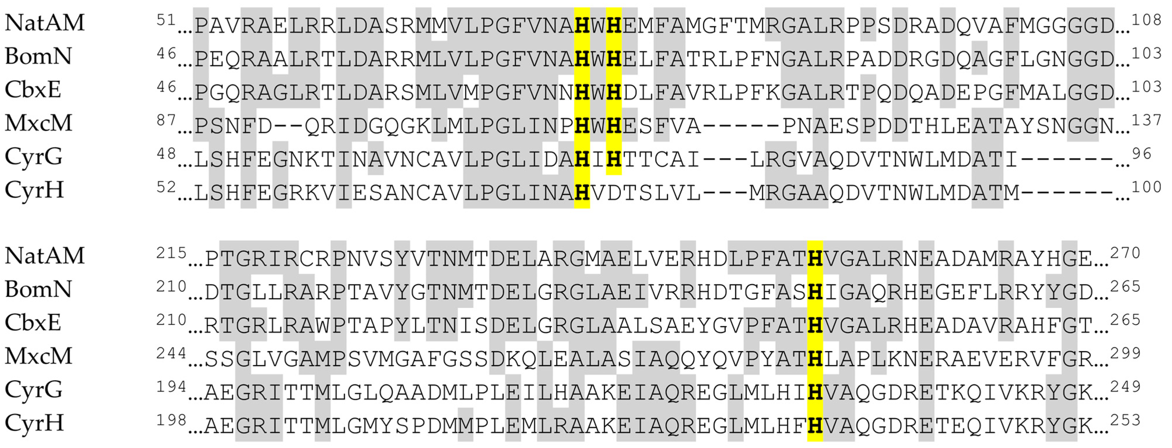
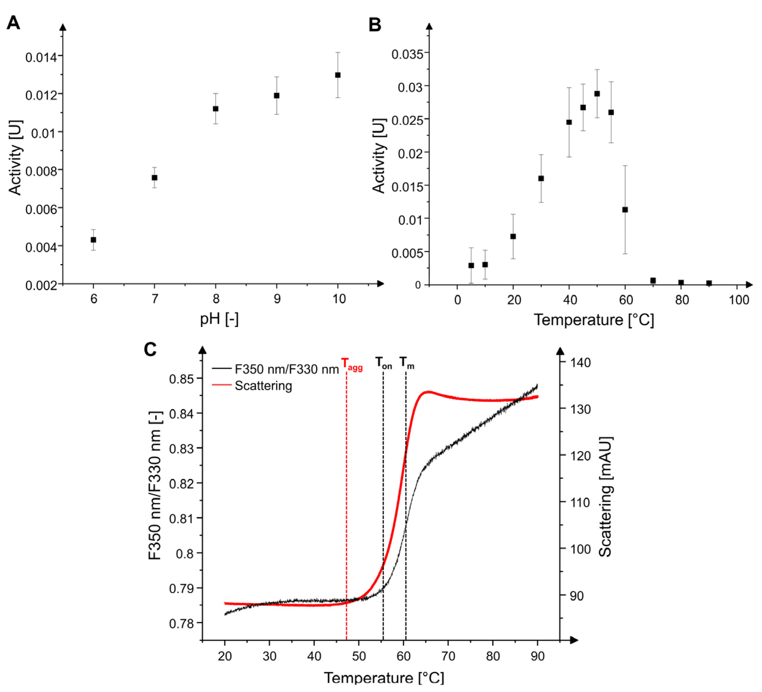
2.1. Characterization of MxcM

Most amidohydrolases possess a catalytic zinc ion in their active center [2,3]. The amidohydrolase NatAM represents the closest homolog of MxcM for which a structure was determined [8]. A sequence alignment shows that the three histidine residues which are responsible for the coordination of the zinc ion in NatAM (His75, His77, and His253), are similar in MxcM and in the other heterocycle-forming amidohydrolases (Figure 2). Only CyrH lacks a histidine residue at position 77 and features an aspartic acid residue instead. Since supplementation of 5 mM EDTA was reported to eliminate the enzymatic activity of NatAM [8], we also evaluated the effect of this complexing agent on the activity of MxcM. Unexpectedly, the addition of EDTA did not significantly affect the enzymatic conversion of the tested MxcM substrate myxochelin B (residual activity 93.6 ± 2.6%). After supplementation of 10 mM ZnCl2 following the EDTA treatment, we did not observe a recovery of the enzymatic activity.
In respect of identifying optimal reaction parameters, the pH and temperature profiles were investigated. MxcM showed activity over the entire analyzed pH range (Figure 3A). The highest activity was observed at pH 10. Due to a degradation of myxochelin B and pseudochelin A above pH 10, it was not easy to the determine the enzymatic activity in this pH range. According to BRENDA (=Braunschweig Enzyme Database), the average pH optimum of 551 previously analyzed amidohydrolases lies between pH 7 and 8 [20].
The activity of MxcM was observed to increase steadily from 5 to 50 °C (Figure 3B). Above 50 °C, the activity rapidly declines until it is completely abolished at temperatures of 75 °C and higher. Among 360 characterized amidohydrolases, the average optimal temperature was between 30 and 40 °C [20]. The relative abundance of amidohydrolases exhibiting a temperature optimum between 50 and 60 °C was calculated to 12.5% [20].
The thermal stability of MxcM was determined by analyzing thermal unfolding and aggregation processes while heating enzyme solutions from 20 to 90 °C (Figure 3C). The shift in the ratio of tryptophan fluorescence at 330 nm and 350 nm during protein unfolding can be used to calculate the unfolding transmission temperature (Tm), which is the temperature point where half of the protein is unfolded. Ton refers to the temperature at which the protein starts to unfold. Increasing light scattering values can be correlated to aggregation of enzyme in the aqueous solution. The aggregation temperature (Tagg) describes the point at which the protein begins to aggregate. Measurements of the tryptophan fluorescence ratio F350 nm/F330 nm of MxcM delivered a Ton of 55.7 ± 0.4 °C and a Tm of 60.6 ± 0.1 °C. The aggregation process is initiated at a temperature of 47.1 ± 2.7 °C.

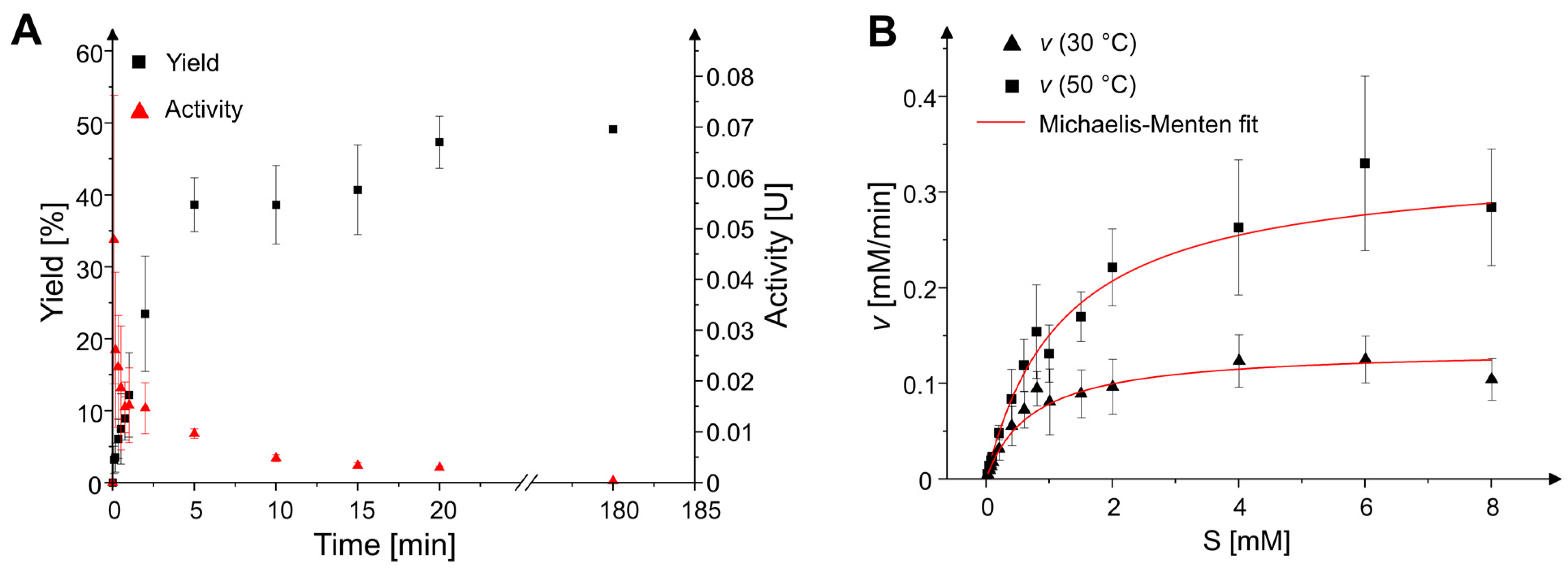
2.2. Reaction Time Course and Kinetic Parameters

The time-dependent course of the MxcM reaction in batch was recorded by varying the incubation time from 5 s to 6 h (Figure 4A). After starting the enzymatic reaction, the product yield increases rapidly from 0 to 38.6% within 5 min. Under standard assay conditions, the equilibrium concentration of 45–50% pseudochelin A is reached after 20 min. The yield-time dependency strongly correlates with the enzymatic activity. The highest activities were observed in the beginning of the reaction. Afterwards, the activity declines with ongoing substrate conversion. The activity approaches to zero with approximating the chemical equilibrium. For determination of the Michaelis–Menten kinetic, the reaction rates for different substrate concentrations after a reaction time of 2 min were calculated and plotted (Figure 4B).
A non-linear fitting of these data against the Michaelis–Menten equation delivered the parameters Km and vmax (Table 1). From these parameters, the enzymatic turnover number (kcat) and the catalytic efficiency (kcat/Km) were calculated. As expected, higher reaction rates and an increased catalytic efficiency were observed at an elevated incubation temperature of 50 °C.

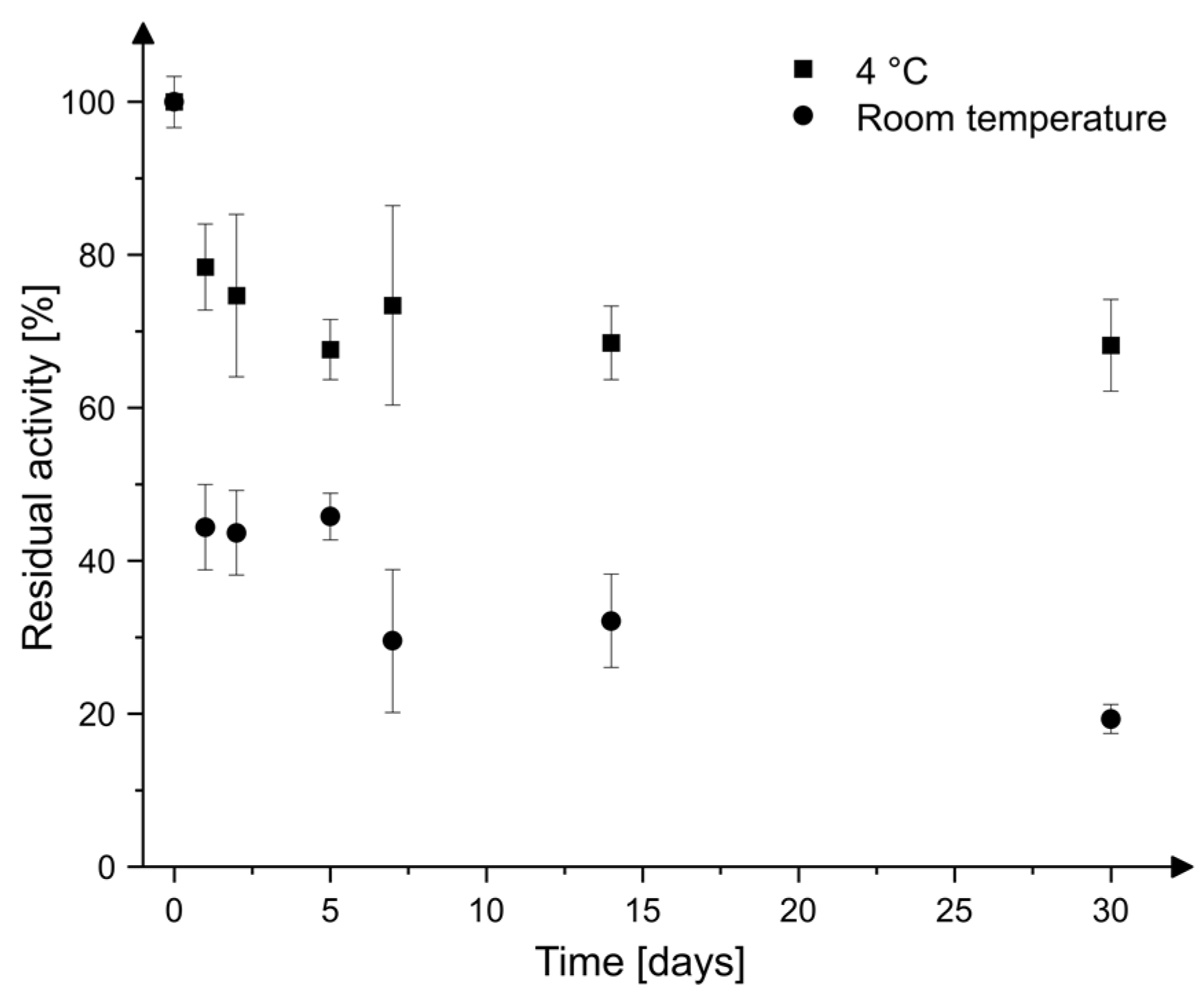
2.3. Storage Stability

When MxcM was stored in phosphate buffer (pH 7), only a slow-paced decline in enzymatic activity was observed. The curve profiles suggest that the time-dependent activity decline is correlated to the storage temperature (Figure 5). For every measured time point, the average residual activity of MxcM was higher at a storage temperature of 4 °C in comparison to room temperature. Even after 30 days of storage, the residual activity remained at 68.2 ± 6.0% (4 °C) or 19.3 ± 1.9% (room temperature).

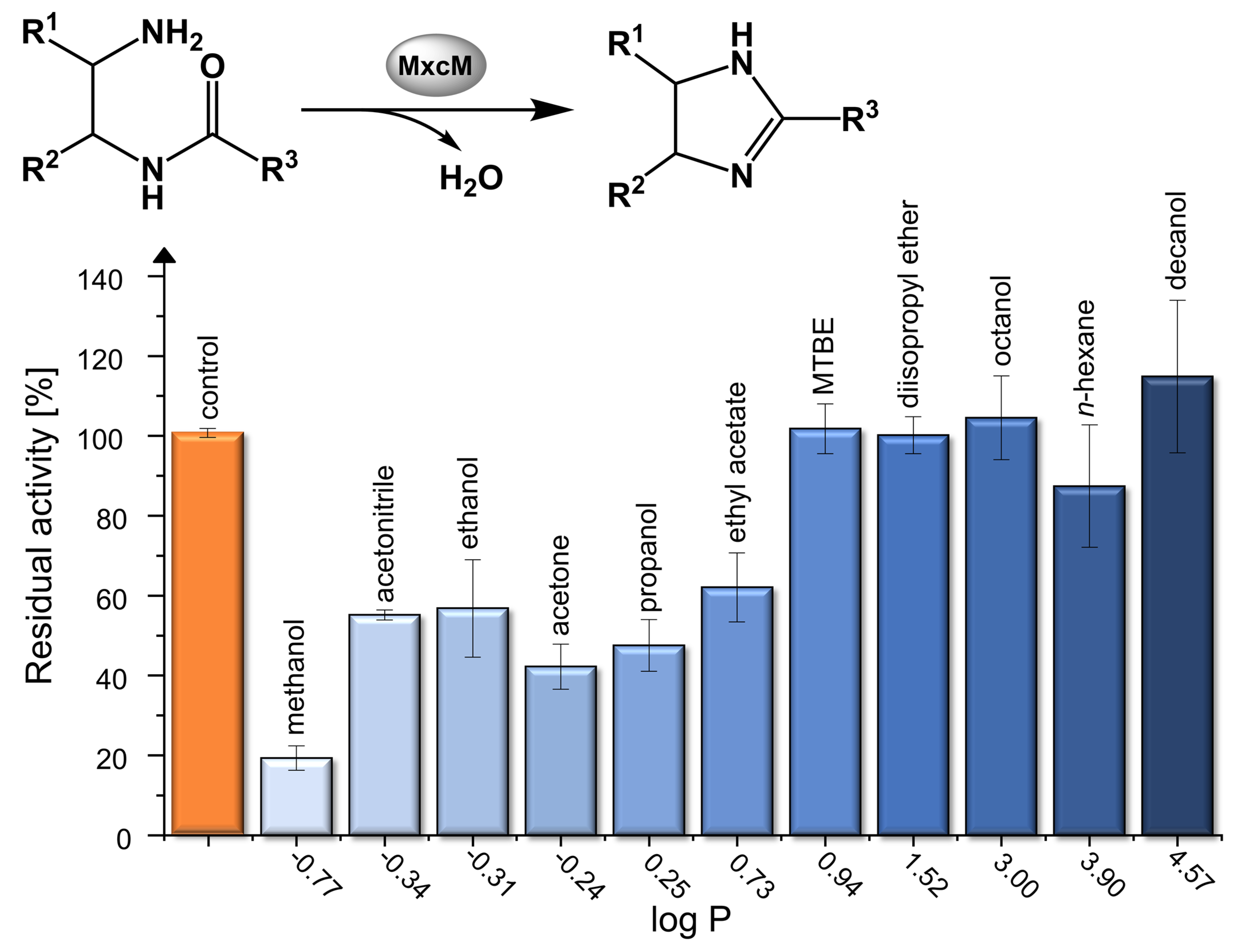
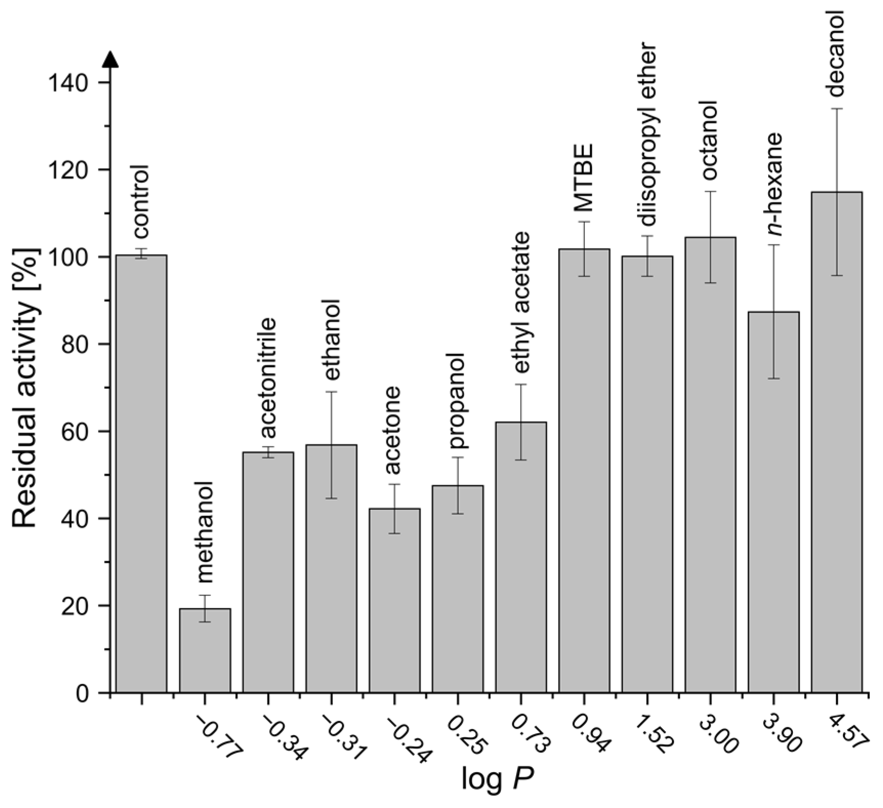
2.4. Activity in Organic Solvents and Salt-Containing Solutions

The use of enzymes in non-aqueous systems can implicate diverse advantages in bio-catalysis, e.g., increased solubility of substrates and products, a directed shift of thermodynamic equilibria or simplified product isolation and purification procedures [21,22,23]. Investigation of the residual enzymatic activity in several organic solvents compared to a control reaction in aqueous buffer revealed that MxcM is active in every tested solvent (Figure 6). Additionally, a dependency between the residual activity and the octanol-water partition coefficient (log P) of the organic solvents was observed. In hydrophilic organic solvents, the activity of MxcM decreases significantly. For instance, the residual activity in methanol, which possesses the highest hydrophilicity among the tested solvents (log P −0.77), was only 19.3 ± 3.1%. In contrast, the activity was elevated to 114.8 ± 19.1% in the hydrophobic solvent decanol (log P 4.57). High salt concentrations were found to have no significant impact on the enzymatic activity. When the enzymatic reaction was conducted in the presence of 2 M NaCl, the residual activity of MxcM amounts to 90.7 ± 5.2%. Notably, the addition of urea elevated the residual activity to 107.4 ± 0.5%.

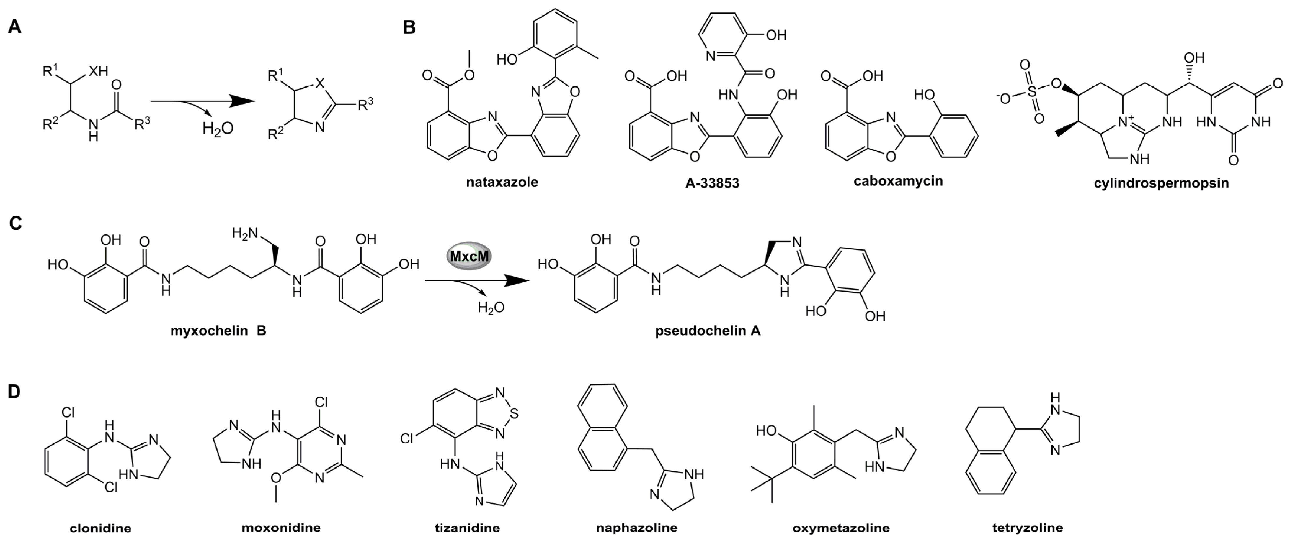
2.5. Substrate Specificity

In vivo studies already revealed that MxcM is capable to process several analogues of its natural substrate myxochelin B [11]. Here, we examined the substrate specificity in vitro for the first time (Table 2). Consistent with our previous investigations [11], we did not find any evidence for the conversion of a substrate featuring a β-hydroxyethyl amide in lieu of the β-aminoethyl amide moiety in myxochelin B. It is thus evident that MxcM is only capable of imidazoline formation and not of oxazoline formation. In contrast, modifications on the aromatic rings of the natural substrate were tolerated. The two substrate analogues myxochelin B1 and B2, which had been produced by precursor-directed biosynthesis [11], were successfully converted, although the residual activity compared to the natural substrate was reduced. While the measured residual activity for myxochelin B1 was 73.3 ± 3.3%, the activity in presence of myxochelin B2 decreased to 10.2 ± 1.9%. When an enzymatic reaction with the synthetic compound N-benzoylethylenediamine was performed, no product formation was observed.

3. Discussion

Most members of the amidohydrolase superfamily catalyze metal-dependent hydrolysis reactions [3,4]. Usually, amidohydrolases require a zinc ion that is considered to activate the target bond and to coordinate a nucleophilic water in their active centers [3,25]. Even condensing enzymes, such as the amidohydrolase NatAM, were reported to depend on a catalytic zinc ion [8]. A sequence alignment verified that the zinc-coordinating residues His75, His77 and His253 of NatAM are highly similar to those in MxcM. Consequently, this finding suggests that MxcM is a metal-dependent amidohydrolase as well. However, the addition of EDTA had only a negligible effect on the MxcM-catalyzed reaction. This experimental result contradicts the previously established hypothesis and indicates that MxcM may belong to the rare metal-independent amidohydrolases. Up to date, only few metal-independent amidohydrolases have been described, among them are the allantoinase PuuE from Pseudomonas fluorescens [26] and the uronate isomerase from Escherichia coli [5]. In case of the uronate isomerase, the enzyme was shown to be capable of binding a zinc ion, but the zinc ion is not required for catalytic activity. In order to identify the role of metal ions for MxcM, further experiments are necessary.
The pH profile of MxcM shows a continuous increase of enzymatic activity from pH 6 to 10. Maximal activity was obtained at pH 10, but for higher pH values, the activity could not be determined due to a degradation of the substrate. Analyzing a dataset of previously described amidohydrolases revealed that highly alkaline pH optima in this class of enzymes are rarely found in nature [20]. MxcM shows activity over a broad temperature range (5–60 °C) with an optimum at 50 °C. Thermal stability testing showed that the unfolding process begins at a temperature of 55.7 ± 0.4 °C. At 60.6 ± 0.1 °C, 50% of the enzyme is unfolded. The starting point for protein aggregation was detected at 47.1 ± 2.7 °C. These data confirm that reaction temperatures above 50 °C lead to a rapid inactivation of MxcM. A comparison of the kinetic parameters at 30 °C and 50 °C demonstrated a significantly higher turnover number (kcat) and catalytic efficiency (kcat/Km) at the enzyme’s temperature optimum. With values of 27.6 s−1 and 22,932 s−1 M−1, MxcM can be defined as an enzyme with moderate catalytic efficiency, according to the categorization by Bar-Even et al. [27].
Aside from that, MxcM possesses an intriguing storage stability in aqueous systems. The residual activity shows an exponential decline with progressing storage time, whereby storing MxcM at 4 °C had lower impact on the enzyme’s activity. After 30 days of storage, the residual activity amounts to 68.3 ± 6.0%. Additionally, MxcM was proven to be highly active in different organic solvents. The activity correlates strongly with the log p value, which classifies organic solvents into different hydrophobicity groups [21,28]. When the log P is lower than 2, an organic solvent is described as hydrophilic and water miscible. Hydrophobic solvents, which are immiscible with water, generally exhibit log p values higher than 4. Since the intramolecular condensation catalyzed by MxcM produces water as by-product, we assume that the reaction equilibrium is shifted to the product side when the reaction is performed in hydrophobic organic solvents. Compared to hydrophilic solvents, the use of water immiscible organic media prevents a disruption of the enzyme’s hydration shell [21,22,29]. Amidohydrolases arrange water molecules in their active centers. This arrangement is crucial for the catalytic activity [22,23]. Hydrophilic solvents can remove these water molecules, causing a loss of catalytic activity. Because salts generally reduce the water activity of an aqueous solution, the tolerance towards organic solvents is often accompanied by a resistance to high salt concentrations [23]. MxcM originates from the marine bacterium P. piscicida S2040 [10,30]. Strains of P. piscicida are able to grow under moderate halophilic conditions [31], suggesting that this correlation might also be true for the amidohydrolase MxcM. Enzymatic reactions in presence of 2 M NaCl or urea did not significantly affect the enzymatic activity. Interestingly, the residual activity of MxcM in the presence of 2 M urea, which is commonly used for the denaturation of proteins [32], raised to 107.4 ± 0.5%. This result validated the hypothesis that MxcM can perform biocatalysis under halophilic conditions.
First evidence for the substrate tolerance of MxcM was obtained from a feeding study of a recombinant M. xanthus strain expressing MxcM [11]. Various benzoic acid derivatives were successfully introduced into myxochelin biosynthesis. The intermediary produced myxochelin B derivatives were further converted into pseudochelin A analogues, presumably due to the activity of MxcM [11]. In this study, the substrate specificity of MxcM was further examined in vitro. In accordance with the in vivo experiments, myxochelin A was not accepted, implying that the β-aminoethyl amide moiety is essential for a successful conversion. Incubation of MxcM with the substrates myxochelin B1 and myxochelin B2 led to the formation of pseudochelin A1 and A2, respectively. We noted, however, a strong discrepancy in the enzymatic conversion of the two non-natural substrates. The residual activity of MxcM was significantly lower for myxochelin B2, which lacks the hydroxyl groups on the benzene ring adjacent to the β-aminoethyl amide moiety. It is possible that these groups facilitate the catalytic conversion due to electronic effects or by mediating additional protein-ligand interactions. This would also explain why incubation of the synthetic compound N-benzoylethylenediamine with MxcM did not result in product formation. Further testing of the substrate specificity of MxcM is certainly advisable.

4. Materials and Methods

4.1. Enzyme Production and Purification

The production of 6xHis-tagged MxcM was conducted using the recombinant Escherichia coli BL21(DE3):pET28a(+)-mxcM according to the previously described protocol [10]. The enzyme was purified from the cell lysate by Ni-nitrilotriacetic acid (NTA) affinity chromatography using Protino Ni-NTA agarose (Macherey–Nagel, Düren, Germany) in a polypropylene column (Qiagen, Hilden, Germany). After equilibrating the stationary phase with 10 mL lysis buffer (50 mM NaH2PO4, 300 mM NaCl, 10 mM imidazole, 10 vol.% glycerol, 20 mM β-mercaptoethanol; pH 8), the MxcM-containing supernatant was applied onto the Ni-NTA matrix. Subsequently, the matrix was washed with 5 mL washing buffer I (lysis buffer including 20 mM imidazole) and 5 mL washing buffer II (lysis buffer including 40 mM imidazole). The 6x-His MxcM was recovered from the Ni-NTA agarose by elution with 2.5 mL elution buffer (lysis buffer including 250 mM imidazole). For desalting the enzyme solution, PD-10 desalting columns (- Cytiva Europe, Freiburg, Germany) were used. After the desalting columns were equilibrated with 25 mL phosphate buffer (100 mM, pH 7), the columns were placed in 50 mL collection tubes. The enzyme solution was applied and eluted by centrifugation (1000× g, 2 min). The purified enzyme solution was analyzed via SDS-PAGE and the concentration of MxcM was determined using the UV-Vis spectrophotometer NanoDrop One (Thermo Fisher Scientific, Dreieich, Germany).

4.2. Activity Assay

In vitro assays were generally performed in 50 mM phosphate buffer (NaH2PO4, pH 9) using 0.2 µM (0.01 mg/mL) MxcM and 0.5 mM myxochelin B. Myxochelin B, which is the natural substrate of MxcM, was isolated and purified from Myxococcus xanthus DSM16526 cultures according to a previously published protocol [10]. After the reaction mixture was incubated at 30 °C for 2 min, MxcM was inactivated with 2 M guanidinium thiocyanate. The reaction products were analyzed by liquid chromatography-mass spectrometry (LC-MS; Agilent 1260 Infinity LC System, Agilent Technologies, Waldbronn, Germany; Bruker Compact Q-TOF mass spectrometer, Bruker Daltonik, Bremen, Germany) using a Nucleoshell RP18 column (Macherey–Nagel, Düren, Germany) or by standard high performance liquid chromatography (HPLC, Shimadzu LC-20AD, Shimadzu, Duisburg, Germany) with a Nucleodur Sphinx RP column (Macherey–Nagel) coupled to a diode array detector (Shimadzu SPD-M20A). The conversion from myxochelin B to pseudochelin A was quantified from the peak areas in the respective chromatograms, which was subsequently correlated to the enzyme activity. One unit (U) MxcM activity is defined as amount of enzyme required for the conversion of 1 µmol myxochelin B per minute. Each experiment was performed in multiple independent replicates (N = number of determinations).

4.3. Determination of Metal Dependency, pH and Temperature Optima, and Thermal Stability

The metal dependency of MxcM was analyzed by incubating the 0.2 µM enzyme with 5 mM ethylenediaminetetraacetic acid (EDTA) for 2 h in phosphate buffer. To restore the enzymatic activity after EDTA treatment, 10 mM ZnCl2 was supplemented and the mixture was incubated for 15 min. Subsequently, the substrate myxochelin B (0.5 mM) was added and the reactions were incubated for 30 min at 30 °C. The pH tolerance of MxcM in phosphate buffer was analyzed in a range from pH 6 to 10 under standard assay conditions. To determine the optimal temperature for MxcM activity, the temperature was varied from 5 °C to 90 °C under standard conditions. The thermal stability of MxcM was investigated using a NanoTemper Prometheus NT.48 (NanoTemper Technologies, Munich, Germany), which is based on differential scanning fluorimetry. The protein samples were analyzed over a temperature range from 20 to 90 °C with an heating rate of 1 °C/min. Thermal unfolding and protein aggregation was recorded by measuring the temperature-dependent tryptophan fluorescence at 330 and 350 nm as well as the light scattering. The experiment was conducted with two independent biological replicates in fivefold determination.

4.4. Enzyme Kinetics

The time course of the MxcM-catalyzed reaction was determined under standard assay conditions by variation of the incubation time. Kinetic parameters of the MxcM-catalyzed reaction were determined by measuring the initial conversion rates for different substrate concentrations under standard assay conditions. The enzyme kinetics were recorded at incubation temperatures of 30 °C and 50 °C. Plotting and calculation of the Michaelis–Menten constant (Km) and the maximal reaction rate (vmax) was performed using the nonlinear curve fitting and the Levenberg-Marquardt algorithm in Origin2020 (OriginLab).

4.5. Storage Stability

For analyzing the storage stability of MxcM in aqueous buffer (phosphate buffer, 50 mM, pH 7), the purified enzyme was stored at 4 °C or at room temperature. After 0, 1, 2, 5, 7, 14 and 30 days of storage, enzymatic reactions were performed with 0.5 µM MxcM and 0.5 mM myxochelin B in phosphate buffer. The reactions were incubated for 2 min at 30 °C and terminated by heating to 99 °C for 5 min.

4.6. Activity in Organic Solvents and Salt-Containing Solutions

To test the influence of organic solvents, purified MxcM (2 µM) was incubated with 0.5 mM myxochelin B for 30 min at 30 °C in methanol, acetonitrile, ethanol, acetone, propanol, ethyl acetate, methyl-tert-butylether (MTBE), diisopropyl ether, octanol, n-hexane or decanol. The concentration of water was adjusted to 5–10 vol.%. Halophilic properties of MxcM were investigated by adding 2 M NaCl or 2 M urea to a standard reaction mixture. The reactions were stopped by heating at 99 °C for 5 min. As control, the reaction was conducted in phosphate buffer (50 mM, pH 9) under similar conditions.

4.7. Substrate Specificity

The substrate tolerance of MxcM was studied using myxochelin A, myxochelin B1, myxochelin B2, and N-benzoylethylenediamine as substrates. Myxochelin A, myxochelin B1 and B2 were isolated from M. xanthus cultures according to previously published protocols [10,11]. Synthetic N-benzoylethylenediamine hydrochloride was obtained from Fluorochem. The enzymatic assays were performed with 0.5 mM substrate and 0.2 µM MxcM in phosphate buffer. The reactions were incubated for 30 min at 30 °C and stopped by heating to 99 °C for 5 min.

5. Conclusions

MxcM is the first enzyme in the amidohydrolase superfamily; it has been shown to catalyze the formation of an imidazoline ring. In the present study, we characterized the MxcM both biochemically and kinetically. We defined the optimal pH and temperature for catalytic activity and analyzed the Michaelis–Menten kinetics as well as its storage stability in aqueous systems. MxcM naturally exhibits an intriguing robustness towards organic solvents and concentrated salt-solutions. The described characteristics and the fact that no (organic) cofactor is required for its action make the amidohydrolase MxcM a promising candidate for biocatalytic imidazoline formation.

Author Contributions

Conceptualization, L.W., D.J.V. and M.N.; methodology, L.W. and D.J.V.; formal analysis, L.W., D.J.V. and J.H.; investigation, L.W., D.J.V. and J.H.; writing—original draft preparation, L.W., D.J.V. and M.N.; writing—review and editing, L.W., D.J.V. and M.N.; visualization, L.W.; supervision, M.N.; project administration, M.N. All authors have read and agreed to the published version of the manuscript.

Funding

Not applicable.

Data Availability Statement

Publicly available datasets were analyzed in this study. This data can be found here: www.brenda-enzymes.org (accessed on 3 June 2021); https://pubchem.ncbi.nlm.nih.gov/ (accessed on 3 June 2021).

Acknowledgments

We gratefully acknowledge the support from Maximilian Wessner and the Chair of Thermodynamics, TU Dortmund University, on the measurement of thermal stability data. We thank our former group members Angela Sester and Juliane Korp for preliminary work and valuable discussions.

Conflicts of Interest

The authors declare no conflict of interest.

References

  1. Akiva, E.; Brown, S.; Almonacid, D.E.; Barber, A.E.; Custer, A.F.; Hicks, M.A.; Huang, C.C.; Lauck, F.; Mashiyama, S.T.; Meng, E.C.; et al. The Structure-Function Linkage Database. Nucleic Acids Res. 2014, 42, D521–D530. [Google Scholar] [CrossRef] [PubMed] [Green Version]
  2. Holm, L.; Sander, C. An evolutionary treasure: Unification of a broad set of amidohydrolases related to urease. Proteins 1997, 28, 72–82. [Google Scholar] [CrossRef]
  3. Seibert, C.M.; Raushel, F.M. Structural and catalytic diversity within the amidohydrolase superfamily. Biochemistry 2005, 44, 6383–6391. [Google Scholar] [CrossRef]
  4. Aimin, L.; Tingfeng, L.; Rong, F. Amidohydrolase Superfamily. In Encyclopedia of Life Sciences; John Wiley & Sons, Ltd.: Chichester, UK, 2001; ISBN 047001590X. [Google Scholar]
  5. Williams, L.; Nguyen, T.; Li, Y.; Porter, T.N.; Raushel, F.M. Uronate isomerase: A nonhydrolytic member of the amidohydrolase superfamily with an ambivalent requirement for a divalent metal ion. Biochemistry 2006, 45, 7453–7462. [Google Scholar] [CrossRef] [PubMed] [Green Version]
  6. Lv, M.; Zhao, J.; Deng, Z.; Yu, Y. Characterization of the Biosynthetic Gene Cluster for Benzoxazole Antibiotics A33853 Reveals Unusual Assembly Logic. Chem. Biol. 2015, 22, 1313–1324. [Google Scholar] [CrossRef] [Green Version]
  7. Losada, A.A.; Cano-Prieto, C.; García-Salcedo, R.; Braña, A.F.; Méndez, C.; Salas, J.A.; Olano, C. Caboxamycin biosynthesis pathway and identification of novel benzoxazoles produced by cross-talk in Streptomyces sp. NTK 937. Microb. Biotechnol. 2017, 10, 873–885. [Google Scholar] [CrossRef] [PubMed]
  8. Song, H.; Rao, C.; Deng, Z.; Yu, Y.; Naismith, J.H. The Biosynthesis of the Benzoxazole in Nataxazole Proceeds via an Unstable Ester and has Synthetic Utility. Angew. Chem. Int. Ed Engl. 2020, 59, 6054–6061. [Google Scholar] [CrossRef] [PubMed]
  9. Mihali, T.K.; Kellmann, R.; Muenchhoff, J.; Barrow, K.D.; Neilan, B.A. Characterization of the gene cluster responsible for cylindrospermopsin biosynthesis. Appl. Environ. Microbiol. 2008, 74, 716–722. [Google Scholar] [CrossRef] [Green Version]
  10. Korp, J.; Winand, L.; Sester, A.; Nett, M. Engineering Pseudochelin Production in Myxococcus xanthus. Appl. Environ. Microbiol. 2018, 84. [Google Scholar] [CrossRef] [Green Version]
  11. Sester, A.; Winand, L.; Pace, S.; Hiller, W.; Werz, O.; Nett, M. Myxochelin- and Pseudochelin-Derived Lipoxygenase Inhibitors from a Genetically Engineered Myxococcus xanthus Strain. J. Nat. Prod. 2019, 82, 2544–2549. [Google Scholar] [CrossRef]
  12. Winand, L.; Sester, A.; Nett, M. Bioengineering of Anti-Inflammatory Natural Products. ChemMedChem 2021, 16, 767–776. [Google Scholar] [CrossRef] [PubMed]
  13. Schieferdecker, S.; König, S.; Koeberle, A.; Dahse, H.-M.; Werz, O.; Nett, M. Myxochelins target human 5-lipoxygenase. J. Nat. Prod. 2015, 78, 335–338. [Google Scholar] [CrossRef]
  14. Sączewski, F.; Kornicka, A.; Balewski, Ł. Imidazoline scaffold in medicinal chemistry: A patent review (2012–2015). Expert Opin. Ther. Pat. 2016, 26, 1031–1048. [Google Scholar] [CrossRef]
  15. Guan, X.; Hu, Y. Imidazoline derivatives: A patent review (2006--present). Expert Opin. Ther. Pat. 2012, 22, 1353–1365. [Google Scholar] [CrossRef]
  16. U.S. Food and Drug Administration. Drug Approvals and Databases. Available online: https://www.accessdata.fda.gov/scripts/cder/daf/index.cfm (accessed on 31 May 2021).
  17. Mehedi, M.S.A.; Tepe, J.J. Recent Advances in the Synthesis of Imidazolines (2009–2020). Adv. Synth. Catal. 2020, 362, 4189–4225. [Google Scholar] [CrossRef]
  18. Lechner, H.; Pressnitz, D.; Kroutil, W. Biocatalysts for the formation of three- to six-membered carbo- and heterocycles. Biotechnol. Adv. 2015, 33, 457–480. [Google Scholar] [CrossRef] [Green Version]
  19. Hemmerling, F.; Hahn, F. Biosynthesis of oxygen and nitrogen-containing heterocycles in polyketides. Beilstein J. Org. Chem. 2016, 12, 1512–1550. [Google Scholar] [CrossRef] [PubMed] [Green Version]
  20. Chang, A.; Jeske, L.; Ulbrich, S.; Hofmann, J.; Koblitz, J.; Schomburg, I.; Neumann-Schaal, M.; Jahn, D.; Schomburg, D. BRENDA, the ELIXIR core data resource in 2021: New developments and updates. Nucleic Acids Res. 2021, 49, D498–D508. [Google Scholar] [CrossRef]
  21. Wang, S.; Meng, X.; Zhou, H.; Liu, Y.; Secundo, F.; Liu, Y. Enzyme Stability and Activity in Non-Aqueous Reaction Systems: A Mini Review. Catalysts 2016, 6, 32. [Google Scholar] [CrossRef] [Green Version]
  22. Klibanov, A.M. Improving enzymes by using them in organic solvents. Nature 2001, 409, 241–246. [Google Scholar] [CrossRef]
  23. Doukyu, N.; Ogino, H. Organic solvent-tolerant enzymes. Biochem. Eng. J. 2010, 48, 270–282. [Google Scholar] [CrossRef]
  24. Kim, S.; Chen, J.; Cheng, T.; Gindulyte, A.; He, J.; He, S.; Li, Q.; Shoemaker, B.A.; Thiessen, P.A.; Yu, B.; et al. PubChem in 2021: New data content and improved web interfaces. Nucleic Acids Res. 2021, 49, D1388–D1395. [Google Scholar] [CrossRef]
  25. Hernick, M.; Fierke, C.A. Zinc hydrolases: The mechanisms of zinc-dependent deacetylases. Arch. Biochem. Biophys. 2005, 433, 71–84. [Google Scholar] [CrossRef] [PubMed]
  26. Ramazzina, I.; Cendron, L.; Folli, C.; Berni, R.; Monteverdi, D.; Zanotti, G.; Percudani, R. Logical identification of an allantoinase analog (puuE) recruited from polysaccharide deacetylases. J. Biol. Chem. 2008, 283, 23295–23304. [Google Scholar] [CrossRef] [Green Version]
  27. Bar-Even, A.; Noor, E.; Savir, Y.; Liebermeister, W.; Davidi, D.; Tawfik, D.S.; Milo, R. The moderately efficient enzyme: Evolutionary and physicochemical trends shaping enzyme parameters. Biochemistry 2011, 50, 4402–4410. [Google Scholar] [CrossRef]
  28. Laane, C.; Boeren, S.; Vos, K.; Veeger, C. Rules for optimization of biocatalysis in organic solvents. Biotechnol. Bioeng. 1987, 30, 81–87. [Google Scholar] [CrossRef]
  29. Zaks, A.; Klibanov, A.M. Enzymatic catalysis in nonaqueous solvents. J. Biol. Chem. 1988, 263, 3194–3201. [Google Scholar] [CrossRef]
  30. Sonnenschein, E.C.; Stierhof, M.; Goralczyk, S.; Vabre, F.M.; Pellissier, L.; Hanssen, K.Ø.; de La Cruz, M.; Díaz, C.; de Witte, P.; Copmans, D.; et al. Pseudochelin A, a siderophore of Pseudoalteromonas piscicida S2040. Tetrahedron 2017, 73, 2633–2637. [Google Scholar] [CrossRef] [Green Version]
  31. Richards, G.P.; Needleman, D.S.; Watson, M.A. Complete Genome Sequence of Pseudoalteromonas piscicida Strain DE2-B, a Bacterium with Broad Inhibitory Activity toward Human and Fish Pathogens. Genome Announc. 2017, 5, e00752-17. [Google Scholar] [CrossRef] [Green Version]
  32. Hua, L.; Zhou, R.; Thirumalai, D.; Berne, B.J. Urea denaturation by stronger dispersion interactions with proteins than water implies a 2-stage unfolding. Proc. Natl. Acad. Sci. USA 2008, 105, 16928–16933. [Google Scholar] [CrossRef] [PubMed] [Green Version]
Figure 1. (A) General reaction mechanism of heterocycle formation catalyzed by amidohydrolases. (B) Examples of natural products containing amidohydrolase-produced heterocycles. (C) MxcM-catalyzed formation of the imidazoline ring in pseudochelin A biosynthesis. (D) Examples of approved drugs featuring an imidazoline scaffold.
Catalysts 11 00892 g001
Figure 2. Alignment of amino acid sequences from heterocycle-forming amidohydrolases. Homologous amino acid residues (threshold ≥ 50%) are highlighted in grey. Amino acid residues involved in the coordination of the Zn2+ ion in NatAM [8] are marked in yellow.
Catalysts 11 00892 g002
Figure 3. Effect of pH and temperature on the activity of MxcM. (A) Activity at different pH values in 50 mM phosphate buffer at 30 °C (N = 5; 0.2 µM MxcM). (B) Activity for varied incubation temperatures in 50 mM phosphate buffer at pH 9 (N = 4; 0.2 µM MxcM). (C) Temperature-dependent unfolding and aggregation curves of MxcM (N = 10).
Catalysts 11 00892 g003
Figure 4. Reaction time course. (A) Dependency of pseudochelin A yield and MxcM activity to the reaction time (N0–20min = 6; N180min = 2; 0.2 µM MxcM) in a batch experiment. (B) Michaelis–Menten plot. Dependency of reaction rate (v) and substrate concentration (S) at temperatures of 30 and 50 °C (N = 6; 0.2 µM MxcM).
Catalysts 11 00892 g004
Figure 5. Storage stability of MxcM in 50 mM phosphate buffer (pH 7) at 4 °C and room temperature (N = 4; 0.5 µM MxcM).
Catalysts 11 00892 g005
Figure 6. Residual activity of MxcM in diverse organic solvents (N = 3; 2 µM MxcM) in comparison to a control reaction in phosphate buffer (pH 9). The hydrophobicity of each solvent is represented by its log p value, which was retrieved from the Pubchem database [24].
Catalysts 11 00892 g006
Table 1. Kinetic parameters of MxcM. Values of vmax and Km are calculated from non-linear fitting to the Michaelis–Menten equation and are provided as arithmetic mean plus standard deviation.
Temperaturevmax [mM/min]Km [mM]kcat [s−1]kcat/Km [s−1 M−1]
30 °C0.13 ± 0.010.74 ± 0.0911.1915,048.01
50 °C0.33 ± 0.021.21 ± 0.0927.6422,932.03
Table 2. Analysis of the substrate specificity of MxcM (N = 3; 0.2 µM MxcM). n.d. not detected.
Catalysts 11 00892 i001
SubstrateR1R2R3R4R5Residual Activity [%]
myxochelin BOHOHOHOHNH2100 ± 1.9
myxochelin AOHOHOHOHOHn.d.
myxochelin B1HHOHOHNH273.3 ± 3.3
myxochelin B2OHOHHHNH210.2 ± 1.9
N-benzoylethylenediamine-----n.d.
Publisher’s Note: MDPI stays neutral with regard to jurisdictional claims in published maps and institutional affiliations.

