Article

Characterization of a Novel Lutein Cleavage Dioxygenase, EhLCD, from Enterobacter hormaechei YT-3 for the Enzymatic Synthesis of 3-Hydroxy-β-ionone from Lutein

1
China Tobacco Guangxi Industrial Co., Ltd., Nanning 530001, China
2
School of Food and Biological Engineering, Zhengzhou University of Light Industry, 5 Dongfeng Rd, Zhengzhou 450002, China
*
Author to whom correspondence should be addressed.
Catalysts 2021, 11(11), 1257; https://doi.org/10.3390/catal11111257
Submission received: 30 September 2021 / Revised: 16 October 2021 / Accepted: 18 October 2021 / Published: 20 October 2021
(This article belongs to the Special Issue Enzyme Catalysis, Biotransformation and Bioeconomy)

Abstract

:
3-Hydroxy-β-ionone, a flavor and fragrance compound with fruity violet-like characteristics, is widely applied in foodstuff and beverages, and is currently produced using synthetic chemistry. In this study, a novel lutein cleavage enzyme (EhLCD) was purified and characterized from Enterobacter hormaechei YT-3 to convert lutein to 3-hydroxy-β-ionone. Enzyme EhLCD was purified to homogeneity by ammonium sulfate precipitation, Q-Sepharose, phenyl-Sepharose, and Superdex 200 chromatography. The molecular mass of purified EhLCD, obtained by SDS-PAGE, was approximately 50 kDa. The enzyme exhibited the highest activity toward lutein, followed by zeaxanthin, β-cryptoxanthin, and β-carotene, suggesting that EhLCD exhibited higher catalytic efficiency for carotenoid substrates bearing 3-hydroxy-ionone rings. Isotope-labeling experiments showed that EhLCD incorporated oxygen from O2 into 3-hydroxy-β-ionone and followed a dioxygenase reaction mechanism for different carotenoid substrates. These results indicated that EhLCD is the first characterized bacterial lutein cleavage dioxygenase. Active EhLCD was also confirmed to be a Fe2+-dependent protein with 1 molar equivalent of non-haem Fe2+. The purified enzyme displayed optimal activity at 45 °C and pH 8.0. The optimum concentrations of the substrate, enzyme, and Tween 40 for 3-hydroxy-β-ionone production were 60 μM lutein/L, 1.5 U/mL, and 2% (w/v), respectively. Under optimum conditions, EhLCD produced 3-hydroxy-β-ionone (637.2 mg/L) in 60 min with a conversion of 87.0% (w/w), indicating that this enzyme is a potential candidate for the enzymatic synthesis of 3-hydroxy-β-ionone in biotechnological applications.

1. Introduction

Lutein (3R, 3′R, 6′R-β,ε-carotene-3,3′-diol) is a member of the naturally abundant carotenoid compounds, and exists in higher plants and other photosynthetic microorganisms, such as algae, bacteria, and some types of fungus [1]. Lutein has been widely used in foods, nutraceuticals, pharmaceuticals, cosmetics, and feed owing to its antioxidant, antitumor, and antiaging activities [2,3,4,5]. Lutein is also a precursor for many flavor and fragrance compounds in raspberries, other fruits, and flowers, and its degradation products generate aromatic molecules, such as β-ionone and 3-hydroxy-β-ionone, which are widely applied in the food industry, including in confectionery, dairy products, beverages, meat, and bakery products [6,7,8,9,10]. Therefore, lutein degradation is an attractive alternative approach to producing flavor and fragrance compounds. Presently, existing lutein degradation methods are mainly physical and chemical. Lutein can be effectively oxidized under the action of light and oxygen to produce a number of apocarotenoids, but the selectivity and specificity of physical degradation methods are poor. Chemical methods for lutein degradation require harsh separation techniques, including high energy consumption, and produce pollutants and undesired byproducts. Therefore, recent studies have focused on biotechnological degradation using microbial production systems to produce natural flavors owing to the versatility, specificity, and sustainability of this method [11,12,13,14].
Although the biotechnological biotransformation of lutein is an alternative approach, only a few lutein-degrading bacteria have been identified to date. A mixed culture of Geotrichum sp. and Bacillus sp. isolated from marigold flowers was found to degrade lutein to produce volatile compounds. Geotrichum sp. was involved in lutein oxidation to produce β-ionone, and Bacillus sp. was responsible for norisoprenoid reduction to produce β-ionone derivatives [15,16]. The mixture of a yeast (Trichosporon asahii) and bacterium (Paenibacillus amylolyticus) converted lutein to β-ionone and its derivatives. T. asahii was responsible for cleaving lutein to produce β-ionone, while P. amylolyticus converted β-ionone into its reduced derivatives [17]. A lutein-degrading bacterium, Pantoea dispersa Y08, was found to cleave the 9′–10′ double bond of lutein to produce aroma compounds β-ionone and 3-hydroxy-β-ionone [18]. Zhong et al. reported that Enterobacter hormaechei A20 degraded lutein to form one new volatile product, 8-methyl-β-ionone [19].
To date, most literature reports on converting lutein to β-ionone and its derivatives have focused on the screening and isolation of lutein-degrading microorganisms. β-Carotene 9′,10′-dioxygenase (BCO2) from mammals and higher vertebrates shows lutein-degrading activity, but also catalyzes various carotenoids, including both provitamin A and non-provitamin A carotenoids, in addition to lutein and zeaxanthin [20,21,22,23]. A degrading enzyme from bacteria that is capable of converting lutein to volatile compounds has not yet been purified and characterized in its native form. In this study, we have purified and biochemically characterized a novel lutein cleavage enzyme (EhLCD) from Enterobacter hormaechei YT-3. This enzyme exhibited high activity in the conversion of lutein to 3-hydroxy-β-ionone, and the cleavage mechanism of lutein was determined.

2. Results and Discussion

2.1. Purification of EhLCD

EhLCD with lutein degradation activity was purified from the supernatant of E. hormaechei YT-3 by fractional ammonium sulfate saturation, column chromatography using Q-Sepharose and phenyl-Sepharose, and Superdex 100 gel filtration chromatography, as described in the Materials and Methods Section. The enzyme purification results are summarized in Table 1. The enzyme was purified 9.90-fold, giving a yield of 5.5% and specific activity of 2.27 U/mg. The molecular mass of the purified EhLCD was approximately 50 kDa, as determined by SDS-PAGE (Figure 1). Eight peptides, including the N-terminal amino acid sequence, were determined by tandem mass spectrometry (MS/MS), and matched those of the DUF3999 domain-containing protein (WP_096204770) of Enterobacter hormaechei, covering 23.1% of the full amino acid sequence (Supplementary Figure S1). The DUF3999 family of proteins has been functionally uncharacterized, and has one single completely conserved residue D that might be functionally important. Sequence alignment demonstrated that the DUF3999 domain-containing proteins shared low sequence identity with the characterized BCO2 (data not shown).

2.2. Substrate Specificity and Kinetic Parameters of EhLCD

Using different carotenoids as substrates, the substrate specificity and kinetic parameters of purified EhLCD were determined, as shown in Table 2. The Km and kcat values of EhLCD were calculated from the Michaelis–Menten equation. EhLCD showed high degradation activity for lutein, zeaxanthin, β-cryptoxanthin, and β-carotene, but no degradation activity was measured for β-carotene, β-carotene, apo-4′-carotenal, and lycopene (data not shown). The Km and Vmax values of EhLCD were 14 μM and 89.5 pmol mg−1·s−1 for lutein, 20 μM and 61.8 pmol mg−1·s−1 for zeaxanthin, and 52 μM and 24.4 pmol mg−1·s−1 for β-cryptoxanthin, respectively. The known carotene-9′,10′-monooxygenase from ferrets showed Km and Vmax values of 51.49 μM and 48.40 pmol mg−1·s−1 for zeaxanthin, and 47.97 μM and 31.91 pmol mg−1·s−1 for lutein, respectively [20]. The human mitochondrial enzyme BCO2 displayed Michaelis kinetics with a Vmax value of 306 pmol mg−1·min−1 and Km value of 16 mM for zeaxanthin [24]. The Km and Vmax values of murine BCO2 were 7.03 μM and 2.01 pmol mg−1·s−1 for β-cryptoxanthin [23]. Therefore, EhLCD showed higher catalytic efficiency compared with previously reported xanthophyll degradation enzymes.
The kcat/Km values for EhLCD toward different carotenoid substrates were in the following order: lutein (0.393 × 10³ s−1 mM−1) > zeaxanthin (0.190 × 10³ s−1 mM−1) >β-cryptoxanthin (0.028 × 10³ s−1 mM−1) >β-carotene (0.005 × 10³ s−1 mM−1). These results indicated that lutein was the best carotenoid substrate for EhLCD among those tested, and that EhLCD exhibited higher catalytic efficiency toward carotenoid substrates bearing 3-hydroxy-ionone rings. Lutein, zeaxanthin, and β-cryptoxanthin are derived from the chemical and enzymatic processing of xanthophyll, and exist in mammalian blood. Our results indicated that EhLCD might have an important function against chronic and degenerative diseases [25,26].

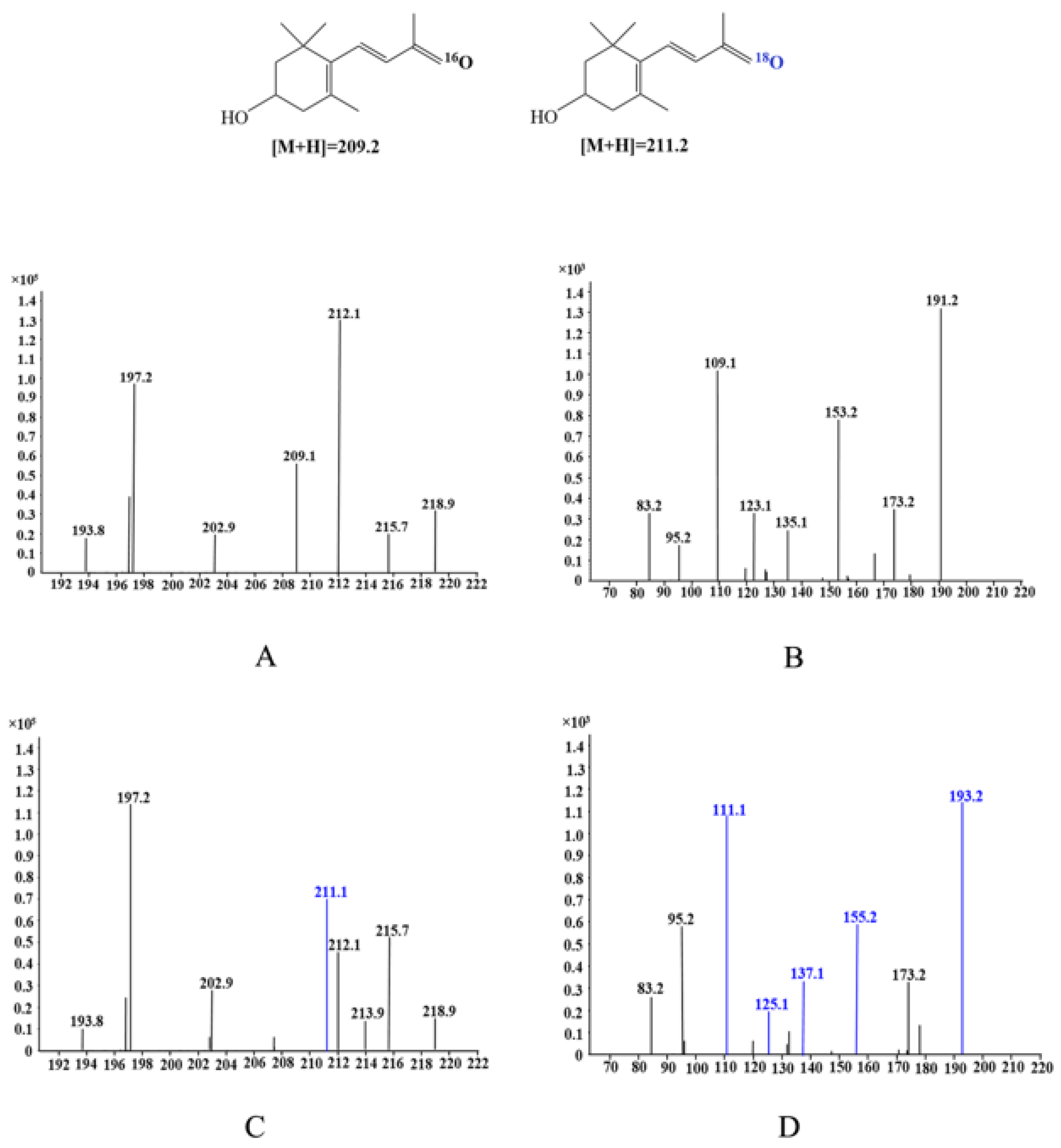
2.3. Reaction Mechanism of Lutein Cleavage by EhLCD

To clarify how the EhLCD enzymatic conversion of lutein proceeded, isotope-labeling experiments were conducted in the presence of 16O2–H218O or 18O2–H216O. Lutein was used as the substrate and the reaction product was monitored by LC-MS to determine isotope incorporation into 3-hydroxy-β-ionone (Table 3 and Figure 2). For the reaction of EhLCD and lutein in 16O2–H218O medium, the reaction product composition was about 95% 16O-3-hydroxy-β-ionone, with a molecular mass of m/z 209.1. This result suggested that the oxygen atom in the 3-hydroxy-β-ionone molecules was not derived from atmospheric H218O. For the reaction of EhLCD and lutein in 18O2–H216O medium, the reaction product composition was about 97% 18O-3-hydroxy-β-ionone, with a molecular mass of m/z 211.1. Owing to oxygen exchange with water, the percentage yields of 18O-3-hydroxy-β-ionone and 16O-3-hydroxy-β-ionone in the control experiment were 4% and 6%, respectively. The molecular masses of labeled 3-hydroxy-β-ionone fragments containing isotope 18O, such as m/z 193.2, 155.2, 137.1, 125.1, and 111.1, were m/z 2 greater than those of unlabeled retinal fragments containing normal 16O, such as m/z 191.2, 153.2, 135.1, 123.1, and 109.1. These results indicated that EhLCD could convert lutein to 3-hydroxy-β-ionone and 3-hydroxy-β-apo-10′-carotenal by the cleavage activity towards the 9′, 10′ double bond of lutein (Figure 3). Isotope-labeling experiments demonstrated that the 18O-3-hydroxy-β-ionone product was produced by enzyme EhLCD incorporating oxygen from O2 into 3-hydroxy-β-ionone during the enzymatic cleavage reaction, and EhLCD followed a dioxygenase reaction mechanism [22,23,27]. Although EhLCD and BCO2 enzymes from mammals and higher vertebrates have been found to be involved in the same mechanism, enzymes have different amino acid sequences, molecular masses, kinetic properties, and substrate specificity [20,21,22,23,27,28]. To our knowledge, EhLCD is the first purified and characterized bacterial lutein cleavage dioxygenase.

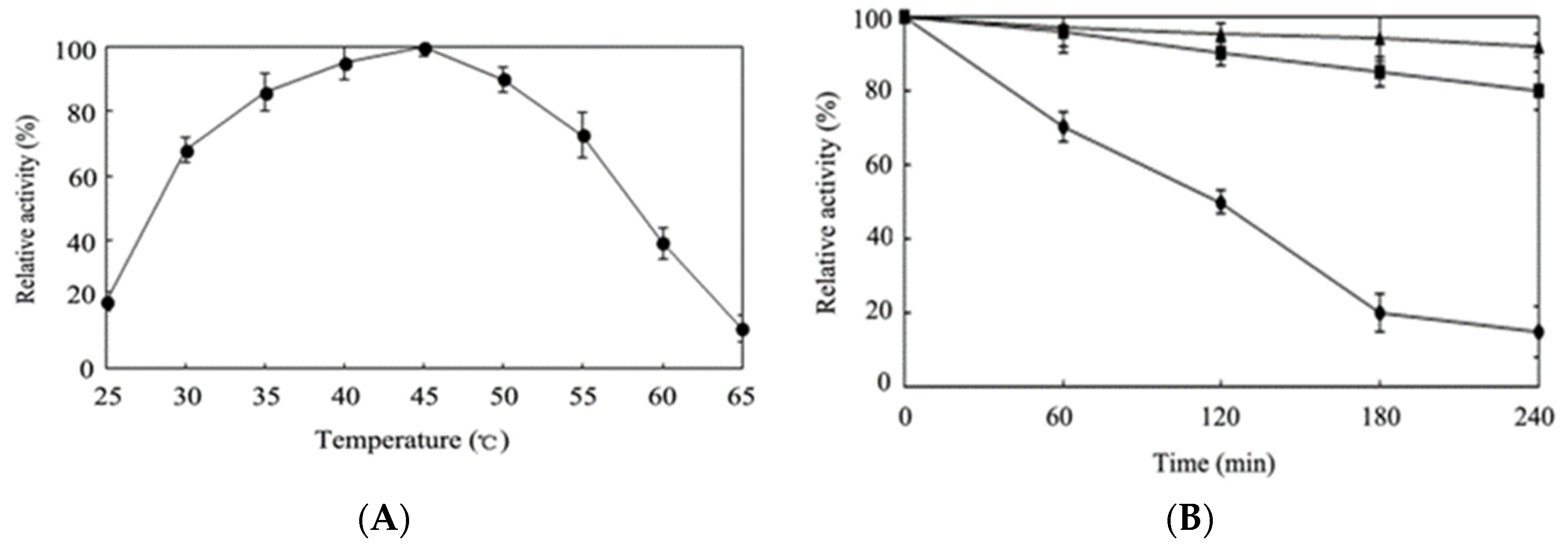
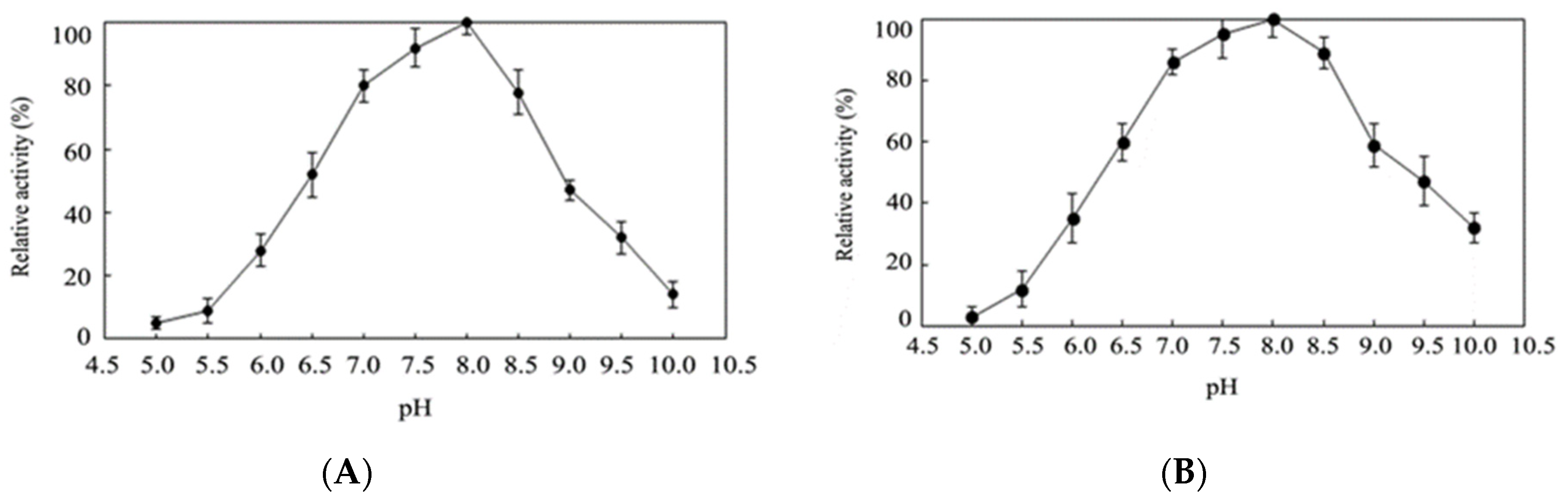
2.4. Effects of Temperature and pH on Activity of Purified EhLCD

The effects of temperature and pH on the activity of purified EhLCD were studied using lutein as a substrate. The enzymatic activity in the temperature range of 25–65 °C was examined using the standard reaction assay (Figure 4A). EhLCD exhibited maximum activity at around 45 °C, and more than half the maximum activity at 30–55 °C, which included temperatures higher than the optimal growth temperature of reported E. hormaechei (37 °C) [29,30,31]. Compared with most reported BCO2 enzymes [20,21,22,23,27,28], which mainly exhibit maximum activity at 37 °C or lower, EhLCD demonstrated a relatively high optimal temperature. The thermostability of EhLCD was examined at 35, 45, and 55 °C with increasing incubation times of up to 240 min. Most enzymatic activity was maintained after incubation at 45 °C for at least 480 min, while the half-life was 120 min at 55 °C (Figure 4B). The activity of purified EhLCD was studied between pH 5.0 and 10.0 using various buffers. The enzyme showed optimal activity at pH 8.0, and more than 50% enzymatic activity in the pH range of 6.5–8.5 (Figure 5A,B). EhLCD exhibited more than 50% activity at pH 6–10, indicating that it is an excellent alkaline enzyme.

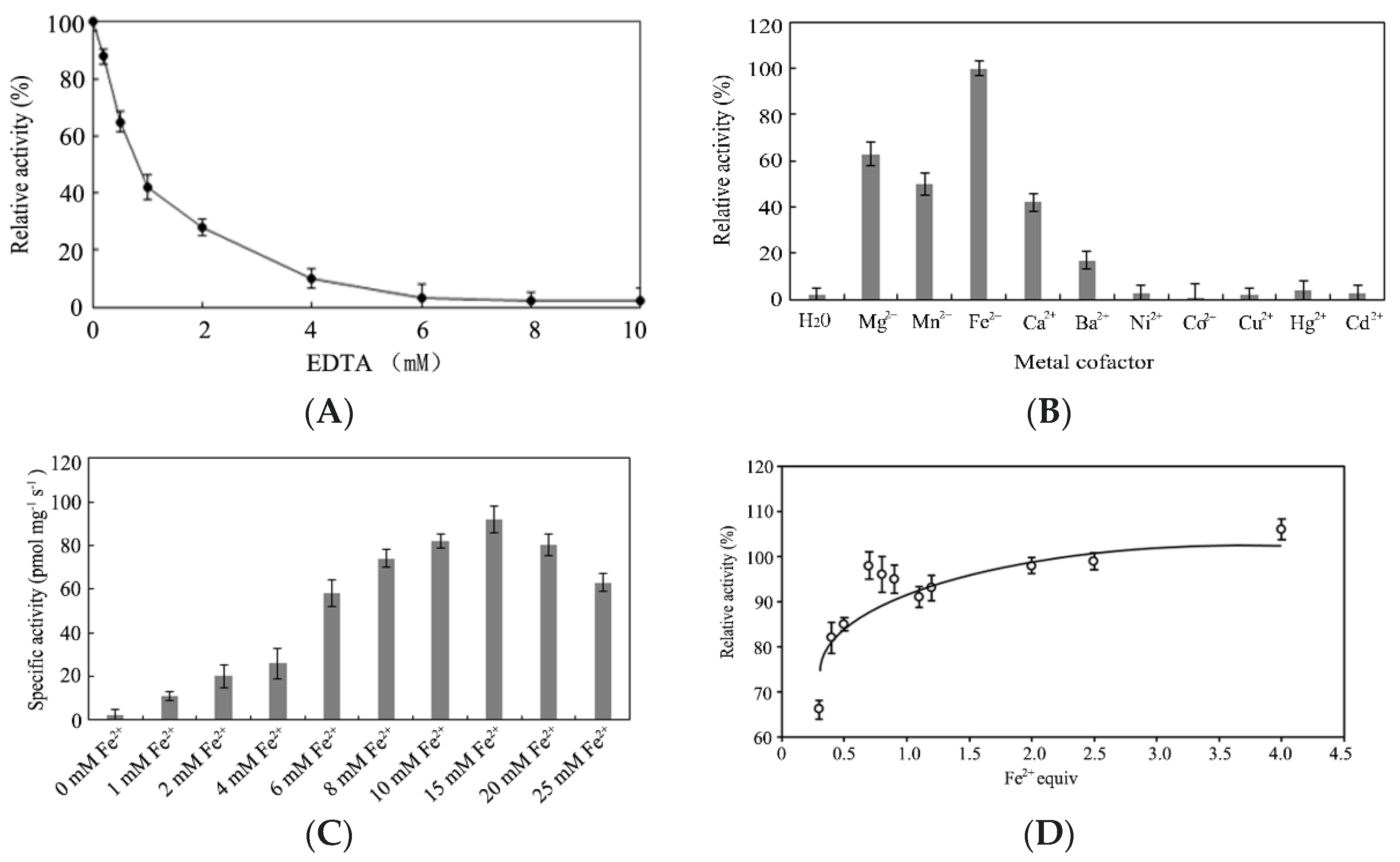
2.5. Effect of Metal Ions on Enzymatic Activity

As some carotenoid cleavage enzymes require metal ions as cofactors, the effect of metal ions on the enzymatic activity of EhLCD was investigated [32,33]. The enzymatic activity of EhLCD protein was measured after incubating with an increasing concentration of ethylenediaminetetraacetic acid (0–10 mM). Approximately 20% inhibition of BCMO7211 activity was observed, indicating that EhLCD might be a metal-dependent protein (Figure 6A). The effect of various metal ions on the activity of EhLCD is shown in Figure 6B. No enzymatic activity was observed in the absence of metal ions in the standard assay, while Fe2+ had the strongest effect on EhLCD activity among the metal ions tested. This was consistent with the carotenoid cleavage enzymes being iron (II)-dependent proteins [33,34]. Hg2+, Cu2+, Ni2+, Cd2+, and Co2+ did not significantly affect the enzymatic activity after incubation for 60 min at room temperature, possibly because these metal ions can bind with the SH, CO, and NH moieties of amino acids in EhLCD [35]. Mg2+, Mn2+, Ca2+, and Ba2+ stimulated enzymatic activity of 63%, 50%, 42%, and 17%, respectively. The effect of Fe2+ concentration on EhLCD activity in the enzymatic reaction was further investigated. As the maximum enzyme activity (92.5 pmol mg−1·s−1) was observed at 15 mM Fe2+, all subsequent experiments were performed in the presence of 15 μM Fe2+ (Figure 6C). To determine the optimal Fe2+ stoichiometry, a Fe2+ titration experiment was performed. To eliminate nonspecific binding of metal ions to the His6-tag, tag-free EhLCD was used. The data shown in Figure 6D indicate a stoichiometry of 0.93. Altogether, these results suggest that EhLCD is an Fe2+-dependent protein with an Fe2+/EhLCD monomer ratio of 1:1, which is consistent with carotenoid cleavage oxygenases having an iron catalytic center coordinated by four conserved histidine residues [32,33,34].

2.6. Effects of Organic Solvent and Detergent on Enzymatic Activity

The effects of various organic solvents and detergents on the enzymatic activity of EhLCD were investigated. As shown in Table 4, acetone, chloroform, DMSO, and DMF at 50% (v/v) markedly inhibited EhLCD activity, while more than 70% of EhLCD activity was retained after incubation with methanol, ethanol, benzene, and toluene. Detergents are used to help dissolve the hydrophobic carotenoid substrates and form micelles under aqueous conditions [36,37]. Of the various detergents, Tween 40 at 2% (w/v) afforded the highest EhLCD enzymatic activity and was selected for use in forming detergent micelles. These results were similar to those obtained with human BCO2 [38]. Incubation with Triton X-100, Span 20, and Span 40 at 1% (w/v) reduced the enzymatic activity by less than 20% (Table 4).

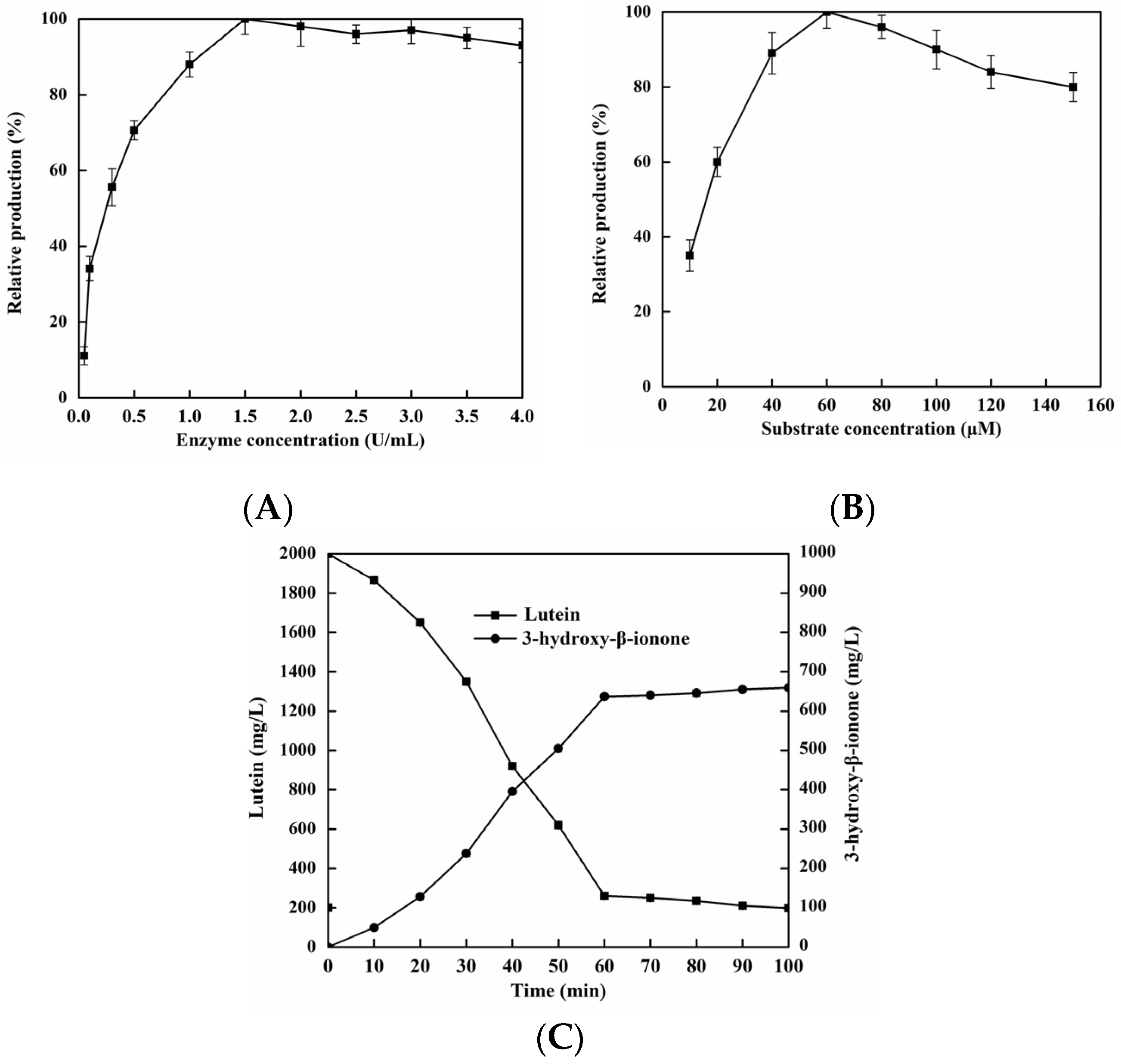
2.7. Production of 3-hydroxy-β-ionone from Lutein by EhLCD

The synthesis of β-ionone catalyzed using synthetic biology and biological enzymes has been reported, but not the synthesis of 3-hydroxy-β-ionone [9,39,40,41]. The effects of enzyme and substrate concentrations on the biocatalytic process of EhLCD are important owing to its practical applications. The effects of enzyme concentration (0.1–4.0 U/mL) and substrate concentration (10–150 μM) on the enzymatic reaction were investigated. Figure 7A shows that 3-hydroxy-β-ionone production increased with increasing the enzyme concentration from 0.1 to 1.5 U/mL. However, a further increase in enzyme concentration (from 1.5 to 4.0 U/mL) showed no obvious increase in the observed production of 3-hydroxy-β-ionone. Therefore, 1.5 U/mL was considered to be the optimal enzyme concentration for 3-hydroxy-β-ionone production. Figure 7B shows that 3-hydroxy-β-ionone production increased with an increasing lutein concentration in the range of 10–60 μM, with maximum 3-hydroxy-β-ionone production occurring with a lutein concentration of 60 μM. By further increasing the lutein loading, 3-hydroxy-β-ionone production showed some decline. Therefore, a high lutein concentration might prevent contact between enzymes at the outer surface of the detergent micelles and lutein located in the hydrophobic core [42]. The production of 3-hydroxy-β-ionone from lutein by EhLCD was investigated under optimum conditions in the standard enzyme assay. This enzyme produced 3-hydroxy-β-ionone (637.2 mg/L) in 60 min with a conversion of 87.0% (w/w) (Figure 7C). Owing to instability and sensitivity to oxidation, the actual amount of 3-hydroxy-β-ionone produced was lower than the theoretical value [43]. These results demonstrated that EhLCD has strong activity for 3-hydroxy-β-ionone production, making it a potential candidate for the enzymatic transformation of lutein into 3-hydroxy-β-ionone in biotechnological applications.

3. Materials and Methods

3.1. Chemicals, Stains, and Plasmids

Using lutein as the sole source of carbon and nitrogen, E. hormaechei YT-3 was isolated from flue-cured tobacco leaves in Sanmenxia City, Henan Province, China. The stain was cultured in culture medium containing K2HPO4 (1.0 g/L), MgSO4·7H2O (0.5 g/L), NaNO3 (3.0 g/L), KCl (0.5 g/L), FeSO4·7H2O (0.01 g/L), sucrose (30 g/L), yeast extract (3.0 g/L), and lutein (20.0 g/L). Q-Sepharose, phenyl-Sepharose, and Superdex 200 gel filtration columns used in this study were purchased from GE Healthcare (Buckinghamshire, UK). Lutein and 3-hydroxy-β-ionone were purchased from J&K Scientific, Ltd. (Beijing, China). All other chemicals used were of analytical grade and purchased from Sangon (Shanghai, China).

3.2. Purification of EhLCD

A culture of E. hormaechei YT-3 (2.0 L) was kept at 28 °C for 2 days, and the cells were collected by centrifugation at 12,000× g for 15 min. The collected cells were suspended in 20 mM Tris-HCl buffer (pH 7.2) and disrupted by sonication. The disrupted cells were incubated with 1% (v/v) of Triton X-100 at 4 °C and 80 rpm for 4 h, and then centrifuged at 12,000× g for 15 min to remove cell debris and denatured proteins. Fractionation using ammonium sulfate at 50%–80% saturation was performed to remove some unwanted proteins [44]. The mixture was allowed to stand overnight, and the precipitate was dissolved in 20 mM Tris-HCl buffer (pH 7.2) and dialyzed overnight against the same buffer. The sample was loaded onto a Q-Sepharose column (1 × 20 cm2) equilibrated with 20 mM Tris-HCl buffer (pH 7.2). The column was then eluted with NaCl solution (100 mL) using a linear concentration gradient ranging from 50 to 600 mM. All active fractions were collected and concentrated using a membrane with a 10 kDa molecular weight cut-off (MWCO). The concentrated protein was applied to a phenyl-Sepharose column (1 × 20 cm2) pre-equilibrated with 20 mM Tris-HCl buffer (pH 7.2) containing 1.0 M (NH4)2SO4, and eluted using 0.7 M (NH4)2SO4 solution. Fractions showing lutein degradation activity were pooled and concentrated using a membrane with a 10 kDa MWCO. The concentrated protein was then loaded onto a Superdex 200 gel filtration (16/60) column equilibrated with 20 mM Tris-HCl (pH 7.2) containing 100 mM NaCl. The fraction size was 1 mL, and the flow rate was 1 mL/min. Fractions showing lutein degradation activity were collected and analyzed by SDS-PAGE. The protein concentration was determined using the Bradford method. The purified protein was stored in 20 mM Tris-HCl (pH 7.2) containing 25% (v/v) glycerol at −80 °C.

3.3. Enzyme Assay

The lutein degradation activity of EhLCD was determined as described previously [22,28]. The standard reaction mixture, containing 60 μM lutein, 2.0% (w/v) Tween 40, 200 mM NaCl, 15 μM FeSO4, 10 mM tris(2-carboxylethyl)phosphine hydrochloride, 1.5% (w/v) 1-S-octyl-β-D-thioglucopyranoside, and 50 mM tricine/KOH buffer (pH 8.0), was preincubated for 2 min and then the reaction was initiated by adding the purified enzyme (0.5 U/mL). The degradation activity was measured at 45 °C for 60 min and the reaction products were monitored by liquid chromatography–mass spectrometry (LC-MS) analysis [23,44]. The above EhLCD-catalyzed samples were characterized using an Agilent 1100 HPLC system and a Bruker Esquire 3000plus mass spectrometer with an electrospray ionization (ESI) source in positive ion mode. For chromatographic separation, a LUNA C18 column (150 cm × 2.1 mm, 1.8 μm) was used, with a mobile phase comprising water (A) and acetonitrile (B), each containing 0.1% formic acid. A linear elution gradient was applied, as follows: 50% B for 1 min, increased from 50% to 80% B for 8 min, increased from 80% to 100% B for 5 min, 100% B for 3 min, and re-equilibrated to 60% B for 5 min. The detection wavelength was 285 nm, and the flow rate was 0.5 mL/min. The MS instrument parameters were as follows: ESI capillary voltage, 4 kV; source and desolvation temperature, 300 °C. Mass spectra were measured at a rate of 0.85 s−1 in the m/z range of 50–500. One unit of enzymatic activity was defined as the amount of enzyme required to liberate 1 μmol min−1 of 3-hydroxy-β-ionone under standard conditions. Measurements were corrected for background hydrolysis in the absence of enzyme.

3.4. Analysis of Amino Acid Sequence of EhLCD

The purified EhLCD protein was loaded onto 12% SDS-PAGE gel and then transferred onto a Hybond-P polyvinylidene difluoride membrane (Amersham Pharmacia Biotech). The corresponding bands from SDS-PAGE were excised, and the N-terminal and internal partial amino acid sequences of EhLCD were determined using Sangon and the ESI-Q-TOF mass spectrometer (Shanghai, China).

3.5. Catalytic Reaction Mechanism of EhLCD

Isotope-labeling experiments were conducted to determine the catalytic reaction mechanism of EhLCD, as described previously [28,44]. For labeling experiments in 16O2–H218O, freeze-dried purified EhLCD and lutein were suspended in H218O solution. For labeling experiments in 18O2–H216O, EhLCD and lutein were suspended in non-isotopically labeled H2O and then the solution was saturated with 18O2 gas over ice for 2 min. The reactions were performed in 50 mM Tricine/KOH buffer (pH 8.0) containing 60 μM lutein and 0.5 U/mL of enzyme at 30 °C for 60 min. To account for oxygen exchange between water and retinal, a 0.1 μM solution of 16O-3-hydroxy-β-ionone (18O-3-hydroxy-β-ionone) was prepared in H218O (H216O) containing EhLCD protein as a control experiment. The resulting solution was centrifuged at 12,000× g for 10 min at 4 °C, and the supernatant was evaporated. The samples were resuspended in acetonitrile and analyzed by LC-MS.

3.6. Catalytic Properties of EhLCD

The optimum pH and temperature for EhLCD degradation activity were determined by LC-MS (AB Sciex, Framingham, MA, USA) in the standard enzymatic assay. The effect of pH on enzymatic activity was examined at 45 °C in the pH range of 5.0–10.0. The effect of temperature (from 25 to 65 °C) on enzymatic activity was investigated at pH 8.0. The effects of several different metal ions, organic solvents, and detergents on the activity of EhLCD were determined by adding each metal salt (5 mM), organic solvent (50%, v/v), and detergent (1% or 2%, w/v) to the standard assay solution. Fe2+ titration (using FeSO4) to determine the metal stoichiometry was performed as described previously [45]. Varying molar ratios of Fe2+/EhLCD were incubated in Tris/NaCl buffer (20 mM Tricine/KOH, 50 mM NaCl, pH 8.0) for 2 h before performing the enzymatic activity assay.

4. Conclusions

In this study, a novel lutein cleavage dioxygenase (EhLCD) was purified and characterized from Enterobacter hormaechei YT-3 for the first time. The enzyme displayed optimal activity at 45 °C and pH 8.0. Isotope-labeling experiments showed that EhLCD followed a dioxygenase reaction mechanism to catalyze the double-bond cleavage of lutein. The active EhLCD was shown to be a Fe2+-dependent protein with 1 molar equivalent of non-haem Fe2+. Under optimum conditions, EhLCD produced 637.2 mg/L of 3-hydroxy-β-ionone from 2000 mg/L of lutein, with a conversion rate of 87.0%. To our knowledge, EhLCD is the first characterized lutein cleavage dioxygenase of bacterial origin. EhLCD is a candidate for use in the enzymatic synthesis of 3-hydroxy-β-ionone in biotechnological applications.

Supplementary Materials

The following are available online at https://www.mdpi.com/article/10.3390/catal11111257/s1, Figure S1: Mass spectrometry analysis of the trypsin-digested EhLCD.

Author Contributions

Z.L. designed and performed experiments, analyzed data, and prepared the manuscript; N.D., Y.X., M.W., J.L., Z.S., Q.L. and D.M. assisted in data analysis; T.W. performed experiments, analyzed data, and assisted in manuscript preparation. All authors have read and agreed to the published version of the manuscript.

Funding

The APC was funded by the Science and Technology Project of China Guangxi Tobacco Industry Co., Ltd. (2019450000340011).

Acknowledgments

This work was supported by grants from Zhongyuan science and technology innovation leading talent project (2022) and the Science and Technology project of overseas Chinese in Henan Province (2019-03).

Conflicts of Interest

The authors declare no conflict of interest.

References

  1. Lin, J.H.; Lee, D.J.; Chang, J.S. Lutein production from biomass: Marigold flowers versus microalgae. Bioresour. Technol. 2015, 184, 421–428. [Google Scholar] [CrossRef]
  2. Barbara, D.A.; William, W.A. Antioxidants in photosynthesis and human nutrition. Science 2002, 298, 2149–2153. [Google Scholar]
  3. Li, X.M.; Li, X.H.; Wu, Z.Z.; Wang, Y.; Cheng, J.S.; Wang, T.; Zhang, B. Chitosan hydrochloride/carboxymethyl starch complex nanogels stabilized Pickering emulsions for oral delivery of β-carotene: Protection effect and in vitro digestion study. Food Chem. 2020, 315, 126288. [Google Scholar] [CrossRef]
  4. Ceron, M.C.; Campos, I.; Sánchez, J.F.; Acien, F.G.; Molina, E.; Fernandez Sevilla, J.M. Recovery of lutein from microalgae biomass: Development of a process for Scenedesmus almeriensis. J. Agric. Food Chem. 2008, 56, 11761–11766. [Google Scholar] [CrossRef]
  5. Carpentier, S.; Knaus, M.; Suh, M. Associations between lutein, zeaxanthin, and age-related macular degeneration: An overview. Crit Rev. Food Sci. Nutr. 2009, 49, 313–326. [Google Scholar] [CrossRef] [PubMed]
  6. Berger, R.G. Biotechnology of flavours—The next generation. Biotechnol. Lett. 2009, 31, 1651–1659. [Google Scholar] [CrossRef] [PubMed]
  7. Lalko, J.; Lapczynski, A.; McGinty, D.; Bhatia, S.; Letizia, C.S.; Api, A.M. Fragrance material review on β-ionone. Food Chem. Toxicol. 2007, 45, S241–S247. [Google Scholar] [CrossRef]
  8. Lalko, J.; Lapczynski, A.; McGinty, D.; Bhatia, S.; Letizia, C.S.; Api, A.M. Fragrance material review on dihydro-β-ionone. Food Chem. Toxicol. 2007, 45, S225–S228. [Google Scholar] [CrossRef]
  9. Nacke, C.; Hüttmann, S.; Etschmann, M.M.W.; Schrader, J. Enzymatic production and in situ separation of natural β-ionone from β-carotene. J. Ind. Microbiol. Biotechnol. 2012, 39, 1771–1778. [Google Scholar] [CrossRef] [PubMed]
  10. Sefton, M.A.; Skouroumounis, G.K.; Elsey, G.M.; Taylor, D.K. Occurrence, sensory impact, formation, and fate of damascenone. in grapes, wines, and other foods and beverages. J. Agric. Food Chem. 2011, 59, 9717–9746. [Google Scholar] [CrossRef]
  11. Serra, S. Recent advances in the synthesis of carotenoid-derived flavours and fragrances. Molecules 2015, 20, 12817–12840. [Google Scholar] [CrossRef] [Green Version]
  12. Gambacorta, G.; Sharley, J.S.; Baxendale, I.R. A comprehensive review of flow chemistry techniques tailored to the flavours and fragrances industries. Beilstein J. Org. Chem. 2021, 17, 1181–1312. [Google Scholar] [CrossRef]
  13. Cataldo, V.F.; López, J.; Cárcamo, M.; Agosin, E. Chemical vs. biotechnological synthesis of C13-apocarotenoids: Current methods, applications and perspectives. Appl. Microbiol. Biotechnol. 2016, 100, 5703–5718. [Google Scholar] [CrossRef] [PubMed]
  14. Zelena, K.; Hardebusch, B.; Hülsdau, B.; Berger, R.G.; Zorn, H. Generation of norisoprenoid flavors from carotenoids by fungal peroxidases. J. Agric. Food Chem. 2009, 57, 9951–9955. [Google Scholar] [CrossRef]
  15. Sanchez-Contreras, A.; Jimenez, M.; Sanchez, S. Bioconversion of lutein to products with aroma. Appl. Microbiol. Biotechnol. 2000, 54, 528–534. [Google Scholar] [CrossRef]
  16. Maldonado-Robledo, G.; Rodrıguez-Bustamante, E.; Sanchez-Contreras, A.; Rodrıguez-Sanoja, R.; Sanchez, S. Production of tobacco aroma from lutein. Specific role of the microorganisms involved in the process. Appl. Microbiol. Biotechnol. 2003, 62, 484–488. [Google Scholar] [CrossRef]
  17. Rodríguez-Bustamante, E.; Maldonado-Robledo, G.; Ortiz, M.A.; Díaz-Avalos, C.; Sanchez, S. Bioconversion of lutein using a microbial mixture-maximizing the production of tobacco aroma compounds by manipulation of culture medium. Appl. Microbiol. Biotechnol. 2005, 68, 174–182. [Google Scholar] [CrossRef] [PubMed]
  18. Zhao, Y.; Zhong, G.F.; Yang, X.P.; Hu, X.M.; Mao, D.B.; Ma, Y.P. Bioconversion of lutein to form aroma compounds by Pantoea dispersa. Biotechnol. Lett. 2015, 37, 1687–1692. [Google Scholar] [CrossRef] [PubMed]
  19. Zhong, G.F.; Wang, F.F.; Sun, J.H.; Ye, J.B.; Mao, D.B.; Ma, K.; Yang, X.P. Bioconversion of lutein by Enterobacter hormaechei to form a new compound, 8-methyl-alpha-ionone. Biotechnol. Lett. 2017, 39, 1019–1024. [Google Scholar] [CrossRef]
  20. Mein, J.R.; Dolnikowski, G.G.; Ernst, H.; Russell, R.M.; Wang, X.D. Enzymatic formation of apo-carotenoids from the xanthophyll carotenoids lutein, zeaxanthin and β-cryptoxanthin by ferret carotene-9′,10′-monooxygenase. Arch. Biochem. Biophys. 2011, 506, 109–121. [Google Scholar] [CrossRef] [PubMed] [Green Version]
  21. Amengual, J.; Lobo, G.P.; Golczak, M.; Li, H.N.M.; Klimova, T.; Hoppel, C.L.; Wyss, A.; Palczewski, K.; von Lintig, J. A mitochondrial enzyme degrades carotenoids and protects against oxidative stress. FASEB J. 2011, 25, 948–959. [Google Scholar] [CrossRef] [PubMed] [Green Version]
  22. Dela Seña, C.; Sun, J.; Narayanasamy, S.; Riedl, K.M.; Yuan, Y.; Curley, R.W.; Schwartz, S.J.; Harrison, E.H. Substrate specificity of purified recombinant chicken β-carotene 9′,10′-oxygenase (BCO2). J. Biol. Chem. 2016, 291, 14609–14619. [Google Scholar] [CrossRef] [Green Version]
  23. Bandara, S.; Thomas, L.D.; Ramkumar, S.; Khadka, N.; Kiser, P.D.; Golczak, M.; von Lintig, J. The structural and biochemical basis of apocarotenoid processing by β-carotene oxygenase-2. ACS Chem. Biol. 2021, 16, 480–490. [Google Scholar] [CrossRef]
  24. Thomas, L.D.; Bandara, S.; Parmar, V.M.; Srinivasagan, R.; Khadka, N.; Golczak, M.; Kiser, P.D.; von Lintig, J. The human mitochondrial enzyme BCO2 exhibits catalytic activity toward carotenoids and apocarotenoids. J. Biol. Chem. 2020, 295, 15553–15565. [Google Scholar] [CrossRef] [PubMed]
  25. Harrison, E.H.; Kopec, R.E. Enzymology of vertebrate carotenoid oxygenases. Biochim. Biophys. Acta (BBA) Mol. Cell Biol. Lipids 2020, 1865, 158653. [Google Scholar] [CrossRef]
  26. Giordano, E.; Quadro, L. Lutein, zeaxanthin and mammalian development: Metabolism, function and implications for health. Arch. Biochem. Biophys. 2018, 647, 33–40. [Google Scholar] [CrossRef]
  27. dela Seña, C.; Riedl, K.M.; Narayanasamy, S.; Curley, R.W., Jr.; Schwartz, S.J.; Harrison, E.H. The human enzyme that converts dietary provitamin A carotenoids to vitamin A is a dioxygenase. J. Biol. Chem. 2014, 289, 13661–13666. [Google Scholar] [CrossRef] [PubMed] [Green Version]
  28. Kelly, M.E.; Ramkumar, S.; Sun, W.Z.; Ortiz, C.C.; Kiser, P.D.; Golczak, M.; von Lintig, J. The biochemical basis of vitamin A production from the asymmetric carotenoid β-cryptoxanthin. ACS Chem. Biol. 2018, 13, 2121–2129. [Google Scholar] [CrossRef]
  29. Pereira, A.G.; Otero, P.; Echave, J.; Carreira-Casais, A.; Chamorro, F.; Collazo, N.; Jaboui, A.; Lourenço-Lopes, C.; Simal-Gandara, J.; Prieto, M.A. Xanthophylls from the Sea: Algae as Source of Bioactive Carotenoids. Mar. Drugs 2021, 19, 188. [Google Scholar] [CrossRef]
  30. Hoffmann, H.; Stindl, S.; Ludwig, W.; Stumpf, A.; Mehlen, A.; Monget, D.; Pierard, D.; Ziesing, S.; Heesemann, J.; Roggenkamp, A.; et al. Enterobacter hormaechei subsp. oharae subsp. nov., E. hormaechei subsp. hormaechei comb. nov., and E. hormaechei subsp. steigerwaltii subsp. nov., three new subspecies of clinical importance. J. Clin. Microbiol. 2005, 43, 3297–3303. [Google Scholar] [CrossRef] [Green Version]
  31. Sutton, G.G.; Brinkac, L.M.; Clarke, T.H.; Fouts, D.E. Enterobacter hormaechei subsp. hoffmannii subsp. nov., Enterobacter hormaechei subsp. xiangfangensis comb. nov., Enterobacter roggenkampii sp. nov., and Enterobacter muelleri is a later heterotypic synonym of Enterobacter asburiae based on computational analysis of sequenced Enterobacter genomes. F1000Research 2018, 7, 521. [Google Scholar]
  32. Sui, X.; Farquhar, E.R.; Hill, H.E.; von Lintig, J.; Shi, W.; Kiser, P.D. Preparation and characterization of metal-substituted carotenoid cleavage oxygenases. J. Biol. Inorg. Chem. 2018, 23, 887–901. [Google Scholar] [CrossRef] [Green Version]
  33. Harrison, P.J.; Bugg, T.D. Enzymology of the carotenoid cleavage dioxygenases: Reaction mechanisms, inhibition and biochemical roles. Arch. Biochem. Biophys. 2014, 544, 105–111. [Google Scholar] [CrossRef] [PubMed]
  34. Daruwalla, A.; Kiser, P.D. Structural and mechanistic aspects of carotenoid cleavage dioxygenases (CCDs). Biochim. Biophys. Acta (BBA) Mol. Cell Biol. Lipids 2019, 1865, 158590. [Google Scholar] [CrossRef]
  35. Li, Y.; Li, W.; Zhang, G.; Lü, X.; Hwang, H.; Aker, W.G.; Guan, H.; Wang, P. Purification and characterization of polysaccharides degradases produced by Alteromonas sp. A321. Int. J. Biol. Macromol. 2016, 86, 86–96. [Google Scholar] [CrossRef] [PubMed]
  36. During, A.; Harrison, E.H. Intestinal absorption and metabolism of carotenoids: Insights from cell culture. Arch. Biochem. Biophys. 2004, 430, 77–88. [Google Scholar] [CrossRef]
  37. Wyss, A.; Wirtz, G.M.; Woggon, W.D.; Brugger, R.; Wyss, M.; Friedlein, A.; Riss, G.; Bachmann, H.; Hunziker, W. Expression pattern and localization of β,β-carotene 15,15′-dioxygenase in different tissues. Biochem. J. 2001, 354, 521–529. [Google Scholar] [CrossRef]
  38. Kim, Y.S.; Yeom, S.J.; Oh, D.K. Production of β-apo-10′-carotenal from β-carotene by human β-carotene-9′, 10′-oxygenase expressed in E. coli. Biotechnol. Lett. 2011, 33, 1195–1200. [Google Scholar] [CrossRef] [PubMed]
  39. López., J.; Bustos, D.; Camilo, C.; Arenas, N.; Saa, P.A.; Agosin, E. Engineering Saccharomyces cerevisiae for the Overproduction of β-Ionone and Its Precursor β-Carotene. Front. Bioeng. Biotechnol. 2020, 8, 578793. [Google Scholar] [CrossRef]
  40. Schempp, F.M.; Drummond, L.; Buchhaupt, M.; Schrader, J. Microbial cell factories for the production of terpenoid flavor and fragrance compounds. J. Agric. Food Chem. 2018, 66, 2247–2258. [Google Scholar] [CrossRef] [PubMed]
  41. Czajka, J.J.; Nathenson, J.A.; Benites, V.T.; Baidoo, E.E.K.; Cheng, Q.; Wang, Y.; Tang, Y.J. Engineering the oleaginous yeast Yarrowia lipolytica to produce the aroma compound β-ionone. Microb. Cell Fact. 2018, 17, 136. [Google Scholar] [CrossRef]
  42. Shikama, K. Nature of the FeO2 bonding in myoglobin and hemoglobin: A new molecular paradigm. Prog. Biophys. Mol. Biol. 2006, 91, 83–162. [Google Scholar] [CrossRef]
  43. Walter, M.H.; Strack, D. Carotenoids and their cleavage products: Biosynthesis and functions. Nat. Prod. Rep. 2011, 28, 663–692. [Google Scholar] [CrossRef] [PubMed]
  44. Wei, T.; Zhao, C.M.; Jia, W.W.; Huang, S.; Mao, D.B. Expression and purification of fructosyl transferase AoFT in Pichia pastoris and study on its enzymatic properties. J. Light Ind. 2020, 35, 1–10. [Google Scholar]
  45. Chai, S.C.; Lu, J.P.; Ye, Q.Z. Determination of binding affinity of metal cofactor to the active site of methionine aminopeptidase based on quantitation of functional enzyme. Anal. Biochem. 2009, 395, 263–264. [Google Scholar] [CrossRef] [PubMed] [Green Version]
Figure 1. SDS-PAGE of purified lutein cleavage dioxygenase EhLCD from E. hormaechei YT-3. Lane 1, molecular weight standards; lanes 2 and 3, Superdex-200 chromatography product (purified enzyme).
Catalysts 11 01257 g001
Figure 2. Mass spectra of products in reaction mixture containing lutein, indicating that EhLCD is a dioxygenase. (A) Mass spectrum of 3-hydroxy-β-ionone formed by EhLCD from lutein in H218O solution. (B) MS/MS traces for fragmentation of 16O-3-hydroxy-β-ionone. Fragment molecular masses from 16O-3-hydroxy-β-ionone were m/z 191.2, 153.2, 135.1, 123.1, and 109.1, respectively. (C) Mass spectrum of 3-hydroxy-β-ionone formed from lutein by EhLCD in the presence of molecular oxygen (18O2). Blue color indicates positions of 18O-3-hydroxy-β-ionone. (D) MS/MS traces for fragmentation of 18O-3-hydroxy-β-ionone. Fragment molecular masses of 18O-3-hydroxy-β-ionone were m/z 193.2, 155.2, 137.1, 125.1, and 111.1, respectively.
Catalysts 11 01257 g002
Figure 3. Enzymatic cleavage reaction and reaction mechanism catalyzed by EhLCD. EhLCD incorporates atoms from O2 into the cleavage products of 3-hydroxy-β-ionone and 3-hydroxy-β-apo-10′-carotenal.
Catalysts 11 01257 g003
Figure 4. Effects of temperature on the activity of purified EhLCD. (A) Optimal temperature of EhLCD was determined using lutein as a substrate in 50 mM Tricine/KOH buffer (pH 8.0) at different temperatures ranging from 25 to 65 °C. (B) Thermostability of EhLCD. Residual enzyme activity was measured after incubation of the purified enzyme at 35 (triangles), 45 (boxes), and 55 °C (diamonds).
Catalysts 11 01257 g004
Figure 5. Optimal (A) pH and (B) pH stability of enzyme EhLCD in the pH range of 5.0–10.0, measured for 60 min at 45 °C using lutein as a substrate. Buffers used were 50 mM of sodium acetate (pH 5.0–6.0), sodium phosphate (pH 6.0–7.5), Tris-HCl (pH 7.5–9.5), and N-cyclohexyl-3-aminopropanesulfonic acid (pH 9.5–10.0). Values are means of three independent experiments.
Catalysts 11 01257 g005
Figure 6. Determination of Fe2+ as a native cofactor for EhLCD. (A) Inhibition of EhLCD activity by EDTA. (B) Effect of metal ions (5 mM) on EhLCD activity. (C) Effect of Fe2+ concentration on purified EhLCD protein. (D) Titration of as-isolated EhLCD with increasing Fe2+ concentration, with activity measured by quantifying 3-hydroxy-β-ionone formation.
Catalysts 11 01257 g006
Figure 7. Production of 3-hydroxy-β-ionone from lutein by EhLCD. (A) Effect of enzyme concentration. (B) Effect of substrate concentration. (C) Time course of 3-hydroxy-β-ionone (circles) production from lutein (boxes) under optimum conditions.
Catalysts 11 01257 g007
Table 1. Purification of EhLCD from E. hormaechei YT-3. Values are means of three replicates.
StepTotal Protein
(mg)
Total Activity
(U)
Specific Activity
(U/mg)
Fold Yield
(%)
Crude cell extract536.3125.60.231100
(NH4)2SO4 precipitation300.5113.90.371.6156.0
Q-Sepharose column129.598.60.763.3024.1
Phenyl Sepharose80.687.71.094.7315.0
Superdex 200 29.566.92.279.905.5
Table 2. Kinetic parameters for hydrolysis of various carotenoid substrates a.
SubstrateKm (μM)kcat (s−1)Vmax (pmol mg−1·s−1)kcat/Km (s−1 mM−1)
Lutein 14 ± 5 5.5 ± 1.489.5 ± 2.60.393 × 103
Zeaxanthin 20 ± 23.8 ± 0.661.8 ± 1.70.190 × 103
β-cryptoxanthin 52 ± 6 1.5 ± 0.924.4 ± 2.10.028 × 103
β-carotene 128 ± 3.5 0.6 ± 0.19.76 ± 1.20.005 × 103
a Specificity was determined using lutein, zeaxanthin, β-cryptoxanthin, and β-carotene as substrates at different substrate concentrations (0.02–0.3 mM).
Table 3. Quantitative analysis of the percentages of 18O-3-hydroxy-β-ionone and 16O-3-hydroxy-β-ionone in reaction products catalyzed by EhLCD.
Enzymatic Reaction18O-3-hydroxy-β-ionone (%)16O-3-hydroxy-β-ionone (%)
EhLCD + 16O-3-hydroxy-β-ionone + H218O5 ± 295 ± 5
EhLCD + 18O-3-hydroxy-β-ionone + H216O97 ± 43 ± 1
EhLCD + lutein (16O2–H218O)4 ± 292 ± 3
EhLCD + lutein e (18O2–H216O)90 ± 56 ± 2
Table 4. Effect of organic solvents and detergents on the enzymatic activity of EhLCD.
Organic Solvents/DetergentsConcentration (%)Relative Activity (%)
None-100
Methanol50 (v/v)89 ± 5
Ethanol50 (v/v)72 ± 6
Acetone50 (v/v)30 ± 2
Toluene50 (v/v)90 ± 4
Benzene50 (v/v)75 ± 3
Chloroform50 (v/v)16 ± 2
DMSO50 (v/v)20 ± 5
DMF50 (v/v)18 ± 4
n-hexane50 (v/v)48 ± 3
Span 201% (w/v)18 ± 2
Span 801% (w/v)18 ± 1
Triton X-1001% (w/v)18 ± 5
Tween 201% (w/v)68 ± 3
2% (w/v)79 ± 6
Tween 401% (w/v)94 ± 2
2% (w/v)108 ± 3
Tween 801% (w/v)78 ± 2
2% (w/v)86 ± 5
Publisher’s Note: MDPI stays neutral with regard to jurisdictional claims in published maps and institutional affiliations.

