Article

Enhancement of S-Adenosylmethionine-Dependent Methylation by Integrating Methanol Metabolism with 5-Methyl-Tetrahydrofolate Formation in Escherichia coli

1
International Center for Biotechnology, Osaka University, 2-1 Yamada-oka, Suita, Osaka 565-0871, Japan
2
Division of Applied Science, School of Engineering, Osaka University, 2-1 Yamada-oka, Suita, Osaka 565-0871, Japan
*
Author to whom correspondence should be addressed.
Catalysts 2020, 10(9), 1001; https://doi.org/10.3390/catal10091001
Submission received: 5 August 2020 / Revised: 24 August 2020 / Accepted: 27 August 2020 / Published: 2 September 2020
(This article belongs to the Special Issue Multi-Step Syntheses in Biology & Chemistry)

Abstract

:
S-Adenosylmethionine (SAM)-dependent methyltransferases are important tools for the biocatalytic methylation of diverse biomolecules. Methylation by a whole-cell biocatalyst allows the utilization of intrinsic SAM and its regeneration system, which consists of a cyclic and multi-step enzymatic cascade. However, low intracellular availability of 5-methyl-tetrahydrofolate (5-methyl-THF), which functions as a methyl group donor, limits SAM regeneration. Here, we integrated methanol metabolism with 5-methyl-THF formation into SAM-dependent methylation system in Escherichia coli, driven by heterologously expressed methanol dehydrogenase (MDH). The coupling of MDH-catalyzed methanol oxidation with the E. coli endogenous reactions enhances the formation of 5-methyl-THF using methanol as a source of methyl group, thereby promoting both the SAM regeneration and methylation reactions. Co-expression of the mutant MDH2 from Cupriavidus necator N-1 with the O-methyltransferase 5 from Streptomyces avermitilis MA-4680 enhanced O-methylation of esculetin 1.4-fold. Additional overexpression of the E. coli endogenous 5,10-methylene-THF reductase, which catalyzes the last step of 5-methyl-THF formation, further enhanced the methylation reaction by 1.9-fold. Together with deregulation of SAM biosynthesis, the titer of methylated compounds was increased about 20-fold (from 0.023 mM to 0.44 mM). The engineered E. coli strain with enhanced 5-methyl-THF formation is now available as a chassis strain for the production of a variety of methylated compounds.

Graphical Abstract

1. Introduction

Biocatalysis is an environmentally friendly alternative to traditional chemical catalysis. The use of enzymes and microbes enables the production of value-added bulk and fine chemicals, as well as pharmaceuticals, under benign conditions [1,2]. Many enzymatic reactions utilize low molecular weight cofactors, such as nicotinamide adenine dinucleotide (NAD+/NADH) and adenosine triphosphate (ATP). These cofactors are typically more expensive than the products of interest and stoichiometric usage of cofactors limits the practical application of biocatalysis. The integration of cofactor regeneration systems is a promising approach to reduce the used amount of cofactor. NADH-dependent biocatalytic reductions were achieved with high turnover by coupling the reactions with NAD+-dependent oxidation of the sacrificial co-substrates [1,3,4]. ATP regeneration from ADP was also achieved by employing various kinds of kinases and phosphate donors [5].
S-Adenosylmethionine (SAM) is a universal cofactor in all domains of life, along with nicotinamide cofactors and ATP. SAM functions as a methyl group donor and is involved in the methylation of diverse biomolecules, including DNA, proteins, and secondary metabolites [6]. Despite its small size, the introduction of a methyl group onto biomolecules can significantly alter their physical and chemical properties, such as binding affinity, solubility, and metabolism [7]. The impact of methylation on biomolecules is so great that it is known as the “magic methyl effect” [8]. The ever-growing numbers of SAM-dependent methyltransferases, which can methylate a wide variety of biomolecules, are important tools for biocatalysis.
Unlike other cofactors, SAM is regenerated through a cyclic and multi-step enzymatic cascade (Figure 1). Direct methylation of S-adenosylhomocysteine (SAH), a demethylated form of SAM, is principally impossible due to the irreversibility of SAM-dependent methylation reactions [9]. Another characteristic of SAM regeneration is its requirement of an additional cofactor, 5-methyl-tetrahydrofolate (5-methyl-THF) as a methyl group donor. SAM regeneration and the subsequent methylation reaction are thus limited by the intracellular availability of 5-methyl-THF. 5-Methyl-THF can be regenerated from THF in a two-step reaction: (i) formation of 5,10-methylene-THF accompanied with the cleavage of serine and glycine; and (ii) following reduction to 5-methyl-THF [10,11]. Meanwhile, both serine and glycine are also utilized in the synthesis of other amino acids, proteins, and lipids [10]. Such a large requirement of serine and glycine in cellular metabolism makes it difficult to direct their carbon flux into 5-methyl-THF regeneration.
In this study, we designed a novel methanol dehydrogenase (MDH)-driven 5-methyl-THF regeneration pathway, where methanol is used as a source of methyl group, and installed it into Escherichia coli expressing a methyltransferase (Figure 1). The use of E. coli as a whole-cell biocatalyst is advantageous as the host-derived SAM regeneration system can be utilized. In this system, a heterologously-expressed MDH catalyzes NAD+-dependent oxidation of methanol to formaldehyde, which is then spontaneously conjugated with THF to yield 5,10-methylene-THF [12]. Finally, 5,10-methylene-THF reductase (MetF) mediates NADH-dependent reduction of 5,10-methylene-THF to 5-methyl-THF. To fully evaluate the potential of this novel pathway, we first optimized culture composition and deregulated SAM biosynthesis by metabolic engineering of E. coli to increase the intracellular pool of SAM. The impact of installation of the methanol metabolism with 5-methyl-THF formation on the methylation reaction was then investigated by employing the methylation of esucletin into its methylated derivatives as a model reaction.

2. Results

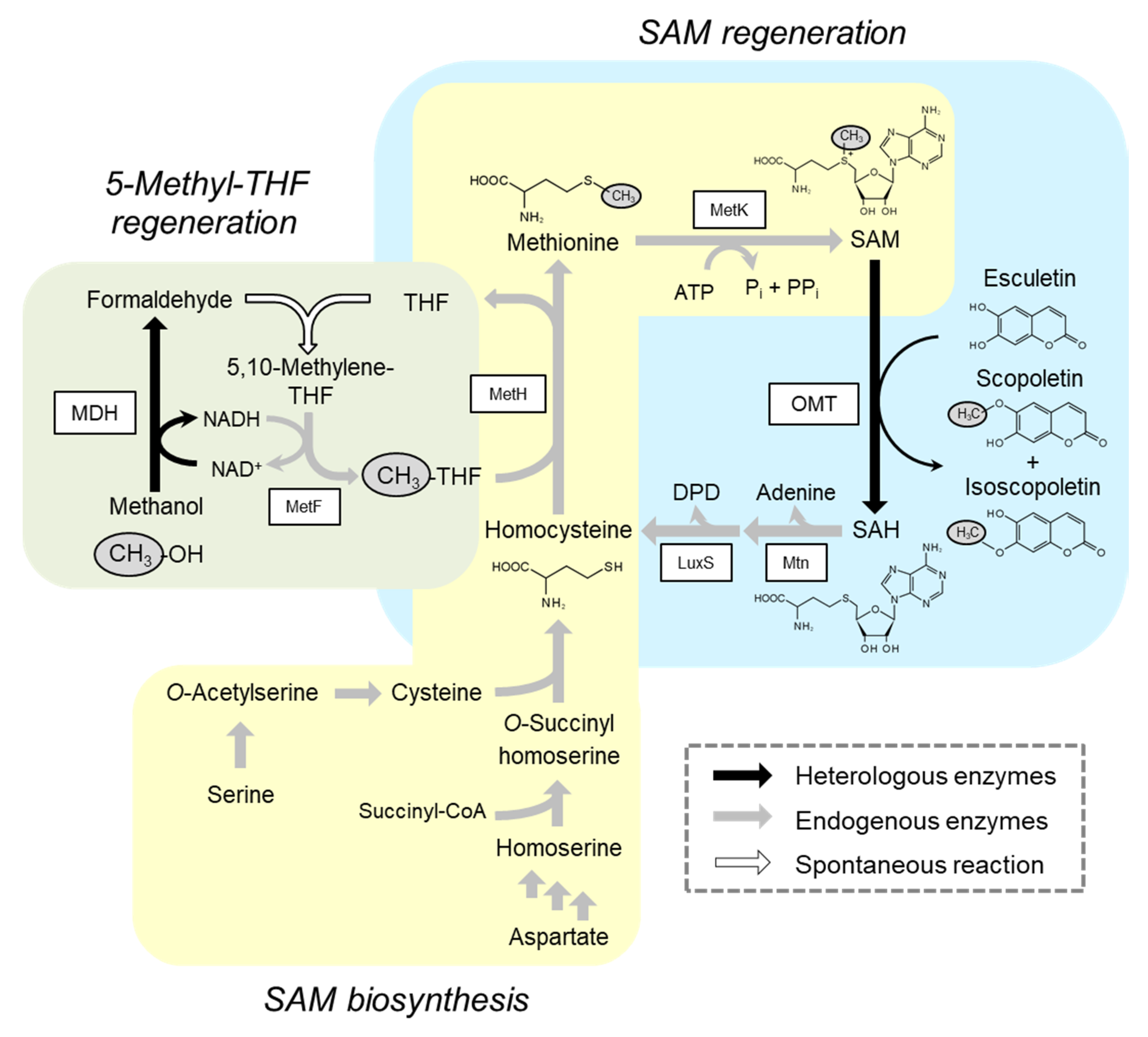
2.1. Deregulation of SAM Biosynthesis

To determine whether intracellularly produced SAM can be used for producing methylated compounds, methylation of esculetin was performed using E. coli BL21 (DE3) harboring pETD-OMT (OMT/BL21) without external SAM input. The pETD-OMT was constructed for expression of O-methyltransferase (OMT) 5 derived from Streptomyces avermitilis MA-4680 [13]. Preliminary experiments showed that OMT5 methylated both 6-OH and 7-OH of esculetin and produced scopoletin and isoscopoletin, respectively (Table S1). Although regioselectivity was low, methylation of esculetin by OMT5 was adopted as a model reaction because of the ease of analysis and the availability of standards. OMT/BL21 cells were cultivated in the nutrient-rich Luria-Bertani (LB) medium and used for the methylation reaction. After 6 h of reaction, 0.023 mM of methylated compounds were produced from 0.5 mM of esculetin (Figure 2a). No further methylation was observed, even after 24 h of the reaction. This indicated that intracellularly produced SAM was available for esculetin methylation, while the yield of methylated compounds was as low as 4.6 mol%. Interestingly, the concentration and yield of methylated compounds was drastically increased by changing the medium for cell preparation. When the cells were prepared in the M9 minimal medium, a 7.4-fold increase was observed in the concentration of methylated compounds (0.17 mM) and yield was 33.3 mol% at 18 h of reaction. However, the increase in the concentration was suppressed to 4.8-fold by adding 1 mM methionine to the M9 medium. These results suggest that SAM biosynthesis is negatively regulated by methionine and the use of the cells prepared in methionine-free medium, such as the M9 minimal medium, is suitable for the methylation reaction.
The methylated compounds yield was further increased by disruption of the gene encoding MetJ, a transcriptional regulator of methionine/SAM biosynthesis (Figure S1) using the CRISPR/Cas9 genome-editing system (Figure S2) [14]. Methylation reactions were performed using the M9-grown ΔmetJ cells harboring pETD-OMT (OMT/ΔmetJ). About 0.24 mM of methylated compounds were produced with a yield of 47.7 mol% at 18 h (Figure 2b). As expected, no significant decrease in the concentration of the methylated compounds (0.22 mM) was observed by the addition of methionine to the medium for the cell preparation. However, the effect of metJ deletion was limited when the cells were prepared in LB medium (0.11 mM of the methylated compounds), implying that SAM biosynthesis is also controlled by factors other than MetJ. To prevent suppression of SAM biosynthesis by unidentified regulatory system(s), the M9-grown ΔmetJ cells were used for methylation reactions in the subsequent experiments.

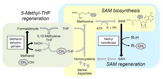
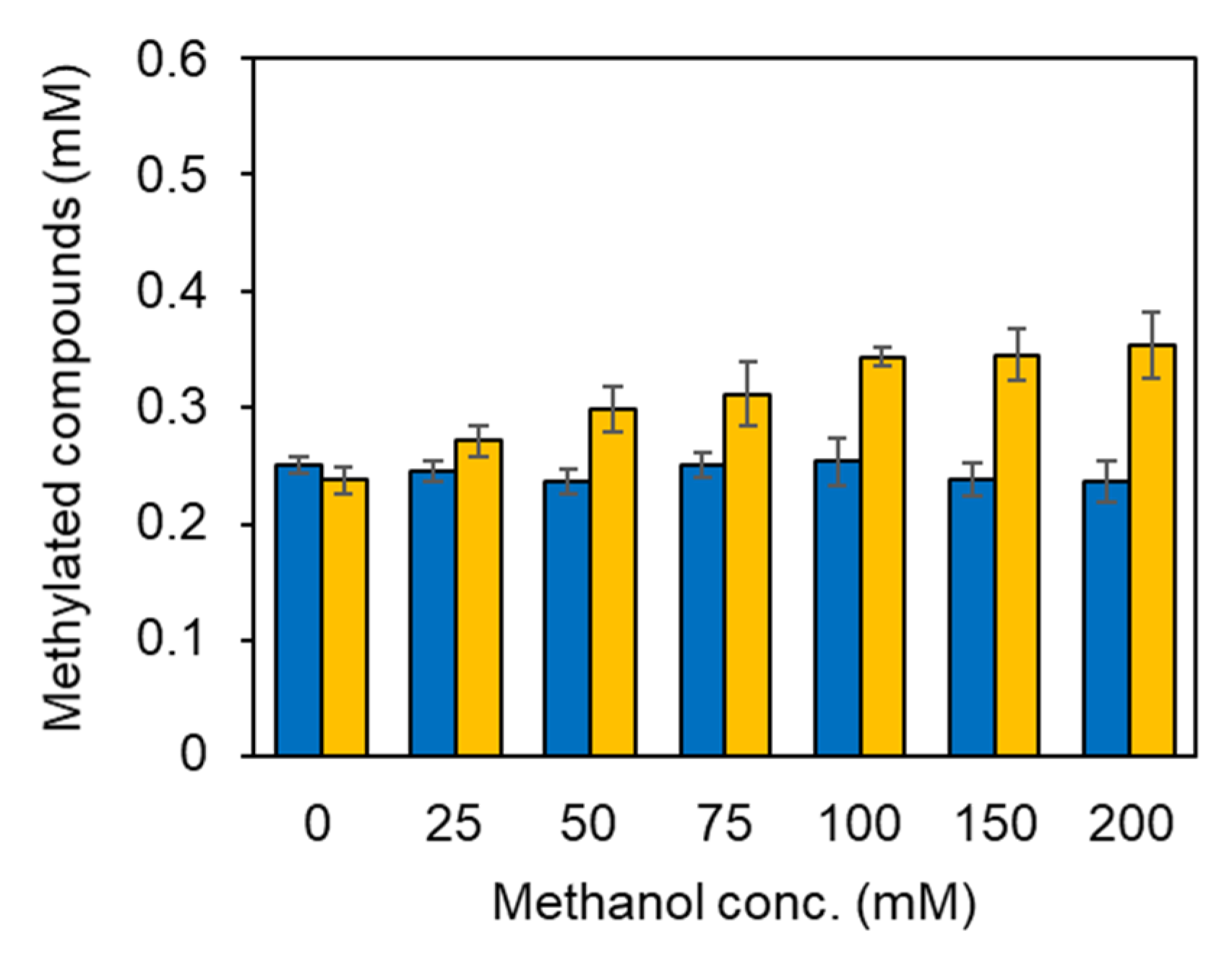
2.2. Integration of Methanol Metabolic Pathway for 5-Methyl-THF Formation into SAM-Dependent Methylation System

The MDH-driven methanol metabolic pathway for 5-methyl-THF formation (Figure 1) was installed into the E. coli ΔmetJ strain by co-expressing an engineered (A26V, A31V, A169V) MDH2, derived from Cupriavidus necator N-1 [15] with OMT. In this pathway, the methyl group derived from methanol is used for the formation of 5-methyl-THF and the resulting 5-methyl-THF promotes the subsequent SAM regeneration and methylation reaction. Methylation reactions were performed using the ΔmetJ cells transformed with pETD-OMT-MDH (OMT-MDH/ΔmetJ) while varying methanol concentrations (Figure 3). OMT-MDH/ΔmetJ cells produced almost the same concentration of the methylated compounds (0.24 mM) as OMT/ΔmetJ cells (0.25 mM), when no methanol was added to the reaction mixtures. Methanol concentration-dependent increase in the methylated compounds was observed only in OMT-MDH/ΔmetJ cells, and the concentration of the methylated compounds increased to 0.34 mM, within the methanol concentration range of 0 to 100 mM. These results show that integration of methanol metabolic pathway for 5-methyl-THF formation is an effective strategy to activate the SAM regeneration and SAM-dependent methylation reactions. No further increase in the concentration of the methylated compounds was observed at methanol concentrations above 100 mM.

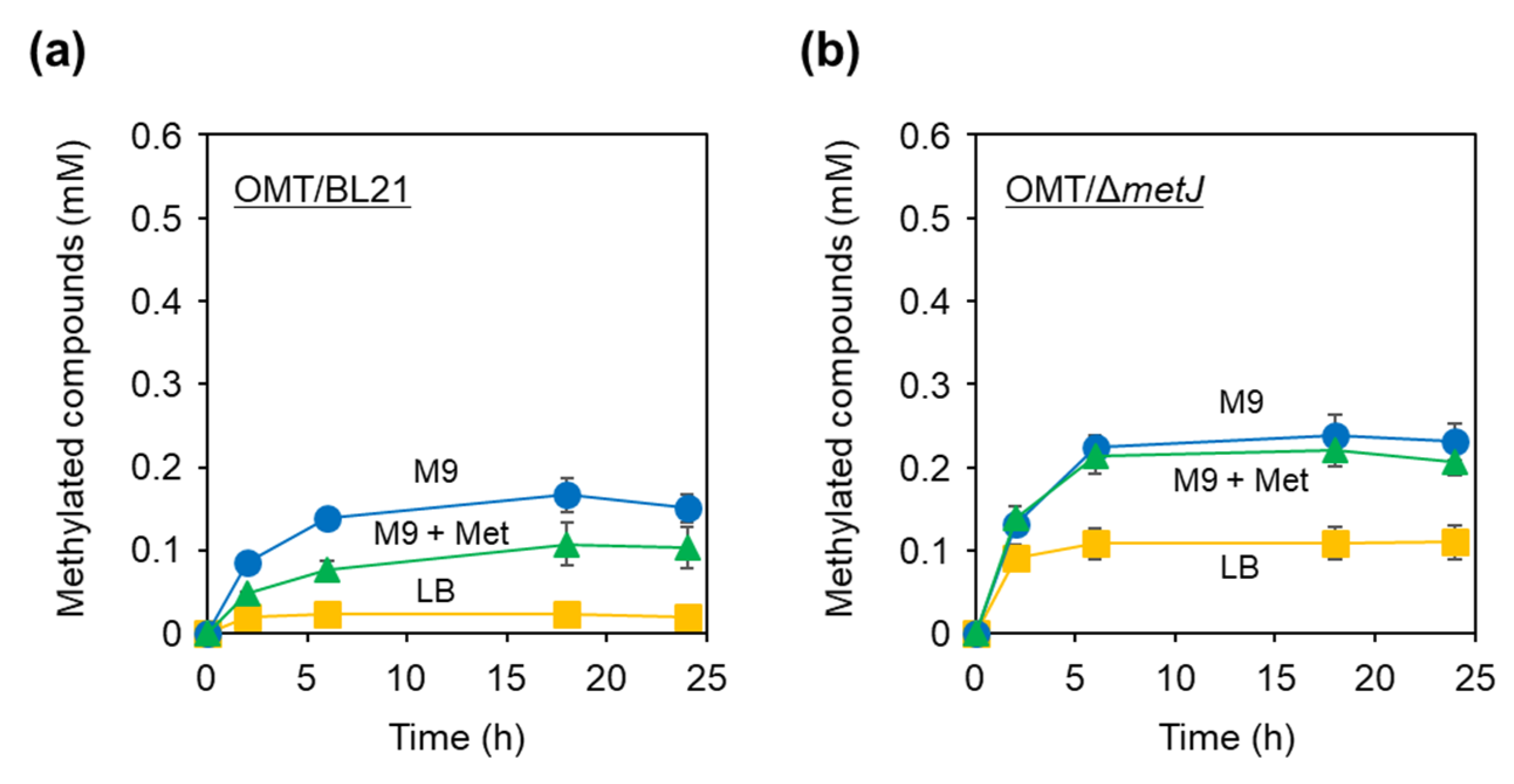
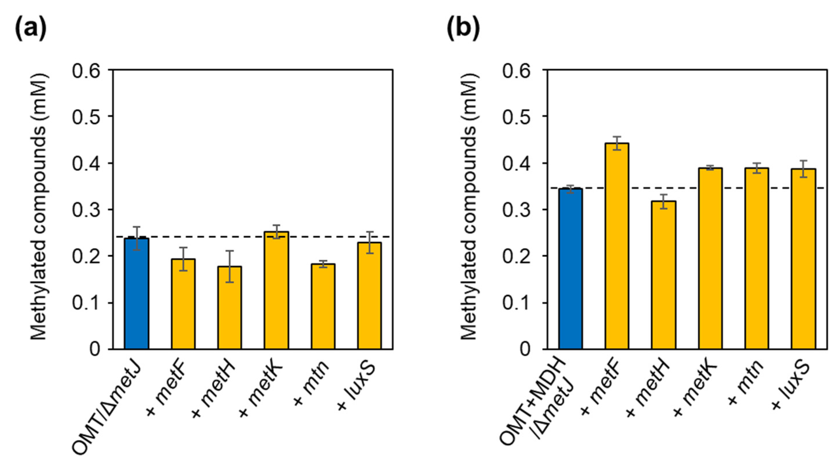
2.3. Alleviation of Bottlenecks in the SAM Regeneration Pathway

Five endogenous enzymatic reactions are involved in the SAM regeneration pathway coupled with the 5-methyl-THF regeneration pathway (Figure 1). Each of the five corresponding genes were overexpressed in OMT/ΔmetJ and OMT-MDH/ΔmetJ cells to alleviate the bottlenecks in the SAM regeneration pathway. No increase in the titer of methylated compounds was observed by overexpression of each of five genes in the OMT/ΔmetJ cells, indicating that the 5,10-methylene-THF supply was the bottleneck in the strain (Figure 4a). In contrast, overexpression of the genes encoding SAM synthase (metK), SAH nucleosidase (mtn), or S-ribosylhomocysteine lyase (luxS) in OMT-MDH/ΔmetJ cells gave a 13% increase in the concentration of the methylated compounds, while a slight decrease (8%) was observed by overexpression of the gene encoding methionine synthase (metH; Figure 4b). The methylation reaction was most enhanced by the overexpression of metF, where the concentration of the methylated compounds was increased by 29%. As a result, 0.44 mM of the methylated compound was produced with a high yield of 88 mol% after 18 h of reaction. These results suggest that 5,10-methylene-THF was efficiently supplied by the oxidation of methanol catalyzed by MDH and following spontaneous condensation with THF, and the reduction of 5,10-methylene-THF to 5-methyl-THF was the bottleneck for the SAM regeneration pathway in OMT-MDH/ΔmetJ cells.

2.4. Methylation Reaction Using Glucose as a Carbon/Energy Source for ATP Regeneration

As well as 5-methyl-THF, ATP is a limiting cofactor for SAM regeneration. ATP is used for adenosylation of methionine to form SAM and an adenosyl moiety of SAM is degraded into adenine (Figure 1). E. coli possess a series of enzymes required for salvage synthesis of ADP from adenine and ribose-5-phosphate; ribose-phosphate pyrophosphokinase, adenine phosphoribosyltransferase, and adenylate kinase [16,17,18]. Therefore, salvage synthesis of ADP is possible by supplying glucose as a carbon source to produce ribose-5-phosphate via the pentose phosphate pathway. In addition, glucose could be used as an energy source to regenerate ATP from ADP. To verify this hypothesis, methylation reactions were performed with the addition of 0–25 mM glucose instead of ATP. The OMT-MDH-MetF/ΔmetJ cells produced 0.30 mM of the methylated products at 18 h without adding glucose, indicating that intracellularly produced ATP is available for SAM regeneration (Figure 5). Furthermore, the concentration of the methylated compounds increased with increasing glucose concentrations. When 25 mM of glucose was added, 0.49 mM of methylated compounds were produced, which is comparable to when 1 mM of ATP is added (0.44 mM). These results show that methylation reactions are possible without adding any exogenous cofactors.

3. Discussion

Many efforts have been dedicated to increase SAM availability in both in vitro and in vivo methylation systems. Recently, significant progress has been made in in vitro methylation systems. Mordhorst et al. [19] first reported an in vitro enzyme cascade, which regenerates SAM from SAH, using five enzymes. In this cascade, hydrolysis of SAH to adenosine, followed by phosphorylation and condensation with externally-added methionine, enabled biomimetic SAM regeneration. By coupling this pathway with SAM-dependent methyltransferases, methylation reactions were achieved with a turnover number of 10–11. Later, Liao and Seebeck [9] reported the simplest SAM regeneration system. Among the SAM-dependent methyltransferases, halide methyltransferase can exceptionally catalyze the reverse reaction. In this reaction, the use of halide methyltransferase with methyl iodide as a methyl group donor allowed direct SAM regeneration from SAH, with higher turnover number of 35–290, and significantly reduced SAM input. However, a major drawback of this elegant SAM regeneration system is the dependence on a hazardous methyl donor.
The advantage of in vivo systems is that intracellularly produced SAM can be utilized for methylation reactions. If sufficient SAM is supplied by enhancement of SAM biosynthesis/regeneration, methylation reactions will proceed without any external SAM input. Overexpression of the genes encoding pathway-related enzymes is the most straightforward way to provide SAM. Overexpression of metK, mtn, or luxS facilitated SAM-dependent methylation in the fermentative production of methylated compounds, such as vanillate and methyl anthranilate [20,21]. Deregulation of SAM biosynthesis is also a frequently used strategy. Expression of several genes involved in SAM biosynthesis are negatively regulated by the MetJ repressor and its corepressor, SAM (Figure S1) [22]. Disruption or repression of metJ improved the titer of methylated compounds [20,23]. In addition, the release of feedback inhibition of homoserine succinyltransferase by methionine, and serine O-acetyltransferase by cysteine, enhanced the methylation reaction [20,21]. Our results also showed that deregulation of the SAM biosynthetic pathway by the use of methionine-free medium and metJ disruption enhanced methylation of esculetin (Figure 2). However, none of these approaches addresses the fundamental problem in SAM regeneration, the limited 5-methyl-THF supply. Our approach integrating the methanol metabolic pathway with 5-methyl-THF formation into the SAM regeneration pathway is a pioneering effort to tackle this problem.
In the design of the 5-methyl-THF regeneration pathway, methanol was used as a methyl group source, which is cheap, abundant, and less toxic compared to methyl iodide, and a mutant MDH from C. necator was selected for methanol oxidation [15]. There are a variety of methanol-oxidizing enzymes, which utilize different electron acceptors, such as NAD+, pyrroloquinoline quinone, and oxygen [24,25,26]. Since the reduction of 5,10-methylene-THF to 5-methyl-THF requires NADH as a co-factor, the use of NAD+-dependent enzymes is suitable for achieving balanced consumption/regeneration of NAD+/NADH in the designed pathway (Figure 1). Among NAD+-dependent enzymes, Bacillus methanolicus MDHs are well-characterized, and parts were utilized for constructing a methanol-fixation pathway [27,28]. However, the affinity of B. methanolicus MDHs to methanol was weak (Km > 170 mM) in the absence of their activator protein, ACT [24]. In our preliminary experiment using the BL21 (DE3) cells co-expressing MDH2 from B. methanolicus MGA3 and OMT, an addition of 100 mM methanol caused only 8% increase in the concentration of methylated compounds (Figure S3). A clear increase in the concentration of the methylated compounds (39%) was observed by the addition of 500 mM methanol compared with that without methanol. In contrast, MDH from C. necator harboring the triple mutation (A26V, A31V, and A169V) showed higher affinity toward methanol (Km = 21.6 mM) without requiring an activator protein [15]. The addition of 100 mM of methanol was enough to facilitate methylation reaction (45% increase) when MDH was co-expressed with OMT (Figure 3). However, the reaction still required an excess amount of methanol to the substrate (0.5 mM). Further screening or improvement of NAD+-dependent MDH is thus required.
Besides optimizing MDH, the methanol metabolic pathway with 5-methyl-THF formation still has room for further improvement. Formaldehyde, a metabolic intermediate in the system, enters its detoxification pathway, except for 5-methyl-THF formation [27]. Detoxification of formaldehyde is initiated by the spontaneous condensation of formaldehyde and glutathione (GSH), and the resulting S-hydroxymethyl-GSH is enzymatically degraded into CO2. Disruption of the gene encoding S-hydroxymethyl-GSH dehydrogenase (frmA) was shown to increase the intracellular concentration of formaldehyde in mdh-expressing E. coli cells [27]. In addition, our results show that reduction of 5,10-methylene-THF was a bottleneck for SAM regeneration in the strain equipped with the methanol metabolic pathway with 5-methyl-THF formation (Figure 4b). Taken together, disruption of frmA and further enhancement of MetF activity will enhance metabolic flux of methyl groups derived from methanol toward 5-methyl-THF formation, thereby promoting SAM regeneration and the methylation reaction.

4. Materials and Methods

4.1. Strains and Media

The strains and plasmids used in this study are listed in Table 1. E. coli TG1 (Zymo Research, Irvine, CA, USA) and BL21 (DE3) (Novagen, Darmstadt, Germany) were used for genetic manipulation and gene expression, respectively. They were grown either in LB medium or in M9 minimal salt medium supplemented with 0.4% (w/v) glucose at 37 °C. For the cultivation of transformants carrying pETDuet-1 (Novagen) and/or pACYCDuet-1 (Novagen) derivatives, 100 μg/mL ampicillin and/or 17 μg/mL chloramphenicol were added to the media, respectively. For solid media, 1.5% (w/v) agar was added to the media described above.

4.2. Plasmid Construction

The oligonucleotide primers used in this study are summarized in Table S2. PCR was carried out using PrimeSTAR HS DNA polymerase (Takara Bio Inc., Shiga, Japan). The codon-optimized omt5 gene derived from S. avermitilis MA-4680 was synthesized by Integrated DNA Technologies, Inc. (Skokie, IL, USA) and its sequence is shown in Table S3 [13]. The omt5 gene was amplified by PCR using the omt_F and omt_R primers. The amplified fragment was digested with NcoI and HindIII, and ligated into the same sites of pETDuet-1. The resulting plasmid was designated as pETD-OMT. Similarly, the codon-optimized gene encoding an engineered (A26V, A31V, A169V) MDH 2, derived from C. necator N-1, was synthesized and amplified with the primers mdh_F and mdh_R [15]. The amplified fragment was inserted into the NdeI and XhoI sites of pETD-OMT, resulting in the pETD-OMT-MDH plasmid.
For overexpression of E. coli endogenous genes encoding MetF, MetH, MetK, Mtn, and LuxS, each of the five genes were amplified from E. coli genomic DNA using the primer pairs metF_F/metF_R, metH_F/metH_R, metK_F/metK_R, mtn_F/mtn_R, and luxS_F/luxS_R, respectively. The amplified fragments were digested with NdeI/XhoI (for metF and metH) or NcoI/HindIII (for metK, mtn, and luxS), followed by insertion into the respective restriction enzyme sites of pACYCDuet-1, which is compatible to pETDuet-1. The resulting plasmids were designated as pACD-MetF, pACD-MetH, pACD-MetK, pACD-Mtn, and pACD-LuxS, respectively.
pCas and pTargetF were used for genome editing based on the CRISPR-Cas9 system and they were gifts from Sheng Yang (Addgene plasmids #62225 and #62226, respectively) [14]. The original N20 sequence, which is a 20-bp target sequence for inducing double strand breaks by the Cas9 nuclease-single guide RNA (sgRNA) complex, of pTargetF was replaced with “CAGCGACGAAATCCCGGAAG” to target metJ by inverse PCR using the primers metJ N20_F and metJ N20_R. The amplified fragment was digested with SpeI and then self-ligated, resulting in pTarget-metJ sgRNA. Subsequently, donor DNA as the genome editing template was introduced into pTarget-metJ sgRNA. The 545-bp upstream and 542-bp downstream regions of metJ were amplified by PCR from E. coli genomic DNA using the primer sets of metJ up_F/metJ up_R and metJ down_F/metJ down_R, respectively. The amplified fragments were connected by overlapping PCR using the primer pair metJ up_F and metJ down_R. The amplified fragment was digested with BglII and XhoI, and subsequently inserted into the same sites of pTarget-metJ sgRNA. The resulting plasmid was designated pTarget-ΔmetJ.

4.3. Genome Editing

The CRISPR-Cas9 two-plasmid system was used for metJ deletion [14]. The plasmid pCas was introduced into E. coli BL21 (DE3). The resulting strain was cultivated overnight at 30 °C in LB medium, supplemented with 10 mM arabinose for induction of λ-Red recombinase, and diluted 1:50 in 5 mL of the same medium [29]. After reaching an OD600 of 0.5, the cells were collected by centrifugation and washed twice using ice-cold 10% (w/v) glycerol, followed by resuspension in 250 μL of ice-cold 10% (w/v) glycerol. For transformation, 100 μL of the cells and 100 ng of pTarget-ΔmetJ were mixed and electroporation was performed in a 2-mm cuvette (Bio-rad Laboratories Inc., Richmond, CA, USA) using a Gene-Pulser (Bio-rad, 2.5 kV, 25 μF, 200 Ω). After electroporation, 1 mL of LB medium supplemented with 10 mM arabinose was added to the cuvette and incubated at 30 °C for 1 h. Then, the cells were spread on LB agar medium containing 10 mM arabinose, 50 μg/mL of kanamycin, and 50 μg/mL of spectinomycin. Deletion of metJ was confirmed by colony PCR using primer pair metJ up 550 bp_F and metJ down 550 bp_R, followed by DNA sequencing analysis. Curing of pCas and pTarget-ΔmetJ were performed as described previously [14].

4.4. Methylation Reaction

Transformants of E. coli BL21 (DE3) and its ΔmetJ mutant were pre-cultivated in 5 mL of LB medium at 37 °C overnight and diluted 1:20 in 50 mL of LB or M9 medium supplemented with appropriate antibiotics. After 2.5 h of cultivation at 37 °C, gene expression was induced by the addition of 0.2 mM of isopropylthiogalactoside. After 2.5 h of induction, the cells were harvested by centrifugation and washed twice with 50 mM Tris-HCl buffer (pH 8.0), and resuspended in the same buffer. Methylation of esculetin to isoscopoletin and scopoletin was performed in a 1.5 mL microtube with 1.0 mL of reaction mixture containing the following: esculetin, 0.5 mM; methanol, 0–200 mM; MgSO4, 10 mM; ATP, 1 mM; Tris-HCl (pH 8.0), 50 mM; dimethyl sulfoxide, 2% (v/v); wet cells of recombinant E. coli, 10 mg/mL. ATP was replaced with 0–25 mM of glucose, if necessary. The mixture was incubated at 37 °C using a Block Bath Shaker (MyBL-100S; AS ONE Co., Osaka, Japan) with shaking at 1500 rpm. After incubation, a portion of the mixture was regularly harvested and centrifuged to remove the cells. Esculetin, isoscopoletin, and scopoletin in the supernatant were quantified by high-performance liquid chromatography equipped with a Develosil ODS-HG-5 column (250 mm × 4.6 mm; Nomura Chemical Co., Ltd., Aichi, Japan). The column was eluted by a mobile phase (20% (v/v) acetonitrile and 1% (v/v) formic acid) at a flow rate of 1.0 mL/min at 40 °C. The eluent was analyzed with a SPD-20AV UV-VIS detector (Shimadzu Co., Kyoto, Japan) at 254 nm.

5. Conclusions

In the present study, the MDH-driven methanol metabolic pathway with 5-methyl-THF formation was constructed and integrated into E. coli expressing the methyltransferase to enhance the SAM-dependent methylation reaction. The combination of deregulation of SAM biosynthesis and the introduction of the methanol metabolic pathway for 5-methyl-THF formation increased the titer of methylated products approximately 20-fold (0.023 mM to 0.44 mM). Importantly, the engineered E. coli strain with enhanced 5-methyl-THF formation can be used as a chassis strain for the production of a variety of methylated compounds simply by exchanging the methyltransferase. The use of different types of methyltransferases, such as N-, C-, P-, S-, Se-, and As-methyltransferases, will enhance the methylation of diverse biomolecules [6]. Moreover, metabolic engineering of the chassis strain will enable effective de novo biosynthesis of complicated natural products, even those requiring multi-steps and different types of methylation reactions [30,31].

Supplementary Materials

The following are available online at https://www.mdpi.com/2073-4344/10/9/1001/s1, Figure S1: Schematic illustration of the regulation of methionine/SAM biosynthesis, Figure S2: Deletion of the metJ gene in E. coli BL21 (DE3), Figure S3: Methylation reactions with Bacillus methanolicus MDH-driven methanol metabolic pathway for 5-methyl-THF formation, Table S1: Concentration of the methylated compounds produced by OMT/BL21 cells, Table S2: Sequence of oligonucleotide primers used in this study, Table S3: Sequences of codon-optimized omt and mdh synthetic genes.

Author Contributions

Conceptualization, K.O. and K.H.; methodology, K.O., Y.S., and S.I.; investigation, K.O., Y.S., S.I., and S.K. (Shizuka Kawakami); writing—original draft preparation, K.O.; writing—review and editing, K.O., Y.S., S.K. (Shigeru Kitani), and K.H.; supervision, K.H.; project administration, K.O.; funding acquisition, K.O. All authors have read and agreed to the published version of the manuscript.

Funding

This research was partly funded by the Japan Society for the Promotion of Sciences (JSPS), KAKENHI Grant (19H04656).

Conflicts of Interest

The authors declare no conflict of interest.

References

  1. Wang, X.; Saba, T.; Yiu, H.H.P.; Howe, R.F.; Anderson, J.A.; Shi, J. Cofactor NAD(P)H regeneration inspired by heterogeneous pathways. Chem 2017, 2, 621–654. [Google Scholar] [CrossRef] [Green Version]
  2. Okano, K.; Honda, K.; Taniguchi, H.; Kondo, A. De novo design of biosynthetic pathways for bacterial production of bulk chemicals and biofuels. FEMS Microbiol. Lett. 2018, 365, fny215. [Google Scholar] [CrossRef] [PubMed] [Green Version]
  3. Uppada, V.; Bhaduri, S.; Noronha, S.B. Cofactor regeneration—An important aspect of biocatalysis. Curr. Sci. 2014, 106, 946–957. [Google Scholar]
  4. Lin, B.; Tao, Y. Whole-cell biocatalysts by design. Microb. Cell Fact. 2017, 16, 106. [Google Scholar] [CrossRef] [Green Version]
  5. Andexer, J.N.; Richter, M. Emerging enzymes for ATP regeneration in biocatalytic processes. ChemBioChem 2015, 16, 380–386. [Google Scholar] [CrossRef] [PubMed]
  6. Struck, A.W.; Thompson, M.L.; Wong, L.S.; Micklefield, J. S-Adenosyl-methionine-dependent methyltransferases: Highly versatile enzymes in biocatalysis, biosynthesis and other biotechnological applications. ChemBioChem 2012, 13, 2642–2655. [Google Scholar] [CrossRef]
  7. Barreiro, E.J.; Kümmerle, A.E.; Fraga, C.A.M. The methylation effect in medicinal chemistry. Chem. Rev. 2011, 111, 5215–5246. [Google Scholar] [CrossRef]
  8. Schönherr, H.; Cernak, T. Profound methyl effects in drug discovery and a call for new C-H methylation reactions. Angew. Chem. Int. Ed. Engl. 2013, 52, 12256–12267. [Google Scholar] [CrossRef]
  9. Liao, C.; Seebeck, F.P. S-adenosylhomocysteine as a methyl transfer catalyst in biocatalytic methylation reactions. Nat. Catal. 2019, 2, 696–701. [Google Scholar] [CrossRef]
  10. Ogawa, H.; Gomi, T.; Fujioka, M. Serine hydroxymethyltransferase and threonine aldolase: Are they identical? Int. J. Biochem. Cell Biol. 2000, 32, 289–301. [Google Scholar] [CrossRef]
  11. Xu, Y.; Meng, H.; Ren, J.; Zeng, A.P. Formaldehyde formation in the glycine cleavage system and its use for an aldolase-based biosynthesis of 1,3-prodanediol. J. Biol. Eng. 2020, 14, 15. [Google Scholar] [CrossRef] [PubMed]
  12. Marx, C.J.; Van Dien, S.J.; Lidstrom, M.E. Flux analysis uncovers key role of functional redundancy in formaldehyde metabolism. PLoS Biol. 2005, 3, e16. [Google Scholar] [CrossRef] [PubMed] [Green Version]
  13. Yoon, Y.; Park, Y.; Yi, Y.S.; Lee, Y.; Jo, G.; Park, J.C.; Ahn, J.H.; Lim, Y. Characterization of an O-methyltransferase from Streptomyces avermitilis MA-4680. J. Microbiol. Biotechnol. 2010, 20, 1359–1366. [Google Scholar] [CrossRef] [PubMed]
  14. Jiang, Y.; Chen, B.; Duan, C.; Sun, B.; Yang, J.; Yang, S. Multigene editing in the Escherichia coli genome via the CRISPR-Cas9 system. Appl. Environ. Microbiol. 2015, 81, 2506–2514. [Google Scholar] [CrossRef] [PubMed] [Green Version]
  15. Wu, T.Y.; Chen, C.T.; Liu, J.T.J.; Bogorad, I.W.; Damoiseaux, R.; Liao, J.C. Characterization and evolution of an activator-independent methanol dehydrogenase from Cupriavidus necator N-1. Appl. Microbiol. Biotechnol. 2016, 100, 4969–4983. [Google Scholar] [CrossRef] [Green Version]
  16. Zhou, W.; Tsai, A.; Dattmore, D.A.; Stives, D.P.; Chitrakar, I.; D’Alessandro, A.M.; Patil, S.; Hicks, K.A.; French, J.B. Crystal structure of E. coli PRPP synthetase. BMC Struct. Biol. 2019, 19, 1. [Google Scholar] [CrossRef]
  17. Hershey, H.V.; Gutstein, R.; Taylor, M.W. Cloning and restriction map of the E. coli apt gene. Gene 1982, 19, 89–92. [Google Scholar] [CrossRef]
  18. Brune, M.; Schumann, R.; Wittinghofer, F. Cloning and sequencing of the adenylate kinase gene (adk) of Escherichia coli. Nucleic Acids Res. 1985, 13, 7139–7151. [Google Scholar] [CrossRef] [Green Version]
  19. Mordhorst, S.; Siegrist, J.; Müller, M.; Richter, M.; Andexer, J.N. Catalytic alkylation using a cyclic S-adenosylmethionine regeneration system. Angew. Chem. Int. Ed. 2017, 56, 4037–4041. [Google Scholar] [CrossRef]
  20. Kunjapur, A.M.; Hyun, J.C.; Prather, K.L. Deregulation of S-adenosylmethionine biosynthesis and regeneration improves methylation in the E. coli de novo vanillin biosynthesis pathway. Microb. Cell Fact. 2016, 15, 61. [Google Scholar] [CrossRef] [Green Version]
  21. Luo, Z.W.; Cho, J.S.; Lee, S.Y. Microbial production of methyl anthranilate, a grape flavor compound. Proc. Natl. Acad. Sci. USA 2019, 116, 10749–10756. [Google Scholar] [CrossRef] [PubMed] [Green Version]
  22. Usuda, Y.; Kurahashi, O. Effects of deregulation of methionine biosynthesis on methionine excretion in Escherichia coli. Appl. Environ. Microbiol. 2005, 71, 3228–3234. [Google Scholar] [CrossRef] [PubMed] [Green Version]
  23. Cress, B.F.; Leitz, Q.D.; Kim, D.C.; Amore, T.D.; Suzuki, J.Y.; Linhardt, R.J.; Koffas, M.A.G. CRISPRi-mediated metabolic engineering of E. coli for O-methylated anthocyanin production. Microb. Cell Fact. 2017, 16, 10. [Google Scholar] [CrossRef] [PubMed] [Green Version]
  24. Krog, A.; Heggeset, T.M.B.; Müller, J.E.N.; Kupper, C.E.; Schneider, O.; Vorholt, J.A.; Ellingsen, T.E.; Brautaset, T. Methylotrophic Bacillus methanolicus encodes two chromosomal and one plasmid born NAD+ dependent methanol dehydrogenase paralogs with different catalytic and biochemical properties. PLoS ONE 2013, 8, e59188. [Google Scholar] [CrossRef] [Green Version]
  25. Keltjens, J.T.; Pol, A.; Reimann, J.; Op den Camp, H.J.M. PQQ-dependent methanol dehydrogenases: Rare-earth elements make a difference. Appl. Microbiol. Biotechnol. 2014, 98, 6163–6183. [Google Scholar] [CrossRef]
  26. Couderc, R.; Baratti, J. Oxidation of methanol by the yeast, Pichia pastoris. Purification and properties of the alcohol oxidase. Agric. Biol. Chem. 1980, 44, 2279–2289. [Google Scholar] [CrossRef]
  27. Müller, J.E.N.; Meyer, F.; Litsanov, B.; Kiefer, P.; Potthoff, E.; Heux, S.; Quax, W.J.; Wendisch, V.F.; Brautaset, T.; Portais, J.C.; et al. Engineering Escherichia coli for methanol conversion. Metab. Eng. 2015, 28, 190–201. [Google Scholar] [CrossRef]
  28. Meyer, F.; Keller, P.; Hartl, J.; Gröninger, O.G.; Kiefer, P.; Vorholt, J.A. Methanol-essential growth of Escherichia coli. Nat. Commun. 2018, 9, 1508. [Google Scholar] [CrossRef]
  29. Datsenko, K.A.; Wanner, B.L. One-step inactivation of chromosomal genes in Escherichia coli K-12 using PCR products. Proc. Natl. Acad. Sci. USA 2000, 97, 6640–6645. [Google Scholar] [CrossRef] [Green Version]
  30. Galanie, S.; Thodey, K.; Trenchard, I.J.; Interrante, M.F.; Smolke, C.D. Complete biosynthesis of opioids in yeast. Science 2018, 349, 1095–1100. [Google Scholar] [CrossRef] [Green Version]
  31. Nakagawa, A.; Matsumura, E.; Koyanagi, T.; Katayama, T.; Kawano, N.; Yoshimatsu, K.; Yamamoto, K.; Kumagai, H.; Sato, F.; Minami, H. Total biosynthesis of opiates by stepwise fermentation using engineered Escherichia coli. Nat. Commun. 2016, 7, 10390. [Google Scholar] [CrossRef] [PubMed] [Green Version]
Figure 1. Schematic illustration of the SAM biosynthetic/regeneration and the MDH-driven 5-methyl-THF regeneration pathways with esculetin methylation. Methyl groups originating from methanol can be transferred to SAM via 5-methyl-THF and methionine. SAM is then utilized for esculetin methylation to scopoletin and isoscopoletin. Abbreviations used: SAM, S-adenosylmethionine; SAH, S-adenosylhomocysteine; THF, tetrahydrofolate; DPD, 4,5-dihydroxypentane-2,3-dione; ATP, adenosine triphosphate; NAD+/NADH, nicotinamide adenine dinucleotide; Pi, phosphate; PPi, pyrophosphate; MDH, methanol dehydrogenase; OMT, O-methyltransferase; MetF, 5,10-methylene-THF reductase; MetH, methionine synthase; MetK, SAM synthase; Mtn, SAH nucleosidase; LuxS, S-ribosylhomocysteine lyase.
Catalysts 10 01001 g001
Figure 2. Effect of the medium used for cell preparation on methylation reaction. Time course data of esculetin methylation using OMT/BL21 cells (a) and OMT/ΔmetJ cells (b). Each strain was cultivated in LB medium (orange), M9 medium (blue), or M9 medium supplemented with 1 mM methionine (green) prior to the methylation reaction. The concentration of the methylated products is shown as the sum of the concentrations of scopoletin and isoscopoletin. Data points represent the mean ± standard deviations for three independent experiments.
Catalysts 10 01001 g002
Figure 3. Methylation reactions with the MDH-driven methanol metabolic pathway for 5-methyl-THF formation. Methylation of esculetin was performed by adding various concentrations of methanol for 18 h using OMT/ΔmetJ cells (blue bars) and OMT-MDH/ΔmetJ cells (orange bars). Data bars represent the mean ± standard deviations for three independent experiments.
Catalysts 10 01001 g003
Figure 4. Alleviation of bottleneck in the SAM regeneration pathway. Each of the five genes related to SAM regeneration were overexpressed in OMT/ΔmetJ cells (a) and OMT-MDH/ΔmetJ cells (b). The methylation reaction of esculetin was performed for 18 h with the addition of 100 mM methanol. Data bars represent the mean ± standard deviations for three independent experiments.
Catalysts 10 01001 g004
Figure 5. Methylation reaction using glucose as a carbon/energy source for ATP regeneration. The methylation reaction of esculetin was performed for 18 h using OMT-MDH-MetF/ΔmetJ cells with various concentration of glucose instead of ATP (orange bars). The methanol concentration was fixed at 100 mM. The result obtained by 1.0 mM of ATP addition (blue bar) was also shown as a control. Data bars represent the mean ± standard deviations for three independent experiments.
Catalysts 10 01001 g005
Table 1. Strains and plasmids used in this study.
Strain or PlasmidRelevant DescriptionSource
Escherichia coli strain
TG1supE thi-1 Δ(lac-proAB) Δ(mcrB-hsdSM)5 (rK mK) [F’ traD36 proAB Zymo Research
lacIqZΔM15]
BL21(DE3)F ompT hsdSB (rB mB) gal dcm (DE3)Novagen
ΔmetJBL21 (DE3) ΔmetJThis study
Plasmid
pETDuet-1Dual T7 promoters, pBR322 ori, bla, lacINovagen
pETD-OMTpETDuet-1 derivative for expressing omt5 from S. avermitilis MA-4680This study
pETD-OMT-MDHpETDuet-1 derivative for co-expressing omt5 and mdh2 (A26V, A31V,This study
A169V) from C. necator N-1
pACYCDuet-1Dual T7 promoter, p15A ori, cat, lacINovagen
pACD-MetFpACYCDuet-1 derivative for expressing endogenous metFThis study
pACD-MetHpACYCDuet-1 derivative for expressing endogenous metHThis study
pACD-MetKpACYCDuet-1 derivative for expressing endogenous metKThis study
pACD-MtnpACYCDuet-1 derivative for expressing endogenous mtnThis study
pACD-LuxSpACYCDuet-1 derivative for expressing endogenous luxSThis study
pCasrep101(Ts), kan, Pcas-cas9, ParaB-Red, lacIq, Ptrc-sgRNA-pMB1[14]
pTargetFpMB1 ori, aadA[14]
pTarget-metJ sgRNApMB1 ori, aadA, metJ-sgRNAThis study
pTarget-ΔmetJpMB1 ori, aadA, metJ-sgRNA, metJ-donor DNAThis study

