Article

Catalytic Oxidation of HCHO over the Sodium-Treated Sepiolite-Supported Rare Earth (La, Eu, Dy, and Tm) Oxide Catalysts

1
Key Laboratory of Beijing on Regional Air Pollution Control, Department of Environmental Science, College of Environmental and Energy Engineering, Beijing University of Technology, Beijing 100124, China
2
Beijing Key Laboratory for Green Catalysis and Separation, Key Laboratory of Beijing on Regional Air Pollution Control, Laboratory of Catalysis Chemistry and Nanoscience, Department of Chemistry and Chemical Engineering, College of Environmental and Energy Engineering, Beijing University of Technology, Beijing 100124, China
*
Authors to whom correspondence should be addressed.
Catalysts 2020, 10(3), 328; https://doi.org/10.3390/catal10030328
Submission received: 15 February 2020 / Revised: 9 March 2020 / Accepted: 10 March 2020 / Published: 14 March 2020
(This article belongs to the Section Catalytic Materials)

Abstract

:
The sodium-treated sepiolite (NaSep)-supported rare earth oxide (RE/NaSep; RE = La, Eu, Dy, and Tm) samples were prepared using the rotary evaporation method. Physicochemical properties of these materials were characterized by XRD, SEM, BET, FTIR, XPS, H2–TPR, NH3–TPD, and in situ DRIFTS, and their catalytic activities for formaldehyde (HCHO) (2000 ppm) oxidation were evaluated. The results show that loading of the rare earth oxide on NaSep improved its catalytic performance. Among all the samples, Eu/NaSep performed the best, and complete HCHO conversion was achieved at a temperature of 150 °C and a gas hourly space velocity of 240,000 mL/(g h); a good catalytic activity was still maintained after 45 h of stability test. The catalytic oxidation mechanism of HCHO was studied using the in situ DRIFTS technique. As a result, the effective and stable catalytic performance of the Eu/NaSep sample was mainly due to the presence of hydroxyl groups on the sepiolite surface and the doped rare earth oxides, which contributed to its high performance. HCHO oxidation underwent via the steps of HCHO + O2 → HCOO + OH → H2O + CO2. It is concluded that the optimal catalytic activity of Eu/NaSep was associated with the highest Oads/Olatt atomic ratio, the largest amount of hydroxyl groups, the highest acidity, and the best reducibility. The present work may provide new insights into the application in the removal of high-concentration HCHO over the rare earth oxides supported on natural low-cost clays.

Graphical Abstract

1. Introduction

Formaldehyde (HCHO) is a harmful pathogenic gas emitted from decorative materials and ordinary furniture [1]. Prolonged exposure to HCHO may cause various discomforts, diseases, and even cancer. People are easily exposed to HCHO in their daily lives, and a series of diseases and even deaths caused by HCHO pollution have received widespread attention. Therefore, it is particularly necessary to control HCHO pollution, and finding a way to effectively remove formaldehyde is an urgent problem to be solved. At present, many effective methods have been developed for the removal of HCHO, such as catalytic oxidation [2,3,4], adsorption [5], and plasma purification [6], among which catalytic oxidation is currently considered to be the most promising and most effective method [7,8].
Since HCHO is widely present and easy to produce, it is a current hot research topic to develop an efficient, low-temperature, and low-cost catalyst for HCHO removal. There are many reports on catalytic oxidation of HCHO to CO2 and H2O. Supported noble metals (e.g., Au, Ag, Pd, and Pt) have the advantages of high activity and good stability. For example, Imamura et al. [9] found that the Ag–Ce composite oxide showed a temperature of less than 150 °C for the total conversion of HCHO. A’lvarez et al. [10] reported that 0.4 wt % Pd–Mn/Al2O3 catalyst could completely oxidize HCHO to CO2 and H2O at 90 °C. However, these catalysts are difficult to be widely utilized due to their high cost. Therefore, it is necessary to develop cheap, active, and highly stable catalysts. Loading of rare earth is expected to overcome shortcomings of the supported noble metal catalysts and possess several advantages of lower cost and better thermal stability.
According to the report by Gheorghita et al. [11], γ–Al2O3-supported rare earth (Y, Dy, Tb, Yb, Ce, Tm, Ho, and Pr) oxides were active for propane oxidative dehydrogenation, and the results showed that propylene selectivity was improved. Zhu et al. [12] synthesized the Au–CeO2 catalysts for the oxidation of HCHO and observed good activities of the materials at low temperatures.
Sepiolite (Si12Mg8O30(OH)4(OH2)4·8H2O) is a natural mineral clay with specific functional groups and fibrous shapes. There are alternating blocks in the direction of the fibers as well as the internal tunnels. The block structure is surrounded by two tetrahedral SiO2, and the center is composed of MgO [13], which has the advantages of good plasticity, high temperature resistance, large surface area, and good adsorption behaviors. In fact, sepiolite possesses higher adsorption capacity and better stability than the other silicate clays, and has been used to prepare the emerging catalysts due to its low cost, easy availability, and environmental friendliness. For example, Song et al. [14] studied the oxidation of CO over the Cu–Mn/Ti–Sep catalysts at low temperatures. Ma et al. [15] pointed out that HCHO could completely be converted over the 1 wt % Pt/Sep catalyst at room temperature. Zhang et al. [16] observed that the TiO2/Sep catalyst was active for the decomposition of HCHO.
Modification of sepiolite with sodium can improve its thermal stability and cation exchange capacity, but the original structure of sepiolite is not changed. Moreover, the rare earth elements are much cheaper and exhibit better stability than the noble metals. In this work, we used the Na-treated sepiolite (NaSep) as a support to prepare the RE/NaSep (RE = La, Eu, Dy, and Tm) catalysts, and explored their physicochemical properties, catalytic activities, reaction mechanisms, and stability for HCHO (2000 ppm) oxidation at a gas hourly space velocity (GHSV) of 240,000 mL/(g h).

2. Results and Discussion

2.1. Crystal Structure

XRD patterns of the NaSep and RE/NaSep samples are shown in Figure 1. The diffraction peaks of each sample appeared at 2θ = 20.6°, 28.0°, 35.2°, 40.4°, and 50.2°, which could be attributed to the characteristic XRD signals of the sepiolite [17]. The results indicate that the structure of NaSep was not changed after loading of rare earth oxides. In the RE/NaSep samples, no obvious diffraction peaks of rare earth oxides were observed, indicating that the loaded rare earth oxides were homogeneously dispersed on the surface of NaSep.
FTIR is usually used to analyze the structure of amorphous crystals and short-range ordered materials, which can show the structure of a material more comprehensively. Figure 2 shows the FTIR spectra of the NaSep and RE/NaSep samples. There were absorption bands at 3670, 3580, 3420, 1630, 1040, 680, and 480 cm−1 for each of the samples. The bands at 3670 and 3580 cm−1 were assigned to the stretching vibrations of the sepiolite hydroxyl group [18,19]; the ones at 3420 and 1630 cm−1 were attributed to the tensile and bending vibrations of OH in bound water of the zeolitic and sepiolite nanofibrous structure [20,21]; the ones at 1040 and 480 cm−1 were caused by the tensile vibration of Si–O in the tetrahedral Si–O–Si group and the bending vibration of Si–O–Si [22], respectively; and the one at 680 cm−1 was attributable to the bending vibration of Mg3OH [23]. Compared with characteristic bands of NaSep, those of the RE/NaSep samples did not change significantly, and the loading of rare earth oxides had no effect on the structure of sepiolite, coinciding with the XRD results of the samples.

2.2. Textural Property and Morphology

Figure 3 shows N2 adsorption–desorption isotherms of the NaSep and RE/NaSep samples. The isotherm of each sample corresponded to the type I in the range of low relative pressures, which was due to the existence of a microporous structure [24]. When the relative pressure was in the range of 0.8–1.0, there was a type IV adsorption isotherm with a type H3 hysteresis loop, indicating the presence of the slit-like mesopores [25]. BET surface areas of the NaSep and RE/NaSep samples were listed in Table 1. The surface area of RE/NaSep (31–64 m2/g) was much lower than that (96 m2/g) of NaSep, which was due to the fact that the loaded rare earth oxide might partially block the pores of NaSep [26]. Surface area decreased in the order of NaSep (96 m2/g) > Tm/NaSep (64 m2/g) > La/NaSep (47 m2/g) > Eu/NaSep (43 m2/g) > Dy/NaSep (31 m2/g).
Figure 4 shows the morphologies of the NaSep and RE/NaSep samples. Each sample possessed a fibrous morphology, indicating the formation of a typical sepiolite structure. Although the RE/NaSep samples showed different surface areas, there was no significant morphological difference between RE/NaSep and NaSep, indicating that loading of rare earth oxides did not change the physical structure of sepiolite.

2.3. Surface Element Composition and Adsorbed Oxygen Species

XPS was used to analyze surface compositions and chemical states of the samples, and their XPS spectra and surface compositions are presented in Figure 5 and Table 2, respectively. The Dy 4d5/2 spectrum of the Dy-loaded sample exhibited two components at binding energy (BE) = 153.7 and 155.9 eV, which were assigned to the surface Dy3+ species [27]. The Eu 4d spectrum of the Eu-loaded sample showed two asymmetric characteristic signals at BE = 141.4 and 135.1 eV, which indicates the presence of surface Eu3+ species [28]. The main peak at BE = 834.5 eV as well as its satellite at BE = 838.0 eV was attributed to the final state of La 3d5/2 on La2O3 [29], indicating that La was present in the form of La3+ on the sample surface. The signal at BE = 176.1 eV was attributed to the Tm3+ species on the surface of Tm/NaSep [30]. Therefore, all of the rare earth ions in the oxides were present in the form of RE3+.
Two peaks in O 1s XPS spectrum of each of the NaSep and RE/NaSep samples appeared at BE = 531.2 and 532.4 eV, representing the surface lattice oxygen (Olatt) and adsorbed oxygen (Oads, for example, O2, O22−, O and/or OH) species [31]. The Oads/Olatt atomic ratios are summarized in Table 2. After loading of rare earth oxides, the Oads/Olatt atomic ratio increased significantly. The Oads/Olatt atomic ratio decreased in the order of Eu/NaSep (7.33) > Dy/NaSep (5.66) > La/NaSep (4.53) > Tm/NaSep (3.93) > NaSep (2.33). According to the literature [32,33], the Oads species were closely related to the catalytic activity of a sample. The higher the Oads concentration, the better was the catalytic activity. Obviously, the Eu/NaSep sample is expected to show the best performance for HCHO oxidation, as confirmed in the activity evaluation.

2.4. Reducibility

H2–TPR was used to evaluate the reducibility of the samples, and their profiles are illustrated in Figure 6. For the NaSep sample, a reduction peak was observed at 633 °C. According to the XRF analysis results, we can see that in addition to SiO2, Al2O3, and MgO in sepiolite which were difficult to be reduced, the iron oxide (2.33 wt %) could be reduced from Fe2O3 to Fe0 in NaSep [34].
The reduction peaks of RE/NaSep appeared at 454–474 and 619–626 °C, respectively. The low-temperature peak at 454–474 °C was attributed to the removal of the surface adsorbed oxygen species, while the high-temperature peak at 619–626 °C was due to reduction of the iron oxide species. The RE/NaSep samples displayed a lower reduction temperature than the NaSep support (633 °C), which might be due to the synergistic effect of the rare earth oxide and iron oxide [35].
Peak temperatures and H2 consumption of the samples are listed in Table 2. After loading of the rare earth oxides, the total hydrogen consumption significantly increased and followed an order of Eu/NaSep (0.708 mmol/g) > Dy/NaSep (0.645 mmol/g) > La/NaSep (0.607 mmol/g) > Tm/NaSep (0.496 mmol/g) > NaSep (0.128 mmol/g). Combining the previous results, we can realize that after loading of the rare earth oxide, the reduction peak was shifted to a lower temperature and the peak area increased. The Eu/NaSep sample showed the lowest reduction temperature and the largest hydrogen consumption, indicating that this sample possessed the best reducibility, which was consistent with the catalytic activity sequence of HCHO oxidation.

2.5. Surface Acid Property

Sepiolite is rich in the hydroxyl group, which can produce a more amount of the Brönsted acid sites. According to the literature [36], adsorption of NH3 at the Brönsted acid sites (–OH) to form the –NH4+ species, and the result of NH3–TPD characterization proved the presence of hydroxyl groups in the sample. A larger amount of hydroxyl groups could offer more opportunity for HCHO adsorption on the sample surface and improve the catalytic activity of HCHO oxidation.
NH3–TPD is frequently used to determine the surface acidity of a catalyst. Figure 7 shows NH3–TPD profiles of the NaSep and RE/NaSep samples. Each profile could be decomposed into three desorption stages of 150–220, 220–300, and 300–450 °C, corresponding to the weak, medium, and strong acid sites. The weak peak at 150–220 °C was due to desorption of the physically adsorbed NH3 and some NH4+ bound to the weak Brønsted acid sites, the medium one at 220–300 °C was attributed to desorption of the NH4+ bound to the strong Bronsted acid sites [37], and the last strong peak above 300 °C was assigned to desorption of the NH3 coordinated with the Lewis acid sites [38].
After the rare earth oxide was loaded, the NH3 adsorption amount obviously increased, especially on the samples with weak and medium acid sites, indicating that the rare earth oxide provided a more amount of the Bronsted acid sites. The amount of the Lewis acid sites above 300 °C also increased, which was also due to loading of the rare earth oxide. Therefore, loading of the rare earth oxide led to a significant increase in amounts of the medium and strong acid sites, thus increasing the total amount of the acid sites. As shown in Table 3, the order of total acidity was Eu/NaSep (53 μmol/g) > Dy/NaSep (42 μmol/g) > La/NaSep (32 μmol/g) > Tm/NaSep (28 μmol/g) > NaSep (24 μmol/g). As compared with the other samples, Eu/NaSep possessed the highest total acidity. The sequence in acid amount also coincided with the catalytic activity order of these samples for HCHO oxidation.

2.6. Catalytic Performance

Catalytic activities over xEu/NaSep (x = 1.0, 3.0, 5.0, and 7.0 wt %) for HCHO oxidation are shown in Figure S1. Among all of the xEu/NaSep catalysts, the one at a 5.0 wt % Eu loading exhibited the best activity. Therefore, an Eu loading of 5.0 wt % was selected for further study.
Figure 8a shows catalytic activities of the NaSep and TM/NaSep samples for the oxidation of HCHO. The reaction temperatures (T100%) required to achieve an HCHO conversion of 100% were applied to evaluate the catalytic activities of the samples. The blank test (in which only quartz sand was loaded in the microreactor) showed that no significant HCHO conversions were detected below 250 °C. In other words, HCHO and O2 did not remarkably react below 250 °C in the absence of a catalyst.
It can be seen from Figure 8a that the complete conversion of HCHO over the NaSep sample was achieved at 260 °C. After loading of the rare earth oxide, catalytic activity was obviously improved, and its sequence was Eu/NaSep > Dy/NaSep > La/NaSep > Tm/NaSep > NaSep. Among all of the samples, Eu/NaSep exhibited the highest catalytic activity, with a T100% of 150 °C for HCHO oxidation.
According to the previous study [39], we used the reaction rate (rcat) to compare the inherent catalytic activities of the samples. The rcat (mol/(gcat s)) is defined as the molar amount of HCHO converted per gram of catalyst per second. The rcat values of HCHO oxidation at 100 °C over the samples are summarized in Table 4. The rcat values (3.54 × 10⁻⁶–1.47 × 10⁻⁶ mol/(gcat s)) for HCHO oxidation at 100 °C over RE/NaSep were much higher than those of the other rare earth oxide-based catalysts, such as (9.20 × 10⁻⁸ mol/(gcat s)) over 24Mn-19Co-1Ce at 100 °C, (5.11 × 10⁻⁷ mol/(gcat s)) over 2.5Au/CeO2 at 100 °C [40], and (4.77 × 10⁻⁸ mol/(gcat s) over Mn–Cu–Ce at 100 °C [41]. Compared with the other supported transition metal catalysts, the rcat values (1.47 × 10⁻⁶–3.54 × 10⁻⁶ mol/(gcat s)) of the RE/NaSep samples were much higher than that (2.55 × 10⁻⁸ mol/(gcat s)) of Cu–Mn/TiO2 and that (3.06 × 10⁻⁸ mol/(gcat s)) of Cu–Mn/γ–Al2O3 [42]. Moreover, the rcat value decreased in the order of Eu/NaSep > Dy/NaSep > La/NaSep > Tm/NaSep > NaSep, which was in good consistency with the changing trend in the catalytic activity of the samples.
Figure 8b shows the catalytic stability of all of the samples after 45 h of HCHO oxidation. It can be seen that catalytic activities over all of the samples were not altered significantly within 45 h of reaction, indicating that these materials possessed good catalytic stability under the adopted reaction conditions.

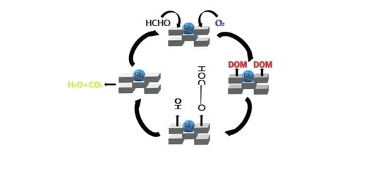
2.7. In Situ DRIFTS and Catalytic Oxidation Mechanism

In order to study the reaction mechanism, the in situ DRIFTS technique was employed to investigate HCHO oxidation over the Eu/NaSep sample. Figure 9a shows the in situ DRIFT spectra of the Eu/NaSep sample exposed to 2000 ppm HCHO + N2 without O2 at 30 °C for 1 h. The absorption bands at 3507, 2983, 1713, 1633, 1590, 1453, and 1390cm−1 were observed. As the adsorption time increased, the band strength began to increase and reached a stable level after 1 h. According to the literature [43], the band at 3507 cm−1 was assigned to the stretching vibration of the hydroxyl group, the ones at 2983, 1590, and 1390 cm−1 were attributed to the formate species on the surface of the sample, the band at 2983 cm−1 was ascribed to the stretching vibration of C–H bond, and the ones at 1590 and 1390 cm−1 were due to the vas(COO) and δ(CH), respectively [44]. The band at 1633 cm−1 was due to adsorbed water on the catalyst surface, and the band at 1713 cm−1 was ascribed to the v(C=O) of carbonate species [45]. Moreover, the band at 1453 cm−1 was due to the dioxymethylene (DOM) [46]. Therefore, the adsorbed HCHO on the sample surface could form the formate species in the absence of O2, which might be due to the fact that the sample surface contained a large amount of the adsorbed oxygen species, which was consistent with the above results.
Figure 9b shows the intermediate species on the surface of the sample after exposure to 2000 ppm HCHO + N2 + O2 at 30 °C. Almost all of the characteristic bands increased in intensity, as compared with the above HCHO adsorption without O2 on the sample surface. Among them, the intensity of the bands (2983 and 1590 cm−1) assignable to the formate species increased significantly, as compared with that of the other characteristic bands. This was because, in the presence of oxygen, HCHO could be more adsorbed on the sample surface and form the formate species. However, the DOM species were hardly changed (i.e., no DOM species (1453 cm−1) formation), which indicates that the Eu/NaSep sample was capable of rapidly converting the HCHO adsorbed on the sample surface. According to the literature [47], when HCHO was oxidized, it was first converted to the DOM species, and then the formate species was generated immediately. Moreover, with the oxidation of HCHO, the intensity of the band (3507 cm−1) of the hydroxyl group increased significantly. Therefore, HCHO could be rapidly oxidized to the formate species on the surface of the sample, demonstrating that the adsorbed HCHO was converted to the formate species according to Equations (1) and (2) [46]:
[HCHO] s + [O] s → [HCOO] s + [H] s
[HCHO] s + [OH] s → [HCOO] s + 2[H]s
Figure S2 shows the in situ DRIFTS spectra of the Eu/NaSep and NaSep samples for the oxidation of HCHO at 30 °C. It can be realized that the intermediate products formed over the NaSep sample (Figure S2b) were the same as those formed over the Eu/NaSep sample (Figure S2a), but absorption band intensity (i.e., the corresponding amounts) of the intermediate products over the former was lower than that over the latter.
Figure 10 shows the oxidation of the intermediates formed via HCHO oxidation over the Eu/NaSep sample at different temperatures. Catalytic oxidation of HCHO at 30 °C is shown in Figure 10b. The HCHO was rapidly decomposed into the formate (1590 and 1390 cm−1) and carbonate (1713 cm−1) species on the Eu/NaSep surface. When the temperature rose to 100 °C, a significant decrease in band intensity of the formate species (1590 and 1390 cm−1) was observed, indicating a rapid increase in the decomposition rate of the formate species. The characteristic band (3507 cm−1) intensity of the hydroxyl group also began to drop, indicating that the hydroxyl group played an important role in the oxidation of HCHO. Generally, abundant hydroxyl groups can provide a more amount of the sites for HCHO adsorption and promote the oxidation of formate species. The characteristic band intensity of the carbonate species increased at the same time, indicating that the formate species were decomposed on the surface of the sample to form the carbonate species. When the temperature reached 150 °C, the intermediate products (e.g., the formate (1590 and 1390 cm−1) and carbonate (1713 cm−1) species) disappeared almost completely, indicating that HCHO was totally oxidized at this temperature, which was in good agreement with the reaction temperature in HCHO oxidation of the Eu/NaSep sample.
As a result, compared with NaSep, loading of the rare earth oxide significantly improved the catalytic activity of HCHO oxidation. It was confirmed by the FTIR results that sepiolite was rich in the hydroxyl group. From the in situ DRIFTS results, it was found that hydroxyl groups could quickly adsorb HCHO on the sample surface, and loading of the rare earth oxide could generate a more amount of oxygen vacancies that served as the active sites for HCHO oxidation, as evidenced by its highest Oads/Olatt atomic ratio revealed by the XPS analysis. The above results show that Eu/NaSep exhibited the best catalytic activity, which was due to the large number of hydroxyl species (Figure 3), the lowest reduction temperature (ca. 456 °C), the highest Oads/Olatt atomic ratio (7.33), and the highest acidity (53 μmol/g).
A possible mechanism for HCHO oxidation over Eu/NaSep is presented in Figure 11. Firstly, HCHO was adsorbed on the surface of the sample, then the surface adsorbed HCHO was oxidized to the DOM intermediate by the adsorbed oxygen species, and finally oxidized to the formate species rapidly. The hydroxyl groups in sepiolite could not only promote the adsorption of HCHO on the sample surface, but also accelerate the formation and decomposition of the formate species. Finally, the carbonate species derived from the oxidation of the formate species were eventually converted to CO2 and H2O. Therefore, the total oxidation of HCHO over the sample surface was as follows:
HCHO + O2 → HCOO + OH → H2O + CO2.

3. Experimental

3.1. Catalyst Preparation

The sepiolite raw material was bought from Liuyang Sepiolite Mining Industry Company (China). The support was obtained via leaching of the sepiolite with sodium. Sodium modification of sepiolite is expected to improve the thermal stability of the sample, the cation exchange capacity was significantly enhanced, and the original structure of sepiolite was not changed. The chemical composition of sepiolite and the sodium-treated procedures are described in the Supplementary material. It is well known that different raw sepiolites may differ slightly in composition. However, after the sepiolite was modified with sodium, its physical property was determined. Therefore, the present study used the sodium-modified sepiolite to load the rare earth elements, and explored the effect of different rare earth elements on the catalytic activity of the sodium-modified sepiolite for HCHO oxidation.
The xEu/NaSep (x = 1.0, 3.0, 5.0, and 7.0 wt %.) catalysts were prepared using the rotary evaporation method. The wet samples were dried for 24 h and calcined at 400 °C for 2 h, and the obtained samples were denoted as xEu/NaSep, and their catalytic activities for HCHO oxidation are shown in Figure S1.
The NaSep-supported rare earth oxides (theoretical rare earth oxide loading = 5.0 wt %) were prepared using the rotary evaporation method with nitrates of La, Eu, Dy, and Tm (A.R., Beijing Chemical Co., Ltd.) as metal precursors, respectively. The samples were at 80 °C dried for 24 h and calcined at 400 °C for 2 h. The obtained samples were donated as RE/NaSep (RE = La, Eu, Dy, and Tm). The synthetic yield of all catalyst samples was 92% to 94%.

3.2. Catalyst Characterization

X-ray diffraction (XRD) patterns of the samples were measured on a Bruker D8-Advance apparatus in the 2θ range of 5–80° (λ = 0.15404 nm at a scan rate of 5 °/min). Nitrogen sorption isotherms were obtained at liquid nitrogen temperature (−196 °C) on a JW-BK200C apparatus (Beijing, China). Specific surface areas of the samples were calculated using the Brunauer–Emmett–Teller (BET) equation, and their pore-size distributions were determined using the desorption branch data of the isotherms by the Barrett–Joyner–Halenda (BJH) method.
Hydrogen temperature-programmed reduction (H2–TPR) of the samples was conducted on a Builder PCA-1200 analyzer. Before the H2–TPR experiment, the sample (100 mg) was first treated in a flow (30 mL/min) of the 5 vol % O2/N2 mixture at 400 °C for 1 h, and then cooled to room temperature (RT). The sample was finally reduced in a flow (30 mL/min) of the 5 vol % H2/N2 mixture at a heating rate of 10 °C/min from RT to 1000 °C. The reduction peaks were calibrated against that of the reduction of a powdered CuO sample (Aldrich, 99.995%).
X-ray photoelectron spectroscopic (XPS) spectra of the samples were recorded on a Thermo ESCALAB 250Xi electron spectrometer (Shanghai, China) equipped with an Al X-ray source and a hemispherical electron analyzer operating at constant pass energy (30.0 eV).
Fourier transform infrared (FTIR) spectra of the samples were obtained on an FTIR spectrometer (Thermo Scientific Nicolet 6700, Waltham. America). The in situ diffuse reflectance infrared spectroscopic (DRIFTS) experiments were conducted on a Bruker Tensor II spectrometer (Karlsruhe. Germany), in which there was a high-temperature reaction chamber (Harrick Praying Mantis). The sample packed in a cup of the reaction chamber was heated to 400 °C at a total N2 flow of 200 mL/min for 1 h to remove the adsorbed impurities. Each of the spectra had a resolution of 4 cm−1 and accumulated 32 scans.

3.3. Catalytic Activity Evaluation

HCHO oxidation activity was evaluated by loading the sample (50 mg) well mixed with quartz sand (150 mg) in a fixed-bed quartz tubular microreactor. The reactant mixture was 2000 ppm HCHO + 20 vol % O2 + N2 (balance) and the GHSV was 240,000 mL/(g h). The HCHO concentration was analyzed online on a Techcomp GC-7900 gas chromatograph (Shanghai, China) equipped with a flame ion detector (FID). The conversion of HCHO (XHCHO) was calculated according to the changes in HCHO concentration (CHCHO,inlet and CHCHO,outlet) of the inlet and outlet gas mixture, as shown below:
X HCHO = C HCHO , inlet C HCHO , outlet C HCHO , inlet × 100 %
The detailed measurement procedures are stated in the Supplementary Materials.

4. Conclusions

After loading the rare earth oxide, the redox properties of the NaSep sample changed greatly, especially for the Eu/NaSep sample. According to the characterization results, we can know that loading of Eu2O3 greatly increased the amount of the surface oxygen vacancies, which enabled gas-phase oxygen molecules to form the adsorbed oxygen species on the sample surface and promoted the oxidation of HCHO. This work provided evidence that the Eu/NaSep sample possessed the highest atomic ratio of Oads/Olatt among all of the samples. As revealed by the H2–TPR characterization results, the Eu/NaSep sample exhibited the best reducibility, which contributed to the good catalytic activity for HCHO oxidation of the sample. HCHO oxidation took place via the steps of HCHO + O2 → HCOO + OH → H2O + CO2. Therefore, the highest Oads/Olatt atomic ratio, the largest amount of hydroxyl groups, the highest acidity, and the best reducibility was accountable for the optimal catalytic activity of Eu/NaSep.

Supplementary Materials

The following are available online at https://www.mdpi.com/2073-4344/10/3/328/s1, Figure S1. HCHO conversion as a function of temperature over the x wt % Eu/NaSep sample under the conditions of 2000 ppm HCHO + 20 vol % O2 + N2 (balance) and GHSV = 240,000 mL/(g h). Figure S2. In situ DRIFTS spectra of (a) Eu/NaSep, and (b) NaSep exposed to the flow of 2000 ppm HCHO + 20 vol % O2 + N2 (balance) at 30 °C and different time.

Author Contributions

Conceptualization, Q.Y.; methodology, N.D. and M.C.; software, N.D. and M.C.; investigation, N.D. and M.C.; resources, Q.Y.; data curation, N.D. and M.C.; writing—original draft preparation, N.D.; writing—review and editing, Q.Y. and H.D.; visualization, S.C. and T.K.; supervision, Q.Y. and H.D.; project administration, Q.Y.; funding acquisition, Q.Y. All authors have read and agreed to the published version of the manuscript.

Funding

This work was supported by the National Natural Science Foundation of China (21277008 and 20777005), the National Key Research and Development Program of China (2017YFC0209905), and the Natural Science Foundation of Beijing (8082008). We also thank Prof. Ralph T. Yang (University of Michigan) for his helpful discussion and encouragement.

Conflicts of Interest

The authors declare no conflicts of interest.

References

  1. Lin, M.; Yu, X.; Yang, X.; Ma, X.; Ge, M. Exploration of the active phase of the hydrotalcite-derived cobalt catalyst for HCHO oxidation. Chin. J. Catal. 2019, 40, 703–712. [Google Scholar] [CrossRef]
  2. Weng, X.; Long, Y.; Wang, W.; Shao, M.; Wu, Z. Structural effect and reaction mechanism of MnO2 catalysts in the catalytic oxidation of chlorinated aromatics. Chin. J. Catal. 2019, 40, 638–646. [Google Scholar] [CrossRef]
  3. Bai, B.; Qiao, Q.; Li, Y.; Peng, Y.; Li, J. Effect of pore size in mesoporous MnO2 prepared by KIT-6 aged at different temperatures on ethanol catalytic oxidation. Chin. J. Catal. 2018, 39, 630–638. [Google Scholar] [CrossRef]
  4. Liu, Y.; Deng, J.; Xie, S.; Wang, Z.; Dai, H. Catalytic removal of volatile organic compounds using ordered porous transition metal oxide and supported noble metal catalysts. Chin. J. Catal. 2016, 37, 1193–1205. [Google Scholar] [CrossRef]
  5. Zhang, M.; Li, C.; Qu, L.; Fu, M.; Zeng, G.; Fan, C.; Ma, J.; Zhan, F. Catalytic oxidation of NO with O2 over FeMnOx/TiO2: Effect of iron and manganese oxides loading sequences and the catalytic mechanism study. Appl. Surf. Sci. 2014, 300, 58–65. [Google Scholar] [CrossRef]
  6. Zhu, X.; Gao, X.; Qin, R.; Zeng, Y.; Qu, R.; Zheng, C.; Tu, X. Plasma-catalytic removal of formaldehyde over Cu–Ce catalysts in a dielectric barrier discharge reactor. Appl. Catal. B 2015, 170, 293–300. [Google Scholar] [CrossRef] [Green Version]
  7. Sun, Y.; Qu, Z.; Chen, D.; Wang, H.; Zhang, F.; Fu, Q. Formaldehyde catalytic oxidation over hydroxyapatite modified with various organic molecules. Chin. J. Catal. 2014, 35, 1927–1936. [Google Scholar] [CrossRef]
  8. Bai, B.; Qiao, Q.; Li, J.; Hao, J. Progress in research on catalysts for catalytic oxidation of formaldehyde. Chin. J. Catal. 2016, 37, 102–122. [Google Scholar] [CrossRef]
  9. Imamura, S.; Uchihori, D.; Utani, K.; Ito, T. Oxidative decomposition of formaldehyde on silver-cerium composite oxide catalyst. Catal. Lett. 1994, 24, 377–384. [Google Scholar] [CrossRef]
  10. Álvarez-Galván, M.C.; De la Peña O’Shea, V.A.; Fierro, J.L.G.; Arias, P.L. Alumina-supported manganese- and manganese–palladium oxide catalysts for VOCs combustion. Catal. Commun. 2003, 4, 223–228. [Google Scholar] [CrossRef]
  11. Guo, H.; Zhang, H.; Peng, F.; Yang, H.; Xiong, L.; Wang, C.; Huang, C.; Chen, X.; Ma, L. Effects of Cu/Fe ratio on structure and performance of attapulgite supported CuFeCo-based catalyst for mixed alcohols synthesis from syngas. Appl. Catal. A 2015, 503, 51–61. [Google Scholar] [CrossRef]
  12. Bu, Y.; Chen, Y.; Jiang, G.; Hou, X.; Li, S.; Zhang, Z. Understanding of Au-CeO2 interface and its role in catalytic oxidation of formaldehyde. Appl. Catal. B 2020, 260, 118–138. [Google Scholar] [CrossRef]
  13. Yu, S.; Liu, X.; Xu, G.; Qiu, Y.; Cheng, L. Magnetic Fe3O4/sepiolite composite synthesized by chemical co-precipitation method for efficient removal of Eu(III). Desalin. Water Treat. 2016, 57, 16943–16954. [Google Scholar] [CrossRef]
  14. Song, Y.; Liu, L.; Fu, Z.; Ye, Q.; Cheng, S.; Kang, T.; Dai, H. Excellent performance of Cu-Mn/Ti-sepiolite catalysts for low-temperature CO oxidation. Front. Environ. Sci. Eng. 2017, 11, 5. [Google Scholar] [CrossRef]
  15. Ma, Y.; Zhang, G. Sepiolite nanofiber-supported platinum nanoparticle catalysts toward the catalytic oxidation of formaldehyde at ambient temperature: Efficient and stable performance and mechanism. Chem. Eng. J. 2016, 288, 70–78. [Google Scholar] [CrossRef]
  16. Zhang, G.; Xiong, Q.; Xu, W.; Guo, S. Synthesis of bicrystalline TiO2 supported sepiolite fibers and their photocatalytic activity for degradation of gaseous formaldehyde. Appl. Clay Sci. 2014, 102, 231–237. [Google Scholar] [CrossRef]
  17. Ye, Q.; Yan, L.; Wang, H.; Cheng, S.; Wang, D.; Kang, T.; Dai, H. Enhanced catalytic performance of rare earth-doped Cu/H-Sep for the selective catalytic reduction of NO with C3H6. Appl. Catal. A 2012, 431, 42–48. [Google Scholar] [CrossRef]
  18. Rytwo, G.; Tropp, D.; Serban, C. Adsorption of diquat, paraquat and methyl green on sepiolite: Experimental results and model calculations. Appl. Clay Sci. 2002, 20, 273–282. [Google Scholar] [CrossRef]
  19. Sabah, E.; Çelik, M.S. Interaction of Pyridine Derivatives with Sepiolite. J. Colloid Interf. Sci. 2002, 251, 33–38. [Google Scholar] [CrossRef]
  20. Alkan, M.; Tekin, G.; Namli, H. FTIR and zeta potential measurements of sepiolite treated with some organosilanes. Micropor. Mesopor. Mat. 2005, 84, 75–83. [Google Scholar] [CrossRef]
  21. Casal, B.; Merino, J.; Serratosa, J.; Ruiz-Hitzky, E. Sepiolite-based materials for the photo-and thermal-stabilization of pesticides. Appl. Clay Sci. 2001, 18, 245–254. [Google Scholar] [CrossRef]
  22. Gao, Y.; Gan, H.; Zhang, G.; Guo, Y. Visible light assisted Fenton-like degradation of rhodamine B and 4-nitrophenol solutions with a stable poly-hydroxyl-iron/sepiolite catalyst. Chem. Eng. J. 2013, 217, 221–230. [Google Scholar] [CrossRef]
  23. Özcan, A.; Özcan, A.S. Adsorption of Acid Red 57 from aqueous solutions onto surfactant-modified sepiolite. J. Hazard. Mater. 2005, 125, 252–259. [Google Scholar] [CrossRef] [PubMed]
  24. Yuan, P.; Yin, X.; He, H.; Yang, D.; Wang, L.; Zhu, J. Investigation on the delaminated-pillared structure of TiO2-PILC synthesized by TiCl4 hydrolysis method. Micropor. Mesopor. Mat. 2006, 93, 240–247. [Google Scholar] [CrossRef] [Green Version]
  25. Qiao, N.; Zhang, X.; He, C.; Li, Y.; Zhang, Z.; Cheng, J.; Hao, Z. Enhanced performances in catalytic oxidation of o-xylene over hierarchical macro-/mesoporous silica-supported palladium catalysts. Front. Environ. Sci. Eng. 2016, 10, 458–466. [Google Scholar] [CrossRef]
  26. Belessi, V.; Lambropoulou, D.; Konstantinou, I.; Katsoulidis, A.; Pomonis, P.; Petridis, D.; Albanis, T. Structure and photocatalytic performance of TiO2/clay nanocomposites for the degradation of dimethachlor. Appl. Catal. B 2007, 73, 292–299. [Google Scholar] [CrossRef]
  27. Barreca, D.; Gasparotto, A.; Milanov, A.; Tondello, E.; Devi, A.; Fischer, R.A. Nanostructured Dy2O3 films: An XPS Investigation. Surf. Sci. Spectra. 2007, 14, 52–59. [Google Scholar] [CrossRef]
  28. Cario, L.; Palvadeau, P.; Lafond, A. Mixed-valence state of europium in the misfit layer compound (EuS)1.173 NbS2. Chem. Mater. 2003, 15, 943–950. [Google Scholar] [CrossRef]
  29. Liu, B.; Wan, Z.; Zhan, Y. Desulfurization of hot coal gas over high-surface-area LaMeOx/MCM-41 sorbents. Fuel 2012, 98, 95–102. [Google Scholar] [CrossRef]
  30. Kabongo, G.L.; Mhlongo, G.H.; Mothudi, B.M.; Hillie, K.T.; Mbule, P.S.; Dhlamini, M.S. Structural, photoluminescence and XPS properties of Tm3+ ions in ZnO nanostructures. J. Lumin. 2017, 187, 141–153. [Google Scholar] [CrossRef]
  31. Yu, L.; Diao, G.; Ye, F.; Sun, M.; Zhou, J.; Li, Y.; Liu, Y. Promoting Effect of Ce in Ce/OMS-2 Catalyst for Catalytic Combustion of Dimethyl Ether. Catal. Lett. 2011, 141, 111–119. [Google Scholar] [CrossRef]
  32. Setvin, M.; Aschauer, U.; Scheiber, P.; Li, Y.F.; Hou, W.; Schmid, M.; Selloni, A.; Diebold, U. Reaction of O2 with subsurface oxygen vacancies on TiO2 anatase (101). Science 2013, 341, 988–991. [Google Scholar] [CrossRef] [PubMed] [Green Version]
  33. Panov, G.; Dubkov, K.; Starokon, E. Active oxygen in selective oxidation catalysis. Catal. Today 2006, 117, 148–155. [Google Scholar] [CrossRef]
  34. Oliveira, L.C.A.; Rios, R.V.R.A.; Fabris, J.D.; Sapag, K.; Garg, V.K.; Lago, R.M. Clay–iron oxide magnetic composites for the adsorption of contaminants in water. Appl. Clay Sci. 2003, 22, 169–177. [Google Scholar] [CrossRef]
  35. Ferrandon, M.; Carnö, J.; Järås, S.; Björnbom, E. Total oxidation catalysts based on manganese or copper oxides and platinum or palladium I: Characterisation. Appl. Catal. A 1999, 180, 141–151. [Google Scholar] [CrossRef]
  36. Topsoe, N.Y.; Topsoe, H.; Dumesic, J.A. Vanadia/Titania Catalysts for Selective Catalytic Reduction SCR of Nitric-Oxide by Ammonia I. Combined Temperature-Programmed in-Situ FTIR and On-line Mass-Spectroscopy Studies. J. Catal. 1995, 151, 226–240. [Google Scholar] [CrossRef]
  37. Roy, S.; Viswanath, B.; Hegde, M.S.; Madras, G. Low-Temperature Selective Catalytic Reduction of NO with NH3 over Ti0.9M0.1O2-δ(M = Cr, Mn, Fe, Co, Cu). J. Phys. Chem. C 2008, 112, 6002–6012. [Google Scholar] [CrossRef]
  38. Peña, D.A.; Uphade, B.S.; Smirniotis, P.G. TiO2-supported metal oxide catalysts for low-temperature selective catalytic reduction of NO with NH3I. Evaluation and characterization of first row transition metals. J. Catal. 2004, 221, 421–431. [Google Scholar]
  39. Ye, Q.; Zhao, J.; Huo, F.; Wang, D.; Cheng, S.; Kang, T.; Dai, H. Nanosized Au supported on three-dimensionally ordered mesoporous β-MnO2: Highly active catalysts for the low-temperature oxidation of carbon monoxide, benzene, and toluene. Micropor. Mesopor. Mat. 2013, 172, 20–29. [Google Scholar] [CrossRef]
  40. Li, H.; Zhang, N.; Chen, P.; Luo, M.; Lu, J. High surface area Au/CeO2 catalysts for low temperature formaldehyde oxidation. Appl. Catal. B 2011, 110, 279–285. [Google Scholar] [CrossRef]
  41. Jiang, X.; Li, X.; Wang, J.; Long, D.; Ling, L.; Qiao, W. Three-dimensional Mn-Cu-Ce ternary mixed oxide networks prepared by polymer-assisted deposition for HCHO catalytic oxidation. Catal. Sci. Technol. 2018, 8, 274–2749. [Google Scholar] [CrossRef]
  42. Pang, G.; Wang, D.; Zhang, Y.; Ma, C.; Hao, Z. Catalytic activities and mechanism of formaldehyde oxidation over gold supported on MnO2 microsphere catalysts at room temperature. Front. Environ. Sci. Eng. 2016, 10, 447–457. [Google Scholar] [CrossRef]
  43. Xu, Z.; Yu, J.; Jaroniec, M. Efficient catalytic removal of formaldehyde at room temperature using AlOOH nanoflakes with deposited Pt. Appl. Catal. B 2015, 163, 306–312. [Google Scholar] [CrossRef]
  44. Shi, C.; Chen, B.; Li, X.; Crocker, M.; Wang, Y.; Zhu, A. Catalytic formaldehyde removal by “storage-oxidation” cycling process over supported silver catalysts. Chem. Eng. J. 2012, 200, 729–737. [Google Scholar] [CrossRef]
  45. Chen, M.; Lin, Y.; Lin, Y.; Lin, H.; Lin, J. Dissociative adsorption of HCOOH, CHOH, and CHO on MCM-41. J. Catal. 2004, 228, 259–263. [Google Scholar] [CrossRef]
  46. Chen, B.; Zhu, X.; Crocker, M.; Wang, Y.; Shi, C. FeOx-supported gold catalysts for catalytic removal of formaldehyde at room temperature. Appl. Catal. B 2014, 154, 73–81. [Google Scholar] [CrossRef]
  47. Zhu, L.; Wang, J.; Rong, S.; Wang, H.; Zhang, P. Cerium modified birnessite-type MnO2 for gaseous formaldehyde oxidation at low temperature. Appl. Catal. B 2017, 211, 212–221. [Google Scholar] [CrossRef]
Figure 1. XRD patterns of sodium-treated sepiolite (NaSep), Eu/NaSep, Dy/NaSep, La/NaSep, and Tm/NaSep.
Catalysts 10 00328 g001
Figure 2. FTIR spectra of NaSep, Eu/NaSep, Dy/NaSep, La/NaSep, and Tm/NaSep.
Catalysts 10 00328 g002
Figure 3. N2 adsorption–desorption isotherms of the samples.
Catalysts 10 00328 g003
Figure 4. SEM images of (a) Eu/NaSep, (b) Dy/NaSep, (c) La/NaSep, (d) Tm/NaSep and (e) NaSep.
Catalysts 10 00328 g004
Figure 5. (a) La 3d, (b) Dy 4d5/2, (c) Eu 4d, (d) Tm 3d, and (e) O 1s XPS spectra of the samples.
Catalysts 10 00328 g005
Figure 6. H2–TPR profiles of the samples.
Catalysts 10 00328 g006
Figure 7. NH3–TPD profiles of the samples.
Catalysts 10 00328 g007
Figure 8. (a) HCHO conversion as a function of temperature and (b) HCHO conversion versus reaction time over the samples under the conditions of 2000 ppm HCHO + 20 vol % O2 + N2 (balance) and GHSV = 240,000 mL/(g h).
Catalysts 10 00328 g008
Figure 9. (a) In situ DRIFTS spectra of HCHO adsorption on the Eu/NaSep sample under the condition of 30 °C and 2000 ppm HCHO + N2 (balance), and (b) in situ DRIFTS spectra of the Eu/NaSep sample in a flow of 2000 ppm HCHO + 20 vol % O2 + N2 (balance) at 30 °C and different time.
Catalysts 10 00328 g009aCatalysts 10 00328 g009b
Figure 10. In situ DRIFTS spectra of the Eu/NaSep sample after 1 h of HCHO oxidation at 30, 100, and 150 °C.
Catalysts 10 00328 g010
Figure 11. Proposed mechanism of HCHO oxidation over the Eu/NaSep sample.
Catalysts 10 00328 g011
Table 1. BET surface areas, pore volumes, and pore diameters.
SampleSurface Area (m2/g)Pore Volume (cm3/g)Pore Diameter (nm)
NaSep960.2148.9
La/NaSep470.20717.8
Eu/NaSep430.12610.7
Dy/NaSep310.20326.9
Tm/NaSep640.35424.6
Table 2. Reduction temperatures, H2 consumption, and Oads/Olatt atomic ratios of the samples.
SampleTemperature (°C)H2 Consumption (mmol/gcat)Oads/Olatt Atomic Ratio
Peak 1Peak 2Peak 1Peak 2Total
NaSep6330.1280.1282.33
La/NaSep4586210.1480.4590.6074.53
Eu/NaSep4566200.1790.5290.7087.33
Dy/NaSep4566190.1690.4760.6455.66
Tm/NaSep4746260.1660.330.4963.93
Table 3. Ammonia uptakes of the samples.
SampleAmmonia Uptake (μmol/g)
Weak (150–220 °C)Medium (220–300 °C)Strong (300–450 °C)Total
NaSep149124
La/NaSep1712332
Eu/NaSep2522653
Dy/NaSep1919442
Tm/NaSep1511228
Table 4. A comparison on specific reaction rates (rcat) of formaldehyde (HCHO_ oxidation at 100 °C over the catalysts studied in the present work and reported in the literature.
Catalyst rcat (mol/(gcat s))Ref.
Eu/NaSep3.54 × 10−6This study
Dy/NaSep2.29 × 10−6This study
La/NaSep1.74 × 10−6This study
Tm/NaSep1.47 × 10−6This study
NaSep4.36 × 10−7This study
24Mn-19Co-1Ce9.20 × 10−8[40]
2.5Au/CeO25.11 × 10−7[40]
Mn–Cu–Ce mixed oxide4.77 × 10−8[41]
Cu–Mn/TiO22.55 × 10−8[42]
Cu–Mn/γ–Al2O33.06 × 10−8[42]

