Article

Controllable Hydrothermal Synthesis and Photocatalytic Performance of Bi2MoO6 Nano/Microstructures

1
College of Health Science and Environmental Engineering, Shenzhen Technology University, Shenzhen 518118, China
2
College of Chemistry and Chemical Engineering, Shanghai University of Engineering Science, Shanghai 201620, China
3
Institute of Innovation & Application, Zhejiang Ocean University, Zhoushan 316022, China
*
Authors to whom correspondence should be addressed.
Catalysts 2020, 10(10), 1161; https://doi.org/10.3390/catal10101161
Submission received: 15 August 2020 / Revised: 17 September 2020 / Accepted: 1 October 2020 / Published: 10 October 2020

Abstract

:
Bi2MoO6 with a tunable morphology was synthesized by a facile hydrothermal route using different surfactants, including nanosheet-assembled microspheres, smooth microspheres, nanoparticle aggregates and nanoparticles. The morphology, crystal structure and photocatalytic activity of as-obtained Bi2MoO6 were characterized by scanning electron microscopes (SEM), X-ray diffraction (XRD), photoelectron spectroscopy (XPS), Brunauer–Emmett–Teller (BET) and UV–Vis spectrophotometer. Bi2MoO6 flower-like microspheres using cetyl-trimethyl-ammonium bromide (BET) as the surfactant exhibited much better photocatalytic activity than Bi2MoO6 with the other morphologies, with a degradation efficiency of 98.4%. It can be summarized that the photocatalytic activity of Bi2MoO6 samples depends on their morphology and composition.

Graphical Abstract

1. Introduction

The properties of most materials are strongly dependent on their compositions, structures and morphologies, and different morphologies of materials with the same composition could exhibit varied performances [1,2,3,4,5,6,7]. Additionally, the properties of nanomaterials can be tuned effectively by adjusting their size and shape [8,9,10,11,12]. The facile synthesis of photocatalytic nanomaterials with controllable morphology and structure is necessary and has a profound influence on their photocatalytic performances [13,14,15,16,17,18,19]. Therefore, controlling morphology and structure of the photocatalysts has been regarded as one of the most effective methods in fundamental scientific interest for exploring practical applications.
As a typical photocatalyst, Bi2MoO6 [20,21,22,23], its activity was affected by the morphology, structure, and size of the sample. Much progress has been made in the preparation of various nanostructures of Bi2MoO6. For example, Li et al. [24], reported bismuth molybdates with three crystal structures created by tuning the experimental parameters using a hydrothermal method, and an aurivillius structure of Bi2MoO6 showed a better photocatalytic property than other bismuth molybdates. Shi et al. [25] have prepared Bi2MoO6 with flower-like and hierarchical microspheres by hydrothermal synthesis using ethylene glycol solution, and found that the shape and size of Bi2MoO6 have an influence on the photocatalytic activities. Zheng et al. [26] have obtained Bi2MoO6 samples with different morphologies and surface structures via the hydrothermal route, and showed that the photocatalytic activity of Bi2MoO6 had a close relationship with the crystal structure of the exposed surface plane. Yang et al. [27] hydrothermally synthesized Bi2MoO6 nanosheets with varied morphologies, crystal phases, and light absorption by adjusting the hydrothermal preparation conditions, and found that the photocatalytic activity of the Bi2MoO6 materials was influenced by the pH value, reaction temperature and the surfactant [28,29]. Furthermore, despite this recent progress, there are still some difficulties in Bi2MoO6 morphology-controlled synthesis, although nanosheets/nanospheres of Bi2MoO6 and Bi2MoO6-based composites [30,31,32,33,34,35,36] have been synthesized. Hence, it is necessary to develop a simple and economical technique to meet the demand for exploring the photocatalytic property of Bi2MoO6 materials.
Herein, we present a simple hydrothermal route using different surfactants for synthesizing varied Bi2MoO6 nano/microstructures with excellent reproducibility. The crystal structure and morphology of the Bi2MoO6 samples were characterized by X-ray diffraction (XRD), Brunauer–Emmett–Teller (BET) and scanning electron microscope (SEM), and the energy band characteristics of Bi2MoO6 samples were measured by photoelectron spectroscopy (XPS) and ultraviolet–visible absorption spectroscopy. We investigated the photocatalytic activity of nanosheet-assembled Bi2MoO6 microspheres compared to Bi2MoO6 nano powders under visible light irradiation.

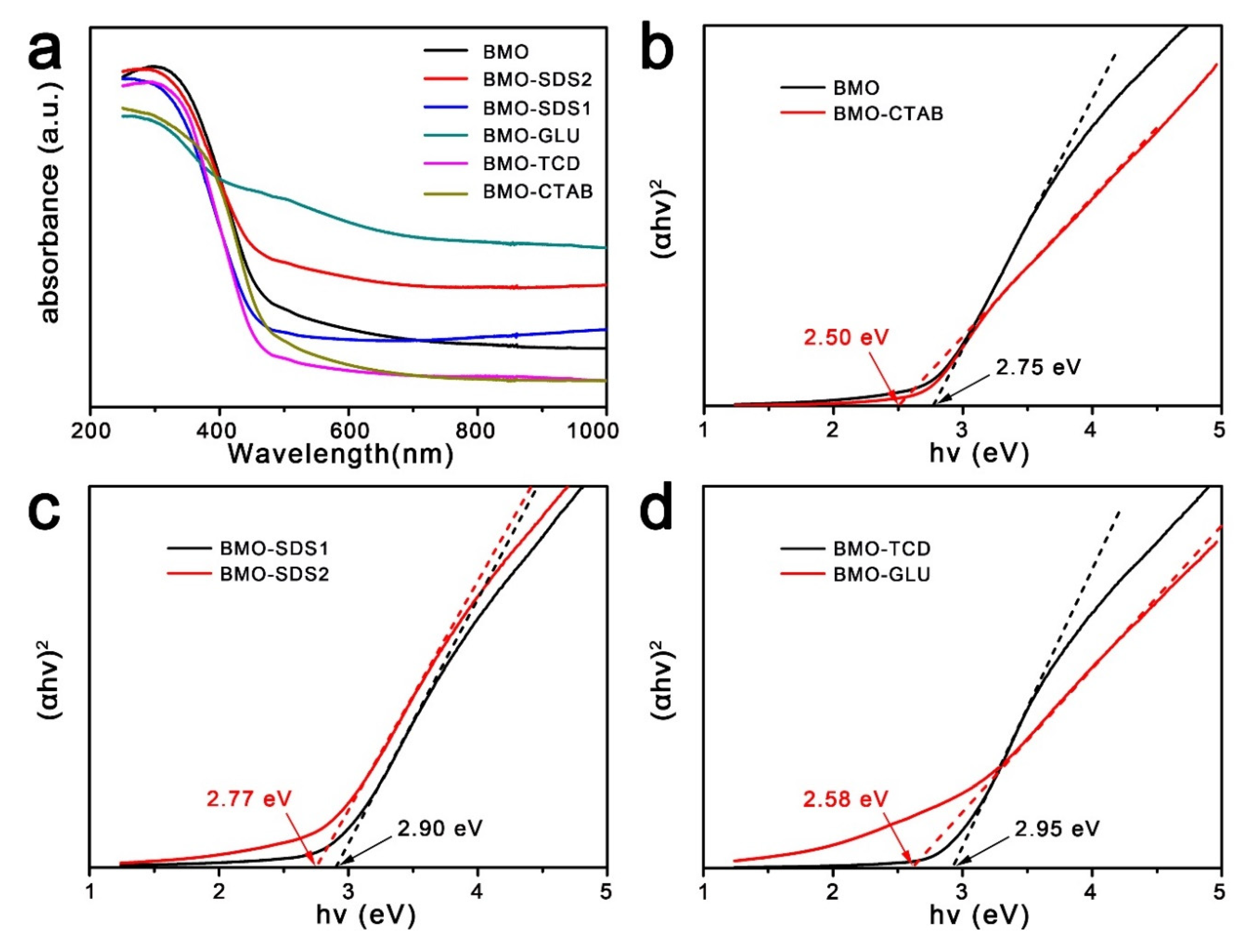
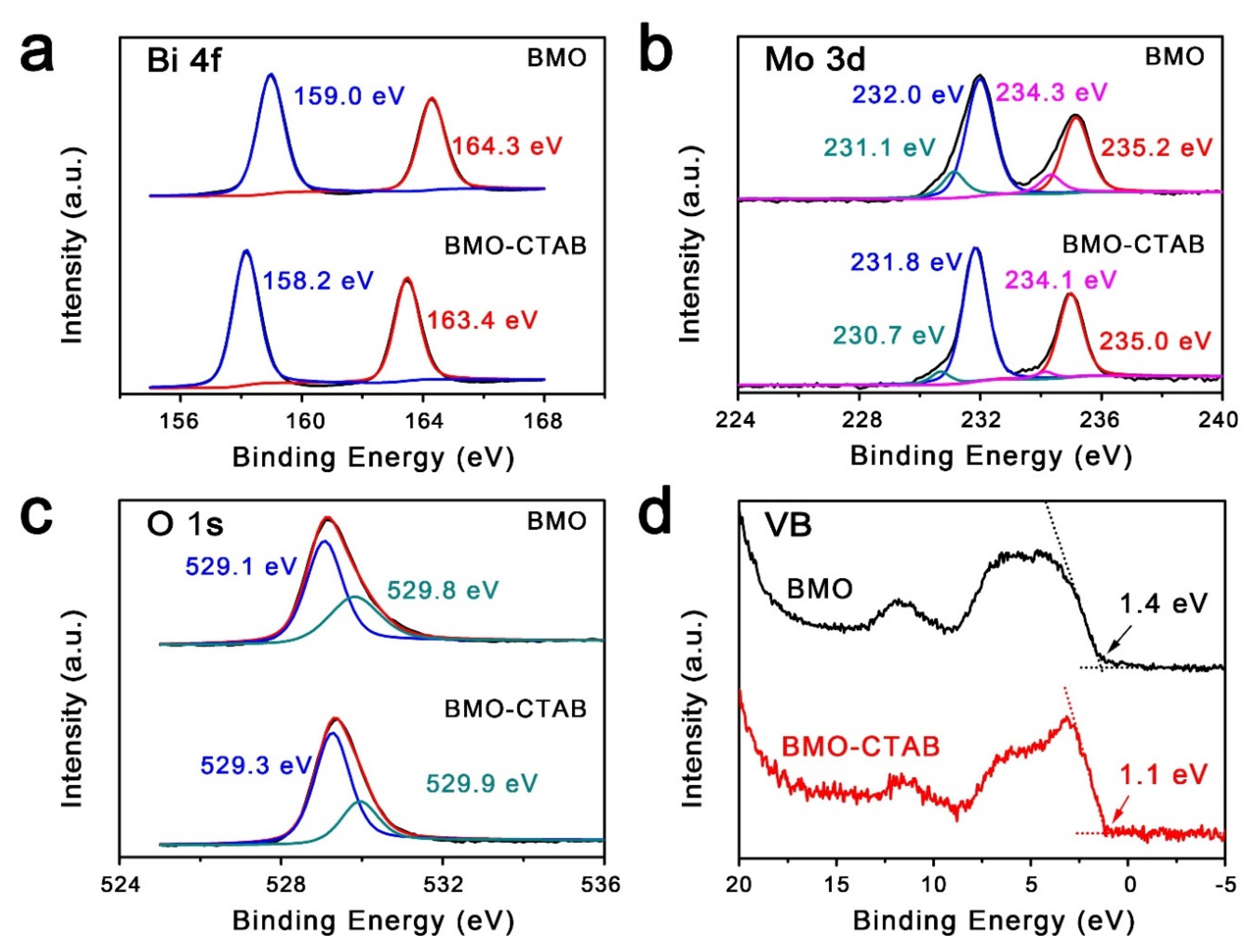
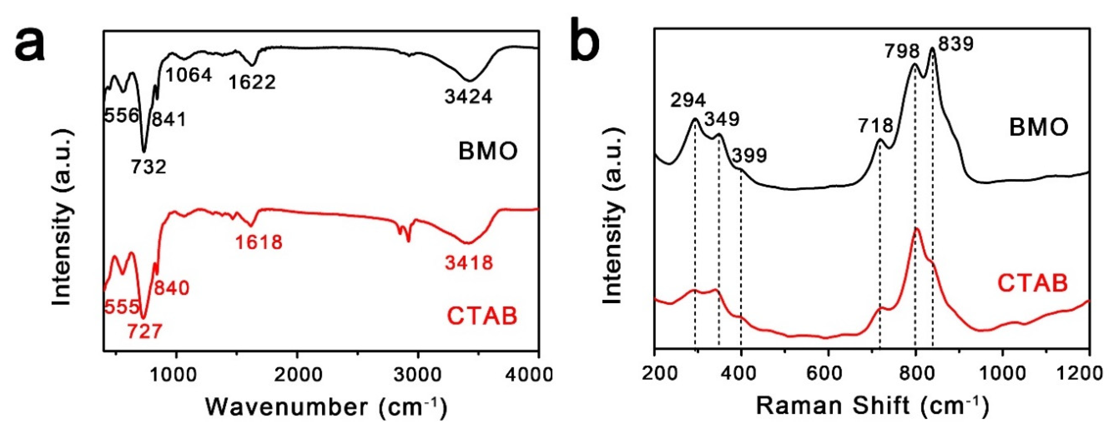
2. Results and Discussion

Figure 1 shows the crystal structures of as-fabricated Bi2MoO6 samples using different surfactants confirmed by X-ray diffraction (XRD) patterns. In the XRD pattern of Bi2MoO6 sample without using any surfactant, it is easy to observe three typical peaks (2θ = 28.3 °C, 32.6 °C and 46.7 °C), which are characteristic peaks of Bi2MoO6 powder crystals (JCPDS card no. 21-0102), and indicate that high-quality orthorhombic-phase Bi2MoO6 was obtained. In the XRD pattern of Bi2MoO6 samples using CTAB, SDS-1 and SDS-2 as the surfactants, respectively, no new peak was observed in these Bi2MoO6 samples. Careful examination of Figure 1 indicates that the intensity of the diffraction peaks at 28.3 °C and 32.6 °C from the Bi2MoO6 sample using CTAB, SDS-1 and SDS-2 as the surfactants changes in comparison to the Bi2MoO6 sample without a surfactant. When using GLU and TCD as the surfactants, the diffraction peaks at 32.6 °C disappeared, and two peaks at around 37.6 °C and 39.6 °C are seen. The diffraction peaks at 28.3 °C, 37.9 °C, 39.6 °C, 44.6 °C, 46.0 °C and 48.3 °C correspond to the (131), (221), (042), (080), (152) and (222) planes of Bi2MoO6 crystal (JCPDS card no. 21-0102), and indicate that different structures of the Bi2MoO6 samples are obtained using GLU and TCD as the surfactants. The XRD analysis results confirm that the crystal structures of Bi2MoO6 are varied in a hydrothermal process using different surfactants.
The morphologies and sizes of as-prepared BMO, BMO-CTAB, BMO-TCD, BMO-GLU, BMO-SDS-1 and BMO-SDS-2 samples were observed by scanning electron microscopy (SEM). SEM images of the Bi2MoO6 samples prepared by a hydrothermal process without using the surfactant are shown in Figure 2a,b. The Bi2MoO6 material is composed of flower-like microspheres with diameters of 1~3 μm, which are built from two-dimensional (2D) nanosheets with a width of ~300 nm and a thickness of ~30 nm. When preparing the Bi2MoO6 samples by the hydrothermal process with the surfactants of CTAB and TCD, the BMO-CTAB samples (Figure 2c,d) display the flower-like microspheres similar to the Bi2MoO6 sample without using the surfactant, and the BMO-TCD samples are composed of different-sized microspheres constructed by irregular nanosheets (Figure 2e). When preparing the Bi2MoO6 samples with the surfactant of GLU, SDS-1 and SDS-2, the BMO-GLU (Figure 2f), BMO-SDS-1 (Figure 2g) and BMO-SDS-2 (Figure 2h) samples show different morphologies, such as microspheres with smooth surfaces, aggregated nanoparticles and nanoparticles, respectively. The SEM results suggest that the morphology of the Bi2MoO6 samples is different when different surfactants are used in the hydrothermal process. It is a generally agreed result that the surfactants affect the morphology of nanostructures; for example, Sun et al. reported changes of the nanostructures in the hydrothermal process with different surfactants [37,38].
The specific surface areas of the products were analyzed by the BET method. N2 adsorption-desorption measurement was performed to obtain the parameter of specific surface area. The specific surface area data of the BMO samples are shown in Table 1. The surface area of the material plays an important role, which leads to the difference in photocatalytic performance. Porous materials with a high surface area are beneficial to the photocatalytic reaction efficiency and promote electron–hole transport. The Bi2MoO6 samples prepared by hydrothermal process without surfactants, and with the surfactants of CTAB, TCD, SDS-1 and SDS-2, show high specific surface area. When GLU is used as a surfactant, the surface of BMO-GLU is smooth (Figure 2f), and the specific surface area is relatively small, which is not conducive to improving the photocatalytic activity.
The UV–Vis-NIR absorption spectra of a series of the BMO samples prepared with different surfactants are shown in Figure 3. All samples showed a sharp absorption edge of the spectra in the violet region of visible light (400–480 nm), which is related to the transition of the intrinsic band. The band gap of the Bi2MoO6 samples can be calculated using the following equation [9,39]
α h v = A ( h ν E g ) 1 / 2
where α, h, ν, A and Eg are the absorption coefficient, Planck constant, light frequency, constant and band gap, respectively. The band gap is determined by plotting (αhν)2 versus for the BMO samples, extrapolating the linear region of the plot toward low energies, as shown in Figure 3b–d. The estimated band gap of BMO and BMO-CTAB is 2.75 eV and 2.50 eV, respectively. The values of the band gap of other samples are 2.77 eV, 2.90 eV, 2.58 eV and 2.95 eV, respectively. The slightly smaller band gap of the BMO-CATB sample may have been caused by the adjustment and doping of surfactants, and it has extended the spectrum absorption range.
The surface electronic state and chemical states of the Bi2MoO6 samples were investigated by the X-ray photoelectron spectroscopy (XPS). Figure 4 shows the high-resolution XPS spectra of the Bi 4f, Mo 3d, O 1s and VB spectra. The two sharp peaks centered at 158.2 eV and 163.4 eV for the BMO-CTAB correspond to Bi 4f7/2 and Bi 4f5/2 of Bi3+ ions [35,36] (Figure 4a). Two strong peaks located at 231.8 eV and 234.1 eV were indexed to Mo 3d5/2 and Mo 3d3/2 orbitals of Mo6+ ions, respectively (Figure 4b). It is evident that O 1s exhibited a distinct peak at the binding energy of 529.3 eV (Figure 4c), which was associated with the Bi-O bond in Bi2MoO6. The atomic percentage of nitrogen in BMO-CTAB measured by XPS is greater than 18% (Supporting Information, Table S1), indicating that nitrogen was successfully doped into BMO when CTAB was used as a surfactant. Through the doping of nitrogen, the binding energy of each element in BMO-CTAB changed slightly compared with the BMO. In order to further investigate the band bending, the valence band XPS patterns of the BMO and BMO-CTAB were also tested, as shown in Figure 4d. The value of valence band maximum (VBM) has shifted toward the lower energy (~1.1 eV) of BMO-CTAB (BMO, ~1.5 eV). It demonstrates that the process helps to reduce the separation of the valence band energy level from the Fermi level, which may reduce the concentration of electrons, and promote the generation of hydroxyl radicals (OH) in the photocatalytic process [40,41,42].
In order to further study the information of the crystal structure, infrared spectroscopy and Raman spectroscopy were performed, and the results are presented in Figure 5. Figure 5a shows Fourier transform infrared (FTIR) spectra of BMO and BMO-CTAB over the wave number of 400–4000 cm−1. The FTIR band at near 727 cm−1 corresponds to the asymmetric stretching of Mo–O relating to the vibration of the equatorial oxygen atoms in the MoO6 octahedron. The band 841 cm−1 is assigned to the asymmetric and symmetric stretching modes of Mo–O vibration of the apical oxygen atoms, respectively [43]. The absorption peak at 3424 cm−1 is caused by the stretching vibration of the OH group in the water of the physical adsorption. The intensity of the absorption bands at 727, 841 and 3424 cm−1 appear to slightly increase with the use of CTAB as the surfactant. This suggests that CTAB could be successfully attached to the surface of Bi2MoO6. Figure 5b shows the Raman spectra of BMO and BMO-CTAB. Both pure BMO and BMO-CATB show many identical characteristic peaks at 294, 349, 399, 718, 798 and 839 cm−1, which illustrates the consistency of the crystal structure of the prepared samples [44]. The characteristic peaks at 718, 798 and 839 cm−1 correspond to the asymmetric, symmetric and asymmetric tensile vibrations of the MoO6 octahedron, respectively. The characteristic peaks below 400 cm−1 are due to Bi–O stretching and lattice modes.
RhB is a popular probe molecule in heterogeneous catalytic reactions and displays a characteristic absorption peak at a wavelength of around 554 nm. The photocatalytic activity of BMO-CTAB as the catalyst was evaluated under visible light. With 0.2 g of the BMO-CTAB as the catalyst after exposure to visible light, the time evolution of the absorption spectrum of RhB solution is shown in Figure 6. It can be seen that the intensity of the absorption peak at 554 nm decreases rapidly with increased exposure time. The degradation efficiencies for RhB under visible light irradiation were 75.3% after 40 min and 98.4% after 120 min, confirming that the RhB solution can be efficiently photodegraded by the BMO-CTAB.
For a comparison, the photocatalytic activities of the BMO, BMO-TCD, BMO-GLU, BMO-SDS-1 and BMO-SDS-2 were evaluated under the same conditions. Figure 7 depicts the variation of RhB relative concentration C/C0 with time over different catalysts with visible light, as well as for blank RhB solution in the dark. It can be seen from Figure 6 that the RhB solution without a catalyst did not degrade in the dark. When BMO, BMO-TCD, BMO-GLU, BMO-SDS-1 and BMO-SDS-2 were used as the catalysts, after 120 min of visible light irradiation, the degradation rate of RhB solution was 54.3%, 59.8%, 14.7%, 35.6% and 65.8%, respectively. Among them, BMO-CTAB displays the highest photocatalytic activity, suggesting that the surfactant in the hydrothermal process for preparing the Bi2MoO6 sample could affect the photocatalytic performances of catalysts. In comparison with other Bi2MoO6 morphologies for the degradation of RhB [40,45,46], Bi2MoO6-CTAB exhibited enhanced photocatalytic activity.
Bi2MoO6-CTAB has better degradation activity, which indicates that the surfactant in the hydrothermal process for preparing the Bi2MoO6 sample played a key role in the improved photocatalytic performance. The enhancement of the photocatalytic properties may be due to the following reasons. Firstly, the higher photocatalysis efficiency of Bi2MoO6-CTAB samples could be explained in terms of the enhancement of UV–vis absorbance spectra because of the surfactant. The enhancement of UV–Vis absorbance spectra of the Bi2MoO6-CTAB samples represents their optical absorption property, offering the higher photocatalytic activity. Secondly, Bi2MoO6-CTAB has the larger surface area due to nanosphere-like structures with nanosheets building blocks. The special hierarchical nanospheres absorb more RhB-molecules, leading to enhanced photocatalytic performance. Therefore, Bi2MoO6-CTAB exhibits higher catalytic activity than BMO, BMO-TCD, BMO-GLU, BMO-SDS-1 and BMO-SDS-2.

3. Experimental Section

3.1. Preparation of Different Morphologies of Bi2MoO6

All reagents were used without further purification. Different morphologies of Bi2MoO6 were prepared by a simple hydrothermal method. In the typical hydrothermal synthesis, firstly, 2 mmol of Bi(NO3)3·5H2O (0.97 g) was weighed and mixed with 20 mL of ethylene glycol, which was magnetically stirred until dissolved. Then, 1 mmol of Na2MoO4·2H2O (0.242 g) was added and stirred for 20 min, and 20 mL of anhydrous ethanol and the surfactant (0.15 g) were added to the mixture solution and stirred for 30 min at room-temperature. After that, the mixed solution was transferred into a 50 mL Teflon-lined stainless-steel autoclave. After the reaction had occurred at 160 °C for 20 h, the autoclave was naturally cooled to room-temperature, the sample was collected, washed with ethanol and deionized water three times, and dried at 60 °C for 12 h. Bi2MoO6 with different morphologies were obtained, and named the samples respectively, i.e., BMO (no surfactant), BMO-CTAB (cetyl-trimethyl-ammonium bromide), BMO-SDS-1 (sodium-dodecyl sulfate), BMO-SDS-2 (sodium-dodecyl sulfonate), BMO-TCD (trisodium citrate dehydrate) and BMO-GLU (glucose).

3.2. Characterizations

X-ray diffraction was performed with an X-ray powder diffractometer (XRD, D/max-2550 PC, Rigaku, Japan) to determine the phase composition and crystal structure of the as-obtained samples. The morphology of the products was investigated through a scanning electron microscope (SEM, Hitachi S-4800, Tokyo, Japan). X-ray photoelectron spectroscopy (XPS) spectra were studied on an XPS spectrometer (Thermo SCIENTIFIC, Waltham, MA, USA) using Mg-K radiation excitation. UV–Vis spectra were recorded using a UV–Vis spectrophotometer (PerkinElmer Phoenix-1901, NEZSCH, Germany).

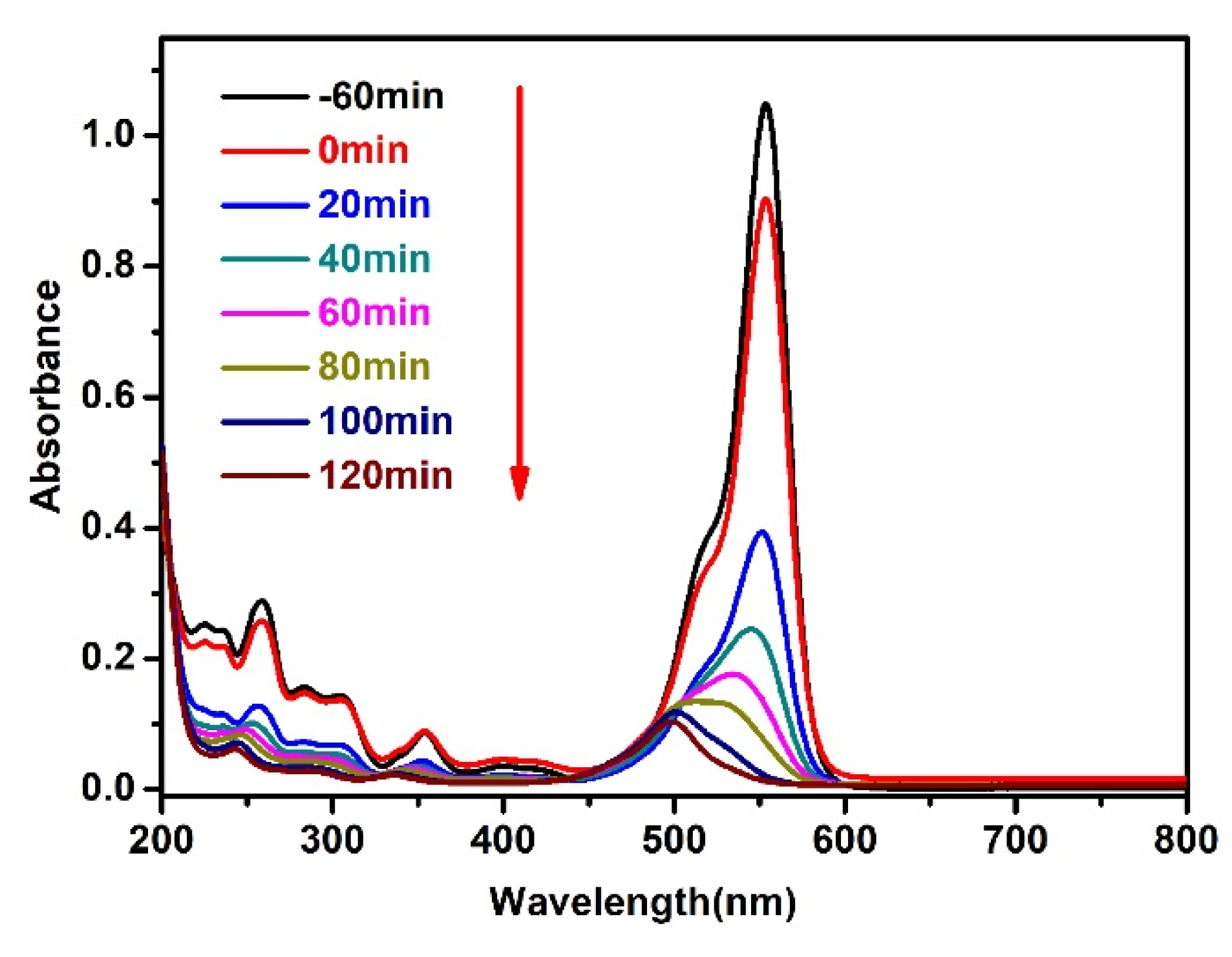
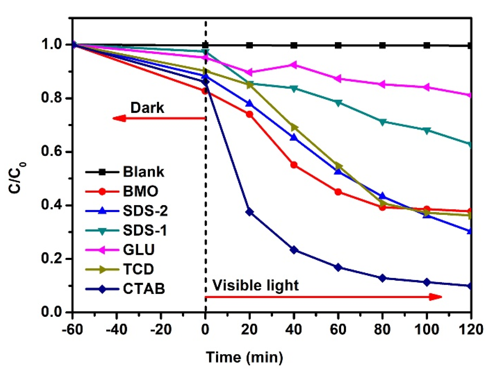
3.3. Photocatalytic Property Experiments

In order to evaluate the photocatalytic activity, degradation experiments of rhodamine B (RhB) dye were carried out under visible light at room-temperature. The RhB is a typical probe molecule for heterogeneous catalytic reactions, because it is a dye resistant to biodegradation and direct photolysis. A xenon lamp (500 W, Model PLS-SXE300) with a cut-off filter (λ > 400 nm) was used as the visible light source. The experimental procedures were performed as follows: 20 mg of catalysts was dispersed in 50 mL of RhB solution (5 mg L−1). Afterwards, the dispersion was stirred for 60 min in the dark to reach adsorption/desorption equilibrium. Then, dispersion under magnetic stirring took place approximately 10 cm below a xenon lamp. At each sampling time (20 min), a dispersion of about 3.5 mL was taken, and the RhB and the catalysts were separated by a centrifugation process. The concentration of RhB solution was analyzed using a spectrophotometer (UV-1901).

4. Conclusions

In summary, the Bi2MoO6 materials with different morphologies have been prepared by a simple hydrothermal process with different surfactants. Using different surfactants greatly changed the morphology of the Bi2MoO6, from nanosheet-assembled microspheres, to the smooth microspheres and aggregated nanoparticles. The photocatalytic efficiency was greatly improved with the conversion of Bi2MoO6 nanostructures from the nanoparticles to the nanosheet-assembled microspheres. The BMO-CTAB of nanosheet-assembled microspheres, especially, displayed high photocatalytic activity; meanwhile, nitrogen was doped during this process, which improved the photocatalytic performance. This work not only provides a low-cost and simple production method for the preparation of the Bi2MoO6 materials with various morphologies in design, but also increases their photocatalytic degradation efficiency in organic pollutant treatment.

Supplementary Materials

The following are available online at https://www.mdpi.com/2073-4344/10/10/1161/s1, Figure S1. N 1s (a) and Br 3d (b) spectra of the BMO-CTAB, respectively, Figure S2. UV-visible spectra of rhodamine B (RhB) solution with time over BMO-TCD under visible light, Figure S3. UV-visible spectra of rhodamine B (RhB) solution with time over BMO-GLU under visible light, Figure S4. UV-visible spectra of rhodamine B (RhB) solution with time over BMO-SDS-1 under visible light, Figure S5. UV-visible spectra of rhodamine B (RhB) solution with time over BMO-SDS-2 under visible light, Figure S6. UV-visible spectra of rhodamine B (RhB) solution with time over BMO under visible light, Table S1. The atomic percentage of each element of the BMO-CTAB sample, measured by XPS.

Author Contributions

Conceptualization, Y.S. and J.H.; Data curation, M.W. and J.W.; Formal analysis, T.J. and M.W.; Funding acquisition, T.J. and J.H.; Investigation, M.W. and X.H.; Project administration, J.W.; Resources, Y.S. and J.H.; Supervision, J.H.; Writing—original draft, T.J., Y.S. and S.L.; Writing—review and editing, T.J., E.H., Y.S. and J.H. All authors have read and agreed to the published version of the manuscript.

Funding

This research was funded by the Shenzhen Science and Technology Research Project (Grant No. JCYJ20170818093553012), the Shenzhen Pengcheng Scholar Program, the Guangdong Basic and Applied Basic Research Foundation (Grant No. 2020A1515010258), and the National Natural Science Foundation of China (Grant Nos. 51972055, 21561031 and 21701135).

Conflicts of Interest

The authors declare no conflict of interest.

References

  1. He, X.Y.; Zhang, C.L. Recent advances in structure design for enhancing photocatalysis. J. Mater. Sci. 2019, 54, 8831–8851. [Google Scholar] [CrossRef]
  2. Xiao, M.; Wang, Z.L.; Lyu, M.Q.; Luo, B.; Wang, S.C.; Liu, G.; Cheng, H.M.; Wang, L.Z. Hollow nanostructures for photocatalysis: Advantages and challenges. Adv. Mater. 2019, 31, 1801369. [Google Scholar] [CrossRef] [PubMed]
  3. Kong, J.J.; Yang, T.; Rui, Z.B.; Ji, H.B. Perovskite-based photocatalysts for organic contaminants removal: Current status and future perspectives. Catal. Today 2019, 327, 47–63. [Google Scholar] [CrossRef]
  4. Na, J.S.; Gong, B.; Scarel, G.; Parsons, G.N. Surface polarity shielding and hierarchical ZnO nano-architectures produced using sequential hydrothermal crystal synthesis and thin film atomic layer deposition. ACS Nano 2009, 3, 3191–3199. [Google Scholar] [CrossRef]
  5. Jiang, H.; Ma, J.; Li, C.Z. Hierarchical porous NiCo2O4 nanowires for high-rate supercapacitors. Chem. Commun. 2012, 48, 4465–4467. [Google Scholar] [CrossRef]
  6. Akbari, A.; Amini, M.; Tarassoli, A.; Eftekhari-Sis, B.; Ghasemian, N.; Jabbari, E. Transition metal oxide nanoparticles as efficient catalysts in oxidation reactions. Nano Struct. Nano Objects 2018, 14, 19–48. [Google Scholar] [CrossRef]
  7. Rodríguez, J.A.; Hrbek, J. Inverse oxide/metal catalysts: A versatile approach for activity tests and mechanistic studies. Surf. Sci. 2010, 604, 241–244. [Google Scholar] [CrossRef]
  8. He, R.G.; Xu, D.F.; Cheng, B.; Yu, J.G.; Ho, W.K. Review on nanoscale Bi-based photocatalysts. Nanoscale Horiz. 2018, 3, 464–504. [Google Scholar] [CrossRef]
  9. Bai, J.W.; Li, X.M.; Hao, Z.W.; Liu, L. Enhancement of 3D Bi2MoO6 mesoporous spheres photocatalytic performance by vacancy engineering. J. Colloid Interface Sci. 2020, 560, 510–518. [Google Scholar] [CrossRef]
  10. Sun, Y.G.; Zhang, Y.Q.; Xu, Y.L.; Yu, X.F.; Ding, D.R.; Liu, X.J. Synthesis and visible-light photocatalytic Properties of ZnO flake-like ensembles. Micro Nano Lett. 2012, 11, 1147–1150. [Google Scholar] [CrossRef]
  11. Gupta, N.K.; Ghafari, Y.; Kim, S.; Bae, J.; Kim, K.S.; Saifuddin, M. Photocatalytic Degradation of Organic Pollutants over MFe2O4 (M = Co, Ni, Cu, Zn) Nanoparticles at Neutral pH. Sci. Rep. 2020, 10, 4942. [Google Scholar] [CrossRef] [PubMed]
  12. Pino, E.; Calderón, C.; Herrera, F.; Cifuentes, G.; Arteaga, G. Photocatalytic Degradation of Aqueous Rhodamine 6G Using Supported TiO2 Catalysts. A Model for the Removal of Organic Contaminants from Aqueous Samples. Front. Chem. 2020, 8. [Google Scholar] [CrossRef]
  13. Yu, J.G.; Low, J.X.; Xiao, W.; Zhou, P.; Jaroniec, M. Enhanced photocatalytic CO2-reduction activity of anatase TiO2 by coexposed {001} and {101} facets. J. Am. Chem. Soc. 2014, 136, 8839–8842. [Google Scholar] [CrossRef] [PubMed]
  14. Stelo, F.; Kublik, N.; Ullah, S.; Wender, H. Recent advances in Bi2MoO6 based Z-scheme heterojunctions for photocatalytic degradation of pollutants. J. Alloys Comp. 2020, 829, 154591. [Google Scholar] [CrossRef]
  15. Zhang, L.W.; Xu, T.G.; Zhao, X.; Zhu, Y.F. Controllable synthesis of Bi2MoO6 and effect of morphology and variation in local structure on photocatalytic activities. Appl. Catal. B 2010, 98, 138–146. [Google Scholar] [CrossRef]
  16. Li, S.J.; Chen, J.L.; Hu, S.W.; Wang, H.L.; Jiang, W.; Chen, X.B. Facile construction of novel Bi2WO6/Ta3N5 Z-scheme heterojunction nanofibers for efficient degradation of harmful pharmaceutical pollutants. Chem. Eng. J. 2020, 402, 126165. [Google Scholar] [CrossRef]
  17. Li, S.J.; Hu, S.W.; Jiang, W.; Zhang, J.L.; Xu, K.B. In situ construction of WO3 nanoparticles decorated Bi2MoO6 microspheres for boosting photocatalytic degradation of refractory pollutants. J. Colloid Interface Sci. 2019, 556, 335–344. [Google Scholar] [CrossRef]
  18. Li, S.J.; Xue, B.; Chen, J.L.; Liu, Y.P.; Zhang, J.L.; Wang, H.W.; Liu, J.S. Constructing a plasmonic p-n heterojunction photocatalyst of 3D Ag/Ag6Si2O7/Bi2MoO6 for efficiently removing broad-spectrum antibiotics. Sep. Purif. Technol. 2020, 254, 117579. [Google Scholar] [CrossRef]
  19. Li, S.J.; Hu, S.W.; Jiang, W.; Zhou, Y.T.; Liu, J.S.; Wang, Z.H. Facile synthesis of cerium oxide nanoparticles decorated flower-like bismuth molybdate for enhanced photocatalytic activity toward organic pollutant degradation. J. Colloid Interface Sci. 2018, 530, 171–178. [Google Scholar] [CrossRef]
  20. Ma, Y.; Jia, Y.L.; Wang, L.N.; Yang, M.; Bi, Y.P.; Qi, Y.X. Hierarchical nanosheetbased Bi2MoO6 nanotubes with remarkably improved electrochemical performance. J. Power Sour. 2016, 331, 481–486. [Google Scholar] [CrossRef]
  21. Zhang, M.; Shao, C.; Mu, J.; Huang, X.; Zhang, Z.; Guo, Z.; Zhang, P.; Liu, Y. Hierarchical heterostructures of Bi2MoO6 on carbon nanofibers: Controllable solvothermal fabrication and enhanced visible photocatalytic properties. J. Mater. Chem. 2012, 22, 577–584. [Google Scholar] [CrossRef]
  22. Wu, M.H.; Wang, Y.X.; Xu, Y.; Ming, J.; Zhou, M.; Xu, R.; Fu, Q.; Lei, Y. Self-supported Bi2MoO6 nanowall for photoelectrochemical water splitting. ACS Appl. Mater. Inter. 2017, 9, 23647–23653. [Google Scholar] [CrossRef] [PubMed]
  23. Wang, J.; Sun, Y.G.; Wu, C.C.; Cui, Z.; Rao, P.H. Enhancing photocatalytic activity of Bi2MoO6 via surface co-doping with Ni2+ and Ti4+ ions. J. Phys. Chem. Solid. 2019, 129, 209–216. [Google Scholar] [CrossRef]
  24. Li, H.; Li, K.; Wang, H. Hydrothermal synthesis and photocatalytic properties of bismuth molybdate materials. Mater. Chem. Phys. 2009, 116, 134–142. [Google Scholar] [CrossRef]
  25. Shi, Y.; Feng, S.; Cao, C. Hydrothermal synthesis and characterization of Bi2MoO6 and Bi2WO6. Mater. Lett. 2000, 44, 215–218. [Google Scholar] [CrossRef]
  26. Zheng, Y.; Duan, F.; Wu, J.; Liu, L.; Chen, M.; Xie, Y. Enhanced photocatalytic activity of bismuth molybdates with the preferentially exposed {010} surface under visible light irradiation. J. Mol. Catal. A Chem. 2009, 303, 9–14. [Google Scholar] [CrossRef]
  27. Yang, Z.X.; Shen, M.; Dai, K.; Zhang, X.H.; Chen, H. Controllable synthesis of Bi2MoO6 nanosheets and their facet-dependent visible-light-driven photocatalytic activity. Appl. Surf. Sci. 2018, 430, 505–514. [Google Scholar] [CrossRef]
  28. Ghasemian, M.B.; Mayyas, M.; Idrus-Saidi, S.A.; Jamal, M.A.; Yang, J.; Mofarah, S.S.; Adabifiroozjaei, E.; Tang, J.B.; Syed, N.; O’Mullane, A.P.; et al. Self-Limiting Galvanic Growth of MnO2 Monolayers on a Liquid Metal—Applied to Photocatalysis. Adv. Funct. Mater. 2019, 29, 1901649. [Google Scholar] [CrossRef]
  29. Idrus-Saidi, S.A.; Tang, J.B.; Ghasemian, M.B.; Yang, J.; Han, J.L.; Syed, N.; Daeneke, T.; Abbasi, R.; Koshy, P.; O’Mullane, A.P.; et al. Liquid metal core–shell structures functionalised via mechanical agitation: The example of Field’s metal. J. Mater. Chem. A 2019, 7, 17876–17887. [Google Scholar] [CrossRef]
  30. Cao, D.D.; Wang, Q.Y.; Wu, Y.; Zhu, S.X.; Jia, Y.; Wang, R.L. Solvothermal synthesis and enhanced photocatalytic hydrogen production of Bi/Bi2MoO6 co-sensitized TiO2 nanotube arrays. Sep. Purif. Technol. 2020, 250, 117132. [Google Scholar] [CrossRef]
  31. Kasinathan, M.; Thiripuranthagan, S.; Sivakumar, A.; Ranganathan, S.; Vembuli, T.; Kumaravel, S.; Erusappan, E. Fabrication of novel Bi2MoO6/N-rGO catalyst for the efficient photocatalytic degradation of harmful dyes. Mater. Res. Bull. 2020, 125, 110782. [Google Scholar] [CrossRef]
  32. Khazaee, Z.; Khavar, A.H.C.; Mahjoub, A.R.; Motaee, A.; Srivastava, V.; Sillanpää, M. Template-confined growth of X-Bi2MoO6 (X: F, Cl, Br, I) nanoplates with open surfaces for photocatalytic oxidation; experimental and DFT insights of the halogen doping. Solar Energy 2020, 196, 567–581. [Google Scholar] [CrossRef]
  33. Suebsom, P.; Phuruangrat, A.; Suwanboon, S.; Thongtem, S.; Thongtem, T. Enhanced visible-light-driven photocatalytic activity of heterostructure Ag/Bi2MoO6 nanocomposites synthesized by photoreduction method. Inorg. Chem. Commun. 2020, 119, 108120. [Google Scholar] [CrossRef]
  34. Wang, Y.J.; Wang, Q.Y.; Zhang, H.; Wu, Y.; Jia, Y.; Jin, R.C.; Gao, S.M. CTAB-assisted solvothermal construction of hierarchical Bi2MoO6/Bi5O7Br with improved photocatalytic performances. Sep. Purif. Technol. 2020, 242, 116775. [Google Scholar] [CrossRef]
  35. Li, S.J.; Shen, X.F.; Liu, J.S.; Zhang, L.S. Synthesis of Ta3N5/Bi2MoO6 core-shell fiber-shaped heterojunctions as efficient and easily recyclable photocatalysts. Environ. Sci. Nano 2017, 4, 1155–1167. [Google Scholar] [CrossRef]
  36. Li, N.; Gao, H.; Wang, X.; Zhao, S.J.; Lv, D.; Yang, G.Q.; Gao, X.Y.; Fan, H.K.; Gao, Y.Q.; Ge, L. Novel indirect Z-scheme g-C3N4/Bi2MoO6/Bi hollow microsphere heterojunctions with SPR-promoted visible absorption and highly enhanced photocatalytic performance. Chin. J. Chem. 2020, 41, 426–434. [Google Scholar] [CrossRef]
  37. Sun, Y.G.; Zou, R.J.; Tian, Q.W.; Wu, J.H.; Chen, Z.G.; Hu, J.Q. Hydrothermal synthesis, growth mechanism, and properties of three-dimensional micro/nanoscaled hierarchical architecture films of hemimorphite zinc silicate. CrystEngComm 2011, 13, 2273–2280. [Google Scholar] [CrossRef]
  38. Li, W.F.; Sun, Y.G.; Xu, J.L. Controllable hydrothermal synthesis and properties of ZnO hierarchical micro/nanostructures. Nano-Micro Lett. 2012, 4, 98–102. [Google Scholar] [CrossRef] [Green Version]
  39. Sun, M.Z.; Guo, P.Y.; Wang, M.; Ren, F.Y. The effect of calcination temperature on the photocatalytic performance of Bi2MoO6 for the degradation of phenol under visible light. Opt. Int. J. Light Electron Opt. 2019, 199, 163319. [Google Scholar] [CrossRef]
  40. Wang, M.; Han, J.; Guo, P.; Sun, M.; Zhang, Y.; Tong, Z.; You, M.; Lv, C. Hydrothermal synthesis of B-doped Bi2MoO6 and its high photocatalytic performance for the degradation of rhodamine B. J. Phys. Chem. Solid. 2018, 113, 86–93. [Google Scholar] [CrossRef]
  41. Rangel, R.; Cedeno, V.J.; Espino, J.; Bartolo-Perez, P.; Rodriguez-Gattorno, G.; Alvarado-Gil, J.J. Comparing the Efficiency of N-Doped TiO2 and N-Doped Bi2MoO6 Photo Catalysts for MB and Lignin Photodegradation. Catalysts 2018, 8, 668. [Google Scholar] [CrossRef] [Green Version]
  42. Ji, T.; Cui, Z.; Zhang, W.L.; Cao, Y.J.; Zhang, Y.F.; He, S.A.; Xu, M.D.; Sun, Y.G.; Zou, R.J.; Hu, J.Q. UV and visible light synergetic photodegradation using rutile TiO2 nanorod arrays based on a p–n Junction. Dalton Trans. 2017, 46, 4296. [Google Scholar] [CrossRef] [PubMed]
  43. Imani, M.; Farajnezhad, M.; Tadjarodi, A. 3D hierarchical flower-like nanostructure of Bi2MoO6: Mechanochemical synthesis, the effect of synthesis parameters and photocatalytic activity. Mater. Res. Bull. 2017, 87, 92. [Google Scholar] [CrossRef]
  44. Shen, H.D.; Xue, W.W.; Fu, F.; Sun, J.F.; Zhen, Y.Z.; Wang, D.J.; Shao, B.; Tang, J.W. Efficient Degradation of Phenol and 4-Nitrophenol by Surface Oxygen Vacancies and Plasmonic Silver Co-Modified Bi2MoO6 Photocatalysts. Chem. Eur. J. 2018, 24, 18463–18478. [Google Scholar] [CrossRef] [PubMed] [Green Version]
  45. Phuruangrat, A.; Dumrongrojthanath, P.; Thongtem, S.; Thongtem, T. Synthesis and characterization of visible light-driven W-doped Bi2MoO6 hotocatalyst and its photocatalytic activities. Mater. Lett. 2017, 194, 114–117. [Google Scholar] [CrossRef]
  46. Li, H.D.; Li, W.J.; Gu, S.N.; Wang, F.Z.; Zhou, H.L.; Liu, X.T.; Ren, C.J. Enhancement of photocatalytic activity in Tb/Eu co-doped Bi2MoO6: The synergistic effect of Tb-Eu redox cycles. RSC Adv. 2016, 6, 48089–48098. [Google Scholar] [CrossRef]
Figure 1. X-ray diffraction patterns of different Bi2MoO6 samples, including, BMO-SDS-2 (sodium-dodecyl sulfonate), BMO-SDS-1 (sodium-dodecyl sulfate), BMO-CTAB (cetyl-trimethyl-ammonium bromide), BMO (no surfactant), BMO-GLU (glucose), and BMO-TCD (trisodium citrate dehydrate).
Catalysts 10 01161 g001
Figure 2. SEM images of BMO (a,b), BMO-CTAB (c,d), BMO-TCD (e), BMO-GLU (f), BMO-SDS-1 (g) and BMO-SDS-2 (h).
Catalysts 10 01161 g002
Figure 3. (a) UV–Vis-NIR absorption spectra of the Bi2MoO6 samples. (αhν)2 versus plots for the samples of BMO and BMO-CTAB (b), BMO-SDS1 and BMO-SDS2 (c), BMO-TCD and BMO-GLU (d).
Catalysts 10 01161 g003
Figure 4. The Bi 4f (a), Mo 3d (b), O 1s (c) and VB (d) XPS spectra of the BMO and BMO-CTAB.
Catalysts 10 01161 g004
Figure 5. Fourier transform infrared spectra (a) and Raman spectra (b) of BMO and BMO-CTAB.
Catalysts 10 01161 g005
Figure 6. UV–visible spectra of rhodamine B (RhB) solution with time over BMO-CTAB under visible light.
Catalysts 10 01161 g006
Figure 7. The photocatalytic activities for RhB over different samples under visible light irradiation.
Catalysts 10 01161 g007
Table 1. The specific surface area data of Bi2MoO6 samples.
SurfactantSDS-1BMO (None)TCDSDS-2CTABGLU
Surface area (m2/g)45.12048.95972.19145.21341.1704.892

