Article

Entomopathogenic Filamentous Fungi as Biocatalysts in Glycosylation of Methylflavonoids

by
Agnieszka Krawczyk-Łebek
*,
Monika Dymarska
,
Tomasz Janeczko
and
Edyta Kostrzewa-Susłow
*
Department of Chemistry, Faculty of Biotechnology and Food Science, Wrocław University of Environmental and Life Sciences, 50-375 Wrocław, Poland
*
Authors to whom correspondence should be addressed.
Catalysts 2020, 10(10), 1148; https://doi.org/10.3390/catal10101148
Submission received: 5 September 2020 / Revised: 29 September 2020 / Accepted: 30 September 2020 / Published: 4 October 2020
(This article belongs to the Special Issue Catalysis in the Synthesis of Biologically Active Compounds)

Abstract

:
Flavonoids are known for their numerous biological activities; however, their pharmacological application is limited by poor bioavailability. Glycosides are usually more stable and more soluble in water and in this form, flavonoids are present in nature. Likewise, the presence of the methyl group in the flavonoid skeleton results in facilitated absorption and greater bioavailability. Entomopathogenic filamentous fungi are effective in the biotransformation of flavonoids; they are known especially for efficient glycosylation. In the current study we used strains of Beauveria bassiana KCH J1.5 and Isaria fumosorosea KCH J2 to biotransform flavonoids with a single methyl group. 2′-Hydroxy-5′-methylchalcone was biotransformed by both strains into 2′-hydroxy-5′-methylchalcone 3-O-β-D-(4″-O-methyl)-glucopyranoside. In the culture of B. bassiana KCH J1.5 four products were obtained from 6-methylflavanone: 4′-hydroxy-6-methylflavanone 3′-O-β-D-(4″-O-methyl)-glucopyranoside; 4′-hydroxyflavanone 6-methylene-O-β-D-(4″-O-methyl)-glucopyranoside; 6-hydroxymethylflavanone 3′-O-β-D-(4″-O-methyl)-glucopyranoside and 4′-hydroxy-6-hydroxymethylflavanone 3′-O-β-D-(4″-O-methyl)-glucopyranoside. Biotransformation with I. fumosorosea KCH J2 as a biocatalyst resulted in the formation of 6-methylflavanone 4′-O-β-D-(4″-O-methyl)-glucopyranoside and 2-phenyl-6-methylchromane 4-O-β-D-(4″-O-methyl)-glucopyranoside. All of these flavonoids can be used in biological activity tests and can be useful in studies concerning structure—bioactivity relationships.

1. Introduction

Flavonoid compounds are plant secondary metabolites engaged in various plants’ interactions with the environment [1]. They are able to exert a wide range of biological activities: antioxidant, anti-inflammatory, antiallergic, antimicrobial, antiplatelet, anticancer and neuroprotective [1,2,3]. Their occurrence in a plant based diet is ubiquitous [4]. However, pharmacological application of flavonoid aglycons is limited by their poor bioavailability [5,6]. In nature, flavonoid derivatives—products of glycosylation, methylation, prenylation, acetylation and polymerization—are very common [7] and the most abundant are glucosides. Addition of sugar moiety (or moieties) to flavonoid aglycone is catalyzed by uridine diphosphate (UDP) glycosyltransferases (UGTs) [8,9]. Flavonoids glycosylation results in the modulation of their physicochemical and biological properties, most of all by improving their aqueous solubility and facilitating their intracellular and intercellular transportation. Nonetheless, the impact of the glycosylation on the biological properties of flavonoids is complicated, because it depends on the structure of the flavonoid core, the position of the sugar attachment and the number of sugar moieties [10,11,12,13,14,15,16].
Many flavonoid glycosides with potential biological activity occur in plants in small amounts and therefore their extraction is difficult. Chemical synthesis is inefficient in the production of flavonoid glycosides, because of laborious procedures necessary to achieve regio- and stereo-selectivity, and low isolated yields due to drastic reaction conditions resulting in the decomposition of aglycones. However, flavonoid glycosides, both well-known and novel, can be obtained by microbial glycosylation of already existing flavonoid aglycons. Enzymatic glycosylation allows to overcome these obstacles. It can be carried out by a few different methods: whole-cell biotransformation, in vivo total biosynthesis and in vitro enzymatic reaction [11,12,17,18,19,20]. Among these methods biotransformation is promising, because it offers an eco-friendly, one-pot and one-step process [21].
Methylation at the ring position of flavonoids leads to derivatives with increased metabolic stability due to insusceptibility to glucuronic acid or sulfate conjugation. Methylation also leads to greatly improved intestinal absorption, and bioavailability [1,7,22]. Methylated flavonoids are widely present in plants, but still less abundant than unmethylated flavonoid aglycons and flavonoid glycosides. Among two types of methylation in flavonoids, O-methylation and C-methylation, the second one is less common and has been found in limited plant species, such as Cleistocalyx operculatus [23,24,25], Picea neoveitchii [26], Corymbia torelliana [27,28] and Campomanesia xanthocarpa [29], and occurs especially at C-6 of the flavonoid skeleton.
There is some evidence of biological activity of C-methylated flavonoids isolated from plants. From the twigs and leaves of P. neoveitchii Mast, two C-methylated flavonoids were isolated: 5,7-dihydroxy-3-methoxy-6-methylflavone 8,4′-di-O-β-D-glucopyranoside and 4′,5,7,8-tetrahydroxy-3-methoxy-6-methylflavone 8-O-β-D-glucopyranoside. The first one exhibited moderate antifungal activity against several plant pathogens (Pyricularia grisea (Cooke) Sacc., Sclerotium rocfsii Sacc. and Alternaria mali Roberts). The second one showed a significant inhibitory effect against S. rocfsii. Both compounds exhibited potent cytotoxicity against Spodoptera litura Fabricius [26].
Methylated chalcones from C. operculatus also showed biological activity. (E)-2′,4,4′-Trihydroxy-6′-methoxy-3′,5′-dimethylchalcone, 2′,4′-dihydroxy−6′-methoxy-3′,5′- dimethylchalcone, 2′,4′-dihydroxy-3′-methyl-6′-methoxychalcone and 2,2′,4′-trihydroxy-6′-methoxy- 3′,5′-dimethylchalcone displayed notable inhibitory activity on the viral neuraminidases from two influenza viral strains, H1N1 and H9N2. The first one exhibited strong inhibition against the neuraminidases from novel influenza H1N1 (WT) and oseltamivir-resistant novel H1N1 (H274Y mutant) expressed in 293T cells. Kinetic studies have indicated that tested chalcones acted as noncompetitive inhibitors to neuraminidases which are responsible for enabling viruses to leave infected cells by decomposition of their cell membranes [25].
In studies conducted on oocytes of Xenopus laevis with expressed human recombinant GABAA receptors 6-methylflavanone acted as a positive allosteric modulator. GABAA receptors are the main inhibitory neurotransmitters in the central nervous system and are classified under six subfamilies of protein subunits: α, β, γ, δ, ε and θ. Anxiolysis and sedation are considered to be mediated via GABAA receptors (α2 and α1, respectively). The tested compound was much more effective at α2β2γ2L than at α1β2γ2L and α1β2 GABAA receptors. This may be quite a unique type of selectivity, significant in seeking new targeted drugs, acting as GABAA receptors agonists and producing certain sedative effects without the unwanted side effects of benzodiazepines [30].
B. bassiana is an entomopathogenic filamentous fungi that affects a broad range of arthropods. It has been used as a biological control agent of various arthropod pests for many years [31,32,33]. The pathogenesis and virulence of Beauveria isolates is connected with the production of toxicogenic metabolites, antioxidant enzymes and primarily extracellular cuticle-degrading enzymes such as chitinases, lipases, and proteases [33,34]. Since B. bassiana genome sequence is already known [35] there is a lot of interest in recognition of genes involved in its pathogenesis and virulence [32,36]. Enzymatic systems of B. bassiana, that are responsible for a cuticle-degradation are also applied to the biotransformations. B. bassiana has the ability to transform a broad range of substrates such as cyclic and aliphatic ketones, flavonoids and steroids [37]. This fungus is capable of performing reactions such as hydroxylation, glycosylation or deglycosylation, acetylation and reduction [37]. Most of the enzymes responsible for these reactions have not been described until now [37]. Likewise I. fumosorosea serves as a biological control agent and alternative to chemical pesticides [38]. Its secondary metabolites exhibit a great variety of biological activities [39]. This genus was used as a biocatalyst by our team in previous studies of flavonoid biotransformations [12,13,14,15].
The current studies are a continuation of our previous work with 6-methylflavone as the biotransformation substrate [12]. In the present study we synthesized two flavonoid compounds with methyl group at C-6 of flavanone skeleton (C-5′ in the case of chalcone) and afterwards biotransformed them with entomopathogenic filamentous fungi strains of B. bassiana KCH J1.5 and I. fumosorosea KCH J2 as biocatalysts. As a result of 2′-hydroxy-5′-methylchalcone biotransformation in both cultures one main product 2′-hydroxy-5′-methylchalcone 3-O-β-D-(4″-O-methyl)-glucopyranoside was obtained. 6-Methylflavanone was efficiently biotransformed by both used biocatalysts. Each strain formed different biotransformation products. Six flavanones 4″-O-methylglucopyranosides were obtained. All biotransformation products have not been previously described in the scientific literature. The biotransformation products may be utilized in further studies as potentially biologically active compounds and can be useful in investigations concerning structure—bioactivity relationships.

2. Results and Discussion

The following results are a continuation of studies focused on biotransformations of flavonoid compounds with a methyl group at C-6 in cultures of entomopathogenic filamentous fungi. In the previous paper about biotransformation of 6-methylflavone by the strain I. fumosorosea KCH J2, two new products were obtained: 6-methylflavone 8-O-β-D-(4″-O-methyl)-glucopyranoside (6.6% yield) and 6-methylflavone 4′-O-β-D-(4″-O-methyl)-glucopyranoside (13.2% yield) [12].
In the present paper two flavonoid substrates (chalcone and flavanone), that are analogs of the 6-methylflavone, were utilized. Two strains of the enthompathogenic filamentous fungi I. fumosorosea KCH J2 and B. bassiana KCH J1.5, isolated from the environment and characterized previously [12,37] were used as biocatalysts. Experiments performed in a semi-preparative scale enabled us to determine the chemical structures of biotransformation products and their isolated yields unambiguously. Hereby, seven biotransformation products that have not yet been described in the scientific literature were obtained: 2′-hydroxy-5′-methylchalcone 3-O-β-D-(4″-O-methyl)-glucopyranoside (3a), 4′-hydroxy-6-methylflavanone 3′-O-β-D-(4″-O-methyl)-glucopyranoside (4a), 4′-hydroxyflavanone 6-methylene-O-β-D-(4″-O-methyl)-glucopyranoside (4b), 6-hydroxymethylflavanone 3′-O-β-D- (4″-O-methyl)-glucopyranoside (4c), 4′-hydroxy-6-hydroxymethylflavanone 3′-O-β-D-(4″-O-methyl)- glucopyranoside (4d), 6-methylflavanone 4′-O-β-D-(4″-O-methyl)-glucopyranoside (4e) and 2-phenyl-6-methylchromane 4-O-β-D-(4″-O-methyl)-glucopyranoside (4f).
The substrates for biotransformation were obtained by a two-step synthesis (Scheme 1).

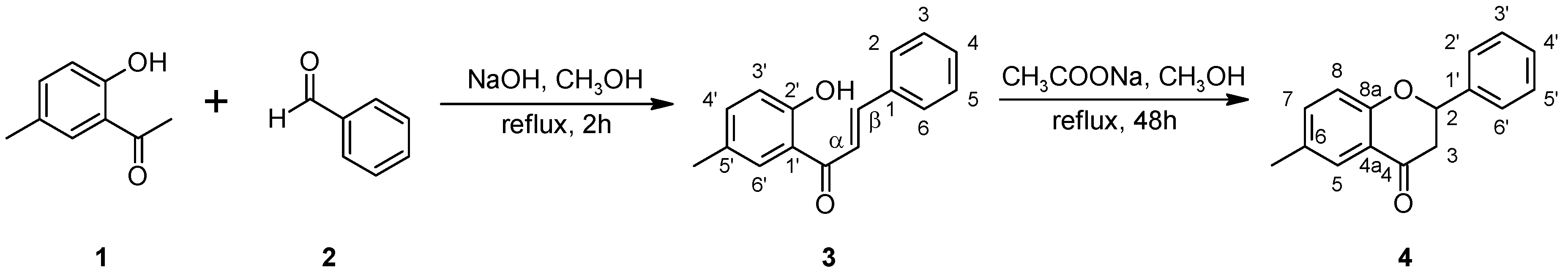
2.1. Biotransformations of 2′-hydroxy-5′-methylchalcone (3)

In the experiment 2′-hydroxy-5′-methylchalcone (3) was synthesized in a Claisen–Schmidt condensation reaction between 2′-hydroxy-5′-methylacetophenone (1) and benzaldehyde (2) with a 50.1% yield. As a result of its 10-day biotransformation, in the B. bassiana KCH J1.5 culture one main product 2′-hydroxy-5′-methylchalcone 3-O-β-D-(4″-O-methyl)-glucopyranoside (3a) with a 29.5% yield (Scheme 2) was isolated and purified by means of the preparative Thin Layer Chromatography (TLC) method with chloroform and methanol (9:1 v/v) as eluents. The same product was obtained using I. fumosorosea KCH J2 as a biocatalyst.
The product 3a was analyzed by NMR spectroscopy that allowed to establish its chemical structure (Table 1 and Table 2, Scheme 3).
The presence of a glucose unit in the compound 3a was confirmed by five characteristic carbon signals observed in the region from δ = 80.5 ppm to δ = 62.3 ppm in the Carbon-13 Nuclear Magnetic Resonance (13C-NMR) spectrum, as well as proton signals of δH ranging from δ = 3.92 ppm to δ = 3.19 ppm in the Proton Nuclear Magnetic Resonance (1H-NMR) spectrum. Moreover, the attachment of a sugar unit to substrate 3 was confirmed by a one-proton doublet from the proton at the anomeric carbon atom at δ = 5.03 ppm in the 1H-NMR spectrum. The β-configuration of the glucose unit was proved by the coupling constant (J = 7.8 Hz) for the anomeric proton. A three-proton singlet at δ = 3.57 ppm in the 1H-NMR spectrum and the corresponding signal at δ = 60.6 ppm in the 13C-NMR spectrum evidences that one of the hydroxyl groups of sugar unit has been methylated. O-methylation occurred in the C-4″ hydroxyl group of the glucose unit. It was detected in the Heteronuclear Multiple Bond Coherence (HMBC) spectrum, where the proton signal due to -OCH3 was correlated with the signal of C-4″ (about δ = 80 ppm) in the glucose unit. Sugar moiety was attached to C-3, because in the HMBC spectrum signal due to the proton at hemiacetal carbon atom (δ = 5.03 ppm) was correlated with the C-3 signal (δ = 159.2 ppm), which was shifted from δ = 129.2 ppm, indicating the attachment of an electronegative atom. Additionally, protons in the 1H-NMR spectrum at C-2 and C-6 became non-equivalent, and the proton at C-3 disappeared. Shifted signals can be observed: one multiplet from the proton at C-2 (δ = 7.61 ppm), which became isolated in the correlation spectrum, one doublet of doublets of doublets from the proton at C-4 (δ = 7.16 ppm) and one doublet from the proton at C-6 (δ = 7.47 ppm). Signal from proton at C-5 was slightly shifted and merged with signal from the proton at C-4′. Signals from one proton of the hydroxyl group at C-2′ and three protons of the methyl group at C-5 remained intact.
Glycosylation of chalcones is rarely reported. Among reported microbial transformations of the chalcones into their glycosides, there are known reactions with xanthohumol as a substrate. These biotransformations led to the production of: xanthohumol 4′-O-β-D-glucopyranoside in the cultures of Penicillium chrysogenum 6933 [40], Absidia coerulea AM93 and Rhizopus nigricans UPF701 [41], xanthohumol 4,4′- O-β-diglucopyranoside in the culture of P. chrysogenum 6933 [40], xanthohumol 4′-O-β-D-(4″-O-methyl)-glucopyranoside in the cultures of B. bassiana AM278 [42] and B. bassiana AM446 [41], and isoxanthohumol 7-O-β-D-glucopyranoside in the cultures of Cunninghamella elegans 6992 [40] and Mortierella mutabilis AM404 [41]. Several studies with enzymes of the glycosyltransferase family led to the formation of the chalcone glycosides at three sites: 2′, 4′ and less often 4 [43]. However, there is no evidence of the glycosylation at C-3 of the chalcone skeleton and also no reports of 2′-hydroxy-5′-methylchalcone (3) biotransformations.

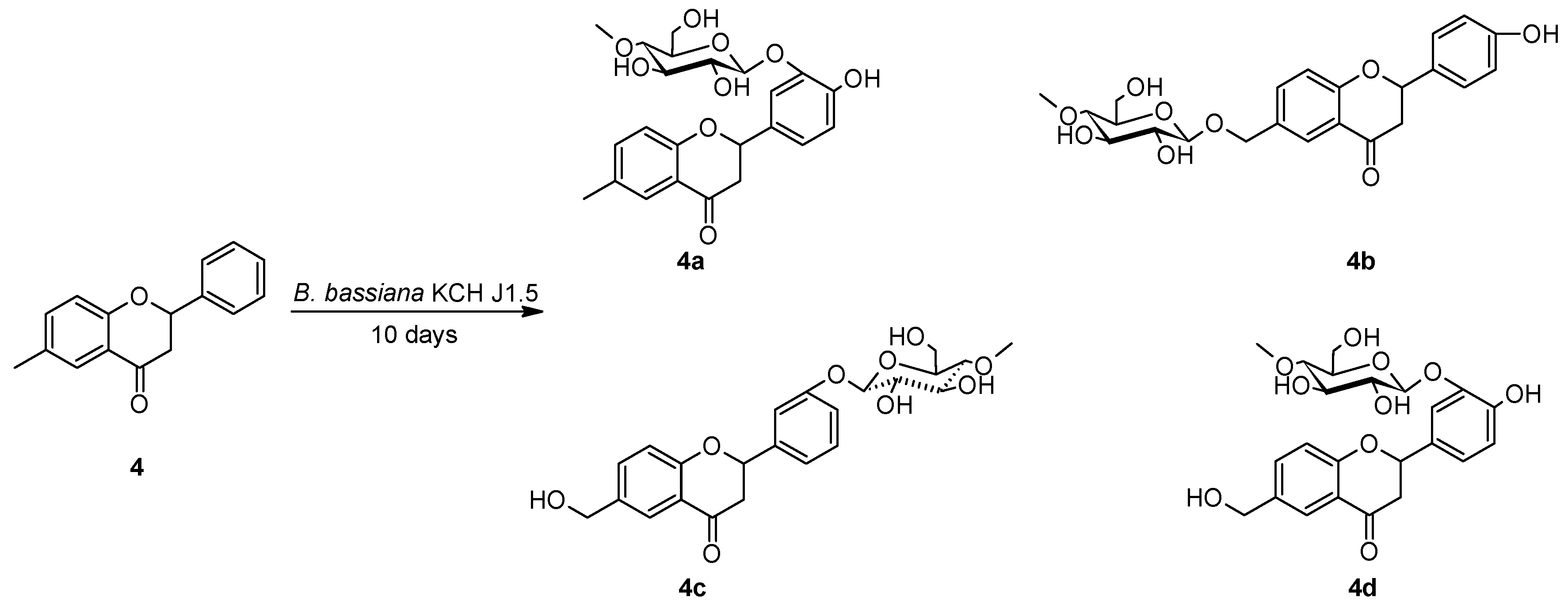
2.2. Biotransformations of 6-methylflavanone (4) in the culture of B. bassiana KCH J1.5

6-Methylflavanone (4) was synthesized from 2′-hydroxy-5′-methylchalcone (3) with 62.3% yield and utilized in 10-day biotransformation in the culture of B. bassiana KCH J1.5. Four biotransformation products were obtained and isolated: 4′-hydroxy-6-methylflavanone 3′-O-β-D-(4″-O-methyl)-glucopyranoside (4a) with 6.9% yield, 4′-hydroxyflavanone 6-methylene-O-β-D-(4″-O-methyl)-glucopyranoside (4b) with 7.9% yield, 6-hydroxymethylflavanone 3′-O-β-D-(4″-O-methyl)-glucopyranoside (4c) with 3.4% yield and 4′-hydroxy-6-hydroxymethylflavanone 3′-O-β-D-(4″-O-methyl)-glucopyranoside (4d) with 8.0% yield (Scheme 4).
The products 4a4d were analyzed by NMR spectroscopy which allowed for the establishment of their chemical structures (Table 3 and Table 4, Scheme 5, Scheme 6, Scheme 7 and Scheme 8).
The presence of a glucose unit in compounds 4a4d was established by five characteristic carbon signals observed in the region from about δ = 80.0 ppm to about δ = 62.0 ppm in the 13C-NMR spectra, as well as proton signals of δH ranging from about δ = 3.9 ppm to δ = 3.1 ppm in the 1H-NMR spectra. Additionally, the attachment of a sugar unit to substrate 4 was confirmed by a one-proton doublet from the proton at the anomeric carbon atom in the 1H-NMR spectrum of 4a at δ = 4.79 ppm, 4b at δ = 4.36 ppm, 4c at δ = 4.98 ppm and 4d at δ = 4.82 ppm. The β-configuration of the glucose unit was proved by the coupling constant (about J = 7.9 Hz) for the anomeric proton. A three-proton singlet at about δ = 3.6 ppm in the 1H-NMR spectra and the corresponding signal at about δ = 60.6 ppm in the 13C-NMR spectra of 4a4d evidences that one of the hydroxyl groups of sugar unit has been methylated. O-methylation occurred in all products at the C-4″ hydroxyl group of the glucose unit. It was detected in the HMBC spectra, that the proton signals due to -OCH3, were correlated with the signals of C-4″ (about δ = 80 ppm) in the glucose units.
Based on the HMBC spectrum, a sugar moiety was attached to C-3′ in the case of compound 4a, due to the proton at the anomeric carbon atom (δ = 4.79 ppm) being coupled with the C-3′ signal (δ = 146.1 ppm), which was shifted from δ = 129.5 ppm, indicating the attachment of an electronegative atom. Moreover, protons in the 1H-NMR spectrum at C-2′ and C-6′ became non-equivalent and protons at C-3′ and C-4′ were not present, suggesting substitution. The C-4′ signal at the 13C-NMR spectrum (δ = 149.0 ppm), which was shifted from δ = 129.3 ppm, indicates the attachment of an electronegative oxygen atom from a hydroxyl group. In the 1H-NMR spectrum shifted signals of flavanone ring B can be observed: one multiplet from the proton at C-2′ (δ = 7.39 ppm), one doublet from the proton at C-5′ (δ = 6.91 ppm), and one doublet of triplets at C-6′ (δ = 7.16 ppm). In the COSY spectrum protons at C-5′ and at C-6′correlate, and the proton at C-2′ does not couple with any proton, which also confirms substitution at C-3′ and C-4′. In the HMBC spectrum, a strong cross-peak between C-3′ and H-2′ and between C-3′ and H-5′, while no peak between C-3′ and H-6′, can be observed. At the same time a strong cross-peak between C-4′ and H-2′ (and H-5′ and H-6′) can be seen. Cross-peak by three chemical bonds occurs less likely than by two chemical bonds in HMBC experiments. Therefore, we assigned the C-3′ signal as having cross-peaks by one, and two chemical bonds, and the C-4′ signal as having cross-peaks by one and two chemical bonds [44,45,46]. Chemical shifts of the other signals in the 1H-NMR and 13C-NMR spectra have only slightly changed. The signal from the three protons of the methyl group at C-6 remained at the same position as in the 1H-NMR spectrum of substrate 4 which indicates the presence of the methyl group in product 4a.
In the case of compound 4b, the sugar moiety was attached to the hydroxyl group—previously formed by microbial oxidation of the methyl group—because in the HMBC spectrum the proton at the anomeric carbon atom (δ = 4.78 ppm) was coupled with the C-6-CH2- signal (δ = 70.3 ppm). Moreover, the signal from the three protons of the methyl group at C-6 present in the 1H-NMR spectrum (δ = 2.32 ppm) of substrate 4 disappeared and two signals appeared from protons at 4.86 ppm and 4.61 ppm (unequal protons from methylene group), which confirms attachment of the glucose unit to the methylene bridge at C-6. In the 1H-NMR spectrum, proton signals of the flavanone ring A became only slightly shifted because of it. Moreover, signals from protons of the ring B became shifted, indicating another substitution in ring B. Two multiplets from the protons at C-2’ and C-6’ (δ = 7.41 ppm) and at C-3’ and C-5’ (δ = 6.90 ppm) were observed and the signal from proton at C-4’ was missing which indicates a substitution at C-4’. In the 13C-NMR spectrum C-4′ signal (δ = 158.7 ppm), which was shifted from δ = 129.3 ppm, indicates the attachment of a hydroxyl group. Chemical shifts of the other signals in the 1H-NMR and 13C-NMR spectra have only slightly changed that signalize that the flavanone skeleton remained intact.
Based on the HMBC spectrum, in compound 4c, a sugar moiety was attached at C-3’, due to the proton at the anomeric carbon atom (δ = 4.98 ppm) being coupled with the C-3’ signal (δ = 158.9 ppm), which was shifted from δ = 129.5 ppm, and points out the attachment of an electronegative atom. Moreover, protons in the 1H-NMR spectrum at C-2′ and C-6′ became non-equivalent. In the 1H-NMR spectrum shifted proton signals of ring B can be observed: multiplet from proton at C-2’ (δ = 7.29 ppm), triplet from proton at C-5’ (δ = 7.35 ppm) and doublet from proton at C-6’ (δ = 7.21 ppm), and signal from proton at C-4’ that overlapped with the signal from proton at C-8 (δ = 7.07 ppm). In the COSY spectrum proton at C-2’ became isolated, confirming substitution at C-3′. Moreover, the signal from the three protons of the methyl group at C-6 present in the 1H-NMR spectrum (δ = 2.32 ppm) of substrate 4 disappeared. The presence of another moiety—a hydroxymethyl group at C-6 can be noticed in the 13C-NMR spectrum, because the signal from C-6 was shifted from δ = 131.6 ppm to δ = 136.7 ppm. It was also confirmed by the appearance of a signal from two protons at 4.61 ppm [47] and slightly shifted signals from protons of ring A: doublet from the proton at C-5 (δ = 7.82 ppm), doublet of doublets from the proton at C-7 (δ = 7.58 ppm) and doublet of doublets from the proton at C-8 (δ = 7.07 ppm). Chemical shifts of the other signals in the 1H-NMR and 13C-NMR spectra have only slightly changed which indicates that the flavanone skeleton remained unaffected.
Based on the HMBC spectrum, a sugar moiety was attached to C-3′ in the case of compound 4d, due to the proton at the anomeric carbon atom (δ = 4.81 ppm) being coupled with the C-3′ signal (δ = 146.1 ppm), which was shifted from δ = 129.5 ppm. Additionally, protons in the 1H-NMR spectrum at C-2′ and C-6′ became non-equivalent. Shifted signals can be observed: one doublet of doublets from the proton at C-2’ (δ = 7.41 ppm), one doublet of triplets at C-6′ (δ = 7.17 ppm), and one doublet from the proton at C-5’ (δ = 6.92 ppm). In the 13C-NMR spectrum signal from C-4’ (δ = 149.0 ppm) and signal from C-6 (δ = 136.5 ppm) was also shifted compared with the signals from substrate 4 δ = 129.3 ppm and δ = 131.6 ppm, respectively. These changes indicate the introduction of a hydroxyl group at C-4′ and hydroxymethyl group at C-6. Methylglucosyl moiety was attached to C-3′ and hydroxyl moiety to C-4′ same as in product 4b, which was confirmed by cross-peaks in the HMBC spectrum [44,45,46]. Signals from the protons of ring A were only slightly changed, but exchange of a methyl group on the hydroxymethyl group at C-6 was confirmed by the disappearance of the signal from the three protons of the methyl moiety that was present in the 1H-NMR spectrum (δ = 2.32 ppm) of substrate 4 and the appearance of a signal from two protons at 4.61 ppm[47].
Glycosylations of flavanones performed by B. bassiana strains have already been described in the scientific literature. Isoxanthohumol and 8-prenylnaringenin were transformed by B. bassiana AM278 into their 7-O-β-D-(4″-O-methyl)-glucosides [48]. Other flavanones, like pinocembrin, naringenin, eriodicytol and hesperetin were glycosylated and 4″-O-methyl-glycosylated at C-7 in the cultures of B. bassiana AM278. Moreover, hesperetin biotransfromation resulted in additional products glycosylated and 4″-O-methyl-glycosylated at C-3′ [49]. The strain B. bassiana KCH J1.5 used in the currently presented studies showed a different regioselectivity of glycosylation. Interestingly B. bassiana KCH J1.5 was also able to hydroxylate 6-methyl moiety in products 4b4d. Presence of the four biotransformation products with different moieties attached indicates the involvement of a few enzymes in the transformation of 6-methylflavanone (4) in the culture of B. bassiana KCH J1.5. In the case of product 4b (4′-hydroxyflavanone 6-methylene-O-β-D-(4″-O-methyl)-glucopyranoside) hydroxylation took place at C-4′ and C-6-CH3, followed by attachment of the glucose unit to hydroxylmethyl group (-CH2-OH) at C-6. The presumable course of microbial transformation of 6-methylflavanones is shown in the Scheme 9.

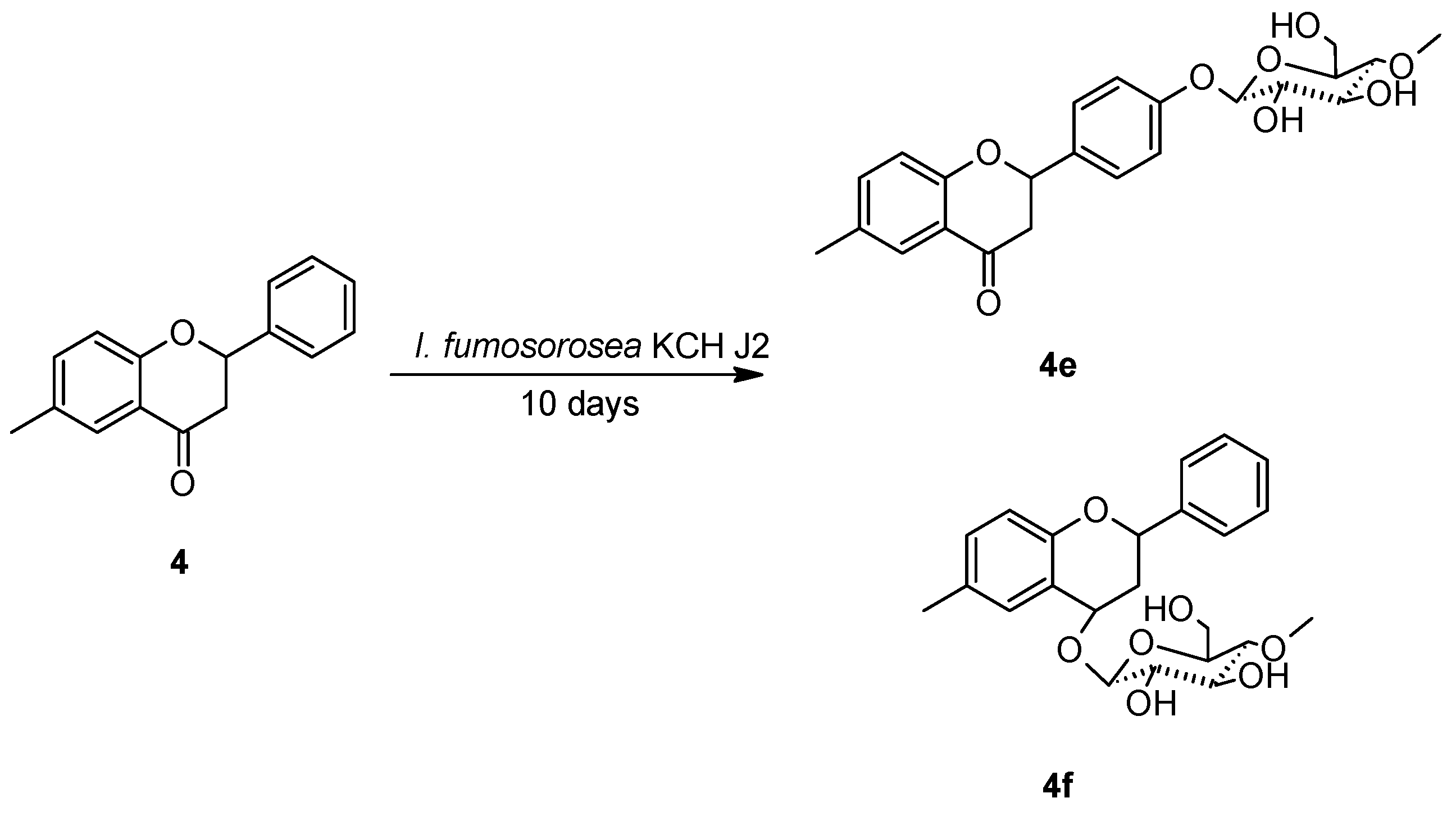
2.3. Biotransformations of 6-methylflavanone (4) in the culture of I. fumosorosea KCH J2

6-Methylflavanone (4) was also utilized in a 10-day biotransformation in the culture of I. fumosorosea KCH J2 and gave two biotransformation products: 6-methylflavanone 4′-O-β-D-(4″-O-methyl)-glucopyranoside (4e) with 22.1% yield and 2-phenyl-6-methylchromane 4-O-β-D-(4″-O-methyl)-glucopyranoside (4f) with 23.3% yield (Scheme 10).
The products 4e and 4f were analyzed by 1H-NMR and 13C-NMR spectroscopy which allowed for the establishment of their chemical structures (Table 3 and Table 4, Scheme 11 and Scheme 12).
The presence of a glucose unit in compounds 4e and 4f was confirmed by five characteristic carbon signals observed in the region from about δ = 80.0 ppm, to about δ = 62.0 ppm, in the 13C-NMR spectra, as well as proton signals of δH ranging from about δ = 3.9 ppm, to δ = 3.1 ppm in the 1H-NMR spectra. Additionally, the attachment of a sugar unit to substrate 4 was confirmed by a one-proton doublet at the anomeric carbon atom at δ = 4.49 ppm in the 1H-NMR spectrum of 4e, δ = 4.97 ppm in the 1H-NMR spectrum of 4f. The β-configuration of the glucose unit was proved for 4e and 4f by the coupling constant (J = 7.8 Hz) for the anomeric proton. A three-proton singlet at about δ = 3.6 ppm in the 1H-NMR spectra and the corresponding signal at δ = 60.5 ppm in the 13C-NMR spectra of 4e and 4f evidences that one of the hydroxyl groups of sugar unit has been methylated. O-methylation occurred at the C-4″ hydroxyl group of the glucose unit. It was detected in the HMBC spectrum, where the proton signal due to -OCH3 was correlated with the signal of C-4″ (about δ = 80 ppm) in the glucose unit.
Based on the HMBC spectrum, a sugar moiety was attached to C-4′ in the case of compound 4e, due to the proton at the anomeric carbon atom (δ = 4.78 ppm) being coupled with the C-4′ signal (δ = 158.8 ppm), which was shifted from δ = 129.3 ppm. In the 1H-NMR spectrum, multiplets from protons at C-2′ and C-6′ (δ = 7.49 ppm) and from protons at C-3′ and C-5′ (δ = 7.12 ppm) can be observed, which is characteristic of an AA’BB’ coupling system, signalizing the presence of flavanone ring B substitution at C-4′. Chemical shifts of the other signals in the 1H-NMR and 13C-NMR spectra have only slightly changed, which indicates that the flavanone skeleton remained intact. The signal from the three protons of the methyl group at C-6 remained at the same position as in the 1H-NMR spectrum of substrate 4, which means that the methyl group in product 4e was present.
Based on the HMBC spectrum, in the case of product 4f, glycosylation occurred at C-4, due to the proton at the anomeric carbon atom (δ = 4.49 ppm) being coupled with the C-4 signal (δ = 70.2 ppm), which was shifted from δ = 191.9 ppm, indicating reduction of the carbonyl group at C-4. Furthermore, in the 1H-NMR spectrum, a triplet at δ = 4.92 ppm appeared from one-proton at C-4. Moreover, only slightly shifted signals from all ten protons from substrate 4 were visible, confirming the inability of another position of substitution. A doublet of doublets at δ = 5.36 ppm from one proton at C-2 became shifted from δ = 5.59 ppm, the same as the signals from the pseudo-axial and pseudo-equatorial protons at C-3, respectively (δ =2.43 ppm shifted from δ = 3.11 ppm and δ = 2.07 ppm shifted from δ = 2.85 ppm). In the COSY spectrum there was a correlation between proton 3ax and 3eq with a new signal from the proton at C-4. In the HMBC spectrum correlations between the proton at C-5 and the carbon at C-4, and between the pseudo-axial proton at C-3 and the carbon at C-4, and between the proton at C-1″ and the carbon at C-4 were also present, which confirms that reduction and glycosylation occurred at C-4. The signal from the three protons of the methyl group at C-6 remained at the same position as in the 1H-NMR spectrum of substrate 4 which indicates the presence of the methyl group in product 4f.
The first product of the biotransformations presented above—6-methylflavanone 4′-O-β-D-(4″-O-methyl)-glucopyranoside (4e)—is analogous to the 6-methoxyflavanone 4′-O-β-D-(4”-O-methyl)-glucopyranoside that was obtained in the microbial transformations of 6-methoxyflavanone by the same strain, I. fumosorosea KCH J2 [15]. Glycosylation at C-4′ also occurred in the biotransformation of naringenin in the culture of I. fumosorosea ACCC 37814, performed by Dou et al. [16]. More unusual, is the second biotransformation product—2-phenyl-6-methylchromane 4-O-β-D-(4″-O-methyl)-glucopyranoside (4f). We assume that attachment of the glycosyl moiety at C-4 was preceded by the carbonyl group reduction, which was previously observed as a result of 2′-methoxyflavanone and 3′-methoxyflavanone biotransformations [15]. However, subsequent glycosylation at the same position has never been observed before. In each biotransformation product a methyl moiety at C-6 was retained, indicating that I. fumosorosea KCH J2 was not able to remove this moiety, unlike B. bassiana KCH J1.5.

3. Materials and Methods

3.1. Substrates

The substrates for biotransformations were obtained by two-step synthesis (Scheme 1). The first step was Claisen–Schmidt condensation between 2′-hydroxy-5′-methylacetophenone (1) and benzaldehyde (2) (purchased from Sigma-Aldrich (St. Louis, MO, USA)) dissolved in methanol, under alkaline conditions at high temperature, according to the method described previously [50,51,52,53]. 2′-Hydroxy-5′-methylchalcone (3) was obtained with a 50.1% yield and was used as the biotransformation substrate, and also as substrate in the second step of the synthesis. 6-Methylflavanone (4) was obtained by cyclization of 2′-hydroxy-5′-methylchalcone (3) in the presence of sodium acetate [54,55] with 62.3% yield.
The physical data, including the retention time tR (min), the optical rotation [ α ]   D 20 , and concentration c (M), as well as NMR spectral data of the resulting compounds 3 and 4 are presented below, and in Table 1, Table 2, Table 3 and Table 4, and in the Supplementary Materials.

3.1.1. 2′-Hydroxy-5′-methylchalcone (3)

C16H14O2, Mw = 238.28 [M + H]+ 239.1054; 1H-NMR, see Table 1, 13C-NMR, see Table 2.

3.1.2. 6-Methylflavanone (4)

C16H14O2, Mw = 238.28, [M + H]+ 239.1086;   [ α ]   D 20 = 0 (c = 0.65, acetone); 1H-NMR, see Table 3, 13C-NMR, see Table 4.

3.2. Microorganisms

The studies were carried out using two strains of entomopathogenic filamentous fungi I. fumosorosea KCH J2 and B. bassiana KCH J1.5, that were collected from the Department of Chemistry of Wrocław University of Environmental and Life Sciences, Poland. The description of material collection, propagation of structures of the fungi, and genetic identification have already been described in our previous papers [12,37]. The microorganisms were maintained on potato slants at 4°C and subcultured before use in the experiments.

3.3. Analysis

The course of the biotransformation was assessed by chromatographic methods (TLC, HPLC). TLC analysis was carried out using TLC Silica gel 60/Kieselguhr F254 (0.2 mm thick) plates (Merck, Darmstadt, Germany). The developing system was a mixture of chloroform and methanol (9:1 v/v). The products were observed (without additional visualization) under the ultraviolet lamp at the two wavelengths 254 nm and 365 nm.
HPLC analyses were performed on a Dionex Ultimate 3000 instrument (Thermo Fisher Scientfic, Waltham, MA, USA) with a diode array detector using an analytical octadecyl silica (ODS) 2 column (4.6 × 250 mm, Waters, Milford, MA, USA) and pre-column. The gradient program was as follows: initial conditions—32.5% B in A, 4 min—40% B in A, 8 min—40% B in A, 10 min—45% B in A, 15 min—95% B in A, 18 min—95% B in A, 19 min—32.5% B in A, 23 min—32.5% B in A. The flow rate was 1 mL/min, the injection volume was 5 µL, and detection wavelength 280 nm.
Separation of the products obtained by the scale-up biotransformation was attained using 500 and 1000 µm preparative TLC silica gel plates (Analtech, Gehrden, Germany). After elution of the compounds from the adsorbent on TLC plates with chloroform and methanol (9:1 v/v) as eluents, compounds were extracted from the selected gel fractions using 20 mL ethyl acetate 3 times. The extracts from a single fraction were combined and ethyl acetate was evaporated under reduced pressure.
NMR analyses (1H-NMR, 13C-NMR, COSY, Heteronuclear Multiple Quantum Correlation (HSQC), HMBC) were performed using a DRX AvanceTM 600 MHz NMR spectrometer (Bruker, Billerica, MA, USA). The prepared samples were dissolved in deuterated acetone, except 3 dissolved in deuterated chloroform.
Optical rotation was measured using digital polarimeter P-2000-Na (ABL & E-JASCO, Kraków, Poland).
Molecular formulas of products were confirmed by UPLC-qTOF-MS/MS analysis. Identification of compounds was performed as described previously, with slight modifications [56], on the Acquity ultra-performance liquid chromatography (UPLC) system, coupled with a quadrupole-time of flight (q-TOF) MS instrument (UPLC/Synapt q-TOF MS, Waters Corp., Milford, MA, USA), with an electrospray ionization (ESI) source. The separation was achieved on the Cadenza CD-C18 UP (75 mm × 2.0 mm i.d., 3 µm) column (Imtakt, Kyoto, Japan). The mobile phase was a mixture of 0.1% aqueous formic acid v/v (A) and acetonitrile (B). The gradient program was as follows: initial conditions—1% B in A, 12 min—25% B in A, 19 min—100% B, 20 min—1% B in A. The flow rate was 0.45 mL/min, and the injection volume was 5 µL. The column was operated at 30°C. The major operating parameters for the q-TOF MS were set as follows: capillary voltage 2.0 kV, cone voltage 40 V, cone gas flow of 11 L/h, collision energy 28–30 eV, source temperature 100 °C, desolvation temperature 250 °C, collision gas, argon; desolvation gas (nitrogen) flow rate, 600 L/h; data acquisition range, m/z 100–2000 Da; ionization mode, negative and positive. The data were collected with Mass-Lynx V 4.1 software. The runs were monitored at a wavelength of 254 nm.

3.4. Screening Procedure

Experiments were carried out using Sabouraud medium (10 g aminobac, 30 g glucose, 1 L distilled water). The microorganism was transferred to a 300 mL Erlenmeyer flask with 100 mL medium. Pre-incubation was carried out on a rotary shaker (140 rpm) at 25 °C for 72 h. The screening was also performed in 300 mL Erlenmeyer flasks with 100 mL of Sabouraud liquid medium. The pre-grown culture (0.5 mL) was transferred to an Erlenmeyer flask, and after 72 h incubation, 10 mg of substrate 3 or 4, dissolved in 0.5 mL of dimethyl sulfoxide, was added. The molar concentrations of substrates 3 and 4 were 0.42 mM. The biotransformation was run under the same conditions as pre-incubation. After 3, 6 and 9 days of substrate incubation samples were collected and extracted once with 30 mL of ethyl acetate. The extracts were dried with anhydrous magnesium sulfate for 5 min, concentrated in vacuo and analyzed by TLC and HPLC methods. Stability of the substrate was evaluated under identical conditions, without using a biocatalyst. Control cultivation with no substrate has also been performed.

3.5. The Semi-Preparative Biotransformations

The semi-preparative biotransformations were performed in 2 L flasks with 500 mL of the Sabouraud medium (10 g aminobac, 30 g glucose, 1 L distilled water) each. The pre-incubation culture (1 mL) was transferred to the flask and incubated for 72 h same as during the screening procedure. Afterwards, 50 mg of substrate 3 or 4, dissolved in 2.5 mL of dimethyl sulfoxide, was added and the cultures were shaken on a rotary shaker for 10 days. The molar concentrations of substrates 3 and 4 were 0.42 mm. After the confirmation of complete substrate conversion (or lack of further substrate conversion) metabolites were extracted 2 times using each time 300 mL of ethyl acetate. The combined extracts were dried with anhydrous magnesium sulfate for 5 min and then concentrated using a rotary evaporator. Biotransformation products were separated using preparative TLC plates and analyzed by NMR and UPLC-qTOF-MS/MS.
The physical data, including the retention time tR (min), the optical rotation [ α ]   D 20 , and concentration c (M), as well as NMR spectral data of the resulting compounds 3a, 4a4f are presented below, and in Table 1, Table 2, Table 3 and Table 4, and in the Supplementary Materials.

3.5.1. 2′-Hydroxy-5′-methylchalcone 3-O-β-D-(4″-O-methyl)-glucopyranoside (3a)

C23H26O8, Mw = 430.45, [M − H]⁻ 429.1574; 1H-NMR, see Table 1, 13C-NMR, see Table 2.

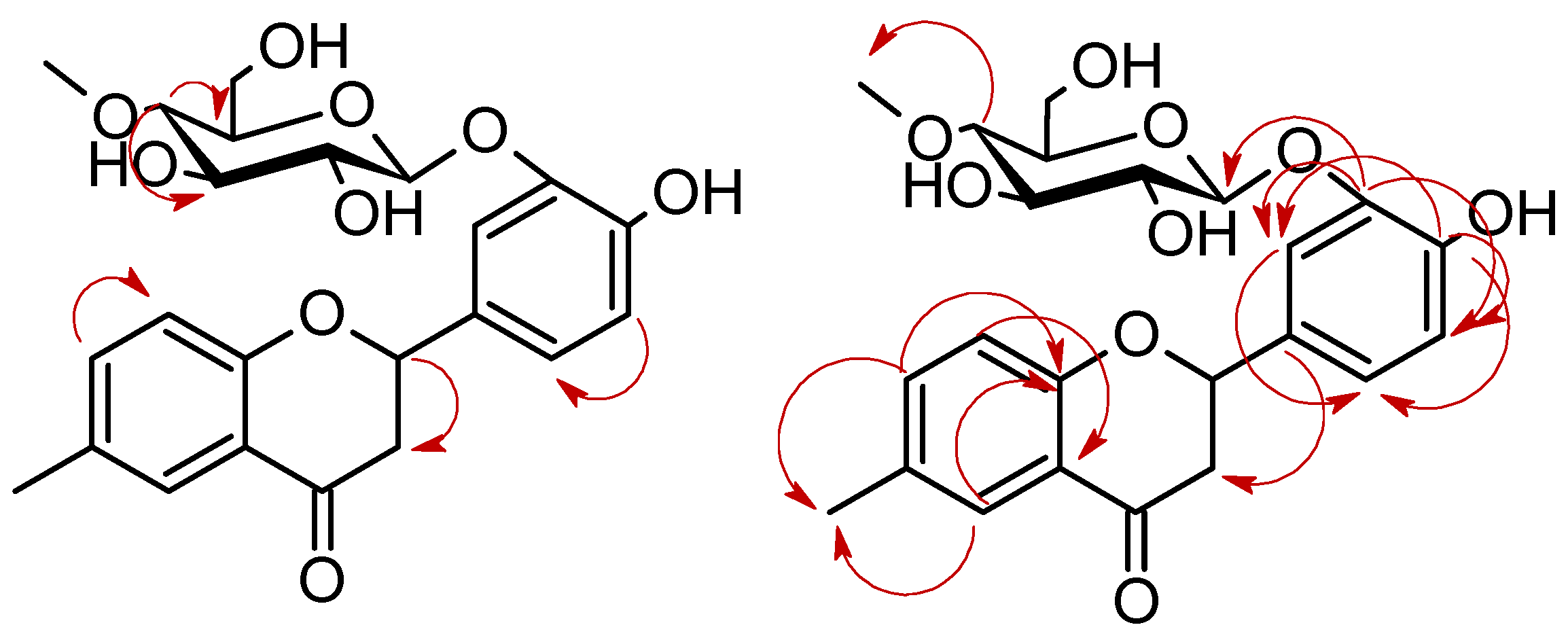
3.5.2. 4′-Hydroxy-6-methylflavanone 3′-O-β-D-(4″-O-methyl)-glucopyranoside (4a)

C23H26O9, Mw = 446.45, [M − H]⁻ 445.1463;   [ α ]   D 20 = −10.5 (c = 0.325, acetone); 1H-NMR, see Table 3, 13C-NMR, see Table 4.

3.5.3. 4′-Hydroxyflavanone 6-methylene-O-β-D-(4″-O-methyl)-glucopyranoside (4b)

C22H24O9, Mw = 446.45, [M − H]⁻ 445.1549;   [ α ]   D 20 = −6.1 (c = 0.365, acetone); 1H-NMR, see Table 3, 13C-NMR, see Table 4.

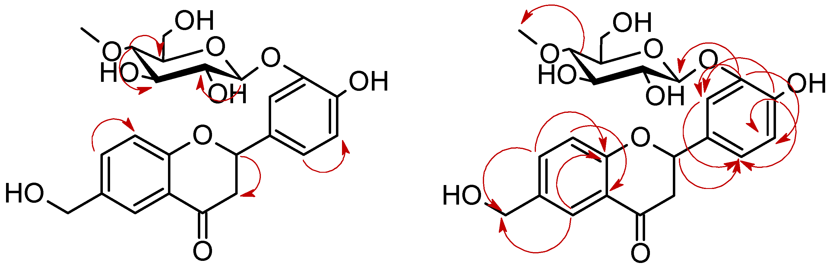
3.5.4. 6-Hydroxymethylflavanone 3′-O-β-D-(4″-O-methyl)-glucopyranoside (4c)

C22H24O9, Mw = 446.45, [M − H]⁻ 445.1506;   [ α ]   D 20 = 9.1 (c = 0.175, acetone); 1H-NMR, see Table 3, 13C-NMR, see Table 4.

3.5.5. 4′-Hydroxy-6-hydroxymethylflavanone 3′-O-β-D-(4″-O-methyl)-glucopyranoside (4d)

C22H24O10, Mw = 462.45, [M − H]⁻ 461.1418;   [ α ]   D 20 = 54.3 (c = 0.390, acetone); 1H-NMR, see Table 3, 13C-NMR, see Table 4.

3.5.6. 6-Methylflavanone 4′-O-β-D-(4″-O-methyl)-glucopyranoside (4e)

C23H26O8, Mw = 430.45, [M − H]⁻ 429.1532;   [ α ]   D 20 = −50.3 (c = 0.950, acetone); 1H-NMR, see Table 3, 13C-NMR, see Table 4.

3.5.7. 2-Phenyl-6-methylchromane 4-O-β-D-(4″-O-methyl)-glucopyranoside (4f)

C23H28O7, Mw = 416.46, [M − H]⁻ 415.1776 (adduct [M + 46− H]⁻ [M + HCOO]⁻ 461.1814 [57]);   [ α ]   D 20 = −25.3 (c = 0.390, acetone); 1H-NMR, see Table 3, 13C-NMR, see Table 4.

4. Conclusions

In this paper we present the results of the glycosylation of a flavanone and chalcone with methyl moiety in entomopathogenic filamentous fungi cultures. The strain B. bassiana KCH J1.5 was able to hydroxylate methyl moiety at C-6 of 6-methylflavanone and also to attach to the glycosyl moiety at this site. These microbial transformations have not yet been described in the scientific literature. Moreover, B. bassiana KCH J1.5 glycosylated 2′-hydroxyl-5′-methylchalcone at C-3 and 6-methylflavanone at C-3′. The B. bassiana KCH J1.5 enzymatic system also catalyzed hydroxylation of the flavanone skeleton at C-4′. The second utilized strain—I. fumosorosea KCH J2—glycosylated 2′-hydroxyl-5′-methylchalcone at C-3 and 6-methylflavanone at C-4′ and C-4. It can be assumed that attachment of the glycosyl moiety at C-4 was preceded by the carbonyl group reduction. This strain was not able to hydroxylate the methyl moiety. All biotransformation products have not been previously described in the scientific literature and may be used in studies assessing their biological activity and bioavailability.

Supplementary Materials

The Supplementary Materials are available online: https://www.mdpi.com/2073-4344/10/10/1148/s1.

Author Contributions

Conceptualization, A.K.-Ł. and E.K.-S.; methodology, A.K.-Ł., E.K.-S. and M.D.; validation, A.K.-Ł.; formal analysis, A.K.-Ł., E.K.-S., M.D. and T.J.; investigation, A.K.-Ł.; resources, E.K.-S.; data curation, A.K.-Ł. and E.K.-S.; writing—original draft preparation, A.K.-Ł.; writing—review and editing, A.K.-Ł., E.K.-S. and M.D.; visualization, A.K.-Ł.; supervision, E.K.-S.; project administration, A.K.-Ł. and E.K.-S.; funding acquisition, A.K.-Ł. and E.K.-S. All authors have read and agreed to the published version of the manuscript.

Funding

This research received no external funding.

Acknowledgments

The research is financed/co-financed under the Leading Research Groups support project from the subsidy increased for the period 2020–2025 in the amount of 2% of the subsidy referred to Art. 387 (3) of the Law of 20 July 2018 on Higher Education and Science, obtained in 2019.

Conflicts of Interest

The authors declare no conflict of interest.

References

  1. Koirala, N.; Thuan, N.H.; Ghimire, G.P.; Thang, D.V.; Sohng, J.K. Methylation of flavonoids: Chemical structures, bioactivities, progress and perspectives for biotechnological production. Enzym. Microb. Technol. 2016, 86, 103–116. [Google Scholar] [CrossRef] [PubMed]
  2. Raffa, D.; Maggio, B.; Raimondi, M.V.; Plescia, F.; Daidone, G. Recent discoveries of anticancer flavonoids. Eur. J. Med. Chem. 2017, 142, 213–228. [Google Scholar] [CrossRef] [PubMed]
  3. Wang, T.Y.; Li, Q.; Bi, K. Bioactive flavonoids in medicinal plants: Structure, activity and biological fate. Asian J. Pharm. Sci. 2018, 13, 12–23. [Google Scholar] [CrossRef] [PubMed]
  4. Vogiatzoglou, A.; Mulligan, A.A.; Lentjes, M.A.H.; Luben, R.N.; Spencer, J.P.E.; Schroeter, H.; Khaw, K.T.; Kuhnle, G.G.C. Flavonoid intake in European adults (18 to 64 years). PLoS ONE 2015, 10, e0128132. [Google Scholar] [CrossRef] [PubMed]
  5. Kostrzewa-Susłow, E.; Dymarska, M.; Białońska, A.; Janeczko, T. Enantioselective conversion of certain derivatives of 6-hydroxyflavanone. J. Mol. Catal. B Enzym. 2014, 102, 59–65. [Google Scholar] [CrossRef]
  6. Gullón, B.; Lú-Chau, T.A.; Moreira, M.T.; Lema, J.M.; Eibes, G. Rutin: A review on extraction, identification and purification methods, biological activities and approaches to enhance its bioavailability. Trends Food Sci. Technol. 2017, 67, 220–235. [Google Scholar] [CrossRef]
  7. Wen, L.; Jiang, Y.; Yang, J.; Zhao, Y.; Tian, M.; Yang, B. Structure, bioactivity, and synthesis of methylated flavonoids. Ann. N. Y. Acad. Sci. 2017, 1398, 120–129. [Google Scholar] [CrossRef]
  8. Wang, X. Structure, mechanism and engineering of plant natural product glycosyltransferases. FEBS Lett. 2009, 583, 3303–3309. [Google Scholar] [CrossRef] [Green Version]
  9. Plaza, M.; Pozzo, T.; Liu, J.; Ara, K.Z.G.; Turner, C.; Nordberg Karlsson, E. Substituent effects on in vitro antioxidizing properties, stability, and solubility in flavonoids. J. Agric. Food Chem. 2014, 62, 3321–3333. [Google Scholar] [CrossRef]
  10. Thilakarathna, S.H.; Rupasinghe, V.H.P. Flavonoid bioavailability and attempts for bioavailability enhancement. Nutrients 2013, 5, 3367–3387. [Google Scholar] [CrossRef]
  11. Xiao, J.; Muzashvili, T.S.; Georgiev, M.I. Advances in the biotechnological glycosylation of valuable flavonoids. Biotechnol. Adv. 2014, 32, 1145–1156. [Google Scholar] [CrossRef] [PubMed]
  12. Dymarska, M.; Grzeszczuk, J.; Urbaniak, M.; Janeczko, T.; Pląskowska, E.; Stępień, Ł.; Kostrzewa-Susłow, E. Glycosylation of 6-methylflavone by the strain Isaria fumosorosea KCH J2. PLoS ONE 2017, 12, e0184885. [Google Scholar] [CrossRef] [PubMed]
  13. Dymarska, M.; Janeczko, T.; Kostrzewa-Susłow, E. Biotransformations of flavones and an isoflavone (daidzein) in cultures of entomopathogenic filamentous fungi. Molecules 2018, 23, 1356. [Google Scholar] [CrossRef] [PubMed] [Green Version]
  14. Dymarska, M.; Janeczko, T.; Kostrzewa-Susłow, E. Glycosylation of 3-hydroxyflavone, 3-methoxyflavone, quercetin and baicalein in fungal cultures of the genus Isaria. Molecules 2018, 23, 2477. [Google Scholar] [CrossRef] [Green Version]
  15. Dymarska, M.; Janeczko, T.; Kostrzewa-Susłow, E. Glycosylation of methoxylated flavonoids in the cultures of Isaria fumosorosea KCH J2. Molecules 2018, 23, 2578. [Google Scholar] [CrossRef] [Green Version]
  16. Dou, F.; Wang, Z.; Li, G.; Dun, B. Microbial transformation of flavonoids by Isaria fumosorosea ACCC 37814. Molecules 2019, 24, 1028. [Google Scholar] [CrossRef] [Green Version]
  17. Hyung Ko, J.; Gyu Kim, B.; Joong-Hoon, A. Glycosylation of flavonoids with a glycosyltransferase from Bacillus cereus. FEMS Microbiol. Lett. 2006, 258, 263–268. [Google Scholar] [CrossRef] [Green Version]
  18. Gurung, R.B.; Kim, E.H.; Oh, T.J.; Sohng, J.K. Enzymatic synthesis of apigenin glucosides by glucosyltransferase (YjiC) from Bacillus licheniformis DSM 13. Mol. Cells 2013, 36, 355–361. [Google Scholar] [CrossRef]
  19. Xie, L.; Zhang, L.; Wang, C.; Wang, X.; Xu, Y.; Yu, H.; Wu, P.; Li, S.; Han, L.; Gunatilaka, A.A.L.; et al. Methylglucosylation of aromatic amino and phenolic moieties of drug-like biosynthons by combinatorial biosynthesis. Proc. Natl. Acad. Sci. USA 2018, 115, E4980–E4989. [Google Scholar] [CrossRef] [Green Version]
  20. Xie, L.; Zhang, L.; Bai, J.; Yue, Q.; Zhang, M.; Li, J.; Wang, C.; Xu, Y. Methylglucosylation of phenolic compounds by fungal glycosyltransferase-methyltransferase functional modules. J. Agric. Food Chem. 2019, 67, 8573–8580. [Google Scholar] [CrossRef]
  21. Kostrzewa-Susłow, E.; Dymarska, M.; Janeczko, T. Microbial transformations of 3-methoxyflavone by strains of Aspergillus niger. Pol. J. Microbiol. 2014, 63, 111–114. [Google Scholar] [CrossRef] [PubMed]
  22. Wen, X.; Walle, T. Methylated flavonoids have greatly improved intestinal absorption and metabolic stability. Drug Metab. Dispos. 2006, 34, 1786–1792. [Google Scholar] [CrossRef] [PubMed] [Green Version]
  23. Ye, C.L.; Lu, Y.H.; Wei, D.Z. Flavonoids from Cleistocalyx operculatus. Phytochemistry 2004, 65, 445–447. [Google Scholar] [CrossRef]
  24. Ye, C.L.; Liu, Y.; Wei, D.Z. Antioxidant and anticancer activity of 3′-formyl-4′, 6′-dihydroxy-2′-methoxy-5′-methylchalcone and (2S)-8-formyl-5-hydroxy-7-methoxy-6-methylflavanone. J. Pharm. Pharm. 2007, 59, 553–559. [Google Scholar] [CrossRef] [PubMed]
  25. Dao, T.T.; Tung, B.T.; Nguyen, P.H.; Thuong, P.T.; Yoo, S.S.; Kim, E.H.; Kim, S.K.; Oh, W.K. C-methylated flavonoids from Cleistocalyx operculatus and their inhibitory effects on novel influenza A (H1N1) neuraminidase. J. Nat. Prod. 2010, 73, 1636–1642. [Google Scholar] [CrossRef]
  26. Chen, W.Q.; Song, Z.J.; Xu, H.H. A new antifungal and cytotoxic C-methylated flavone glycoside from Picea neoveitchii. Bioorg. Med. Chem. Lett. 2012, 22, 5819–5822. [Google Scholar] [CrossRef]
  27. Nobakht, M.; Grkovic, T.; Trueman, S.J.; Wallace, H.M.; Katouli, M.; Quinn, R.J.; Brooks, P.R. Chemical constituents of kino extract from Corymbia torelliana. Molecules 2014, 19, 17862–17871. [Google Scholar] [CrossRef] [Green Version]
  28. Nobakht, M.; Trueman, S.J.; Wallace, H.M.; Brooks, P.R.; Streeter, K.J.; Katouli, M. Antibacterial properties of flavonoids from kino of the eucalypt tree, Corymbia torelliana. Plants 2017, 6, 39. [Google Scholar] [CrossRef] [Green Version]
  29. Salmazzo, G.R.; Verdan, M.H.; Silva, F.; Cicarelli, R.M.; da Silva Mota, J.; Salvador, M.J.; de Carvalho, J.E.; Cardoso, C.A.L. Chemical composition and antiproliferative, antioxidant and trypanocidal activities of the fruits from Campomanesia xanthocarpa (Mart.) O. Berg (Myrtaceae). Nat. Prod. Res. 2019, 1–5. [Google Scholar] [CrossRef]
  30. Hall, B.J.; Chebib, M.; Hanrahan, J.R.; Johnston, G.A.R. 6-Methylflavanone, a more efficacious positive allosteric modulator of γ-aminobutyric acid (GABA) action at human recombinant α2β2γ2L than at α1β2γ2L and α1β2 GABAA receptors expressed in Xenopus oocytes. Eur. J. Pharm. 2006, 512, 97–104. [Google Scholar] [CrossRef]
  31. Boomsma, J.J.; Jensen, A.B.; Meyling, N.V.; Eilenberg, J. Evolutionary interaction networks of insect pathogenic fungi. Annu. Rev. Entomol. 2014, 59, 467–485. [Google Scholar] [CrossRef] [PubMed]
  32. Valero-Jiménez, C.A.; Wiegers, H.; Zwaan, B.J.; Koenraadt, C.J.M.; van Kan, J.A.L. Genes involved in virulence of the entomopathogenic fungus Beauveria bassiana. J. Invertebr. Pathol. 2016, 133, 41–49. [Google Scholar] [CrossRef] [PubMed]
  33. Mascarin, G.M.; Jaronski, S.T. The production and uses of Beauveria bassiana as a microbial insecticide. World J. Microbiol. Biotechnol. 2016, 32, 177. [Google Scholar] [CrossRef]
  34. Cito, A.; Barzanti, G.P.; Strangi, A.; Francardi, V.; Zanfini, A.; Dreassi, E. Cuticle-degrading proteases and toxins as virulence markers of Beauveria bassiana (Balsamo) Vuillemin. J. Basic Microbiol. 2016, 56, 941–948. [Google Scholar] [CrossRef] [PubMed]
  35. Xiao, G.; Ying, S.H.; Zheng, P.; Wang, Z.L.; Zhang, S.; Xie, X.Q.; Shang, Y.; St. Leger, R.J.; Zhao, G.P.; Wang, C.; et al. Genomic perspectives on the evolution of fungal entomopathogenicity in Beauveria bassiana. Sci. Rep. 2012, 2, 483. [Google Scholar] [CrossRef] [Green Version]
  36. Chen, J.; Lai, Y.; Wang, L.; Zhai, S.; Zou, G.; Zhou, Z.; Cui, C.; Wang, S. CRISPR/Cas9-mediated efficient genome editing via blastospore-based transformation in entomopathogenic fungus Beauveria bassiana. Sci. Rep. 2017, 7, 45763. [Google Scholar] [CrossRef] [PubMed]
  37. Kozłowska, E.; Urbaniak, M.; Hoc, N.; Grzeszczuk, J.; Dymarska, M.; Stępień, Ł.; Pląskowska, E.; Kostrzewa-Susłow, E.; Janeczko, T. Cascade biotransformation of dehydroepiandrosterone (DHEA) by Beauveria species. Sci. Rep. 2018, 8, 13449. [Google Scholar] [CrossRef] [PubMed]
  38. Zimmermann, G. The entomopathogenic fungi Isaria farinosa (formerly Paecilomyces farinosus) and the Isaria fumosorosea species complex (formerly Paecilomyces fumosoroseus): Biology, ecology and use in biological control. Biocontrol Sci. Technol. 2008, 18, 865–901. [Google Scholar] [CrossRef]
  39. Weng, Q.; Zhang, X.; Chen, W.; Hu, Q. Secondary metabolites and the risks of Isaria fumosorosea and Isaria farinosa. Molecules 2019, 24, 664. [Google Scholar] [CrossRef] [Green Version]
  40. Kim, H.J.; Lee, I.S. Microbial metabolism of the prenylated chalcone xanthohumol. J. Nat. Prod. 2006, 69, 1522–1524. [Google Scholar] [CrossRef]
  41. Tronina, T.; Bartmańska, A.; Milczarek, M.; Wietrzyk, J.; Popłoński, J.; Rój, E.; Huszcza, E. Antioxidant and antiproliferative activity of glycosides obtained by biotransformation of xanthohumol. Bioorg. Med. Chem. Lett. 2013, 23, 1957–1960. [Google Scholar] [CrossRef] [PubMed]
  42. Huszcza, E.; Bartmańska, A.; Tronina, T. Glycosylation of xanthohumol by fungi. Z. Nat. C J. Biosci. 2008, 63, 557–560. [Google Scholar] [CrossRef] [PubMed] [Green Version]
  43. Hofer, B. Recent developments in the enzymatic O-glycosylation of flavonoids. Appl. Microbiol. Biotechnol. 2016, 100, 4269–4281. [Google Scholar] [CrossRef] [PubMed]
  44. Júnior, G.M.V.; Sousa, C.M.D.M.; Cavalheiro, A.J.; Lago, J.H.G.; Chaves, M.H. Phenolic derivatives from fruits of Dipteryx lacunifera Ducke and evaluation of their antiradical activities. Helv. Chim. Acta 2008, 91, 2159–2167. [Google Scholar] [CrossRef]
  45. Moon, B.H.; Lee, Y.; Ahn, J.H.; Lim, Y. Complete assignment of 1H and 13C NMR data of dihydroxyflavone derivatives. Magn. Reson. Chem. 2006, 44, 99–101. [Google Scholar] [CrossRef]
  46. Park, Y.; Moon, B.H.; Lee, Y.; Yoon, Y.; Ahn, J.H.; Lim, Y. 1H and 13C-NMR data of hydroxyflavone derivatives. Magn. Reson. Chem. 2007, 45, 488–495. [Google Scholar] [CrossRef]
  47. Cuca-Suárez, L.; Monache, F. 6-C-formyl and 6-C-hydroxymethyl flavonoides from Petiveria Alliacea. Phytochemistry 1992, 31, 2481–2482. [Google Scholar] [CrossRef]
  48. Sordon, S.; Popłoński, J.; Huszcza, E. Microbial glycosylation of flavonoids. Pol. J. Microbiol. 2016, 65, 137–151. [Google Scholar] [CrossRef] [Green Version]
  49. Sordon, S.; Popłoński, J.; Tronina, T.; Huszcza, E. Regioselective O-glycosylation of flavonoids by fungi Beauveria bassiana, Absidia coerulea and Absidia glauca. Bioorg. Chem. 2019, 93, 102750. [Google Scholar] [CrossRef]
  50. Silva, A.M.S.; Tavares, H.R.; Barros, A.I.N.R.A.; Cavaleiro, J.A.S. NMR and structural and conformational features of 2′-hydroxychalcones and flavones. Spectrosc. Lett. 1997, 30, 1655–1667. [Google Scholar] [CrossRef]
  51. Yadav, N.; Dixit, S.K.; Bhattacharya, A.; Mishra, L.C.; Sharma, M.; Awasthi, S.K.; Bhasin, V.K. Potent antimalarial activity of newly synthesized substituted chalcone analogs in vitro. Chem. Biol. Drug Des. 2012, 80, 340–347. [Google Scholar] [CrossRef] [PubMed]
  52. Janeczko, T.; Gładkowski, W.; Kostrzewa-Susłow, E. Microbial transformations of chalcones to produce food sweetener derivatives. J. Mol. Catal. B Enzym. 2013, 98, 55–61. [Google Scholar] [CrossRef]
  53. Palko-Łabuz, A.; Kostrzewa-Susłow, E.; Janeczko, T.; Środa-Pomianek, K.; Poła, A.; Uryga, A.; Michalak, K. Cyclization of flavokawain B reduces its activity against human colon cancer cells. Hum. Exp. Toxicol. 2020, 39, 262–275. [Google Scholar] [CrossRef] [PubMed]
  54. Murti, Y.; Mishra, P. Synthesis and evaluation of flavanones as anticancer agents. Indian J. Pharm. Sci. 2014, 76, 163–166. [Google Scholar]
  55. Gładkowski, W.; Siepka, M.; Janeczko, T.; Kostrzewa-Susłow, E.; Popłoński, J.; Mazur, M.; Żarowska, B.; Łaba, W.; Maciejewska, G.; Wawrzeńczyk, C. Synthesis and antimicrobial activity of methoxy-substituted γ-Oxa-ε -lactones derived from flavanones. Molecules 2019, 24, 4151. [Google Scholar] [CrossRef] [Green Version]
  56. Mizgier, P.; Kucharska, A.Z.; Sokół-Łętowska, A.; Kolniak-Ostek, J.; Kidoń, M.; Fecka, I. Characterization of phenolic compounds and antioxidant and anti-inflammatory properties of red cabbage and purple carrot extracts. J. Funct. Foods 2016, 21, 133–146. [Google Scholar] [CrossRef]
  57. Li, C.; Zhang, X.; Xue, X.; Zhang, F.; Xu, Q.; Liang, X. Structural characterization of iridoid glucosides by ultra-performance liquid chromatography/electrospray ionization quadrupole time-of-flight tandem mass spectrometry. Rapid Commun. Mass Spectrom. 2008, 22, 1941–1954. [Google Scholar] [CrossRef]
Scheme 1. Synthesis of 2′-hydroxy-5′-methylchalcone (3) and 6-methylflavanone (4).
Catalysts 10 01148 sch001
Scheme 2. Microbial transformation of 2′-hydroxy-5′-methylchalcone (3) in B. bassiana KCH J1.5 culture.
Catalysts 10 01148 sch002
Scheme 3. Key Correlation Spectroscopy (COSY), (on the left) and HMBC (on the right) correlations for the structure elucidation of product 3a.
Catalysts 10 01148 sch003
Scheme 4. Microbial transformation of 6-methylflavanone (4) in B. bassiana KCH J1.5 culture.
Catalysts 10 01148 sch004
Scheme 5. Key COSY (on the left) and HMBC (on the right) correlations for the structure elucidation of product 4a.
Catalysts 10 01148 sch005
Scheme 6. Key COSY (on the left) and HMBC (on the right) correlations for the structure elucidation of product 4b.
Catalysts 10 01148 sch006
Scheme 7. Key COSY (on the left) and HMBC (on the right) correlations for the structure elucidation of product 4c.
Catalysts 10 01148 sch007
Scheme 8. Key COSY (on the left) and HMBC (on the right) correlations for the structure elucidation of product 4d.
Catalysts 10 01148 sch008
Scheme 9. The probable course of transformation of 6-methylflavanones.
Catalysts 10 01148 sch009
Scheme 10. Microbial transformation of 6-methylflavanone (4) in I. fumosorosea KCH J2 culture.
Catalysts 10 01148 sch010
Scheme 11. Key COSY (on the left) and HMBC (on the right) correlations for the structure elucidation of product 4e.
Catalysts 10 01148 sch011
Scheme 12. Key COSY (on the left) and HMBC (on the right) correlations for the structure elucidation of product 4f.
Catalysts 10 01148 sch012
Table 1. 1H-NMR chemical shifts δ (ppm) and coupling constants J (Hz) of 2′-hydroxy-5′-methylchalcone (3) in Chloroform-d, and product of its biotransformation 3a in Acetone-d6, 600 MHz (Supplementary Materials).
ProtonCompound
33a
H-27.68 (m)7.61 (m)
H-37.45 (dd)
J = 4.8, 1.8
-
H-47.45 (dd)
J = 4.8, 1.8
7.16 (ddd)
J = 8.1, 2.3, 0.6
H-57.45 (dd)
J = 4.8, 1.8
7.40 (dd)
J = 13.0, 5.0
H-67.68 (m) 7.47 (d)
J = 7.7
H- α7.68 (m) 8.03 (d)
J α, β= 15.5
H- β7.92 (d)
J β, α = 15.5
7.87 (d)
J β, α = 15.5
H-2′--
H-3′6.94 (d)
J = 8.4
6.89 (d)
J = 8.4
H-4′7.32 (dd)
J = 8.4, 2.1
7.40 (dd)
J = 13.0, 5.0
H-5′--
H-6′7.68 (m) 8.12 (d)
J = 1.3
H-1″-5.03 (d)
J = 7.8
H-2″-3.52 (ddd)
J = 15.1, 9.1, 5.1
H-3″-3.66 (m)
H-4″-3.19 (m)
H-5″-3.61 (ddd)
J = 9.7, 5.8, 2.2
H-6″-3,92 (ddd)
J = 11.8, 5.6, 2.1
3.73 (dt)
J = 12.1, 6.2
C4″-OCH3-3.57 (s)
C2′-OH12.63 (s)12.67 (s)
C5′-CH32.36 (s)2.35 (s)
Table 2. 13C-NMR chemical shifts δ (ppm) of 2′-hydroxy-5′-methylchalcone (3) in Chloroform-d, and product of its biotransformation 3a in Acetone-d6, 151 MHz (Supplementary Materials).
CarbonCompound
33a
C-1134.8137.1
C-2128.8116.7
C-3129.2159.2
C-4131.0120.2
C-5129.2130.8
C-6128.8124.3
C- α120.4122.0
C- β145.4145.6
C-1′119.8120.5
C-2′161.7162.5
C-3′118.5118.7
C-4′137.7138.5
C-5′128.1129.3
C-6′129.5131.1
C-1″-101.7
C-2″-75.0
C-3″-78.1
C-4″-80.5
C-5″-77.2
C-6″-62.3
C4″-OCH3-60.6
C = O193.8194.9
C5′-CH320.820.4
Table 3. 1H-NMR chemical shifts δ (ppm) and coupling constants J (Hz) of 6-methylflavanone (4) and products of its biotransformation 4a4f in Acetone-d6, 600 MHz (Supplementary Materials).
ProtonCompound
44a4b4c4d4e4f
H-25.59 (dd)
J2,3ax = 13.0,
J2,3eq = 2.8
5.46 (dd)
J2,3ax = 12.8,
J2,3eq = 2.9
5.52 (dd)
J2,3ax = 13.0,
J2,3eq = 2.8
5.61 (dd)
J2,3ax = 12.5,
J2,3eq = 3.0
5.48 (dd)
J2,3ax = 12.8,
J2,3eq = 2.9
5.53 (dd)
J2,3ax = 12.9,
J2,3eq = 2.9
5.36 (dd)
J = 12.1,
J = 1.9
H-3ax3.11 (dd)
J3ax,2 = 13.0,
J3ax,3eq = 16.7
3.11 (dd)
J3ax,2 = 12.8,
J3ax,3eq = 16.7
3.16 (dt)
J = 8.3,
J3ax,3eq = 16.8
3.13 (dd)
J3ax,2 = 12.5,
J3ax,3eq = 16.8
3.15 (dd)
J3ax,2 = 12.9,
J3ax,3eq = 16.8
3.12 (dd)
J3ax,2 = 12.9,
J3ax,3eq = 16.7
2.43 (dt)
J = 14.1,
J = 2.2
H-3eq2.85 (d)
J3eq,2 =2.9
2.78 (dd)
J3eq,2 = 2.9,
J3eq,3ax = 16.8
2.79 (dd)
J3eq,2 = 2.9,
J3eq,3ax = 16.8
2.90 (d)
J = 3.2
2.80 (dd)
J3eq,2 = 2.9,
J3eq,3ax = 16.7
2.79 (dd)
J3eq,2 = 2.9,
J3eq,3ax = 16.8
2.07 (ddd)
J = 6.0,
J = 3.6,
J = 1.2
H-4------4.92 (t)
J = 2.8
H-57.63 (s)7.61 (d)
J5,7 = 2.3
7.83 (d)
J5,7 =2.2
7.82 (d)
J5,7 =2.1
7.82 (d)
J5,7 = 2.3
7.62 (d)
J5,7 = 1.5
7.22 (d)
J5,7 = 1.9
H-6-------
H-77.40 (dt)
J = 10.9,
J = 4.7
7.39 (m)7.61 (dd)
J7,8 = 8.5,
J7,5 = 2.2
7.58 (dd)
J7,8 = 8.5,
J7,5 = 2.3
7.56 (dd)
J7,8 = 8.5,
J7,5 = 2.1
7.39 (m)7.08 (dd)
J7,8 = 8.3,
J7,5 = 1.9
H-86.98 (d)
J8,7 = 8.4
6.96 (d)
J8,7 = 8.4
7.02 (d)
J8,7 = 8.6
7.07 (dd)
J8,7 = 8.3,
J = 1.7
7.02 (d)
J8,7 = 8.5
6.95 (d)
J8,7 = 8.4
6.80 (d)
J8,7 = 8.3
H-2′7.58 (d)
J2′,3′ = 7.6
7.39 (m)7.41 (m)7.29 (m)7.41 (dd)
J = 4.5,
J = 2.1
7.49 (m)7.50 (d)
J = 7.2
H-3′7.45 (t)
J = 7.5
-6.90 (m)--7.12 (m)7.40 (t)
J = 7.6
H-4′ 7.40 (dt)
J = 10.9,
J = 4.7
--7.07 (dd)
J = 8.3,
J = 1.7
--7.33 (m)
H-5′7.45 (t)
J = 7.5
6.91 (d)
J = 8.2
6.90 (m)7.35 (t)
J = 8.0
6.92 (d)
J = 8.2
7.12 (m)7.40 (t)
J = 7.6
H-6′7.58 (d)
J = 7.6
7.16 (dt)
J = 2.0,
J = 8.2
7.41 (m)7.21 (d)
J = 7.7
7.17 (dd)
J6′,5′ = 8.3,
J = 1.9
7.49 (m)7.50 (d)
J = 7.2
H-1″-4.79 (d)
J = 7.9
4.36 (d)
J = 7.8
4.98 (d)
J = 7.8
4.82 (d)
J = 7.9
4.97 (d)
J = 7.8
4.49 (d)
J = 7.8
H-2″-3.50 (t)
J = 9.4
3.26 (m)3.48 (m)3.50 (t)
J = 8.2
3.48 (ddd)
J = 9.8,
J = 5.0,
J = 2.0
3.30 (m)
H-3″-3.63 (m)3.50 (m)3.62 (ddd)
J = 9.1,
J = 7.6,
J = 4.2
3.63 (d)
J = 6.6
3.63 (t)
J = 9.0
3.52 (m)
H-4″-3.22 (m)3.10 (m)3.21 (m)3.21 (m)3.21 (dd)
J = 9.6,
J = 9.1
3.12 (dd)
J = 9.6,
J = 9.0
H-5″-3.45 (ddd)
J = 9.8,
J = 4.9,
J = 1.9
3.26 (m)3.48 (m)3.46 (m)3.48 (ddd)
J = 9.8,
J = 5.0,
J = 2.0
3.30 (m)
H-6″-3.85 (dt)
J = 8.5,
J = 4.4
3.69 (m)
3.82 (d)
J = 10.0
3.66 (m)
3.84 (dd)
J = 9.3,
J = 5.2
3.68 (m)
3.85 (d)
J = 10.6
3.68 (m)
3.83 (d)
J = 12.2
3.69 (m)
3.89 (m)
3.71 (dt)
J = 11.4,
J = 5.8
-CH2---4.86 (dd)
J = 11.8
J = 2.6
4.61 (dt)
J = 11.7
J =2.7
4.61 (d)
J = 5.2
4.61 (m) --
C6-CH32.32 (s)2.31 (s)---2,31 (s)2.27 (s)
C4″-OCH3-3.55 (s)3.53 (s)3.56 (s)3.55 (s)3.56 (s)3.54 (s)
Table 4. 13C-NMR chemical shifts δ (ppm) of 6-methylflavanone (4) and products of its biotransformation 4a4f in Acetone-d6, 151 MHz (Supplementary Materials).
CarbonCompound
44a4b4c4d4e4f
C-280.380.080.480.080.180.073.8
C-345.144.944.844.944.744.937.5
C-4191.9192.3192.3191.9192.3192.270.2
C-4a121.6121.6121.5121.5121.5121.5120.5
C-5127.0126.9126.7125.2125.2126.9132.7
C-6131.6131.5132.3136.7136.5131.5129.6
C-7137.7137.7136.7135.6135.5137.8131.5
C-8118.8118.8118.8117.1118.8118.7117.5
C-8a160.5160.6162.0161.4161.5160.5154.1
C-1′140.5131.8131.0141.9131.6133.9142.7
C-2′127.3117.9129.0115.4117.7128.7127.1
C-3′129.5146.1116,2158.9146.1117.3129.2
C-4′129.3149.0158.7118.8149.0158.8128.6
C-5′129.5116.8116.2130.5116.8117.3129.2
C-6′127.3123.3129.0120.8123.3128.7127.1
C-1″-104.2103.0101.5104.0101.6101.4
C-2″-75.075.275.074.974.975.3
C-3″-77.578.178.177.577.978.2
C-4″-80.180,580.180.280.180.6
C-5″-77.376.977.077.277.076.9
C-6″-62.062.462.162.062.062.6
C6-CH2-OH--70.863.963.9--
C-4″-OCH3-60.660.560.560.660.560.5
C6-CH320.420.4---20.420.5

