Large Extracellular Vesicle-Derived Latent MMP-8 and Gelatinolytically Active MMP-2 as Potential Circulating Markers for Lymph Node Metastasis in Breast Cancer

 , and
, and 
Simple Summary
Abstract
1. Introduction
2. Materials and Methods
2.1. Clinical Sample Collection
2.2. Isolation of Large Extracellular Vesicles (L-EVs)
2.3. Transmission Electron Microscopy (TEM)
2.4. Dynamic Light Scattering (DLS)
2.5. Dot Blot and Western Blot Analysis
2.6. Proteome Profiler Human Protease Array
2.7. Zymography Assay
2.8. RNA Isolation and Quantitative Real-Time PCR (qRT-PCR) Analysis
2.9. In Silico Exploration Using Available Biological Data Sources
2.10. Statistical Analysis
3. Results
3.1. Clinicopathological Characteristics of Breast Cancer Patients Grouped by LNM
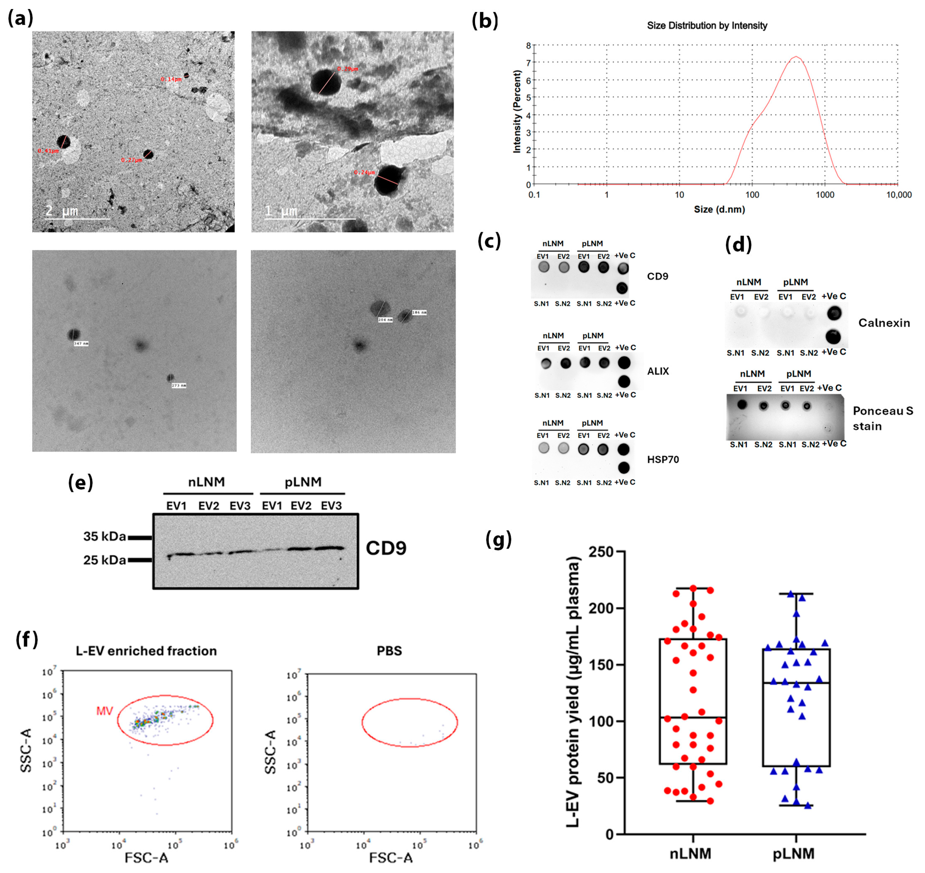
3.2. Characterization of Isolated Plasma-Derived L-EVs
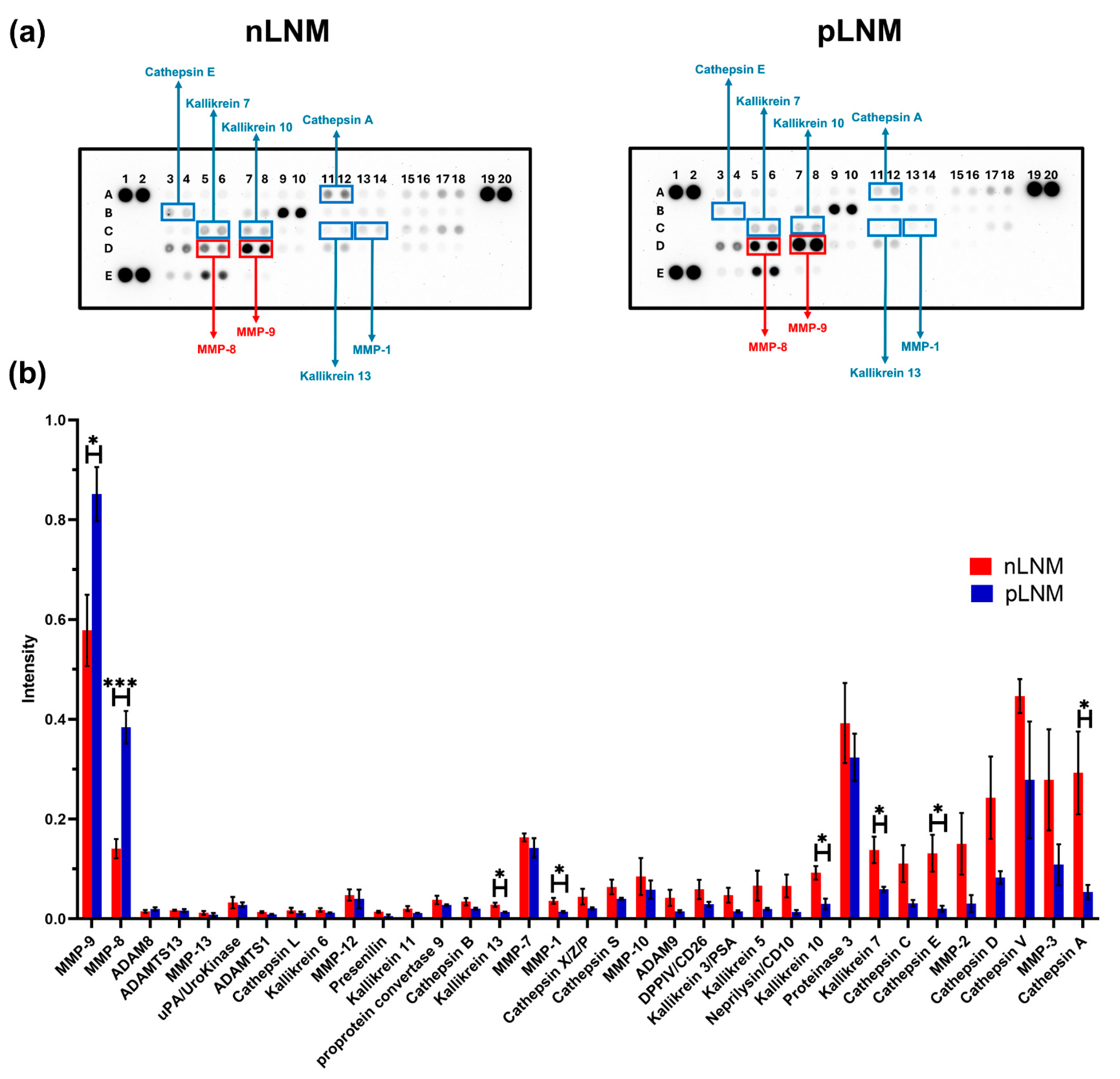
3.3. Protease Profiling Reveals a Significant Elevation of MMP-8 and MMP-9 in L-EVs Breast Cancer Patients with pLNM Compared with nLNM
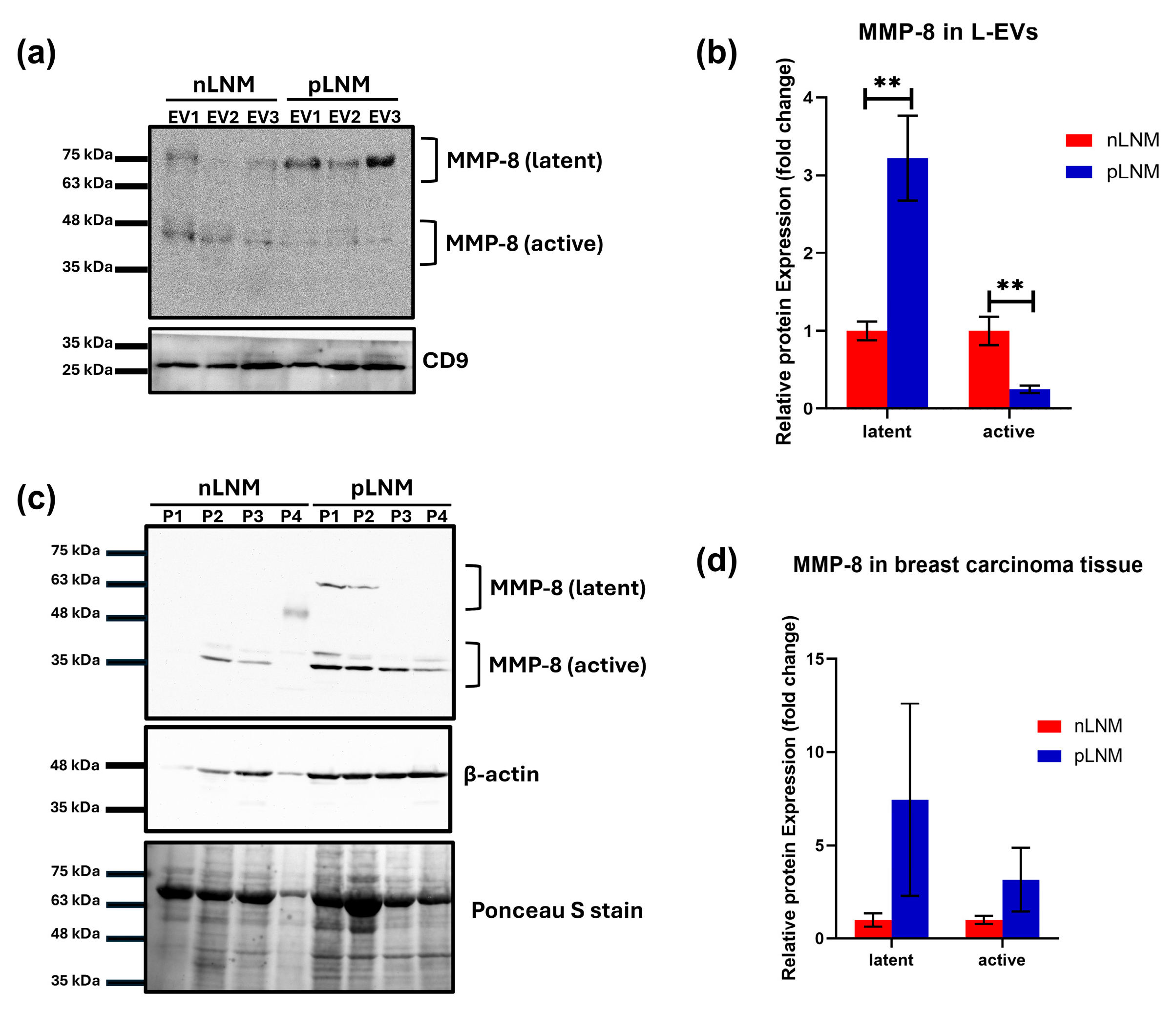
3.4. Differential Expression of Latent and Active MMP-8 in L-EVs from pLNM Compared with nLNM Breast Cancer Patients
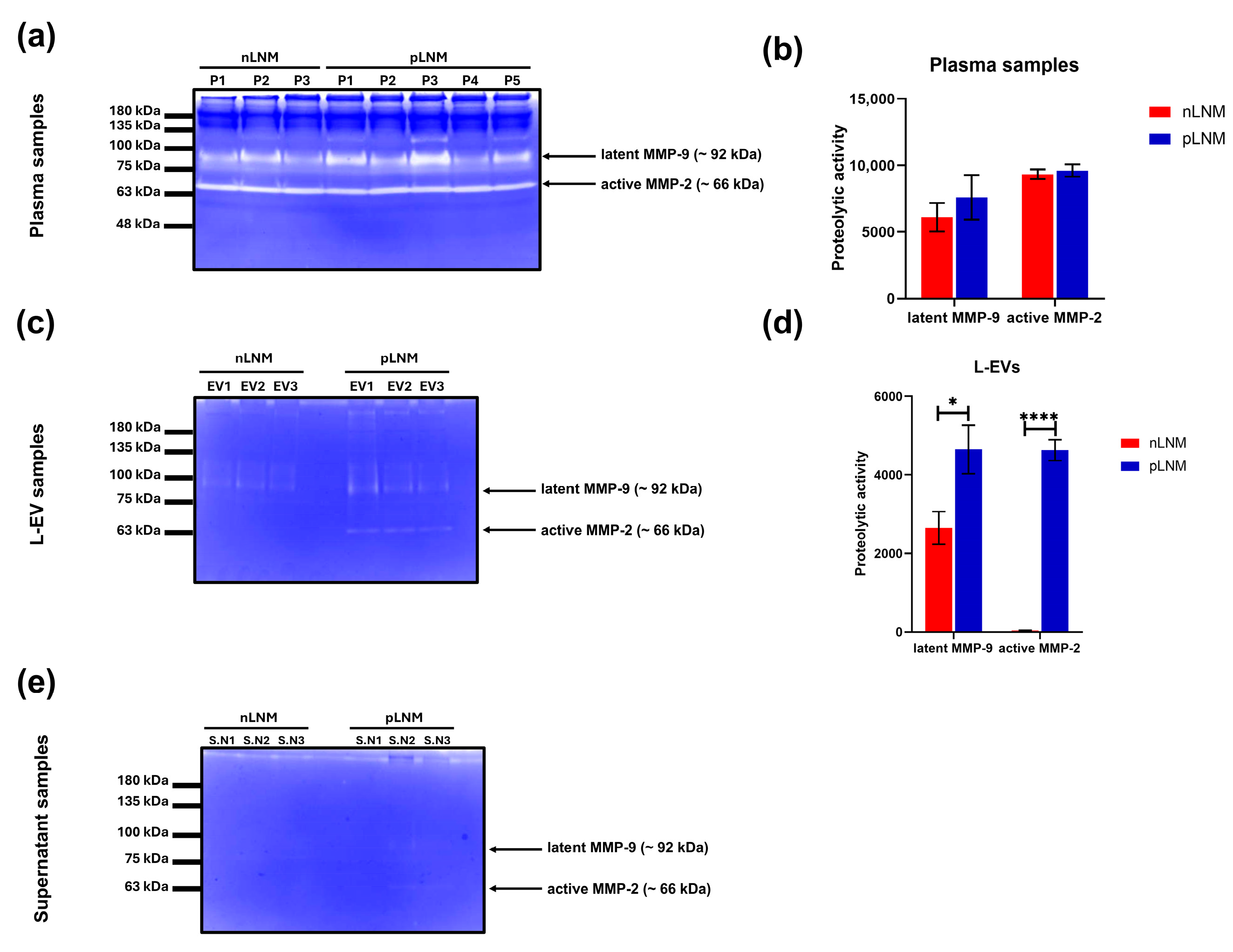
3.5. MMP-2 and MMP-9 Activities Are Elevated in L-EVs from pLNM Compared with nLNM Breast Cancer Patients
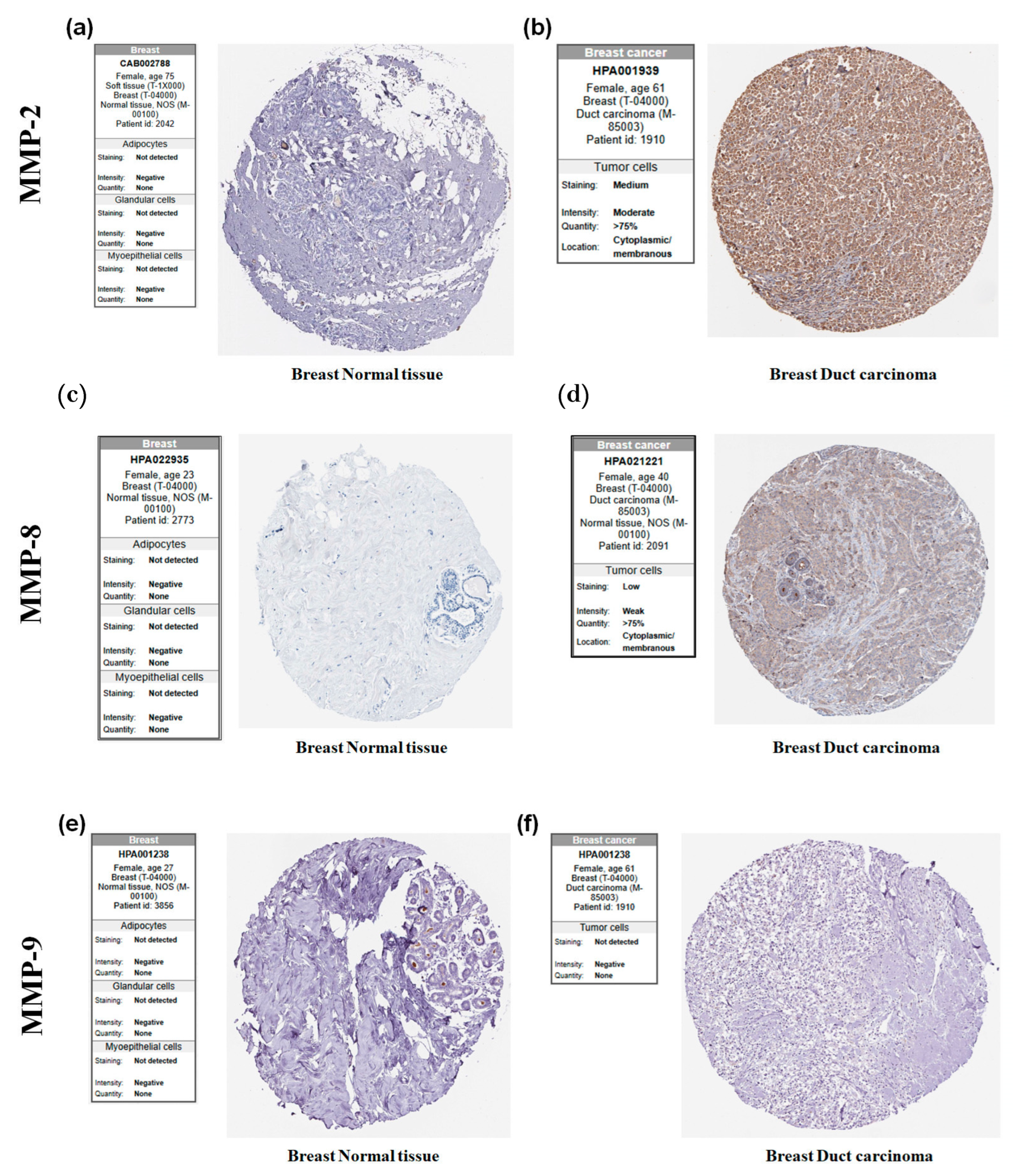
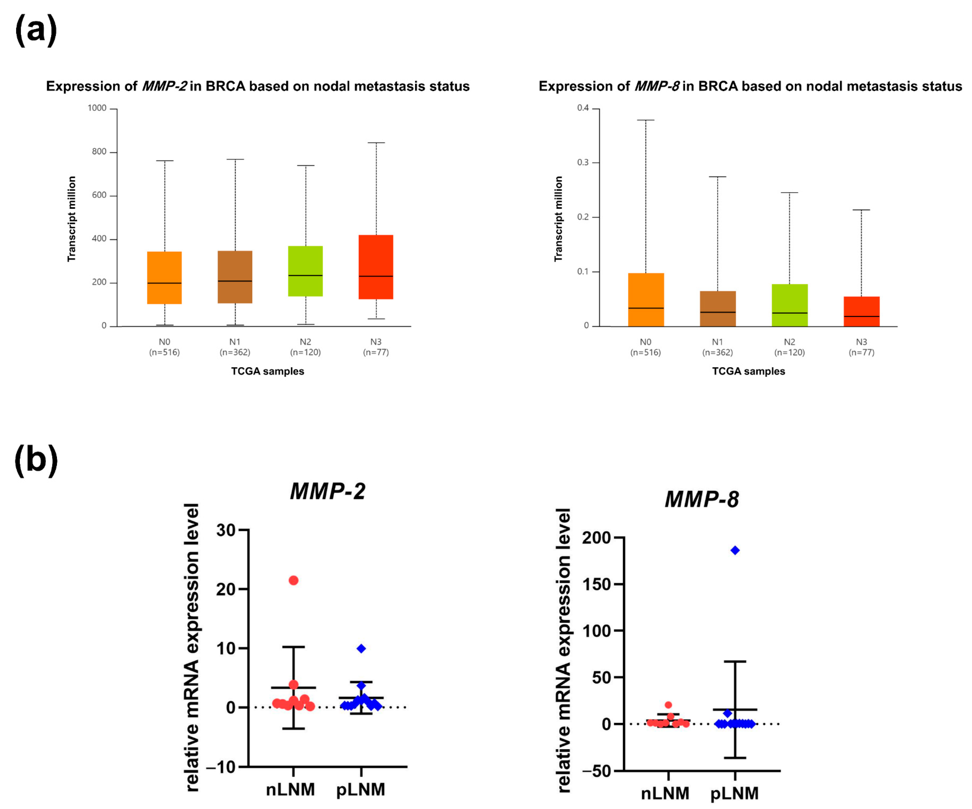
3.6. Expression of MMP-2, MMP-8, and MMP-9 in Breast Cancer and Its Relation with LNM Status
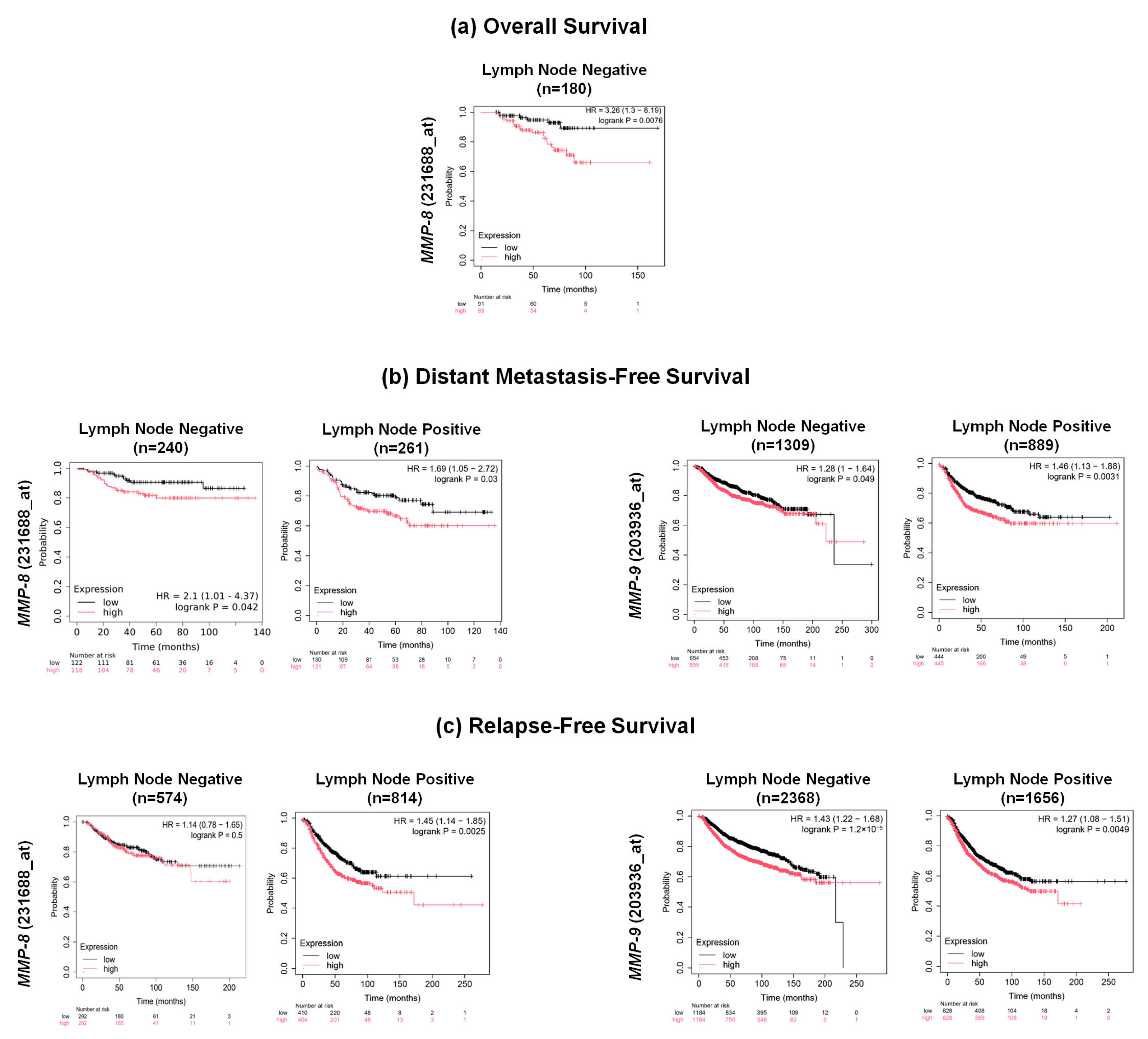
3.7. Prognostic Significance of MMP-2, MMP-8, and MMP-9 Expression in Breast Cancer Patients
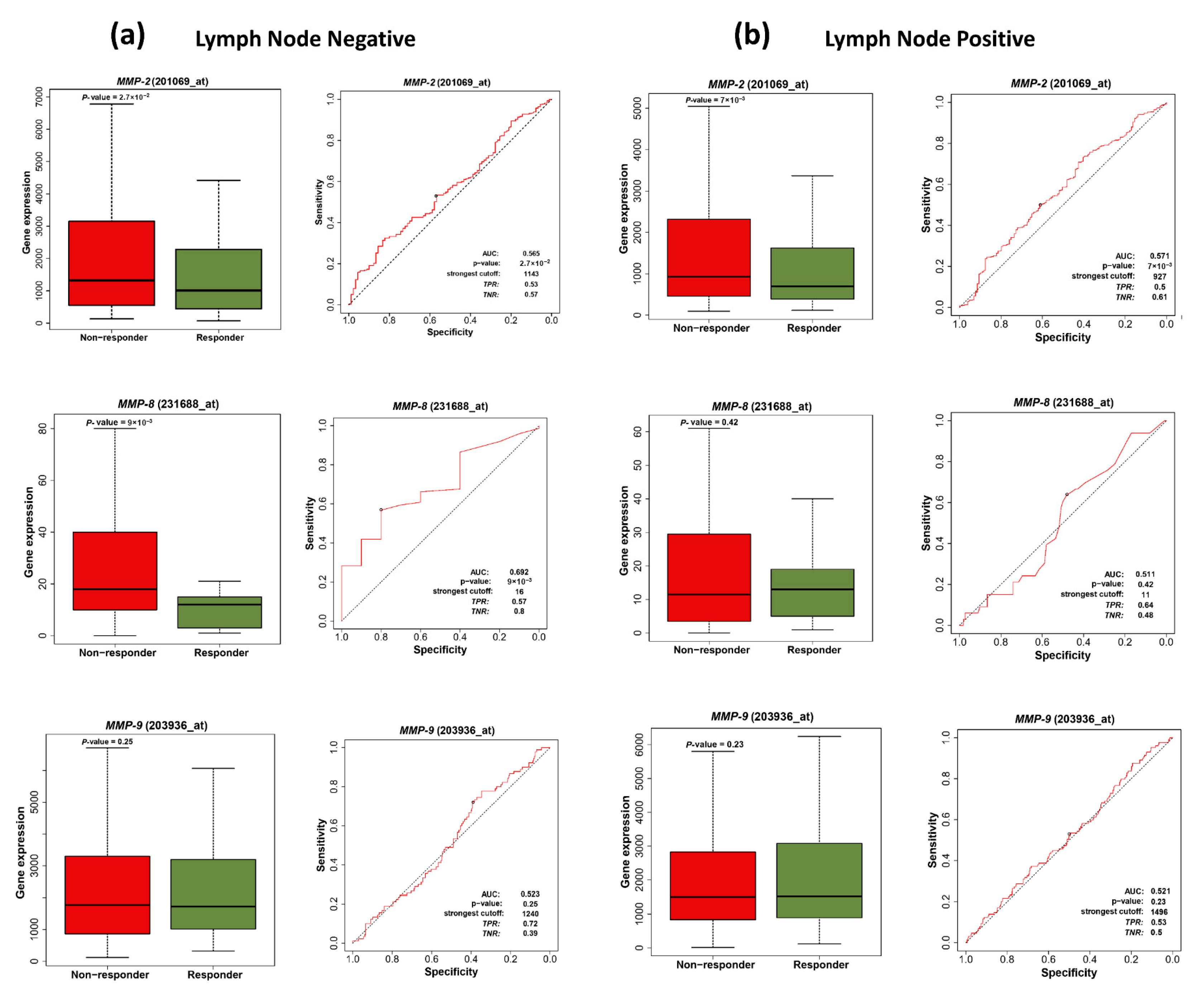
3.8. MMP-2 and MMP-8 as Predictive Biomarkers for Chemotherapy Response in Breast Cancer Patients
4. Discussion
5. Conclusions
Supplementary Materials
Author Contributions
Funding
Institutional Review Board Statement
Informed Consent Statement
Data Availability Statement
Acknowledgments
Conflicts of Interest
Abbreviations
| LNM | Lymph node metastasis |
| EVs | Extracellular vesicles |
| ECM | Extracellular matrix |
| MMPs | Matrix metalloproteinases |
| nLNM | Negative LNM |
| pLNM | Positive LNM |
| PBS | Phosphate-buffered saline |
| L-EVs | Large extracellular vesicles |
| ACD | Acid–citrate–dextrose |
| PPP | Platelet-poor plasma |
| RT | Room temperature |
| ISEV | International Society for Extracellular Vesicles |
| FSC-A | Forward scatter area |
| SSC-A | Side scatter area |
| TEM | Transmission electron microscopy |
| HR-TEM | High-resolution TEM |
| DLS | Dynamic light scattering |
| PDI | Polydispersity index |
| HRP | Horseradish peroxidase |
| ECL | Enhanced chemiluminescent |
| qRT-PCR | Quantitative real-time PCR |
| TCGA | The Cancer Genome Atlas |
| IHC | Immunohistochemistry |
| HPA | Human Protein Atlas |
| LN | Lymph node |
| OS | Overall survival |
| RFS | Relapse-free survival |
| DMFS | Distant metastasis-free survival |
| HRs | Hazard ratios |
| CIs | Confidence intervals |
| ROC | Receiving operating characteristics |
| pCR | Pathological complete response |
| AUC | Area under the curve |
| ANOVA | One-way analysis of variance |
| FEC | Fluorouracil, epirubicin, and cyclophosphamide |
| FAC | Fluorouracil, adriamycin, and cyclophosphamide |
| TME | Tumor microenvironment |
References
- Kim, J.; Harper, A.; McCormack, V.; Sung, H.; Houssami, N.; Morgan, E.; Mutebi, M.; Garvey, G.; Soerjomataram, I.; Fidler-Benaoudia, M.M. Global Patterns and Trends in Breast Cancer Incidence and Mortality across 185 Countries. Nat. Med. 2025, 31, 1154–1162. [Google Scholar] [CrossRef]
- Ji, H.; Hu, C.; Yang, X.; Liu, Y.; Ji, G.; Ge, S.; Wang, X.; Wang, M. Lymph Node Metastasis in Cancer Progression: Molecular Mechanisms, Clinical Significance and Therapeutic Interventions. Signal Transduct. Target. Ther. 2023, 8, 367. [Google Scholar] [CrossRef] [PubMed]
- Cheng, Z.; Ma, J.; Yin, L.; Yu, L.; Yuan, Z.; Zhang, B.; Tian, J.; Du, Y. Non-Invasive Molecular Imaging for Precision Diagnosis of Metastatic Lymph Nodes: Opportunities from Preclinical to Clinical Applications. Eur. J. Nucl. Med. Mol. Imaging 2022, 50, 1111–1133. [Google Scholar] [CrossRef]
- Ma, L.; Guo, H.; Zhao, Y.; Liu, Z.; Wang, C.; Bu, J.; Sun, T.; Wei, J. Liquid Biopsy in Cancer: Current Status, Challenges and Future Prospects. Signal Transduct. Target. Ther. 2024, 9, 336. [Google Scholar] [CrossRef]
- Pandey, S.; Yadav, P. Liquid Biopsy in Cancer Management: Integrating Diagnostics and Clinical Applications. Pract. Lab. Med. 2025, 43, e00446. [Google Scholar] [CrossRef] [PubMed]
- Rayamajhi, S.; Sipes, J.; Tetlow, A.L.; Saha, S.; Bansal, A.; Godwin, A.K. Extracellular Vesicles as Liquid Biopsy Biomarkers across the Cancer Journey: From Early Detection to Recurrence. Clin. Chem. 2024, 70, 206–219. [Google Scholar] [CrossRef] [PubMed]
- Petroni, D.; Fabbri, C.; Babboni, S.; Menichetti, L.; Basta, G.; Del Turco, S. Extracellular Vesicles and Intercellular Communication: Challenges for In Vivo Molecular Imaging and Tracking. Pharmaceutics 2023, 15, 1639. [Google Scholar] [CrossRef]
- Chang, W.H.; Cerione, R.A.; Antonyak, M.A. Extracellular Vesicles and Their Roles in Cancer Progression. In Cancer Cell Signaling; Methods in Molecular Biology; Humana: New York, NY, USA, 2021; Volume 2174, pp. 143–170. [Google Scholar] [CrossRef]
- Kumar, M.A.; Baba, S.K.; Sadida, H.Q.; Marzooqi, S.A.; Jerobin, J.; Altemani, F.H.; Algehainy, N.; Alanazi, M.A.; Abou-Samra, A.-B.; Kumar, R.; et al. Extracellular Vesicles as Tools and Targets in Therapy for Diseases. Signal Transduct. Target. Ther. 2024, 9, 27. [Google Scholar] [CrossRef]
- Lorite, P.; Domínguez, J.N.; Palomeque, T.; Torres, M.I. Extracellular Vesicles: Advanced Tools for Disease Diagnosis, Monitoring, and Therapies. Int. J. Mol. Sci. 2024, 26, 189. [Google Scholar] [CrossRef]
- Dong, Q.; Liu, X.; Cheng, K.; Sheng, J.; Kong, J.; Liu, T. Pre-Metastatic Niche Formation in Different Organs Induced by Tumor Extracellular Vesicles. Front. Cell Dev. Biol. 2021, 9, 733627. [Google Scholar] [CrossRef]
- Li, Y.; Zheng, Y.; Tan, X.; Du, Y.; Wei, Y.; Liu, S. Extracellular Vesicle-Mediated Pre-Metastatic Niche Formation via Altering Host Microenvironments. Front. Immunol. 2024, 15, 1367373. [Google Scholar] [CrossRef] [PubMed]
- Wang, Y.; Jia, J.; Wang, F.; Fang, Y.; Yang, Y.; Zhou, Q.; Yuan, W.; Gu, X.; Hu, J.; Yang, S. Pre-Metastatic Niche: Formation, Characteristics and Therapeutic Implication. Signal Transduct. Target. Ther. 2024, 9, 236. [Google Scholar] [CrossRef] [PubMed]
- Radisky, E.S. Extracellular Proteolysis in Cancer: Proteases, Substrates, and Mechanisms in Tumor Progression and Metastasis. J. Biol. Chem. 2024, 300, 107347. [Google Scholar] [CrossRef]
- Woodward, J.K.L.; Holen, I.; Coleman, R.E.; Buttle, D.J. The Roles of Proteolytic Enzymes in the Development of Tumour-Induced Bone Disease in Breast and Prostate Cancer. Bone 2007, 41, 912–927. [Google Scholar] [CrossRef]
- Tune, B.X.J.; Sim, M.S.; Poh, C.L.; Mac Guad, R.; Woon, C.K.; Hazarika, I.; Das, A.; Gopinath, S.C.B.; Rajan, M.; Sekar, M.; et al. Matrix Metalloproteinases in Chemoresistance: Regulatory Roles, Molecular Interactions, and Potential Inhibitors. J. Oncol. 2022, 2022, 3249766. [Google Scholar] [CrossRef]
- Reunanen, N.; Kähäri, V. Matrix Metalloproteinases in Cancer Cell Invasion; National Institutes of Health: Bethesda, MA, USA, 2013.
- Duffy, M.J. The Role of Proteolytic Enzymes in Cancer Invasion and Metastasis. Clin. Exp. Metastasis 1992, 10, 145–155. [Google Scholar] [CrossRef]
- Proteases as Prognostic Markers in Cancer.|Clinical Cancer Research|American Association for Cancer Research. Available online: https://aacrjournals.org/clincancerres/article-abstract/2/4/613/33562/Proteases-as-prognostic-markers-in-cancer (accessed on 18 May 2025).
- Savage, P.; Pacis, A.; Kuasne, H.; Liu, L.; Lai, D.; Wan, A.; Dankner, M.; Martinez, C.; Muñoz-Ramos, V.; Pilon, V.; et al. Chemogenomic Profiling of Breast Cancer Patient-Derived Xenografts Reveals Targetable Vulnerabilities for Difficult-to-Treat Tumors. Commun. Biol. 2020, 3, 310. [Google Scholar] [CrossRef]
- György, B.; Pálóczi, K.; Kovács, A.; Barabás, E.; Beko, G.; Várnai, K.; Pállinger, É.; Szabó-Taylor, K.; Szabó, T.G.; Kiss, A.A.; et al. Improved Circulating Microparticle Analysis in Acid-Citrate Dextrose (ACD) Anticoagulant Tube. Thromb. Res. 2014, 133, 285–292. [Google Scholar] [CrossRef]
- Talat, L.Y.; Mohamed, G.; Ibraheem, M.H.; WalyEldeen, A.A.; Hassan, H.; Ibrahim, S.A. SDC2 and FN as Cargo Proteins in Circulating Extracellular Vesicles in Obese Breast Cancer Patients with Lymph Node Metastasis. Sci. Rep. 2025, 15, 32498. [Google Scholar] [CrossRef]
- Schank, M.B.; Zhao, J.; Wang, L.; Moorman, J.P.; Yao, Z.Q. Extracellular Vesicles: A Comprehensive Review of Their Origins, Functions, and Therapeutic Potential. Biomedicines 2026, 14, 495. [Google Scholar] [CrossRef] [PubMed]
- Menck, K.; Bleckmann, A.; Schulz, M.; Ries, L.; Binder, C. Isolation and Characterization of Microvesicles from Peripheral Blood. J. Vis. Exp. 2017, 2017, 55057. [Google Scholar] [CrossRef]
- Muller, L.; Hong, C.S.; Stolz, D.B.; Watkins, S.C.; Whiteside, T.L. Isolation of Biologically-Active Exosomes from Human Plasma. J. Immunol. Methods 2014, 411, 55–65. [Google Scholar] [CrossRef]
- Emad, B.; WalyEldeen, A.A.; Hassan, H.; Sharaky, M.; Abdelhamid, I.A.; Ibrahim, S.A.; Mohamed, H.R. Yttrium Oxide Nanoparticles Induce Cytotoxicity, Genotoxicity, Apoptosis, and Ferroptosis in the Human Triple-Negative Breast Cancer MDA-MB-231 Cells. BMC Cancer 2023, 23, 1151. [Google Scholar] [CrossRef]
- Ahmed, S.H.; Espinoza-Sánchez, N.A.; El-Damen, A.; Fahim, S.A.; Badawy, M.A.; Greve, B.; El-Shinawi, M.; Götte, M.; Ibrahim, S.A. Small Extracellular Vesicle-Encapsulated MiR-181b-5p, MiR-222-3p and Let-7a-5p: Next Generation Plasma Biopsy-Based Diagnostic Biomarkers for Inflammatory Breast Cancer. PLoS ONE 2021, 16, e0250642. [Google Scholar] [CrossRef]
- Fahim, S.A.; Abdullah, M.S.; Espinoza-sánchez, N.A.; Hassan, H.; Ibrahim, A.M.; Ahmed, S.H.; Shakir, G.; Badawy, M.A.; Zakhary, N.I.; Greve, B.; et al. Inflammatory Breast Carcinoma: Elevated MicroRNA MiR-181b-5p and Reduced MiR-200b-3p, MiR-200c-3p, and MiR-203a-3p Expression as Potential Biomarkers with Diagnostic Value. Biomolecules 2020, 10, 1059. [Google Scholar] [CrossRef]
- Kalra, H.; Simpson, R.J.; Ji, H.; Aikawa, E.; Altevogt, P.; Askenase, P.; Bond, V.C.; Borràs, F.E.; Breakefield, X.; Budnik, V.; et al. Vesiclepedia: A Compendium for Extracellular Vesicles with Continuous Community Annotation. PLoS Biol. 2012, 10, e1001450. [Google Scholar] [CrossRef]
- Chandrashekar, D.S.; Karthikeyan, S.K.; Korla, P.K.; Patel, H.; Shovon, A.R.; Athar, M.; Netto, G.J.; Qin, Z.S.; Kumar, S.; Manne, U.; et al. UALCAN: An Update to the Integrated Cancer Data Analysis Platform. Neoplasia 2022, 25, 18–27. [Google Scholar] [CrossRef] [PubMed]
- Uhlen, M.; Zhang, C.; Lee, S.; Sjöstedt, E.; Fagerberg, L.; Bidkhori, G.; Benfeitas, R.; Arif, M.; Liu, Z.; Edfors, F.; et al. A Pathology Atlas of the Human Cancer Transcriptome. Science 2017, 357, 2507. [Google Scholar] [CrossRef] [PubMed]
- Uhlén, M.; Fagerberg, L.; Hallström, B.M.; Lindskog, C.; Oksvold, P.; Mardinoglu, A.; Sivertsson, Å.; Kampf, C.; Sjöstedt, E.; Asplund, A.; et al. Tissue-Based Map of the Human Proteome. Science 2015, 347, 1260419. [Google Scholar] [CrossRef]
- Győrffy, B. Survival Analysis across the Entire Transcriptome Identifies Biomarkers with the Highest Prognostic Power in Breast Cancer. Comput. Struct. Biotechnol. J. 2021, 19, 4101–4109. [Google Scholar] [CrossRef]
- Fekete, J.T.; Győrffy, B. ROCplot.Org: Validating Predictive Biomarkers of Chemotherapy/Hormonal Therapy/Anti-HER2 Therapy Using Transcriptomic Data of 3104 Breast Cancer Patients. Int. J. Cancer 2019, 145, 3140–3151. [Google Scholar] [CrossRef]
- Park, K.C.; Dharmasivam, M.; Richardson, D.R. The Role of Extracellular Proteases in Tumor Progression and the Development of Innovative Metal Ion Chelators That Inhibit Their Activity. Int. J. Mol. Sci. 2020, 21, 6805. [Google Scholar] [CrossRef]
- Nawaz, M.; Shah, N.; Zanetti, B.R.; Maugeri, M.; Silvestre, R.N.; Fatima, F.; Neder, L.; Valadi, H. Extracellular Vesicles and Matrix Remodeling Enzymes: The Emerging Roles in Extracellular Matrix Remodeling, Progression of Diseases and Tissue Repair. Cells 2018, 7, 167. [Google Scholar] [CrossRef]
- Pucci, F.; Garris, C.; Lai, C.P.; Newton, A.; Pfirschke, C.; Engblom, C.; Alvarez, D.; Sprachman, M.; Evavold, C.; Magnuson, A.; et al. SCS Macrophages Suppress Melanoma by Restricting Tumor-Derived Vesicle–B Cell Interactions. Science 2016, 352, 242–246. [Google Scholar] [CrossRef]
- Gutiérrez-Fernández, A.; Fueyo, A.; Folgueras, A.R.; Garabaya, C.; Pennington, C.J.; Pilgrim, S.; Edwards, D.R.; Holliday, D.L.; Jones, J.L.; Span, P.N.; et al. Matrix Metalloproteinase-8 Functions as a Metastasis Suppressor through Modulation of Tumor Cell Adhesion and Invasion. Cancer Res. 2008, 68, 2755–2763. [Google Scholar] [CrossRef]
- Moilanen, M.; Pirilä, E.; Grénman, R.; Sorsa, T.; Salo, T. Expression and Regulation of Collagenase-2 (MMP-8) in Head and Neck Squamous Cell Carcinomas. J. Pathol. 2002, 197, 72–81. [Google Scholar] [CrossRef] [PubMed]
- Stadlmann, S.; Pollheimer, J.; Moser, P.L.; Raggi, A.; Amberger, A.; Margreiter, R.; Offner, F.A.; Mikuz, G.; Dirnhofer, S.; Moch, H. Cytokine-Regulated Expression of Collagenase-2 (MMP-8) Is Involved in the Progression of Ovarian Cancer. Eur. J. Cancer 2003, 39, 2499–2505. [Google Scholar] [CrossRef] [PubMed]
- Väyrynen, J.P.; Vornanen, J.; Tervahartiala, T.; Sorsa, T.; Bloigu, R.; Salo, T.; Tuomisto, A.; Mäkinen, M.J. Serum MMP-8 Levels Increase in Colorectal Cancer and Correlate with Disease Course and Inflammatory Properties of Primary Tumors. Int. J. Cancer 2012, 131, E463–E474. [Google Scholar] [CrossRef]
- Qin, G.; Luo, M.; Chen, J.; Dang, Y.; Chen, G.; Li, L.; Zeng, J.; Lu, Y.; Yang, J. Reciprocal Activation between MMP-8 and TGF-Β1 Stimulates EMT and Malignant Progression of Hepatocellular Carcinoma. Cancer Lett. 2016, 374, 85–95. [Google Scholar] [CrossRef]
- Köhrmann, A.; Kammerer, U.; Kapp, M.; Dietl, J.; Anacker, J. Expression of Matrix Metalloproteinases (MMPs) in Primary Human Breast Cancer and Breast Cancer Cell Lines: New Findings and Review of the Literature. BMC Cancer 2009, 9, 188. [Google Scholar] [CrossRef] [PubMed]
- Li, L.; Chen, L.; Zhang, W.; Liao, Y.; Chen, J.; Shi, Y.; Luo, S. Serum Cytokine Profile in Patients with Breast Cancer. Cytokine 2017, 89, 173–178. [Google Scholar] [CrossRef]
- Cavallari, C.; Camussi, G.; Brizzi, M.F. Extracellular Vesicles in the Tumour Microenvironment: Eclectic Supervisors. Int. J. Mol. Sci. 2020, 21, 6768. [Google Scholar] [CrossRef]
- Lee, M.K.; Azizgolshani, N.; Shapiro, J.A.; Nguyen, L.N.; Kolling, F.W.; Zanazzi, G.J.; Frost, H.R.; Christensen, B.C. Identifying Tumor Type and Cell Type-Specific Gene Expression Alterations in Pediatric Central Nervous System Tumors. Nat. Commun. 2024, 15, 3634. [Google Scholar] [CrossRef]
- Zhang, Z.; Jin, W.; Xu, K.; Zheng, X.; Zhou, Y.; Luo, M.; Yan, C.; Zheng, X.; Jin, E. Blood Exosome PD-L1 Is Associated with PD-L1 Expression Measured by Immunohistochemistry, and Lymph Node Metastasis in Lung Cancer. Tissue Cell 2022, 79, 101941. [Google Scholar] [CrossRef] [PubMed]
- Thuault, S.; Ghossoub, R.; David, G.; Zimmermann, P. A Journey on Extracellular Vesicles for Matrix Metalloproteinases: A Mechanistic Perspective. Front. Cell Dev. Biol. 2022, 10, 886381. [Google Scholar] [CrossRef] [PubMed]
- Kessenbrock, K.; Plaks, V.; Werb, Z. Matrix Metalloproteinases: Regulators of the Tumor Microenvironment. Cell 2010, 141, 52–67. [Google Scholar] [CrossRef]
- Van Lint, P.; Libert, C. Matrix Metalloproteinase-8: Cleavage Can Be Decisive. Cytokine Growth Factor Rev. 2006, 17, 217–223. [Google Scholar] [CrossRef] [PubMed]
- Jiang, H.; Li, H. Prognostic Values of Tumoral MMP2 and MMP9 Overexpression in Breast Cancer: A Systematic Review and Meta-Analysis. BMC Cancer 2021, 21, 149. [Google Scholar] [CrossRef]
- Li, H.; Qiu, Z.; Li, F.; Wang, C. The Relationship between MMP-2 and MMP-9 Expression Levels with Breast Cancer Incidence and Prognosis. Oncol. Lett. 2017, 14, 5865–5870. [Google Scholar] [CrossRef]
- Dolo, V.; Ginestra, A.; Ghersi, G.; Nagase, H.; Vittorelli, M.L. Human Breast Carcinoma Cells Cultured in the Presence of Serum Shed Membrane Vesicles Rich in Gelatinolytic Activities. J. Submicrosc. Cytol. Pathol. 1994, 26, 173–180. [Google Scholar]
- Di Vizio, D.; Morello, M.; Dudley, A.C.; Schow, P.W.; Adam, R.M.; Morley, S.; Mulholland, D.; Rotinen, M.; Hager, M.H.; Insabato, L.; et al. Large Oncosomes in Human Prostate Cancer Tissues and in the Circulation of Mice with Metastatic Disease. Am. J. Pathol. 2012, 181, 1573–1584. [Google Scholar] [CrossRef] [PubMed]
- Ginestra, A.; Monea, S.; Seghezzi, G.; Dolo, V.; Nagase, H.; Mignatti, P.; Vittorelli, M.L. Urokinase Plasminogen Activator and Gelatinases Are Associated with Membrane Vesicles Shed by Human HT1080 Fibrosarcoma Cells. J. Biol. Chem. 1997, 272, 17216–17222. [Google Scholar] [CrossRef]
- Dolo, V.; D’Ascenzo, S.; Violini, S.; Pompucci, L.; Festuccia, C.; Ginestra, A.; Vittorelli, M.L.; Canevari, S.; Pavan, A. Matrix-Degrading Proteinases Are Shed in Membrane Vesicles by Ovarian Cancer Cells in Vivo and in Vitro. Clin. Exp. Metastasis 1999, 17, 131–140. [Google Scholar] [CrossRef]
- Waas, E.T.; Lomme, R.M.L.M.; DeGroot, J.; Wobbes, T.; Hendriks, T. Tissue Levels of Active Matrix Metalloproteinase-2 and -9 in Colorectal Cancer. Br. J. Cancer 2002, 86, 1876–1883. [Google Scholar] [CrossRef]
- Lengyel, E.; Schmalfeldt, B.; Konik, E.; Späthe, K.; Härting, K.; Fenn, A.; Berger, U.; Fridman, R.; Schmitt, M.; Prechtel, D.; et al. Expression of Latent Matrix Metalloproteinase 9 (MMP-9) Predicts Survival in Advanced Ovarian Cancer. Gynecol. Oncol. 2001, 82, 291–298. [Google Scholar] [CrossRef] [PubMed]
- Han, K.Y.; Dugas-Ford, J.; Seiki, M.; Chang, J.H.; Azar, D.T. Evidence for the Involvement of MMP14 in MMP2 Processing and Recruitment in Exosomes of Corneal Fibroblasts. Investig. Ophthalmol. Vis. Sci. 2015, 56, 5323–5329. [Google Scholar] [CrossRef]
- Heo, D.S.; Choi, H.; Yeom, M.Y.; Song, B.J.; Oh, S.J. Serum Levels of Matrix Metalloproteinase-9 Predict Lymph Node Metastasis in Breast Cancer Patients. Oncol. Rep. 2014, 31, 1567–1572. [Google Scholar] [CrossRef][Green Version]
- Stankovic, S.; Konjevic, G.; Gopcevic, K.; Jovic, V.; Inic, M.; Jurisic, V. Activity of MMP-2 and MMP-9 in Sera of Breast Cancer Patients. Pathol. Res. Pract. 2010, 206, 241–247. [Google Scholar] [CrossRef]
- Decock, J.; Hendrickx, W.; Wildiers, H.; Christiaens, M.R.; Neven, P.; Drijkoningen, M.; Paridaens, R. Plasma Gelatinase Levels in Patients with Primary Breast Cancer in Relation to Axillary Lymph Node Status, Her2/Neu Expression and Other Clinicopathological Variables. Clin. Exp. Metastasis 2005, 22, 495–502. [Google Scholar] [CrossRef] [PubMed]
- La Rocca, G.; Pucci-Minafra, I.; Marrazzo, A.; Taormina, P.; Minafra, S. Zymographic Detection and Clinical Correlations of MMP-2 and MMP-9 in Breast Cancer Sera. Br. J. Cancer 2004, 90, 1414–1421. [Google Scholar] [CrossRef]
- Decock, J.; Hendrickx, W.; Vanleeuw, U.; Van Belle, V.; Van Huffel, S.; Christiaens, M.R.; Ye, S.; Paridaens, R. Plasma MMP1 and MMP8 Expression in Breast Cancer: Protective Role of MMP8 against Lymph Node Metastasis. BMC Cancer 2008, 8, 77. [Google Scholar] [CrossRef]
- Jonsson, A.; Hjalmarsson, C.; Falk, P.; Ivarsson, M.L. Levels of Matrix Metalloproteinases Differ in Plasma and Serum—Aspects Regarding Analysis of Biological Markers in Cancer. Br. J. Cancer 2016, 115, 703–706. [Google Scholar] [CrossRef] [PubMed]
- Gerlach, R.F.; Demacq, C.; Jung, K.; Tanus-Santos, J.E. Rapid Separation of Serum Does Not Avoid Artificially Higher Matrix Metalloproteinase (MMP)-9 Levels in Serum versus Plasma. Clin. Biochem. 2007, 40, 119–123. [Google Scholar] [CrossRef] [PubMed]
- Loric, S.; Denis, J.A.; Desbene, C.; Sabbah, M.; Conti, M. Extracellular Vesicles in Breast Cancer: From Biology and Function to Clinical Diagnosis and Therapeutic Management. Int. J. Mol. Sci. 2023, 24, 7208. [Google Scholar] [CrossRef] [PubMed]



| Characteristic | nLNM (n = 40) | pLNM (n = 32) | p-Value |
|---|---|---|---|
| Age (years) | |||
| Range | 34–76 | 36–79 | a p > 0.05 |
| Mean ± SEM | 57.92 ± 2.002 | 57.69 ± 1.852 | |
| <50 | 12 (30%) | 8 (25%) | b p > 0.05 |
| ≥50 | 26 (65%) | 24 (75%) | |
| NA | 2 (5%) | - | |
| Menopause status, n (%) | |||
| Premenopausal | 14 (35%) | 10 (31.25%) | b p > 0.05 |
| Postmenopausal | 22 (55%) | 17 (53.125%) | |
| NA | 4 (10%) | 5 (15.625%) | |
| Laterality, n (%) | |||
| Bilateral | 2 (5%) | 1 (3.125%) | b p > 0.05 |
| Right | 17 (42.5%) | 17 (53.125%) | |
| Left | 21 (52.5%) | 14 (43.75%) | |
| Tumor size (cm), n (%) | |||
| ≤4 | 34 (85%) | 28 (87.5%) | b p > 0.05 |
| >4 | 6 (15%) | 4 (12.5%) | |
| Tumor grade, n (%) | |||
| Grade 1 | 4 (10%) | - | b p > 0.05 |
| Grade 2 | 30 (75%) | 27 (84.375%) | |
| Grade 3 | 5 (12.5%) | 5 (15.625%) | |
| NA | 1 (2.5%) | - | |
| ER, n (%) | |||
| Negative | 5 (12.5%) | 1 (3.125%) | b p > 0.05 |
| Positive | 35 (87.5%) | 31 (96.875%) | |
| PR, n (%) | |||
| Negative | 7 (17.5%) | 4 (12.5%) | b p > 0.05 |
| Positive | 33 (82.5%) | 28 (87.5%) | |
| HER2, n (%) | |||
| Negative | 34 (85%) | 26 (81.25%) | b p > 0.05 |
| Equivocal (non-amplified) | 6 (15%) | 6 (18.75%) | |
| Stages, n (%) | |||
| I | 23 (57.5%) | 1 (3.125%) | b p < 0.05 |
| II | 17 (42.5%) | 15 (46.875%) | |
| III | - | 16 (50%) | |
| Lymph node status, n (%) | |||
| N0 | 40 (100%) | - | b p < 0.05 |
| N1 | - | 18 (56.25%) | |
| N2 | - | 8 (25%) | |
| N3 | - | 6 (18.75%) | |
| Tumor size, n (%) | |||
| T1 | 23 (57.5%) | 8 (25%) | b p > 0.05 |
| T2 | 14 (35%) | 22 (68.75%) | |
| T3 | 3 (7.5%) | 1 (3.125%) | |
| T4 | - | 1 (3.125%) |
| Coordinates | Protein Name | Gene Symbol | Vesiclepedia | Fold Change | p-Value | |
|---|---|---|---|---|---|---|
| A11, A12 | Cathepsin A | CTSA | √* | 0.2 | 0.030 | * |
| B3, B4 | Cathepsin E | CTSE | √ | 0.2 | 0.025 | * |
| C5, C6 | Kallikrein 7 | KLK7 | √* | 0.4 | 0.027 | * |
| C7, C8 | Kallikrein 10 | KLK7 | √* | 0.3 | 0.011 | * |
| C11, C12 | Kallikrein 13 | KLK13 | √* | 0.5 | 0.014 | * |
| C13, C14 | Matrix metallopeptidase 1 | MMP1 | √* | 0.4 | 0.011 | * |
| D5, D6 | Matrix metallopeptidase 8 | MMP8 | √* | 2.7 | 0.001 | *** |
| D7, D8 | Matrix metallopeptidase 9 | MMP9 | √* | 1.5 | 0.023 | * |
| Gene | Parameter | nLNM | pLNM | ||||||
|---|---|---|---|---|---|---|---|---|---|
| FEC | Anthracycline | FAC | Taxane | FEC | Anthracycline | FAC | Taxane | ||
| MMP-2 | Responder (expression) | 1178 ± 1398 | 1695 ± 1846 | 2268 ± 2839 | 1497 ± 1830 | 1317 ± 1288 | 1406 ± 1768 | 3326 ± 3303 | 1102 ± 110 |
| (n = 32) | (n = 86) | (n = 18) | (n = 75) | (n = 51) | (n = 129) | (n = 14) | (n = 110) | ||
| Non-responder (expression) | 1691 ± 1281 (n = 56) | 2187 ± 2263 | 2690 ± 3030 | 1738 ± 2016 | 1567 ± 1365 | 1688 ± 1890 | 1400 ± 1736 | 1452 ± 1736 | |
| (n = 276) | (n = 51) | (n = 196) | (n = 100) | (n = 483) | (n = 91) | (n = 379) | |||
| ROC p-value | 4.30 × 10⁻³ | 0.058 | 0.47 | 0.2 | 0.21 | 7.30 × 10⁻³ | 0.077 | 2.60 × 10⁻³ | |
| AUC | 0.665 | 0.554 | 0.507 | 0.533 | 0.539 | 0.57 | 0.639 | 0.586 | |
| Mann–Whitney test p-value | 0.011 | 0.13 | 0.94 | 0.4 | 0.43 | 0.014 | 0.096 | 0.0058 | |
| Fold change | 1.4 | 1.3 | 1.2 | 1.2 | 1.2 | 1.2 | 2.4 | 1.3 | |
| MMP-8 | Responder (expression) | 16 ± 13 | 12 ± 10 | 0 | 16 ± 13 | 12 ± 10 | 25 ± 42 | 0 | 12 ± 10 |
| (n = 4) | (n = 10) | (n = 4) | (n = 23) | (n = 33) | (n = 23) | ||||
| Non-responder (expression) | 10 ± 7 | 30 ± 42 | 0 | 10 ± 7 | 23 ± 93 | 31 ±77 | 0 | 23 ± 93 | |
| (n = 28) | (n = 74) | (n = 28) | (n = 60) | (n = 112) | (n = 60) | ||||
| ROC p-value | 0.25 | 9.00 × 10⁻³ | 0.25 | 0.28 | 0.42 | 0.28 | |||
| AUC | 0.621 | 0.692 | 0.621 | 0.541 | 0.511 | 0.541 | |||
| Mann–Whitney test p-value | 0.46 | 0.051 | 0.46 | 0.57 | 0.85 | 0.57 | |||
| Fold change | 1.6 | 2.5 | 1.6 | 2 | 1.2 | 2 | |||
| MMP-9 | Responder (expression) | 2083 ± 1908 | 3338 ± 6489 | 6154 ± 13,238 (n = 18) | 3301 ± 6901 (n = 75) | 2319 ± 3894 | 2777 ± 3761 | 3570 ± 5513 | 2620 ± 3546 |
| (n = 32) | (n = 86) | (n = 51) | (n = 129) | (n = 14) | (n = 110) | ||||
| Non-responder (expression) | 3164 ± 6229 | 3203 ± 5459 | 3332 ± 5068 | 3310 ± 5928 (n = 196) | 1437 ± 1962 | 2665 ± 5170 | 2350 ± 2526 | 2614 ± 5614 | |
| (n = 56) | (n = 276) | (n = 51) | (n = 100) | (n = 483) | (n = 91) | (n = 379) | |||
| ROC p-value | 0.1 | 0.22 | 0.15 | 0.3 | 3.00 × 10⁻³ | 0.23 | 0.11 | 0.27 | |
| AUC | 0.577 | 0.527 | 0.58 | 0.52 | 0.629 | 0.521 | 0.591 | 0.519 | |
| Mann–Whitney test p-value | 0.23 | 0.45 | 0.32 | 0.61 | 0.0099 | 0.46 | 0.28 | 0.54 | |
| Fold change | 1.5 | 1 | 1.8 | 1 | 1.6 | 1 | 1.5 | 1 | |
Disclaimer/Publisher’s Note: The statements, opinions and data contained in all publications are solely those of the individual author(s) and contributor(s) and not of MDPI and/or the editor(s). MDPI and/or the editor(s) disclaim responsibility for any injury to people or property resulting from any ideas, methods, instructions or products referred to in the content. |
© 2026 by the authors. Licensee MDPI, Basel, Switzerland. This article is an open access article distributed under the terms and conditions of the Creative Commons Attribution (CC BY) license.
Share and Cite
Talat, L.Y.; WalyEldeen, A.A.; Mohamed, G.; Ibraheem, M.H.; Maher, M.M.; Ibrahim, S.A.; Hassan, H.; Götte, M. Large Extracellular Vesicle-Derived Latent MMP-8 and Gelatinolytically Active MMP-2 as Potential Circulating Markers for Lymph Node Metastasis in Breast Cancer. Cancers 2026, 18, 1464. https://doi.org/10.3390/cancers18091464
Talat LY, WalyEldeen AA, Mohamed G, Ibraheem MH, Maher MM, Ibrahim SA, Hassan H, Götte M. Large Extracellular Vesicle-Derived Latent MMP-8 and Gelatinolytically Active MMP-2 as Potential Circulating Markers for Lymph Node Metastasis in Breast Cancer. Cancers. 2026; 18(9):1464. https://doi.org/10.3390/cancers18091464
Chicago/Turabian StyleTalat, Liali Yousef, Amr Ahmed WalyEldeen, Ghada Mohamed, Maher H. Ibraheem, Maysaa Mahmoud Maher, Sherif Abdelaziz Ibrahim, Hebatallah Hassan, and Martin Götte. 2026. "Large Extracellular Vesicle-Derived Latent MMP-8 and Gelatinolytically Active MMP-2 as Potential Circulating Markers for Lymph Node Metastasis in Breast Cancer" Cancers 18, no. 9: 1464. https://doi.org/10.3390/cancers18091464
APA StyleTalat, L. Y., WalyEldeen, A. A., Mohamed, G., Ibraheem, M. H., Maher, M. M., Ibrahim, S. A., Hassan, H., & Götte, M. (2026). Large Extracellular Vesicle-Derived Latent MMP-8 and Gelatinolytically Active MMP-2 as Potential Circulating Markers for Lymph Node Metastasis in Breast Cancer. Cancers, 18(9), 1464. https://doi.org/10.3390/cancers18091464