Chemokines are crucial in regulating immune responses and tumor progression, making them potential biomarkers for both prognostic and diagnostic purposes in cancer [
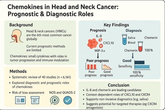
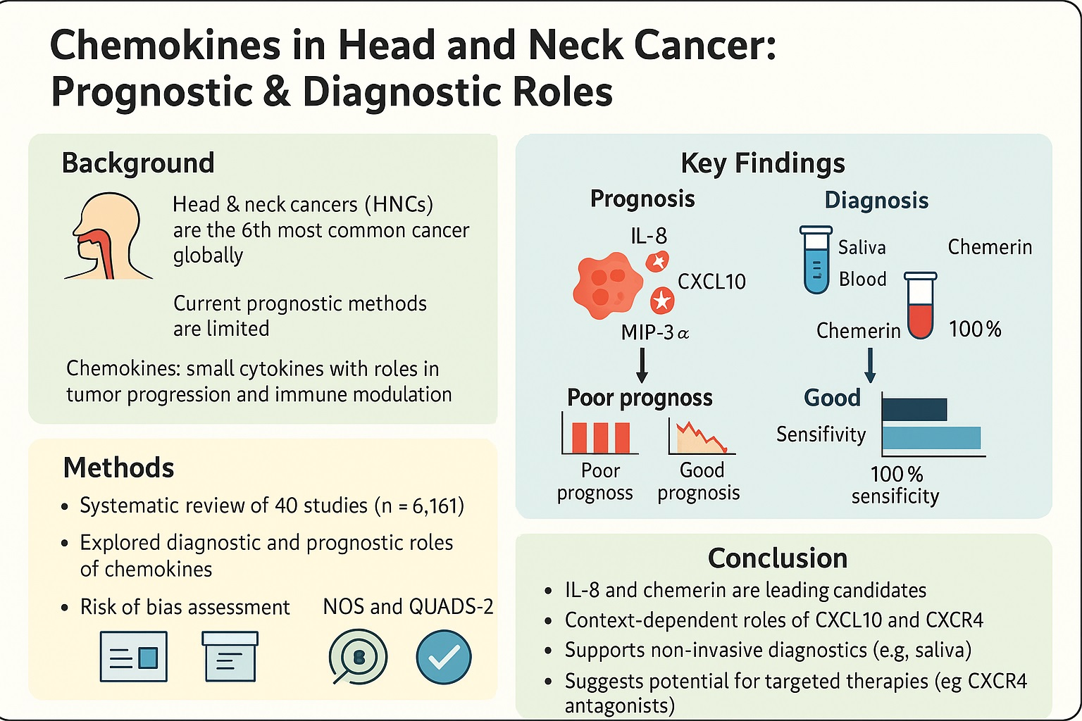
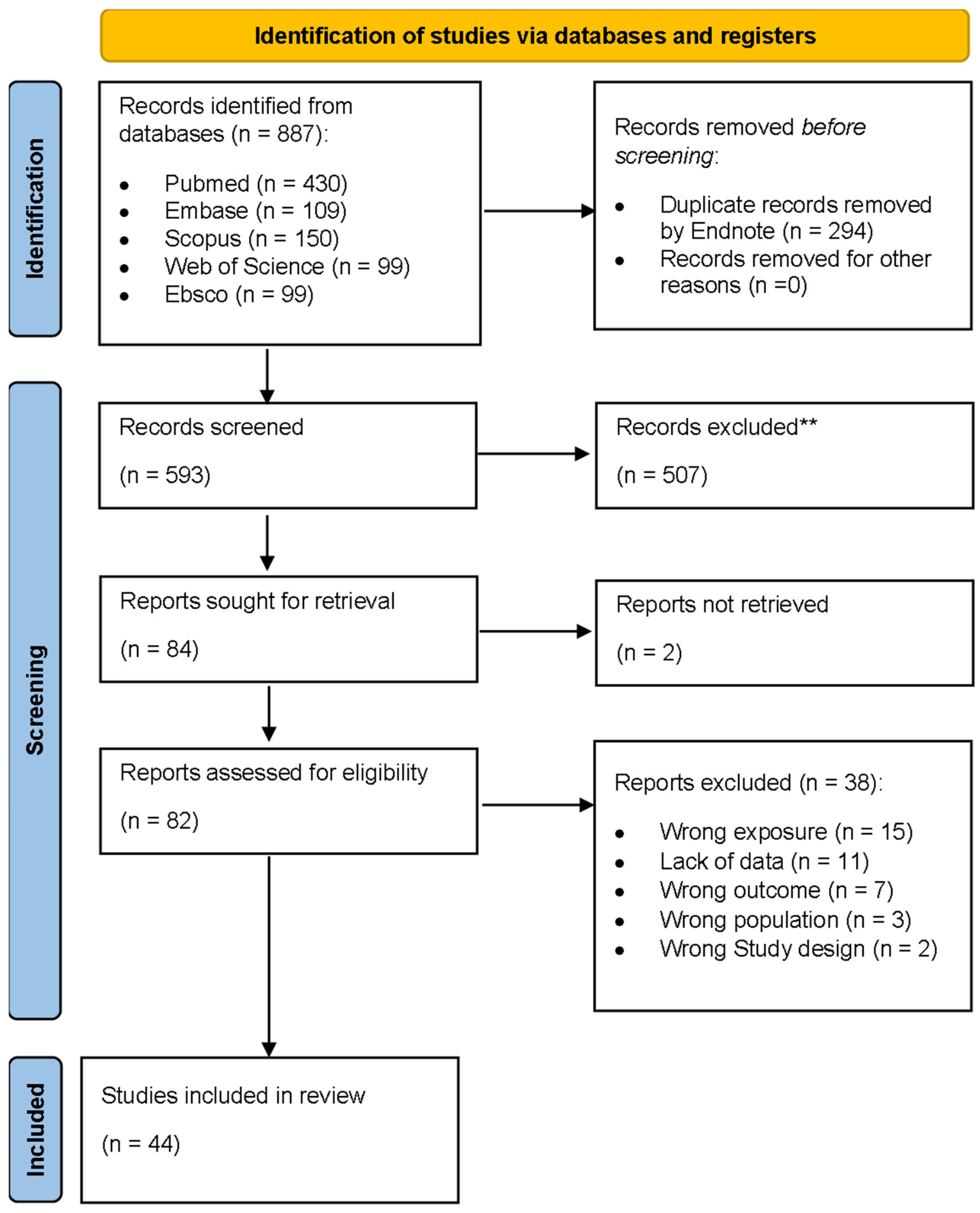
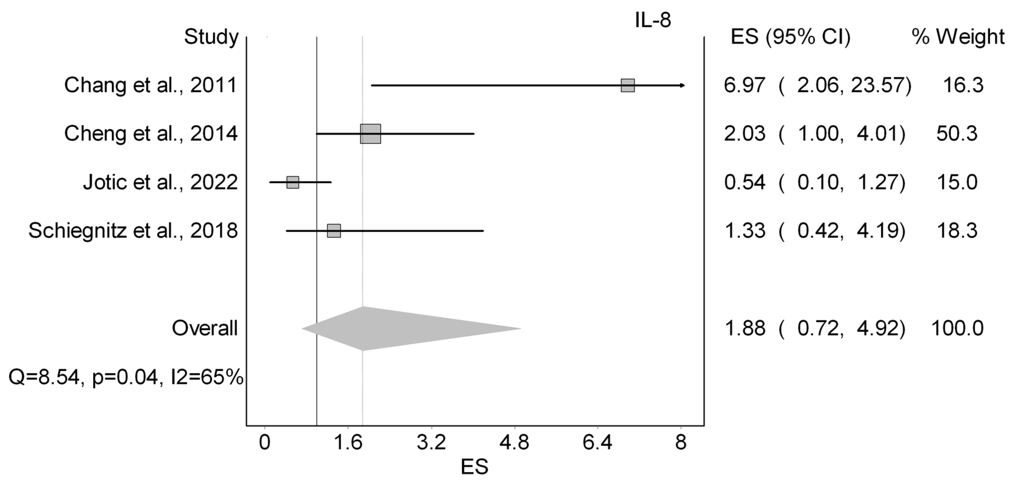
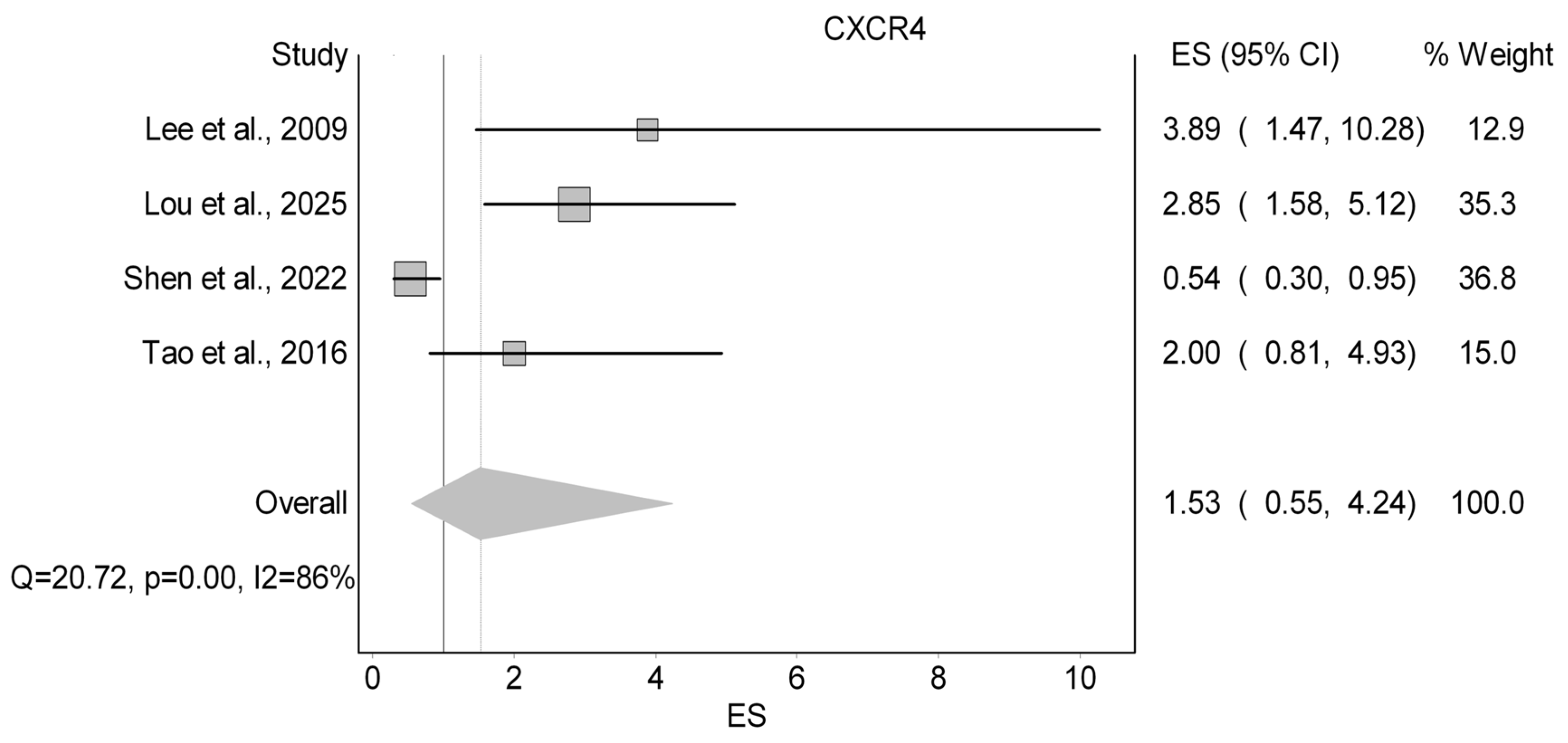
14]. To further investigate their role in the diagnosis and/or prognosis of HNCs, we conducted a comprehensive systematic review of 44 studies, involving 7294 participants. Our findings indicated that high levels of IL-8 were associated with lower survival rates, but the difference is not statistically significant. CXCL10 showed inconsistent results, with some studies linking it to better or worse OS, as well as increased risks for PFS and DFS. CXCR4 also displayed inconsistencies, with increased risks for OS, LRC, and DFS. Finally, MIP-3α was consistently linked to poor prognosis across the studies reviewed. Notably, chemerin and IL-8 were the most extensively studied chemokines in the diagnosis of HNCs. Chemerin demonstrated a promising diagnostic performance in two relatively small studies, achieving 100% sensitivity and specificity, while IL-8 exhibited varying sensitivity and specificity across the included studies.
The overall methodological quality of the studies included supports the credibility of our findings, though some variability was noted. Most of the prognostic studies (21 out of 31) were rated as good quality based on the Newcastle–Ottawa Scale, lending robustness to the observed associations. However, the presence of seven poor-quality and three fair-quality studies introduces some risk of bias—often due to issues such as inadequate comparability between groups, insufficient follow-up, or unclear selection methods—which should be considered when interpreting trends, especially for chemokines with inconsistent findings. In contrast, all 11 diagnostic studies assessed using the QUADAS-2 tool demonstrated a low risk of bias with no applicability concerns, which strengthens our conclusions regarding the diagnostic potential of biomarkers like chemerin and IL-8. Taken together, these quality assessments provide critical context for understanding the prognostic and diagnostic roles of chemokines in HNCs.
In our review, we observed that for each chemokine, different studies employed varying detection techniques, including ELISA, RT-qPCR, immunohistochemistry (IHC), and multiplex assays. This methodological heterogeneity may contribute to the variability in reported diagnostic and prognostic outcomes across studies. For instance, ELISA and multiplex assays detect protein levels, reflecting post-transcriptional modifications and protein abundance, while RT-qPCR measures mRNA expression, which may not directly correlate with protein levels due to regulatory mechanisms [
75,
76]. IHC provides spatial localization of proteins within tissues but is semi-quantitative and highly dependent on antibody specificity and tissue processing [
77]. These differences in detection modalities can influence the sensitivity and specificity of chemokine measurements. Therefore, the observed discrepancies in chemokine-related outcomes across studies may, in part, be attributed to the differences in detection techniques employed, emphasizing the need for methodological standardization in future research.
Altogether, this review illustrates that chemokines exhibit variable prognostic and diagnostic roles in HNCs, with IL-8 and MIP-3α showing more consistent prognostic associations, while chemerin and IL-8 provide a potential diagnostic value, but heterogenicity across studies contributes to the variability in outcomes reported.
4.1. Role of Chemokines and/or Their Receptors in the Prognosis of Head and Neck Cancers
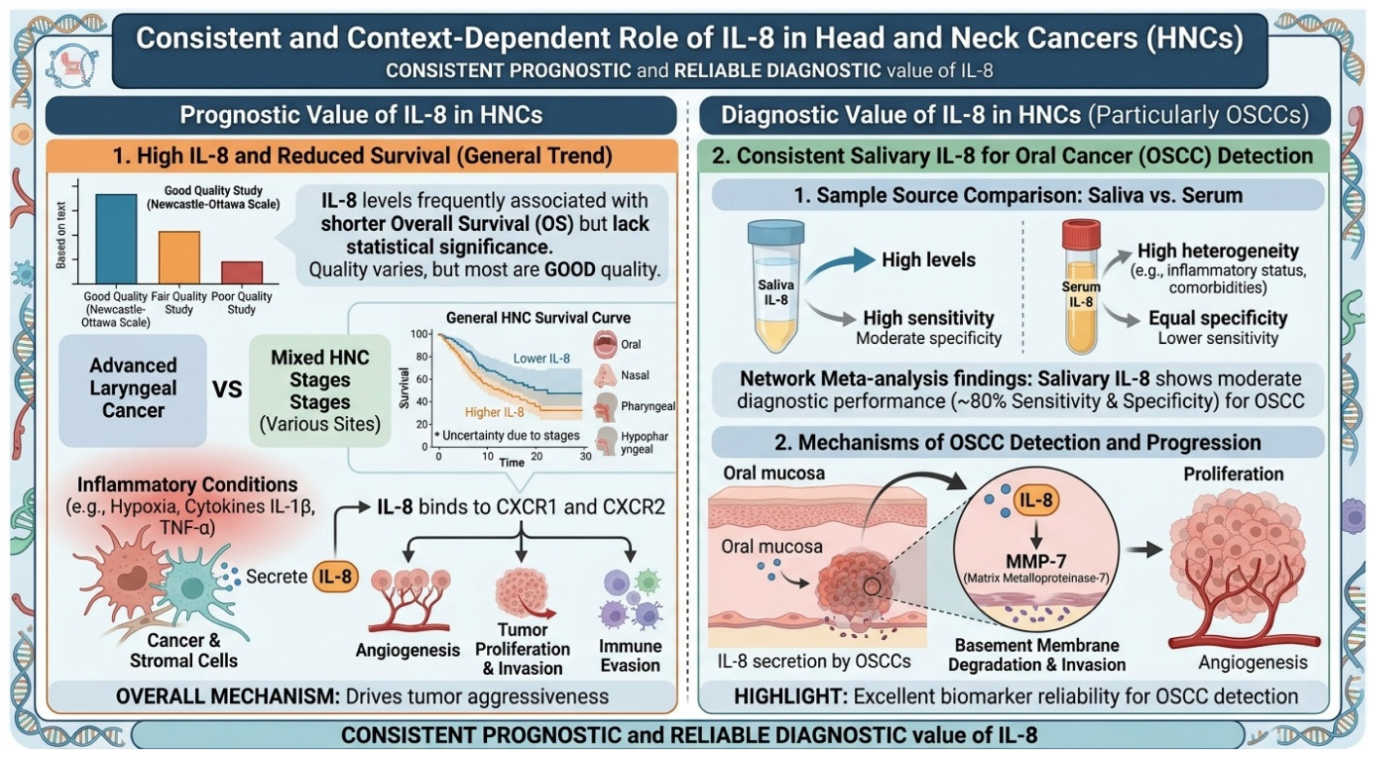
Many chemokines have been associated with cancer progression and prognosis in the literature. IL-8 has been frequently studied for its role in tumor progression and metastasis. Although most included studies showed that IL-8 was associated with reduced OS in patients with HNC, Jotic et al. (2022) identified IL-8 as a significant predictor of 3-year OS and disease-specific survival (DSS) in patients with advanced laryngeal cancer [
65]. This discrepancy could be explained by the fact that Jotic et al. included only patients with advanced laryngeal cancer, whereas other studies included patients at various stages of the disease. Additionally, differences in tumor biology, treatment response, and cytokine interactions in advanced stages may influence the prognostic role of IL-8. These findings highlight the importance of understanding the multifaceted role of IL-8 in tumor progression and its complex prognostic implications. IL-8, also known as CXCL8, is a pro-inflammatory chemokine secreted by tumor and stromal cells under inflammatory conditions like hypoxia and cytokines (e.g., IL-1β, TNF-α), acting via CXCR1 and CXCR2 receptors [
78]. These receptors activate pathways (PI3K/Akt, MAPK, NF-κB) promoting tumor angiogenesis, proliferation, invasion, and immune evasion [
78,
79]. IL-8 also facilitates extracellular matrix degradation and tumor vascularity through endothelial proliferation and MMP expression [
78], leading to greater tumor aggressiveness and worse survival outcomes [
80].
Recent studies have increasingly highlighted the role of CXC chemokines and their receptors in regulating the tumor microenvironment, cancer cell proliferation, and metastasis [
67]. In our study, we identified inconsistencies in the OS results associated with CXCL10. For instance, Li et al. (2021) reported that increased CXCL10 expression was associated with better OS rates in patients with head and neck squamous cell carcinoma (HNSCC), suggesting a potentially favorable role in immune response and tumor suppression [
67]. Conversely, Rentoft et al. (2014) found that high levels of CXCL10 were linked to poor OS in patients with squamous cell carcinoma of the tongue [
72]. Similarly, our results showed that CXCR4 demonstrated contrasting findings with OS. Despite the discrepancy, a meta-analysis by Zhao et al. (2015) revealed that the overexpression of CXCR4 in patients with HNC was associated with worse OS rates [
81].
A number of these conflicting findings may be elucidated by the paradoxical role of CXCL10 and CXCR4 in head and neck cancers. CXCL10, also known as interferon-inducible protein-10, exhibits a bifunctional nature, acting both as an immune recruiter and as a promoter of tumor aggressiveness. While it generally serves to recruit cytotoxic immune cells, such as T lymphocytes and natural killer (NK) cells [
82], its overexpression in HNC is closely associated with more aggressive tumor behavior, immune evasion, and diminished progression-free survival [
58,
66]. Upon binding to its receptor (CXCR3), CXCL10 activates pivotal signaling cascades, including JAK/STAT, ERK1/2, and p38 MAPK, thereby promoting tumor cell proliferation, migration, and epithelial–mesenchymal transition (EMT) [
82,
83]. Similarly, CXCR4 also exhibits dual functionality; in certain malignancies, its activation may induce apoptosis of tumor cells or inhibit their proliferation, thereby functioning as a tumor suppressor [
84]. However, in the context of HNCs, elevated CXCR4 expression has been shown to be correlated with advanced tumor stages and poor survival outcomes through mediating the chemotaxis of tumor cells and their metastatic dissemination to CXCL12-enriched microenvironments [
81], such as lymph nodes, lungs, and liver [
85,
86]. These roles suggest that the prognostic significance of CXCL10 and CXCR4 may vary depending on the cancer subtype as well as the variations within the tumor microenvironment. Factors such as tumor stage and treatment modality may contribute to the observed variability in outcomes, emphasizing the need for further research to understand their context-dependent effects.
Moreover, elevated MIP-3α levels have been consistently linked, in the included studies, to poor survival rates in HNC patients, indicating its potential as a prognostic biomarker. MIP-3α, also known as CCL20, secreted by tumor cells, stromal cells, and TAMs, binds to CCR6 to modulate the tumor microenvironment [
87,
88]. Our findings could be explained by the role MIP-3α plays in the pathogenesis of the HNCs. First, MIP-3α recruits immunosuppressive CCR6+ cells, such as immature dendritic cells and regulatory T cells, suppressing antitumor immunity [
87]. Additionally, it enhances EMT and tumor invasion, further exacerbating the survival rates in HNC patients [
89].
In general, these findings indicate IL-8 and MIP-3α capability as a prognostic marker, as it was consistently correlated with poorer survival. On the other hand, CXCL20 and CXCR4 showed conflicting prognostic effects, possibly due to the influence of tumor subtype and microenvironmental factors.
4.2. Role of Chemokines in the Diagnosis of Head and Neck Cancers
Our results reveal notable differences in the diagnostic performance between IL-8 and Chemerin. Although both chemokines have been implicated in cancer cell proliferation and angiogenesis, underscoring their high diagnostic potential [
90], chemerin remains relatively understudied compared to IL-8.
Our findings suggest that salivary IL-8 is a promising biomarker for diagnosing HNCs, particularly OSCC (
Figure 5). This is supported by studies consistently demonstrating higher salivary IL-8 levels in HNC patients compared to healthy controls [
91,
92]. For example, a network meta-analysis of 40 studies observed that patients with oral cancer had higher salivary IL-8 levels compared to healthy controls, demonstrating a moderate diagnostic performance with 80% sensitivity and specificity [
93]. The strong diagnostic potential of IL-8 stems from its multifaceted role within the tumor microenvironment, particularly in the progression of HNCs. IL-8 secretion by OSCCs has been shown to upregulate matrix metalloproteinase-7 (MMP-7), a key mediator of invasion, while also promoting proliferation, angiogenesis, and the recruitment of immunosuppressive cells [
93,
94,
95]. These diverse mechanisms highlight IL-8’s exceptional reliability as a biomarker for detecting HNCs, especially OSCC.
Upon comparing serum and salivary IL-8, we observed that both exhibited a similar specificity; however, salivary IL-8 showed a greater sensitivity, underscoring its diagnostic advantage for HNCs. In line with our findings, Rezaei et al. (2019) found that salivary IL-8 concentrations of patients with OSCC were 4.8-fold higher than serum levels in their meta-analysis [
91]. It has been suggested that serum IL-8 levels may be influenced by the heterogeneity of HNC, including factors such as tumor progression, inflammatory status, or comorbidities, which limits its reliability as a standalone diagnostic marker [
96]. Hence, salivary IL-8 emerges as a more valuable and stable diagnostic tool, offering a non-invasive, easily accessible, and potentially more reflective measure of local tumor activity and inflammatory responses in the head and neck region.
The relatively high sensitivity and specificity observed for salivary IL-8 in the diagnosis of OSCC highlight its potential clinical relevance as a biomarker. From a translational perspective, IL-8 is particularly attractive due to its detectability in saliva, a biofluid that can be obtained non-invasively. This positions salivary IL-8 as a promising candidate for use in screening and early detection strategies, particularly in high-risk populations. In terms of clinical application, IL-8 may support early detection of OSCC and could be incorporated into multimarker panels to improve diagnostic accuracy when combined with other tumor-related biomarkers. However, given the variability observed across studies, IL-8 is unlikely to function effectively as a standalone biomarker and should instead be considered as part of an integrated diagnostic framework requiring further validation.
Furthermore, despite the limited number of available studies, our paper highlights chemerin as a potentially highly specific and sensitive biomarker for OSCC, although current evidence remains limited. Chemerin achieved 100% specificity and sensitivity across both salivary and serum samples. These findings align with other studies [
36,
97], including Khijmatgar et al. (2024), who identified chemerin as a top-performing biomarker for diagnosing OSCC with a sensitivity of 0.94 (95% CI: 0.78–1.00) [
98]. Chemerin is a multifunctional adipokine with established roles in inflammation, adipogenesis, and glucose homeostasis [
99]. It acts primarily through its receptors CMKLR1 and GPR1, which mediate signaling pathways, including RhoA/ROCK, involved in tumor cell proliferation, migration, invasion, and immune modulation [
100,
101,
102]. The diagnostic relevance of chemerin is highlighted by its elevated levels in biological fluids like saliva in patients with HNCs, particularly OSCCs [
103]. This elevation is associated with key tumor-related processes, such as extracellular matrix remodeling and inflammatory cytokine production [
99]. Additionally, chemerin’s role in recruiting immune cells, such as macrophages and dendritic cells, further supports its utility in differentiating cancerous from non-cancerous states [
100]. These characteristics highlight chemerin as a promising candidate for early detection and diagnostic evaluation in HNCs. Despite these promising findings, several important caveats warrant consideration and cautious interpretation. First, the evidence base for chemerin as a diagnostic biomarker remains limited, with only two small primary studies reporting perfect diagnostic accuracy (
n = 30 and
n = 64 in Ghallab and Shaker (2017) and Susha and Ravindran (2023), respectively) [
36,
47]. Such perfect sensitivity and specificity values are uncommon in biomarker research and may be overestimated due to small sample sizes, potential selection bias, or overfitting, particularly in the absence of validation cohorts. Second, both studies were conducted in specific populations (Egypt and India), and their findings have not yet been validated in larger, independent, multi-center cohorts, limiting generalizability. Third, the absence of external validation cohorts, along with the lack of standardized cutoff values across populations, further constrains the reliability and reproducibility of these results. Additionally, the included studies did not stratify findings by tumor stage or histological grade, leaving uncertainty regarding chemerin’s diagnostic performance across different disease stages. Therefore, while chemerin shows promise as a potential non-invasive biomarker for OSCC detection, these findings should be interpreted as preliminary, and larger prospective studies are needed to validate its diagnostic utility before clinical implementation.
Given that OSCC represented the most frequently investigated tumor subtype in this review, these mechanistic findings are particularly relevant to oral carcinogenesis. OSCC development is strongly influenced by chronic inflammation and tumor microenvironment interactions, where chemokine signaling regulates immune cell recruitment, angiogenesis, epithelial–mesenchymal transition (EMT), and extracellular matrix remodeling. IL-8 promotes OSCC progression through activation of CXCR1/2-mediated pathways that enhance tumor cell proliferation, invasion, and vascularization, while CXCL10 and CXCR4 contribute to immune modulation and metastatic dissemination through tumor–immune crosstalk. Similarly, CCL20 (MIP-3α) has been implicated in OSCC progression through recruitment of CCR6+ regulatory immune cells that facilitate immune evasion and tumor persistence. These findings highlight the biological relevance of chemokine-mediated signaling networks in OSCC pathogenesis and support their investigation as mechanistically informative biomarkers in oral cancer research [
81,
85].
4.2.1. Context-Dependent Performance of Chemokine Biomarkers Across HNC Subtypes
Notably, the diagnostic and prognostic performance of chemokines varied not only between biomarkers but also across tumor subtypes and sample sources, underscoring the biological heterogeneity of HNC. IL-8, for instance, demonstrated relatively consistent adverse prognostic associations across multiple subtypes, including nasopharyngeal carcinoma (NPC) [
53], oral squamous cell carcinoma (OSCC) [
35], and laryngeal squamous cell carcinoma (LSCC) [
63], suggesting a more universal role in promoting tumor progression and inflammation-driven oncogenesis.
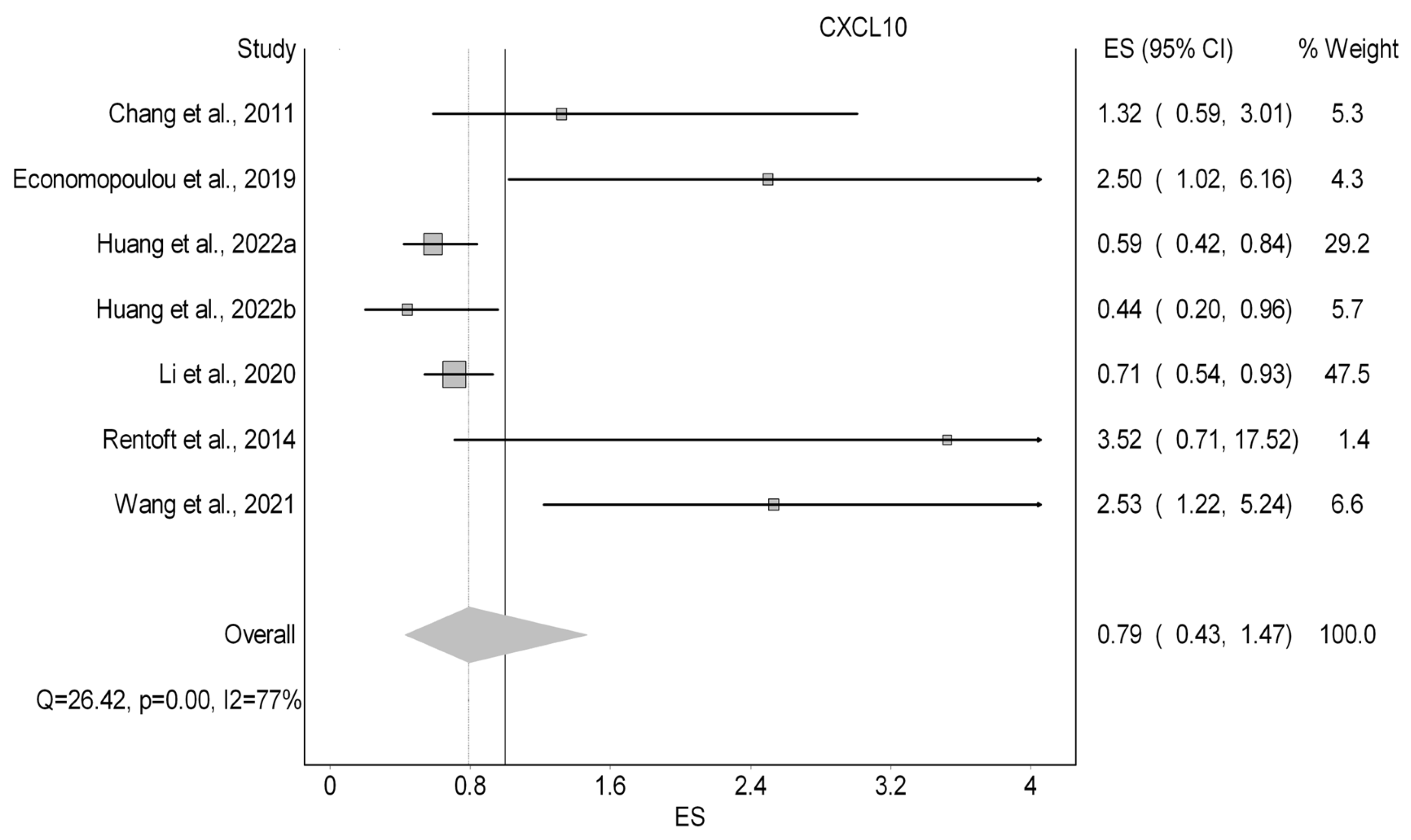
In contrast, CXCL10 and CXCR4 exhibited more context-dependent and, at times, conflicting effects. Elevated CXCL10 expression was associated with improved overall survival in OSCC (HR 0.44–0.59) [
39], yet correlated with poorer outcomes in NPC (HR 2.53) [
58] and HNSCC (HR 2.5) [
66]. Similarly, CXCR4 showed divergent prognostic associations, ranging from protective effects (HR 0.54) [
69] to markedly increased risk (HR 2.00–3.89) [
40,
57]. These inconsistencies likely reflect fundamental differences in tumor biology, including etiological drivers such as human papillomavirus (HPV) in oropharyngeal cancers and Epstein–Barr virus (EBV) in NPC, which differentially shape immune infiltration, chemokine signaling, and tumor–host interactions.
Beyond tumor subtype, sample source also appears to significantly influence biomarker performance. Tissue-based measurements, particularly for CXCL10, were more consistently associated with survival outcomes compared to circulating levels [
39,
58,
67,
72], potentially due to their closer reflection of the localized tumor microenvironment, where chemokine-mediated signaling is most active. This highlights an important methodological consideration, as variability in biospecimen type may contribute to heterogeneity in reported findings across studies.
In contrast to these more extensively studied chemokines, chemerin has been investigated exclusively in OSCC, limiting its applicability to other HNC subtypes and further emphasizing the need for broader evaluation across diverse tumor contexts.
Collectively, these observations reinforce that chemokine biomarkers cannot be interpreted in isolation. Rather, their clinical relevance is highly context-dependent, influenced by tumor subtype, anatomical sites, etiological factors, and methodological differences such as sample source. Future research should therefore prioritize standardized, subtype-stratified analyses and integrative approaches that account for tumor biology, in order to more accurately define the diagnostic and prognostic utility of chemokines in HNC.
4.2.2. Heterogeneity Across Studies and Methodological Considerations
A major finding of this review is the substantial heterogeneity across included studies, complicating direct comparison of results. This heterogeneity was evident across multiple dimensions, including tumor subtypes (OSCC, NPC, LSCC, OPSCC, HNSCC), which differ in molecular profiles and etiologies; sample sources (tissue, serum, saliva, plasma), where biomarker concentrations vary between local and systemic compartments; and outcome measures (OS, DFS, PFS, CSS, DSS, LRC), with differing definitions and follow-up durations. A particularly important contributor to this variability is the wide range of analytical platforms employed, including ELISA, multiplex suspension arrays, immunohistochemistry (IHC), RT-qPCR, gene expression profiling, and immunofluorescence. These techniques differ fundamentally in what they measure. For example, protein-based assays (ELISA, multiplex) quantify circulating or secreted chemokines in biofluids, while RNA-based approaches (RT-qPCR, RNA-seq) measure mRNA transcript abundance, which may not correlate with protein levels due to post-transcriptional regulation, and IHC provides spatial localization but remains semi-quantitative and subject to interpretation variability [
75,
76,
77].
These methodological differences have direct implications for biomarker performance. For instance, tissue-based assessments of CXCL10 using mRNA or IHC demonstrated more consistent prognostic associations, whereas serum-based measurements showed more variable outcomes [
39,
52,
58,
66,
67,
72]. Similarly, IL-8 diagnostic studies employed both ELISA and multiplex assays in saliva, contributing to the wide range of reported sensitivity (58–98%) and specificity (57–100%) [
33,
37,
38,
41,
43,
45,
46].
The observed inconsistencies in prognostic associations for IL-8, CXCL10, and CXCR4 may also reflect the complex and dynamic nature of the tumor microenvironment in head and neck cancers. Chemokine expression is influenced not only by tumor cells but also by stromal and immune components, including macrophages, neutrophils, and endothelial cells, particularly under inflammatory or hypoxic conditions [
78,
79]. For example, IL-8 is produced by multiple cell types within the tumor microenvironment and may reflect both tumor aggressiveness and host inflammatory responsesv [
78,
79,
80]. Similarly, CXCL10 may exhibit context-dependent effects through recruitment of cytotoxic immune cells while also contributing to chronic inflammation and immune dysregulation, potentially explaining its dual prognostic associations [
82,
83]. CXCR4 signaling is likewise influenced by ligand gradients and microenvironmental interactions that regulate tumor cell migration and metastatic dissemination [
85,
86]. In addition, systemic inflammatory conditions and comorbidities may affect circulating chemokine levels, particularly in serum-based studies, further contributing to variability across findings [
91,
96]. Collectively, these observations suggest that heterogeneity in reported associations reflects both biological complexity and methodological differences, which limits comparability across studies, precludes meaningful quantitative synthesis, and complicates the identification of clinically reliable biomarkers. Future research should prioritize methodological harmonization, including standardized sample collection protocols and validated assays, to enable meta-analytic synthesis and clinical translation, while accounting for context-specific tumor microenvironment and immune-related influences on chemokine expression in HNCs.
4.3. Implications and Future Research
The findings from this systematic review highlight the complex and multifaceted roles of chemokines and their receptors in the diagnosis and prognosis of HNCs. By identifying key biomarkers, such as IL-8, CXCL10, CXCR4, MIP-3α, and chemerin, our findings pave the way for future studies to focus on these potentially useful chemokines in HNCs.
From a translational perspective, OSCC represents a particularly suitable model for chemokine biomarker development due to the accessibility of oral lesions and the feasibility of repeated saliva-based sampling. Salivary biomarkers are especially attractive in OSCC because saliva is in direct contact with tumor tissue, allowing detection of locally secreted inflammatory mediators and tumor-derived molecules [
104]. This proximity may partly explain the relatively strong diagnostic performance observed for salivary IL-8 and chemerin in OSCC populations. The integration of salivary chemokine biomarkers into clinical practice could potentially support earlier detection of oral malignancies, improve risk stratification of premalignant lesions, and enable non-invasive monitoring of disease progression or treatment response. Therefore, further validation of chemokine panels in OSCC-specific cohorts may facilitate the development of clinically applicable biomarker strategies in oral oncology therapeutically, targeting chemokine signaling pathways represents a promising strategy for mitigating tumor progression and metastasis. For example, the U.S. Food and Drug Administration has approved AMD3100 (plerixafor, Mozobil
®), a CXCR4 antagonist, while several additional agents targeting chemokine pathways are currently under investigation in clinical trials [
81,
105]. Additionally, combining chemokine-targeted therapies with immunotherapy could potentially enhance anti-tumor immune responses by modulating the tumor microenvironment, creating synergistic therapeutic effects [
106]. This is validated by a recent study conducted by Yoshida et al. (2024) [
107] ho investigated the effects of adding the CXCR4 inhibitor AMD3100 to cisplatin in OSCC cells in vitro and in mouse xenograft models in vivo. The study revealed that this combination improved the anti-tumor effect of cisplatin and reduced the number of CXCR4-positive blood vessels in cisplatin-resistant OSCC xenografts. These findings suggest that the addition of a CXCR4 inhibitor may enhance the anti-tumor effects of cisplatin in patients with refractory OSCC [
107].
Conducting longitudinal studies will be critical for understanding the dynamic changes in chemokine expression during disease progression and treatment, enabling the development of personalized therapeutic strategies. Future research should prioritize large-scale prospective studies to validate the diagnostic utility of chemokines at different stages of HNC. The therapeutic potential of targeting chemokine pathways, such as the CXCL12-CXCR4 axis, also warrants further investigation. Furthermore, meta-analyses to validate the prognostic significance of chemokines across diverse populations and clinical settings will further solidify their role in improving the management and treatment of HNCs.
All in all, these findings underline the clinical potential of chemokines as diagnostic and prognostic biomarkers in HNCs and emphasize the need for future research to support and validate their utility in large-scale studies.
4.4. Limitations
There are several limitations that must be acknowledged. First, the review is confined to studies published in the English language, potentially excluding relevant research published in other languages, which could introduce language bias. Second, although a meta-analysis was performed, it was limited to a small subset of chemokines and restricted to OS outcomes. The included studies demonstrated substantial heterogeneity, as reflected by the high I2 values observed in our analyses. Potential sources of this heterogeneity include differences in tumor subtypes (e.g., OSCC, NPC, OPSCC), sample types (saliva, serum, and tissue), study designs, patient populations, tumor stages, and methodological variations, including differences in analytical and technical techniques used among the included studies. These factors likely contributed to the observed variability and limit the interpretability of the pooled results. Furthermore, the small number of studies contributed to wide confidence intervals and statistically non-significant findings, raising the possibility of type II error. There is also a potential risk of publication bias, although this could not be reliably assessed due to the limited number of studies included in each synthesis. Subgroup and sensitivity analyses were not performed because of the small number of studies and limited variability in reported variables. Another important limitation is the clinical heterogeneity of tumor types included in this review, encompassing oral cavity, oropharyngeal, nasopharyngeal, and laryngeal cancers. These entities differ in their underlying biology, including molecular drivers, HPV status, and tumor immune microenvironment. Due to the limited number of studies available for each chemokine within individual tumor subtypes, stratified or subgroup analyses by tumor site were not feasible. This may limit the interpretability and generalizability of the findings, and future research should aim to provide more standardized, site-specific data to allow more precise analyses. Furthermore, a meta-analysis of diagnostic performance was not feasible, as most studies reported only sensitivity and specificity without sufficient data to reconstruct 2 × 2 contingency tables. Additionally, the included studies themselves have inherent limitations, as many of the studies included in this review are case–control or retrospective cohort studies, which are inherently at risk of selection bias. Such study designs, combined with small sample sizes and varying quality, further constrain the generalizability of the results. Moreover, it is important to note that most of the studies were conducted in China, which may restrict the external validity of the findings to other populations. Additionally, the lack of stratification by Human Papillomavirus (HPV) status, especially in oropharyngeal cancer, is a key limitation, as HPV-positive tumors have distinct chemokine profiles and prognosis. Lastly, due to inconsistent tumor site reporting, we could not perform site-specific analyses; nasopharyngeal carcinoma was included for completeness but differs significantly from other HNC subtypes. Future studies should stratify by HPV status and tumor site to improve biomarker specificity and clinical relevance.




 ,
,