Article

Gene-Specific Outcomes After Central Nervous System Metastases in Germline BRCA1- and BRCA2-Associated Breast Cancer


1
Department of Obstetrics and Gynecology, Comprehensive Cancer Center, Medical University of Vienna, 1090 Vienna, Austria
2
Department of Laboratory Medicine, Medical University of Vienna, 1090 Vienna, Austria
3
Faculty of Medical and Life Sciences, Sunway University, Petaling Jaya 47500, Selangor, Malaysia
4
Cancer Research Malaysia, Subang Jaya 47500, Selangor, Malaysia
5
Department of Medicine I, Division of Oncology, Medical University of Vienna, 1090 Vienna, Austria
6
The Peter MacCallum Cancer Centre, Melbourne, VIC 3010, Australia
7
Sir Peter MacCallum Department of Oncology, University of Melbourne, Parkville, VIC 3010, Australia
*
Author to whom correspondence should be addressed.
Cancers 2026, 18(8), 1240; https://doi.org/10.3390/cancers18081240

Submission received: 12 March 2026 / Revised: 8 April 2026 / Accepted: 11 April 2026 / Published: 14 April 2026
(This article belongs to the Section Cancer Epidemiology and Prevention)

Simple Summary

Breast cancer is one of the leading causes of central nervous system (CNS) metastases, which are associated with reduced overall survival. In patients with inherited BRCA1 or BRCA2 mutations, breast cancers differ in their biology and clinical behavior, yet these groups are often combined in studies of CNS disease. In this multicenter cohort from Austria and Australia, we evaluated outcomes separately for BRCA1 and BRCA2 mutation carriers and compared them with non-carriers. We found that patients with BRCA2 mutations developed CNS metastases later and showed longer survival after CNS diagnosis in unadjusted analyses. However, these differences were attenuated after accounting for tumor subtype and other clinical factors. These findings suggest that BRCA1- and BRCA2-associated breast cancers may follow distinct disease trajectories in the CNS setting, but should be interpreted with caution. Our results highlight the importance of gene-specific analyses and support further investigation in larger, prospective cohorts to better understand how inherited genetic background may influence prognosis and treatment in this setting.

Abstract

Background: Many studies evaluating central nervous system (CNS) metastases in breast cancer (BC) combine germline BRCA1 and BRCA2 pathogenic variant carriers, limiting gene-specific interpretation. We evaluated gene-specific overall survival (OS) after CNS metastasis and time to CNS involvement. Methods: We retrospectively identified BC patients with confirmed CNS metastases (1995–2022) from the Medical University of Vienna and the kConFab consortium. Germline status was classified as gBRCA1 PV, gBRCA2 PV, or non-carrier. Primary endpoint was OS from CNS metastasis diagnosis; secondary endpoint was CNS metastasis-free interval from primary BC diagnosis. Kaplan–Meier/log-rank tests were used for group comparisons. Multivariable Cox models assessed OS in complete cases, stratified by molecular subtype and adjusted for prognostic factors. Sensitivity analyses included subtype-adjusted and time-period models. Results: Among 115 patients (gBRCA1 n = 32, gBRCA2 n = 18, and non-carriers n = 65), median OS differed by germline status (p = 0.019): 20.0 months (95% CI 6.7–60.0) for gBRCA2 versus 7.1 months (95% CI 3.7–10.0) for gBRCA1 and 7.6 months (95% CI 3.4–12.0) for non-carriers. In subtype-stratified analyses, gBRCA1 showed similar mortality to non-carriers (HR 0.90, 95% CI 0.49–1.64, p = 0.730), while gBRCA2 showed a lower but non-significant hazard (HR 0.48, 95% CI 0.18–1.25, p = 0.131). Median CNS metastasis-free interval was longer for gBRCA2 (8.4 years) versus gBRCA1 (3.0 years) and non-carriers (3.1 years; p = 0.020). Sensitivity analyses were consistent. Conclusions: gBRCA2 carriers demonstrated longer unadjusted OS after CNS metastasis and a longer CNS metastasis-free interval compared with gBRCA1 carriers and non-carriers. However, these associations were attenuated and not statistically significant after adjustment, and should therefore be interpreted as hypothesis-generating. These findings supports further investigation of gene-specific CNS disease trajectories in larger cohorts.

1. Background

Central nervous system (CNS) metastases, including parenchymal brain metastases and leptomeningeal disease, occur in about 20–30% of patients with metastatic breast cancer (BC) and remain a major cause of morbidity and mortality, with median survival typically ranging from 6 to 36 months depending on tumor subtype and treatment [1,2,3,4,5]. The risk of CNS involvement is strongly influenced by tumor biology and is highest in triple-negative and HER2-positive disease, with important implications for systemic treatment selection. Current management is multidisciplinary and may include stereotactic radiosurgery (SRS) and, in selected cases, surgical resection with systemic treatment tailored to subtype [6,7,8,9]. Whole-brain radiotherapy (WBRT) is now used more selectively because of its adverse effects on neurocognitive function and quality of life. In parallel, contemporary guidelines increasingly support an initial systemic therapy approach in selected patients when subtype-specific agents with demonstrated intracranial activity are available [2,4,5,8,10].
For HER2-positive BC, tucatinib in combination with trastuzumab and capecitabine has been shown to improve intracranial progression-free survival (PFS) and overall survival (OS) [11], while trastuzumab-deruxtecan (T-DXd) has also demonstrated meaningful intracranial activity [12,13,14]. In germline BRCA1/2 (gBRCA1/2)-associated BC, PARP inhibitors have shown superior efficacy to chemotherapy in metastatic disease [15,16,17,18], with evidence of activity also in patients with stable CNS metastasis [19,20]. However, the impact of gBRCA1/2 status on outcomes after CNS metastasis remains unclear. Interpretation is complicated by the close relationship between BRCA gene status, tumor subtype and baseline CNS risk [21,22,23,24]. In addition, homologous recombination deficiency may influence metastatic patterns, including CNS involvement [25,26].
Although prior studies suggest a higher risk of CNS involvement in gBRCA1-associated BC [27,28,29,30] and possible subtype-independent CNS involvement in gBRCA2-associated disease [29], CNS-specific outcome data remain limited. Existing studies are constrained by small sample sizes, heterogeneous endpoints, and importantly, the frequent aggregation of BRCA1 and BRCA2 carriers, which may obscure clinically meaningful gene-specific differences [29,31].
To address this gap, we analyzed a binational multicenter cohort from Austria and Australia comprising BC patients with CNS metastases to compare OS after CNS metastasis and the CNS metastasis-free interval across gBRCA1, gBRCA2 and non-carriers, while accounting for molecular subtype and key clinical prognostic factors. We hypothesized that gBRCA2 PV carriers would show distinct CNS disease trajectories compared with gBRCA1 carriers.

2. Methods

2.1. Study Design and Population

We conducted a retrospective cohort study of BC patients from the MUV and the national Australasian kConFab consortium with confirmed CNS metastasis (brain parenchymal and/or leptomeningeal disease) diagnosed between 1995 and 2022. CNS involvement was confirmed by computer tomographic and/or magnetic resonance imaging, lumbar puncture or autopsy report. Patients were included in the study if they were 18 years and above, had histologically confirmed BC, known gBRCA1/2 status (gBRCA1/2 PV class 4 or 5 vs. non-carrier class 1 or 2), and provided research consent. Patients were excluded if they had unconfirmed CNS disease, missing genetic status, gBRCA1/2 class 3 variants (variants of uncertain significance), or non-BRCA1/2 PVs. Due to low number of patients with HER2-positive and unknown molecular subtypes, these groups were retained for descriptive analyses but were excluded from Cox models.

2.2. Variables and Data Collection

MUV clinical data were extracted from medical or autopsy records. Germline BRCA1/2 status was derived from clinical genetic testing and retrieved from the Austrian ATHENA hereditary cancer registry, and outcomes data through linkage with Statistics Austria. kConFab data were collected through treating hospitals, clinical pathology centers and cancer registries with additional information obtained from structured follow-up questionnaires administered every three years. In the present analyses, “non-carriers” refers to non-carriers of gBRCA1/2 pathogenic variants. The extent of additional multigene panel testing differed by cohort and calendar period. In the MUV cohort, panel testing beyond BRCA1/2 was introduced from 2015 onwards, whereas in kConFab, a subset of BRCA1/2-negative individuals underwent at least a 10-gene panel test. As such, undetected pathogenic variants in other homologous recombination repair genes (e.g., PALB2, ATM) cannot be fully excluded in earlier testing eras.
Variables collected included sex assigned at birth, menopausal status at BC diagnosis, tumor histology/molecular subtype, local CNS treatments, presence of leptomeningeal disease or other metastases detected by imaging, CNS lesion count, and CNS-directed treatment types.

2.3. Statistical Analysis

Baseline characteristics were summarized by gBRCA status using medians and interquartile ranges (IQRs) for continuous variables and counts with percentages for categorical variables. Group comparisons were performed using a Kruskal–Wallis test for continuous variables and Chi-square or Fisher’s exact tests for categorical variables.
OS was defined as the time from CNS metastasis diagnosis to death from any cause and patients alive at last follow-up were censored. The primary endpoint was OS from CNS metastasis diagnosis. Secondary endpoint was CNS metastasis-free interval, defined as the time from primary BC diagnosis to CNS metastasis. We estimated OS and CNS metastasis-free interval using Kaplan–Meier methods and compared germline groups using log-rank tests, with Bonferroni-adjusted pairwise comparisons. We did not fit multivariable models for this endpoint because all included patients developed CNS metastasis by design and it does not allow valid inference regarding predictors of incident CNS involvement in the broader metastatic population. In addition, several factors such as imaging intensity/surveillance and systemic therapy exposure are not captured with sufficient completeness to support a robust multivariable risk model.
Multivariable associations with OS were evaluated using Cox proportional hazards regression. Prespecified covariates included age at CNS metastasis diagnosis (continuous), gBRCA status (gBRCA1 PV, gBRCA2 PV, non-carrier), presence of leptomeningeal disease (yes/no), number of CNS metastases (1/≥2), presence of extracranial metastases (yes/no), and receipt of local CNS-directed therapy (WBRT-only, SRS-only, WBRT + SRS, any surgery ± WBRT/SRS, and no local therapy). Covariates were selected based on clinical relevance and established prognostic factors for BC brain metastases [1]. We could not implement the diagnosis-specific graded prognostic assessment (DS-GPA) score in the models because performance status (ECOG or Karnofsky) was not available consistently for both cohorts. We therefore adjusted for key prognostic components aligned with those used in DS-GPA that were captured in our dataset, including germline status. Molecular subtype was additionally addressed through stratified analyses and sensitivity-adjusted models to evaluate the robustness of estimates to residual confounding by subtype. Cox models were restricted to cases with known subtype and limited to luminal and triple-negative cases only. HER2-positive cases were excluded because of low numbers (n < 5) to ensure model stability and to avoid unreliable estimates. All analyses were conducted as complete-cases analyses without imputation. Proportional hazards assumptions were evaluated using Schoenfeld residuals. To evaluate whether results were sensitive to temporal changes in CNS imaging and clinical management, we repeated the models using a time-period indicator (diagnoses pre- and post-2010). Year 2010 was selected to approximate a shift toward routine MRI availability and modern stereotactic approaches. Potential cohort effects were explored by comparing OS between cohorts using Kaplan–Meier curves and log-rank tests. Cohort-stratified Cox models were not fitted due to small sample size, but cohort-specific univariable models were estimated to assess whether the direction of gBRCA associations was consistent within each cohort. All analyses were performed using R (version 4.4.1) and two-sided p-values < 0.05 were considered statistically significant.

3. Results

3.1. Descriptive Analysis

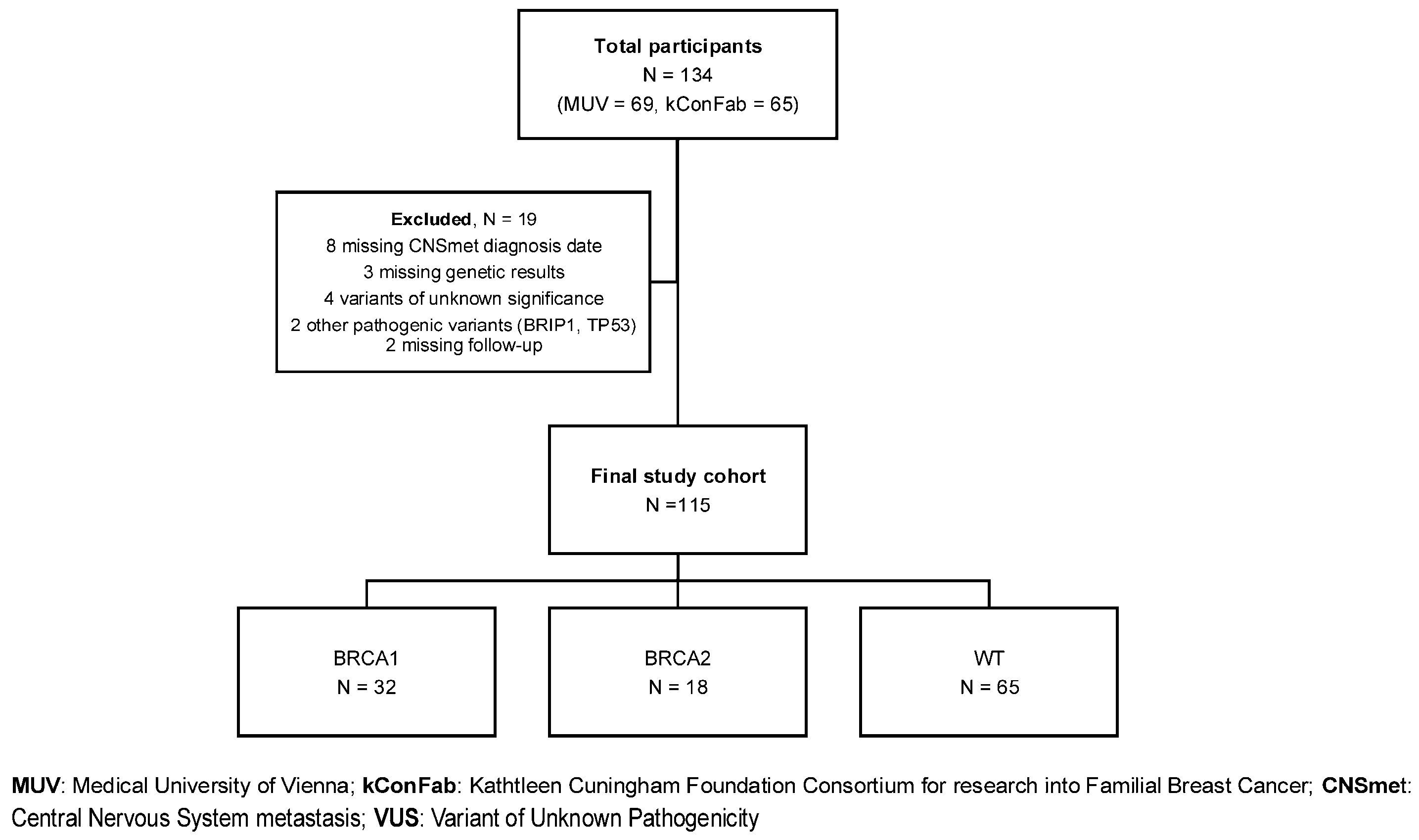
Figure 1 shows patient disposition. Baseline characteristics by gBRCA1 status are presented in Table 1. Of 115 patients, 105 were female, 3 were male, and 7 unspecified. Median age at CNS metastasis diagnosis was 48.4 years (IQR 40.3–56.3) and differed significantly across gBRCA subgroups (p < 0.003), with gBRCA1 PV carriers presenting at a younger age than both gBRCA2 PV carriers and non-carriers (41.1 years vs. 48.9 years vs. 51.6 years, respectively). Bonferroni-adjusted pairwise comparisons showed differences between gBRCA1 and gBRCA2 PV (p = 0.017) and between gBRCA1 PV and non-carriers (p = 0.011), but no difference was observed between gBRCA2 PV and non-carriers (p = 0.999). The younger age at CNS metastasis among gBRCA1 carriers was also observed when restricting descriptively to triple-negative cases. Breast cancer molecular subtype showed borderline differences by gBRCA status (p = 0.053), with gBRCA1 PV carriers enriched for triple-negative disease (65.6%) and gBRCA2 PV carriers enriched for luminal disease (44.4%). Subtype was unknown more often among gBRCA2 PV carriers (38.9%). When restricted to cases with known subtype, baseline distributions also differed between MUV and kConFab cohorts (Supplementary Table S1), so cohort effects on OS were assessed prior to pooling.

3.2. Survival Analysis

During the observation period, 104/115 patients died and eleven were censored alive at last follow-up, giving an overall event rate of 90.4%. Among censored patients, median follow-up was 16.5 months (IQR 4.3–38.5).
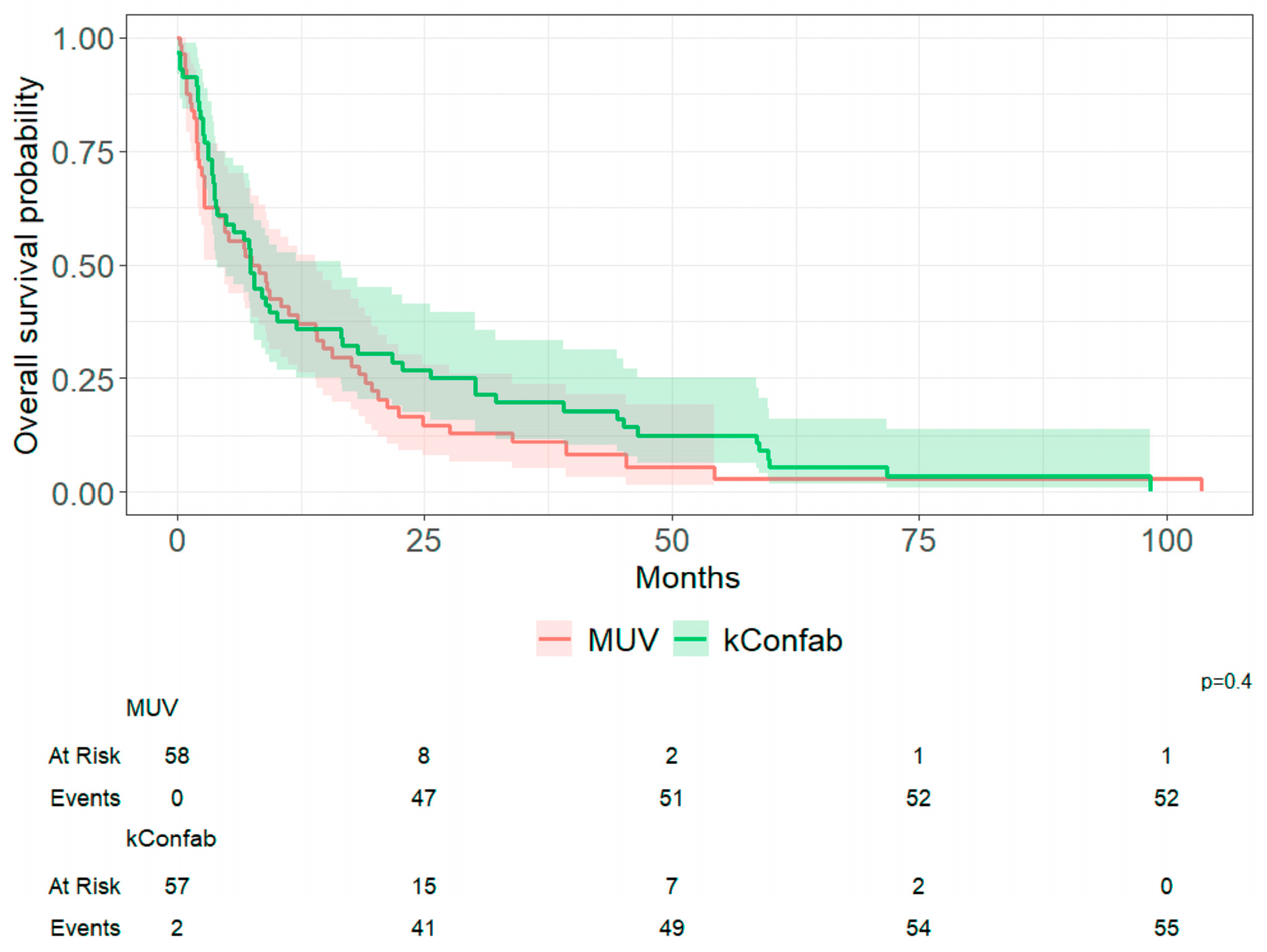
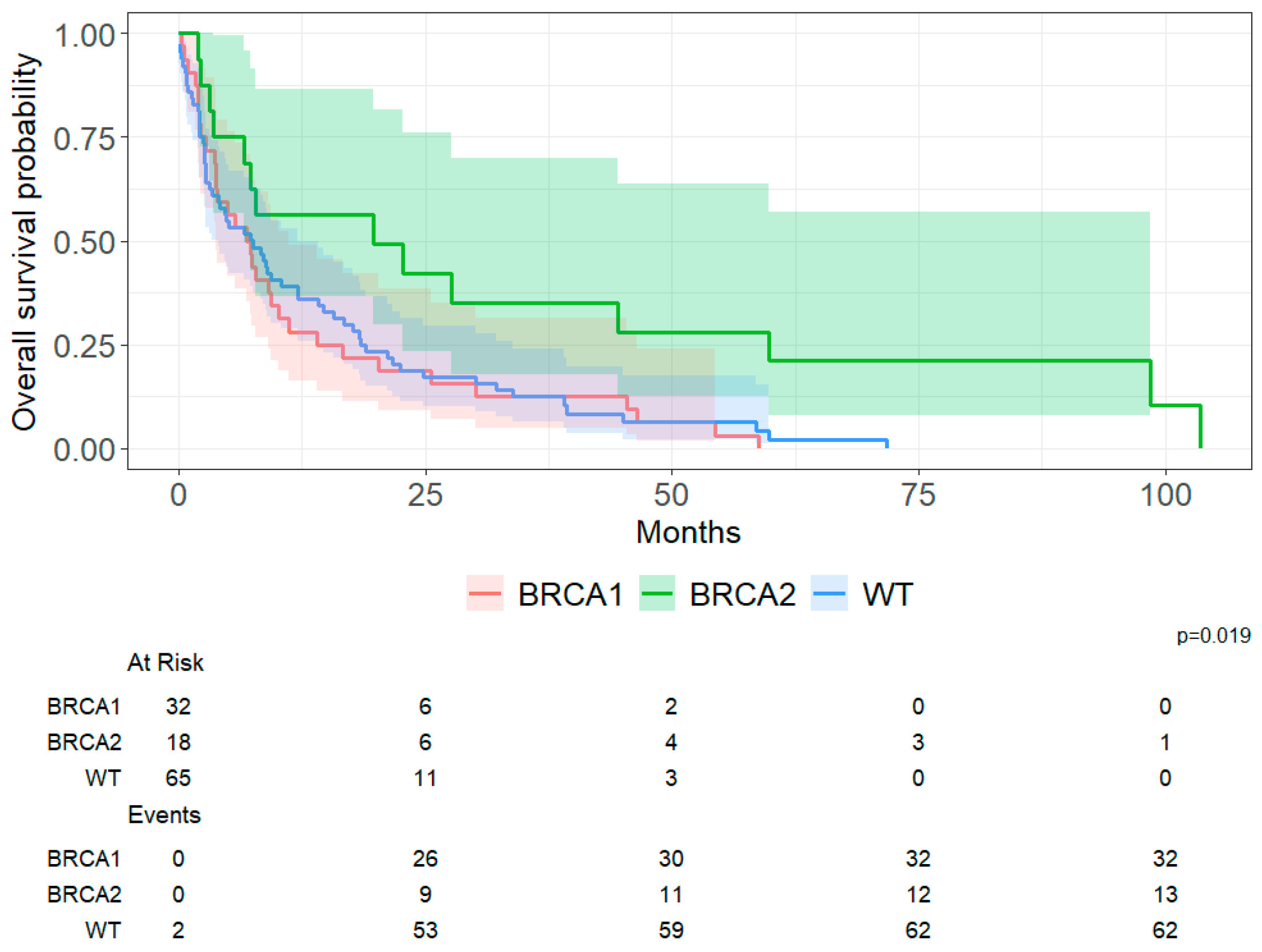
OS did not differ between the MUV and kConFab cohorts on Kaplan–Meier analysis (Figure 2). Cohort-specific Kaplan–Meier curves across germline groups are shown in Supplementary Figures S1 and S2. Cohort-specific univariable Cox models are provided in Supplementary Table S2. Cohort-specific estimates for gBRCA2 carriers versus non-carriers were consistent in direction, although confidence intervals were wide due to small sample size (MUV HR = 0.26, 95% CI 0.06–1.09, p = 0.065; kConFab HR = 0.62, 95% CI 0.29–1.33, p = 0.220). As such, subsequent analyses were performed using the combined dataset. Figure 3 shows that survival differed across gBRCA subgroups. Germline BRCA2 PV carriers had the longest post-CNS metastasis survival (median 20 months; 95% CI 6.7–60.0) compared with gBRCA1 PV carriers (7.1 months; 95% CI 3.7–10.0, p = 0.051) and non-carriers (7.6 months; 95% CI 3.4–12.0, p = 0.029). The overall three-group comparison was significant (log-rank p < 0.019). In Bonferroni-adjusted pairwise comparisons, survival differed between gBRCA2PV carriers and non-carriers (p = 0.029) while the comparison between gBRCA2 and gBRCA1 PV carriers was borderline (p = 0.051).
To contextualize these findings, we also derived absolute survival estimates from the Kaplan–Meier curves. At 12 months after CNS metastasis diagnosis, approximately 56% of gBRCA2 PV carriers remained alive compared to 39% of non-carriers and 28% of gBRCA1 PV carriers. This represents a 17% absolute difference in 1-year survival between gBRCA2 PV carriers and non-carriers.

3.3. Multivariate Analysis

In a multivariable model stratified by subtype (n = 77, restricted to luminal and triple-negative subtype, Table 2), gBRCA2 PV carriers had a lower hazard of death compared with non-carriers (HR = 0.48, 95% CI 0.18–1.25, p = 0.131), although this finding did not reach statistical significance. gBRCA1 PV carriers had hazards like non-carriers (HR 0.90, 95% CI 0.49–1.64, p = 0.730). Subtype effects were not estimated directly in this model due to stratification. Compared to WBRT alone, patients who received SRS only (HR = 0.55, 95% CI 0.23–1.30, p = 0.175), WBRT + SRS (HR = 0.57, 95% CI 0.26–1.28, p = 0.173), and any surgery ±WBRT/SRS (HR = 0.58, 95% CI 0.26–1.25, p = 0.164) had lower hazards, whereas no local therapy had a higher hazard (HR = 2.28, 95% CI 0.84–6.16, p = 0.105). Schoenfeld residuals test did not suggest violations of the proportional hazards assumption for the final subtype-stratified complete-case model.
In the model including molecular subtype as an adjustment covariate (Supplementary Table S3), TNBC was associated with worse OS versus luminal disease (HR = 1.83, 95% CI 1.00–3.36, p = 0.050) while the gBRCA2 estimate continued to favor lower mortality versus non-carriers, although it was not statistically significant (HR = 0.46, 95% CI 0.18–1.17, p = 0.104). In time-period sensitivity analyses, the gBRCA2 association was similar in direction (gBRCA2 PV vs. non-carrier, HR = 0.55–0.57; Supplementary Tables S4 and S5).

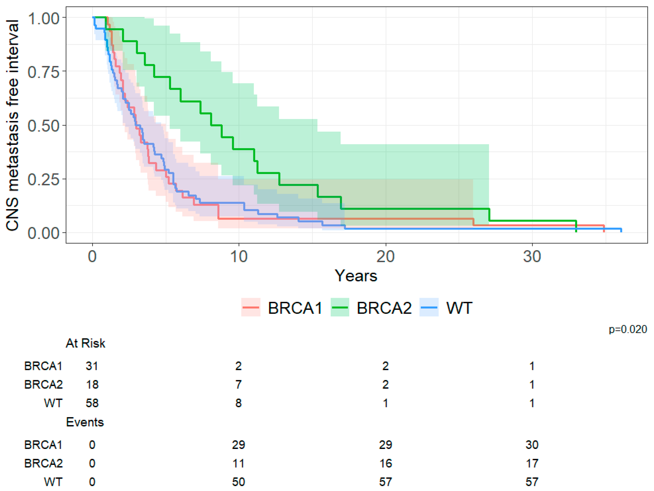
3.4. CNS Metastasis-Free Interval

A total of 107 patients were included in this analysis. Eight were excluded due to missing date of primary BC diagnosis. CNS metastasis-free interval differed by germline group (log-rank, p = 0.020; Figure 4). Median CNS metastasis-free interval was 3.0 years for gBRCA1 PV carriers (95% CI 2.1–5.0), 8.4 years for gBRCA2 PV carriers (95% CI 5.3–15.0) and 3.1 years for non-carriers (95% CI 2.3–4.7). In the pairwise comparisons, gBRCA2 carriers had a significantly longer CNS metastasis-free interval than both gBRCA1 PV carriers (p = 0.006) and non-carriers (p = 0.006), while no significant difference was observed between gBRCA1 PV carriers and non-carriers.

4. Discussion

In this multicenter retrospective cohort of BC patients with CNS metastases, outcomes differed by germline BRCA status, with gBRCA2 PV carriers showing longer OS post-CNS metastasis than gBRCA1 PV carriers and non-carriers in unadjusted analysis. This is clinically relevant because gBRCA1 and gBRCA2-associated BC differ in clinicopathologic features and systemic disease course [21,22,23,32], yet many prior studies have pooled gBRCA1 and gBRCA2 carriers, which can obscure meaningful gene-specific patterns. Our CNS-specific results are timely and provide context in an era when systemic therapies with intracranial activity, such as PARP inhibitors and antibody–drug conjugates, are increasingly used in metastatic BC with CNS involvement (for, e.g., OlympiAD [15], EMBRACA [17] and DESTINY-Breast12 [12]), while CNS-specific evidence remains comparatively limited [5]. In parallel, molecular profiling studies report higher homologous recombination deficiency (HRD) signals in BC brain metastases than in matched primary tumors, raising the possibility that PARP inhibitor benefit may extend beyond germline BRCA-associated disease and supporting prospective evaluation of HRD-guided systemic approaches in CNS metastases [25,33,34].
The apparent gBRCA2 survival advantage observed in the unadjusted analyses was attenuated and no longer statistically significant in multivariable models, both when molecular subtype was handled by stratification or when it was included as an adjustment covariate. This may reflect, in part, the unequal distribution of tumor subtype across gBRCA groups, a major determinant of outcomes after CNS metastasis, with gBRCA1 cases enriched for TNBC and gBRCA2 for hormone-receptor positive or luminal disease. The small number of gBRCA2 cases also limited precision for adjusted estimates. These findings should therefore be interpreted as associations rather than evidence of an independent prognostic effect of gBRCA status after CNS metastasis.
A key consideration in interpreting these findings is the strong correlation between gBRCA status and breast cancer molecular subtype. In our cohort, gBRCA1 carriers were predominantly triple-negative, whereas gBRCA2 carriers were more frequently associated with luminal subtypes. Given the well-established differences in natural history, treatment responsiveness, and survival outcomes between these subtypes, it is likely that part of the observed survival advantage in gBRCA2 carriers reflects subtype-related effects rather than germline status per se. Although we addressed this through stratified and adjusted analyses, residual confounding cannot be excluded, particularly given the limited sample size.
Our gene-specific findings are consistent with a prior report suggesting gBRCA2 PV carriers have longer survival after brain metastasis than gBRCA1 patients [28]. Berliner et al. [31] reported substantially shorter survival in pooled gBRCA1/2 carriers compared with non-carriers after CNS diagnosis, but the sample size was small (n = 75) and the lack of gene-specific stratification limits interpretation. When we applied the same pooled-carrier approach to our dataset (i.e., gBRCA1 + gBRCA2 PV versus non-carriers), survival was similar between carriers and non-carriers (log-rank p = 0.295, HR 0.81, 95% CI 0.55–1.20), illustrating how pooling can obscure gene-specific outcomes and contribute to discrepant findings across studies. Song et al. [29] studied patients at first locoregional recurrence and distant metastasis, and found that CNS involvement was frequent among gBRCA PV carriers, particularly for gBRCA2 PV carriers. However, their survival endpoint was BC-specific survival and measured from first recurrence or metastasis instead of CNS diagnosis, which limited direct compatibility with our study. Moreno et al. [27] restricted to patients diagnosed with TNBC, and likewise, reported higher risk of metastasis among gBRCA2 PV carriers. They also reported a trend toward lower mortality among gBRCA carriers compared to non-carriers (gBRCA1 PV HR 0.52, 95% CI 0.27–1.00, p = 0.053; gBRCA2 PV HR 0.80, 95% CI 0.31–2.09, p = 0.659). Their cohort also had more bone metastases in gBRCA2 PV carriers and lung metastases in gBRCA1 PV carriers, patterns which we similarly observed in our cohort but did not reach statistical significance. These differences in extracranial metastatic patterns may be clinically relevant, as visceral metastases such as lung involvement are generally associated with poorer prognosis compared with bone-dominant disease. As such, variation in metastatic distribution across germline groups may have contributed to the observed survival differences. However, given the limited sample size and the absence of time-dependent modeling of metastatic burden and progression, we are unable determine whether these patterns independently account for differences in OS across groups. These findings are therefore interpreted as descriptive and hypothesis-generating, and warrant further investigation in larger cohorts with longitudinal modeling of metastatic trajectories.
We assessed the CNS metastasis-free interval as a secondary outcome, and we found that gBRCA2 PV carriers had a significantly longer time from primary BC diagnosis to CNS metastasis compared to gBRCA1 and non-carriers. Berliner et al. [31] reported a slightly longer interval among pooled carriers compared to non-carriers, but their pooled analysis did not allow for gene-specific interpretation. In our cohort, the gene-stratified pattern is consistent with known differences in tumor phenotype and overall disease progression patterns between gBRCA2- and gBRCA1-associated BC, but it should be interpreted as a timing comparison among patients who developed CNS metastasis.
Beyond survival and metastasis-free interval, we found that gBRCA1 PV carriers were younger at CNS metastasis diagnosis than gBRCA2 PV and non-carriers, indicating a difference in age distribution at CNS presentation across germline groups in our cohort. This younger age at presentation was also observed when restricting to triple-negative cases. This finding is consistent with the pooled BRCA series from Berliner et al. [31], but in contrast with Song et al. [29] and Moreno et al. [27], who reported age at first distant metastasis or breast metastasis instead of age at CNS metastasis, thereby limiting direct comparison with our findings.
We also observed differences in OS after CNS metastasis across therapy categories. These observations should be interpreted with caution given small subgroup sizes and likely influenced by confounding by indication. Eligibility for surgery or other local therapy depends on factors such as disease extent and importantly performance status [7], which was not captured consistently across both cohorts. Although we adjusted for several prognostic markers, we could not apply a formal disease-specific GPA classification. We therefore interpret therapy-category estimates as descriptive rather than causal. Our results highlight the importance of timely CNS diagnosis and multidisciplinary assessment to preserve eligibility for effective local treatment options.
Several limitations should be considered when interpreting our findings. First, the sample size was small, particularly for gBRCA2 carriers. Pooling MUV and the kConFab cohort increased sample size but introduced heterogeneity. Despite this, the gBRCA2 association with post-CNS survival was directionally consistent when cohorts were examined separately. Second, because the cohort was restricted to patients with confirmed CNS metastases only, we could not compare baseline characteristics or outcomes to a metastatic cohort without CNS involvement. Third, genetic testing depth differed by cohort and over time. While some gBRCA1/2-negative individuals, particularly those within kConFab, underwent multigene panel testing, earlier cases, especially in the MUV cohort, were tested for gBRCA1/2 only. As such, a proportion of patients classified as non-carriers may harbor PVs in other homologous recombination repair genes (e.g., PALB2, ATM). This potential misclassification would be expected to dilute true differences between groups and bias estimates toward the null, suggesting that the observed gene-specific patterns may be conservative. Fourth, the study covers a long time span during which access to neuroimaging, CNS-directed treatments and systemic therapies changed substantially. When we repeated the analyses using a time-period comparing diagnoses pre- and post-2010, the estimates were similar in direction, but with wide confidence intervals (due to a few patients diagnosed before 2010), and with hazard >1. This likely reflects ascertainment differences rather than treatment effect where MRI detects more aggressive CNS disease. We therefore view the time-period as a rough proxy for how the cohort and diagnostic practices changed over time, and not as a causal effect. Differences in imaging practices between centers and across time periods may also have introduced detection bias. MRI was more frequently used in the MUV cohort, whereas CT-based diagnosis was more common in earlier periods and in parts of the kConFab cohort. Given the higher sensitivity of MRI for detecting CNS metastases, this may have led to earlier detection and an apparent prolongation of survival following CNS diagnosis. While the time-period sensitivity analyses partially address temporal changes in imaging availability, they cannot fully account for differences in surveillance intensity or diagnostic thresholds. Residual detection bias should therefore be considered when interpreting these findings. Finally, detailed systemic treatment data were not uniformly available across the full study period, including the use of PARP inhibitors and other agents with potential intracranial activity. Given the established efficacy of PARP inhibitors in gBRCA-associated metastatic breast cancer (e.g., OlympiAD, EMBRACA), unmeasured differences in treatment exposure may have influenced survival outcomes across groups. This is particularly relevant for the time-period analyses, as the uptake of targeted therapies has increased over time. As such, residual confounding by treatment cannot be excluded. Future studies with detailed treatment data will be important to determine whether differences in systemic therapy contribute to the observed survival patterns.

5. Conclusions

In this multicenter retrospective cohort, gBRCA2 PV carriers demonstrated longer unadjusted survival after CNS metastasis and a longer interval to CNS involvement compared with gBRCA1 PV carriers and non-carriers. However, these associations were attenuated after adjustment for molecular subtype and clinical factors and should be interpreted as exploratory rather than evidence of an independent prognostic effect. Larger prospective cohorts with standardized CNS endpoints and detailed treatment data are required to validate these findings.

Supplementary Materials

The following supporting information can be downloaded at: https://www.mdpi.com/article/10.3390/cancers18081240/s1, Figure S1: Overall survival post-CNS metastasis (MUV); Figure S2: Overall survival post-CNS metastasis (kConFab); Table S1. Overall characteristics by cohort; Table S2. Cohort-specific univariable Cox models—OS from CNS metastasis diagnosis. Table S3. Cox model, subtype as a covariate; Table S4. Subtype-stratifed Cox model + treatment time-period; Table S5. Cox model, subtype as covariate + treatment time-period.

Author Contributions

A.D.: project administration, investigation, data curation, formal analysis, visualization, writing—original draft, writing—review and editing. R.S.-P.: resources, writing—review and editing. W.-K.H.: validation, writing—review and editing. D.M.: investigation, writing—review and editing. A.M.S.: investigation, writing—review and editing. A.S.B.: resources, writing—review and editing. R.B.: resources, writing—review and editing. C.F.S.: resources, writing—review and editing. kConFab Investigators: resources, writing—review and editing. Y.Y.T.: conceptualization, methodology, formal analysis, resources, supervision, funding acquisition, writing—original draft, writing—review and editing. All authors have read and agreed to the published version of the manuscript.

Funding

This study is supported by the Medical University of Vienna Comprehensive Cancer Center Research Initiative Grant. ATHENA hereditary cancer registry was supported by Pfizer and AstraZeneca. The sponsors have no role in any aspect related to the study, including study design and conduct, patient recruitment, data analysis and interpretation, and writing or publication of the manuscript.

Institutional Review Board Statement

The study was conducted in line with the principles of the Declaration of Helsinki and study approval was granted by the Ethics Committee of Medical University of Vienna (Protocol EK2190/2019, 10 December 2019). All participants provided informed consent.

Informed Consent Statement

Informed consent was obtained from all subjects involved in the study.

Data Availability Statement

Data is available upon reasonable request from the corresponding author.

Acknowledgments

We thank all the clinicians and research staff at the Medical University of Vienna for their contributions to this resource, as well as the many families in Austria who participated in this study. We also wish to thank Heather Thorne, Eveline Niedermayr, Sharon Guo, all the kConFab research nurses and staff, the heads and staff of the Family Cancer Clinics, and the Clinical Follow Up Study (funded by NHMRC, the National Breast Cancer Foundation, Cancer Australia, and the National Institute of Health (USA)) for their contributions to this resource, plus participating families. kConFab is supported by the National Breast Cancer Foundation, and previously by the National Health and Medical Research Council (NHMRC), the Queensland Cancer Fund, the Cancer Councils of New South Wales, Victoria, Tasmania and South Australia, and the Cancer Foundation of Western Australia. We would like to thank all patients who participated in the study, as well as the research staff and clinicians involved in the care of patients with breast cancer and brain metastasis who are included in this study.

Conflicts of Interest

A.M.S. has received lecture honoraria from Astra Zeneca and travel and congress registration support from PharmaMar, MSD, Lilly, Astra Zeneca and Stemline Menarini. A.S.B. has research support from Daiichi Sankyo, Roche and honoraria for lectures, consultation or advisory board participation from Roche Bristol-Meyers Squibb, Merck, Daiichi Sankyo, AstraZeneca, CeCaVa, Seagen, Alexion, Servier, Pfizer, Ygion as well as travel support from Roche, Gilead, Amgen and AbbVie. R.B. has received honoraria from Amgen, Astra-Zeneca, BMS, Daiichi-Sankyo, Eisai, Eli-Lilly, Gilead, Gruenenthal, MSD, MedMedia, Novartis, Pfizer, Pierre-Fabre, Roche, Stemline, Research Support from Astra-Zeneca, Daiichi-Sanyko, MSD, and travel support from Astra-Zeneca, Daiichi-Sankyo, Eli-Lilly, Gilead, MSD, Novartis, Pfizer, Roche. All other authors declare no competing interests.

Abbreviations

BCbreast cancer 
CNScentral nervous system 
CNSmetcentral nervous system metastasis
CTcomputer tomography 
DCISductal carcinoma in situ 
ERestrogen receptor HER2/ERBB human epithelial growth factor receptor 2 
HRDhomologous recombination deficiency
IDCinvasive ductal carcinoma 
ILCinvasive lobular carcinoma 
kConFabKathleen Cuningham Foundation Consortium for research into familial breast cancer 
LCISlobular carcinoma in situ 
MRImagnetic resonance imaging 
MUVMedical University of Vienna/Vienna General Hospital 
PVpathogenic variant 
TNBCtriple-negative breast cancer 
VUSvariant of uncertain significance 
WBRTwhole-brain radiotherapy 

References

  1. Sperduto, P.W.; Mesko, S.; Li, J.; Cagney, D.; Aizer, A.; Lin, N.U.; Nesbit, E.; Kruser, T.J.; Chan, J.; Braunstein, S.; et al. Survival in Patients With Brain Metastases: Summary Report on the Updated Diagnosis-Specific Graded Prognostic Assessment and Definition of the Eligibility Quotient. J. Clin. Oncol. 2020, 38, 3773–3784. [Google Scholar] [CrossRef] [PubMed]
  2. Raghavendra, A.S.; Ibrahim, N.K. Breast Cancer Brain Metastasis: A Comprehensive Review. JCO Oncol. Pract. 2024, 20, 1348–1359. [Google Scholar] [CrossRef] [PubMed]
  3. Lamba, N.; Wen, P.Y.; Aizer, A.A. Epidemiology of brain metastases and leptomeningeal disease. Neuro Oncol. 2021, 23, 1447–1456. [Google Scholar] [CrossRef]
  4. Corti, C.; Antonarelli, G.; Criscitiello, C.; Lin, N.U.; Carey, L.A.; Cortes, J.; Poortmans, P.; Curigliano, G. Targeting brain metastases in breast cancer. Cancer Treat. Rev. 2022, 103, 102324. [Google Scholar] [CrossRef]
  5. Soffietti, R.; Ahluwalia, M.; Lin, N.; Ruda, R. Management of brain metastases according to molecular subtypes. Nat. Rev. Neurol. 2020, 16, 557–574. [Google Scholar] [CrossRef]
  6. Rostami, R.; Mittal, S.; Rostami, P.; Tavassoli, F.; Jabbari, B. Brain metastasis in breast cancer: A comprehensive literature review. J. Neurooncol. 2016, 127, 407–414. [Google Scholar] [CrossRef] [PubMed]
  7. Vogelbaum, M.A.; Brown, P.D.; Messersmith, H.; Brastianos, P.K.; Burri, S.; Cahill, D.; Dunn, I.F.; Gaspar, L.E.; Gatson, N.T.N.; Gondi, V.; et al. Treatment for Brain Metastases: ASCO-SNO-ASTRO Guideline. J. Clin. Oncol. 2021, 40, 492–516. [Google Scholar] [CrossRef]
  8. Le Rhun, E.; Guckenberger, M.; Smits, M.; Dummer, R.; Bachelot, T.; Sahm, F.; Galldiks, N.; de Azambuja, E.; Berghoff, A.; Metellus, P.; et al. EANO–ESMO Clinical Practice Guidelines for diagnosis, treatment and follow-up of patients with brain metastasis from solid tumours. Ann. Oncol. 2021, 32, 1332–1347. [Google Scholar] [CrossRef] [PubMed]
  9. Lukas, R.V.; Gabikian, P.; Garza, M.; Chmura, S.J. Treatment of brain metastases. Oncology 2014, 87, 321–329. [Google Scholar] [CrossRef]
  10. Alabdulrahman, M.; Daly, G.R.; Naidoo, S.; Stuart, L.; Almasri, S.; Benour, A.; McGrath, J.; Habaka, M.; Dowling, G.P.; Hehir, C.M.; et al. Breast Cancer Brain Metastases: Current Understanding and Future Directions. Curr. Oncol. Rep. 2026, 28, 5. [Google Scholar] [CrossRef]
  11. Lin, N.U.; Borges, V.; Anders, C.; Murthy, R.K.; Paplomata, E.; Hamilton, E.; Hurvitz, S.; Loi, S.; Okines, A.; Abramson, V.; et al. Intracranial Efficacy and Survival With Tucatinib Plus Trastuzumab and Capecitabine for Previously Treated HER2-Positive Breast Cancer With Brain Metastases in the HER2CLIMB Trial. J. Clin. Oncol. 2020, 38, 2610–2619. [Google Scholar] [CrossRef]
  12. Harbeck, N.; Ciruelos, E.; Jerusalem, G.; Muller, V.; Niikura, N.; Viale, G.; Bartsch, R.; Kurzeder, C.; Higgins, M.J.; Connolly, R.M.; et al. Trastuzumab deruxtecan in HER2-positive advanced breast cancer with or without brain metastases: A phase 3b/4 trial. Nat. Med. 2024, 30, 3717–3727. [Google Scholar] [CrossRef] [PubMed]
  13. Bartsch, R.; Berghoff, A.S.; Furtner, J.; Marhold, M.; Bergen, E.S.; Roider-Schur, S.; Starzer, A.M.; Forstner, H.; Rottenmanner, B.; Dieckmann, K.; et al. Trastuzumab deruxtecan in HER2-positive breast cancer with brain metastases: A single-arm, phase 2 trial. Nat. Med. 2022, 28, 1840–1847. [Google Scholar] [CrossRef] [PubMed]
  14. Pérez-García, J.M.; Vaz Batista, M.; Cortez, P.; Ruiz-Borrego, M.; Cejalvo, J.M.; de la Haba-Rodriguez, J.; Garrigós, L.; Racca, F.; Servitja, S.; Blanch, S.; et al. Trastuzumab deruxtecan in patients with central nervous system involvement from HER2-positive breast cancer: The DEBBRAH trial. Neuro Oncol. 2023, 25, 157–166. [Google Scholar] [CrossRef]
  15. Robson, M.; Im, S.A.; Senkus, E.; Xu, B.; Domchek, S.M.; Masuda, N.; Delaloge, S.; Li, W.; Tung, N.; Armstrong, A.; et al. Olaparib for Metastatic Breast Cancer in Patients with a Germline BRCA Mutation. N. Engl. J. Med. 2017, 377, 523–533. [Google Scholar] [CrossRef]
  16. Robson, M.E.; Tung, N.; Conte, P.; Im, S.A.; Senkus, E.; Xu, B.; Masuda, N.; Delaloge, S.; Li, W.; Armstrong, A.; et al. OlympiAD final overall survival and tolerability results: Olaparib versus chemotherapy treatment of physician’s choice in patients with a germline BRCA mutation and HER2-negative metastatic breast cancer. Ann. Oncol. 2019, 30, 558–566. [Google Scholar] [CrossRef]
  17. Litton, J.K.; Rugo, H.S.; Ettl, J.; Hurvitz, S.A.; Goncalves, A.; Lee, K.H.; Fehrenbacher, L.; Yerushalmi, R.; Mina, L.A.; Martin, M.; et al. Talazoparib in Patients with Advanced Breast Cancer and a Germline BRCA Mutation. N. Engl. J. Med. 2018, 379, 753–763. [Google Scholar] [CrossRef] [PubMed]
  18. Litton, J.K.; Hurvitz, S.A.; Mina, L.A.; Rugo, H.S.; Lee, K.H.; Gonçalves, A.; Diab, S.; Woodward, N.; Goodwin, A.; Yerushalmi, R.; et al. Talazoparib versus chemotherapy in patients with germline BRCA1/2-mutated HER2-negative advanced breast cancer: Final overall survival results from the EMBRACA trial. Ann. Oncol. 2020, 31, 1526–1535. [Google Scholar] [CrossRef]
  19. Litton, J.K.; Ettl, J.; Hurvitz, S.A.; Martin, M.; Roche, H.; Lee, K.-H.; Goodwin, A.; Usari, T.; Lanzalone, S.; Guenzel, C.; et al. Clinical outcomes in patients (pts) with a history of central nervous system (CNS) metastases receiving talazoparib (TALA) or physician’s choice of chemotherapy (PCT) in the phase 3 EMBRACA trial. J. Clin. Oncol. 2021, 39, 1090. [Google Scholar] [CrossRef]
  20. Senkus, E.; Delaloge, S.; Domchek, S.M.; Conte, P.; Im, S.-A.; Xu, B.; Armstrong, A.; Masuda, N.; Fielding, A.; Robson, M.; et al. Olaparib efficacy in patients with germline BRCA-mutated, HER2-negative metastatic breast cancer: Subgroup analyses from the phase III OlympiAD trial. Int. J. Cancer 2023, 153, 803–814. [Google Scholar] [CrossRef] [PubMed]
  21. Petrucelli, N.; Daly, M.B.; Pal, T. BRCA1- and BRCA2-Associated Hereditary Breast and Ovarian Cancer. In GeneReviews((R)); Adam, M.P., Feldman, J., Mirzaa, G.M., Pagon, R.A., Wallace, S.E., Amemiya, A., Eds.; Seattle (WA)1993–2025 (Updated 2025 Mar 20); Available online: https://europepmc.org/article/NBK/nbk1247 (accessed on 22 January 2026).
  22. Lakhani, S.R.; Van De Vijver, M.J.; Jacquemier, J.; Anderson, T.J.; Osin, P.P.; McGuffog, L.; Easton, D.F. The pathology of familial breast cancer: Predictive value of immunohistochemical markers estrogen receptor, progesterone receptor, HER-2, and p53 in patients with mutations in BRCA1 and BRCA2. J. Clin. Oncol. 2002, 20, 2310–2318. [Google Scholar] [CrossRef]
  23. Mavaddat, N.; Barrowdale, D.; Andrulis, I.L.; Domchek, S.M.; Eccles, D.; Nevanlinna, H.; Ramus, S.J.; Spurdle, A.; Robson, M.; Sherman, M.; et al. Pathology of breast and ovarian cancers among BRCA1 and BRCA2 mutation carriers: Results from the Consortium of Investigators of Modifiers of BRCA1/2 (CIMBA). Cancer Epidemiol. Biomark. Prev. 2012, 21, 134–147. [Google Scholar] [CrossRef]
  24. Giannoudis, A.; Sokol, E.S.; Bhogal, T.; Ramkissoon, S.H.; Razis, E.D.; Bartsch, R.; Shaw, J.A.; McGregor, K.; Clark, A.; Huang, R.; et al. Breast cancer brain metastases genomic profiling identifies alterations targetable by immune-checkpoint and PARP inhibitors. NPJ Precis. Oncol. 2024, 8, 282. [Google Scholar] [CrossRef]
  25. Diossy, M.; Reiniger, L.; Sztupinszki, Z.; Krzystanek, M.; Timms, K.M.; Neff, C.; Solimeno, C.; Pruss, D.; Eklund, A.C.; Tóth, E.; et al. Breast cancer brain metastases show increased levels of genomic aberration-based homologous recombination deficiency scores relative to their corresponding primary tumors. Ann. Oncol. 2018, 29, 1948. [Google Scholar] [CrossRef]
  26. Woditschka, S.; Evans, L.; Duchnowska, R.; Reed, L.T.; Palmieri, D.; Qian, Y.; Badve, S.; Sledge, G., Jr.; Gril, B.; Aladjem, M.I.; et al. DNA Double-Strand Break Repair Genes and Oxidative Damage in Brain Metastasis of Breast Cancer. JNCI J. Natl. Cancer Inst. 2014, 106, dju145. [Google Scholar] [CrossRef]
  27. Moreno, M.; Oliveira, J.u.S.; Brianese, R.C.; de Castro, D.G.; Sanches, S.M.; Torrezan, G.T.; Santiago, K.M.; De Brot, M.; de Lima, V.C.C.; Makdissi, F.B.A.; et al. Risk of metastasis in BRCA2 carriers diagnosed with triple-negative breast cancer. Cancer Med. 2023, 12, 16129–16141. [Google Scholar] [CrossRef] [PubMed]
  28. Garber, H.R.; Raghavendra, A.S.; Lehner, M.; Qiao, W.; Gutierrez-Barrera, A.M.; Tripathy, D.; Arun, B.; Ibrahim, N.K. Incidence and impact of brain metastasis in patients with hereditary BRCA1 or BRCA2 mutated invasive breast cancer. NPJ Breast Cancer 2022, 8, 46. [Google Scholar] [CrossRef] [PubMed]
  29. Song, Y.; Barry, W.T.; Seah, D.S.; Tung, N.M.; Garber, J.E.; Lin, N.U. Patterns of recurrence and metastasis in BRCA1/BRCA2-associated breast cancers. Cancer 2020, 126, 271–280. [Google Scholar] [CrossRef]
  30. Albiges, L.; Andre, F.; Balleyguier, C.; Gomez-Abuin, G.; Chompret, A.; Delaloge, S. Spectrum of breast cancer metastasis in BRCA1 mutation carriers: Highly increased incidence of brain metastases. Ann. Oncol. 2005, 16, 1846–1847. [Google Scholar] [CrossRef] [PubMed]
  31. Ben-Zion Berliner, M.; Yust-Katz, S.; Lavie, I.; Goldberg, Y.; Kedar, I.; Yerushalmi, R. Central nervous system metastases in breast cancer patients with germline BRCA pathogenic variants compared to non-carriers: A matched-pair analysis. BMC Cancer 2024, 24, 219. [Google Scholar] [CrossRef]
  32. Foulkes, W.D.; Smith, I.E.; Reis-Filho, J.S. Triple-Negative Breast Cancer. N. Engl. J. Med. 2010, 363, 1938–1948. [Google Scholar] [CrossRef] [PubMed]
  33. Tyran, M.; Carbuccia, N.; Garnier, S.; Guille, A.; Adelaïde, J.; Finetti, P.; Touzlian, J.; Viens, P.; Tallet, A.; Goncalves, A.; et al. A Comparison of DNA Mutation and Copy Number Profiles of Primary Breast Cancers and Paired Brain Metastases for Identifying Clinically Relevant Genetic Alterations in Brain Metastases. Cancers 2019, 11, 665. [Google Scholar] [CrossRef] [PubMed]
  34. Cosgrove, N.; Varešlija, D.; Keelan, S.; Elangovan, A.; Atkinson, J.M.; Cocchiglia, S.; Bane, F.T.; Singh, V.; Furney, S.; Hu, C.; et al. Mapping molecular subtype specific alterations in breast cancer brain metastases identifies clinically relevant vulnerabilities. Nat. Commun. 2022, 13, 514. [Google Scholar] [CrossRef] [PubMed]
Figure 1. Study cohort inclusion.
Cancers 18 01240 g001
Figure 2. Overall survival after CNS metastasis by cohort.
Cancers 18 01240 g002
Figure 3. Overall survival after CNS metastasis categorized by gBRCA status.
Cancers 18 01240 g003
Figure 4. CNS metastasis-free interval by gBRCA status.
Cancers 18 01240 g004
Table 1. Overall characteristics of 115 patients categorized by gBRCA status.
gBRCA1 PV
N (%)
32 (28)
gBRCA2 PV
N (%)
18 (16)
Non-Carriers
N (%)
65 (57)
p-Value 1
Age at CNSmet diagnosis, median [IQR] 41.1
[39.1, 50.1]
48.9
[45.0, 56.8]
51.6
[41.1, 60.8]
0.003
Menopausal status at diagnosis   0.119
Premenopausal3 (9.4)2 (11.1)9 (13.8)
Postmenopausal19 (59.4)9 (50.0)20 (30.8)
Unknown10 (31.2)7 (38.9)36 (55.4)
BC histology   0.258
DCIS0 (0.0)0 (0.0)4 (6.2)
IDC29 (90.6)16 (88.9)44 (67.7)
ILC0 (0.0)1 (5.6)5 (7.7)
Inflammatory0 (0.0)0 (0.0)1 (1.5)
Unknown3 (9.4)1 (5.6)11 (16.9)
BC molecular subtype   0.053
Luminal A2 (6.2)5 (27.8)11 (16.9)
Luminal B2 (6.2)3 (16.6)6 (10.8)
HER2+0 (0.0)0 (0.0)5 (7.7)
TN21 (65.6)3 (16.7)33 (50.8)
Unknown7 (21.9)7 (38.9)9 (13.8)
BC therapy    
Chemotherapy28 (87.5)15 (83.3)51 (78.5)0.546
Radiotherapy24 (75.0)13 (72.2)44 (67.7)0.747
Surgery32 (100.0)17 (94.4)52 (80.0)0.012
Number of CNSmet   0.845
16 (18.8)4 (22.2)20 (30.8)
≥222 (68.8)11 (61.1)37 (56.9)
Unknown4 (12.5)3 (16.7)8 (12.3)
Leptomeningeal disease15 (46.9)6 (33.3)26 (40.0)0.631
CNSmet as first metastatic site15 (46.9)9 (50.0)27 (41.5)0.770
Singular CNSmet 27 (21.9)5 (27.8)8 (12.3)0.227
Other sites of metastasis    
Lung 13 (40.6)5 (27.8)36 (55.4)0.081
Liver11 (34.4)4 (22.2)26 (40.0)0.373
Bone13 (40.6)11 (61.1)32 (49.2)0.377
Lymph Nodes10 (31.2)4 (22.2)31 (47.7)0.082
Skin3 (9.4)1 (5.6)10 (15.4)0.450
Other 31 (3.1)1 (5.6)13 (20.0)0.040
Number of distant metastases sites (excluding CNSmet), median [IQR]1.0
[1.0, 2.3]
1.5
[0.3, 2.0]
2.00
[1.00, 3.00]
0.014
CNSmet therapy   0.527
WBRT12 (37.5)9 (50.0)18 (27.7)
SRS4 (12.5)2 (11.1)10 (15.4)
SRS + WBRT5 (15.6)1 (5.6)7 (10.8)
Surgery0 (0.0)2 (11.1)3 (4.6)
Surgery + WBRT2 (6.2)1 (5.6)10 (15.4)
Surgery + SRS + WBRT4 (12.5)1 (5.6)4 (6.2)
None5 (15.6)2 (11.1)13 (20.0)
Abbreviations: BC, breast cancer; CNSmet, CNS metastasis; DCIS, ductal carcinoma in situ; IDC, invasive ductal carcinoma; ILC, invasive lobular carcinoma; TN, triple negative; SRS, stereotactic radiosurgery; WBRT, whole-brain radiation therapy; SD, standard deviation; IQR, interquartile range. p-values in bold: significant (p < 0.05). 1 Kruskal–Wallis test for continuous variables and Chi-squared or Fisher’s exact test for categorical variables; unknown category is excluded from comparison. 2 Patients who presented with CNS metastases only (no other metastatic site). 3 Other sites of metastases recorded were gastrointestinal (7), thyroid (1), bladder (1), adrenal gland (2), orbita (1), connective soft tissue (1), mediastinum (2).
Table 2. Subtype-stratified Cox proportional hazards regression *.
HR95% CI
(Lower–Upper)
p-Value
Age at CNSmet Diagnosis (per year)1.000.97–1.020.776
Number of metastases   
11.00--
≥21.860.94–3.710.076
Leptomeningeal disease1.250.801.970.326
No1.00  
Yes1.500.89–2.540.130
Extracranial metastases   
No1.00  
Yes1.320.54–3.240.540
gBRCA status   
Non-carrier1.00--
gBRCA10.900.49–1.640.730
gBRCA20.480.18–1.250.131
CNS-directed therapy   
WBRT only1.00--
SRS only 0.550.23–1.300.175
SRS + WBRT0.570.26–1.280.173
Any surgery (±WBRT/SRS)0.580.27–1.260.164
No local therapy2.280.84–6.180.105
Abbreviations: HR, hazard ratio; CI, confidence interval; CNS, central nervous system; SRS, stereotactic radiosurgery; WBRT, whole-brain radiation therapy. * No evidence of proportional hazards violations was observed based on Schoenfeld residuals test. Complete-case N = 79 (gBRCA1 PV = 23, gBRCA2 PV: 10, non-carrier: 46), with 69 deaths and 10 censored.
Disclaimer/Publisher’s Note: The statements, opinions and data contained in all publications are solely those of the individual author(s) and contributor(s) and not of MDPI and/or the editor(s). MDPI and/or the editor(s) disclaim responsibility for any injury to people or property resulting from any ideas, methods, instructions or products referred to in the content.