Critical Evaluation of Treatment Response, Driver Mutations, and Circulating Tumor DNA as Markers of Tumor Biology in Colorectal Liver Metastasis

Simple Summary
Abstract
1. Introduction
2. Methods
3. Response to Chemotherapy
3.1. Radiologic Response
3.2. Pathologic Response
3.3. Radiologic–Pathologic Correlation
4. Genetic Mutations
4.1. Frequency, Presentation, and Prognosis
4.2. Co-Mutations and Pathway-Centric Risk Classification
4.3. Local Therapy Considerations
5. Circulating Tumor DNA
6. Future Directions in CLM Biology
7. Conclusions
Author Contributions
Funding
Institutional Review Board Statement
Informed Consent Statement
Data Availability Statement
Conflicts of Interest
References
- Siegel, R.L.; Kratzer, T.B.; Giaquinto, A.N.; Sung, H.; Jemal, A. Cancer statistics, 2025. CA Cancer J. Clin. 2025, 75, 10–45. [Google Scholar] [CrossRef]
- Eng, C.; Yoshino, T.; Ruíz-García, E.; Mostafa, N.; Cann, C.G.; O’Brian, B.; Benny, A.; Perez, R.O.; Cremolini, C. Colorectal cancer. Lancet 2024, 404, 294–310. [Google Scholar] [CrossRef] [PubMed]
- Johannsen, I.R.; Boysen, A.K.; Nielsen, M.F.; Mortensen, F.V.; Kirkegård, J. Liver Metastases and Survival Among Patients with Colorectal Cancer. JAMA Netw. Open 2025, 8, e2550467. [Google Scholar] [CrossRef]
- Fong, Y.; Fortner, J.; Sun, R.L.; Brennan, M.F.; Blumgart, L.H. Clinical score for predicting recurrence after hepatic resection for metastatic colorectal cancer: Analysis of 1001 consecutive cases. Ann. Surg. 1999, 230, 309–318. [Google Scholar] [CrossRef]
- Kawaguchi, Y.; Kopetz, S.; Panettieri, E.; Hwang, H.; Wang, X.; Cao, H.S.T.; Tzeng, C.D.; Chun, Y.S.; Aloia, T.A.; Vauthey, J.N. Improved Survival over Time After Resection of Colorectal Liver Metastases and Clinical Impact of Multigene Alteration Testing in Patients with Metastatic Colorectal Cancer. J. Gastrointest. Surg. 2022, 26, 583–593. [Google Scholar] [CrossRef]
- Adam, R.; Piedvache, C.; Chiche, L.; Adam, J.P.; Salamé, E.; Bucur, P.; Cherqui, D.; Scatton, O.; Granger, V.; Ducreux, M.; et al. Liver transplantation plus chemotherapy versus chemotherapy alone in patients with permanently unresectable colorectal liver metastases (TransMet): Results from a multicentre, open-label, prospective, randomised controlled trial. Lancet 2024, 404, 1107–1118. [Google Scholar] [CrossRef]
- Lam, V.W.; Spiro, C.; Laurence, J.M.; Johnston, E.; Hollands, M.J.; Pleass, H.C.; Richardson, A.J. A systematic review of clinical response and survival outcomes of downsizing systemic chemotherapy and rescue liver surgery in patients with initially unresectable colorectal liver metastases. Ann. Surg. Oncol. 2012, 19, 1292–1301. [Google Scholar] [CrossRef]
- Okuno, M.; Hatano, E.; Toda, R.; Nishino, H.; Nakamura, K.; Ishii, T.; Seo, S.; Taura, K.; Yasuchika, K.; Yazawa, T.; et al. Conversion to complete resection with mFOLFOX6 with bevacizumab or cetuximab based on K-RAS status for unresectable colorectal liver metastasis (BECK study): Long-term results of survival. J. Hepatobiliary Pancreat. Sci. 2020, 27, 496–509. [Google Scholar] [CrossRef]
- Morris, V.K.; Kennedy, E.B.; Baxter, N.N.; Benson, A.B., 3rd; Cercek, A.; Cho, M.; Ciombor, K.K.; Cremolini, C.; Davis, A.; Deming, D.A.; et al. Treatment of Metastatic Colorectal Cancer: ASCO Guideline. J. Clin. Oncol. 2023, 41, 678–700. [Google Scholar] [CrossRef] [PubMed]
- Chun, Y.S.; Vauthey, J.-N.; Boonsirikamchai, P.; Maru, D.M.; Kopetz, S.; Palavecino, M.; Curley, S.A.; Abdalla, E.K.; Kaur, H.; Charnsangavej, C.; et al. Association of Computed Tomography Morphologic Criteria with Pathologic Response and Survival in Patients Treated with Bevacizumab for Colorectal Liver Metastases. JAMA 2009, 302, 2338–2344. [Google Scholar] [CrossRef] [PubMed]
- Miller, A.B.; Hoogstraten, B.; Staquet, M.; Winkler, A. Reporting results of cancer treatment. Cancer 1981, 47, 207–214. [Google Scholar] [CrossRef]
- Eisenhauer, E.A.; Therasse, P.; Bogaerts, J.; Schwartz, L.H.; Sargent, D.; Ford, R.; Dancey, J.; Arbuck, S.; Gwyther, S.; Mooney, M.; et al. New response evaluation criteria in solid tumours: Revised RECIST guideline (version 1.1). Eur. J. Cancer 2009, 45, 228–247. [Google Scholar] [CrossRef]
- Mantatzis, M.; Kakolyris, S.; Amarantidis, K.; Karayiannakis, A.; Prassopoulos, P. Treatment response classification of liver metastatic disease evaluated on imaging. Are RECIST unidimensional measurements accurate? Eur. Radiol. 2009, 19, 1809–1816. [Google Scholar] [CrossRef]
- Fournier, L.; de Geus-Oei, L.F.; Regge, D.; Oprea-Lager, D.E.; D’Anastasi, M.; Bidaut, L.; Bäuerle, T.; Lopci, E.; Cappello, G.; Lecouvet, F.; et al. Twenty Years On: RECIST as a Biomarker of Response in Solid Tumours an EORTC Imaging Group—ESOI Joint Paper. Front. Oncol. 2021, 11, 800547. [Google Scholar] [CrossRef] [PubMed]
- Choi, H.; Charnsangavej, C.; de Castro Faria, S.; Tamm, E.P.; Benjamin, R.S.; Johnson, M.M.; Macapinlac, H.A.; Podoloff, D.A. CT evaluation of the response of gastrointestinal stromal tumors after imatinib mesylate treatment: A quantitative analysis correlated with FDG PET findings. AJR Am. J. Roentgenol. 2004, 183, 1619–1628. [Google Scholar] [CrossRef]
- Blazer, D.G.; Kishi, Y.; Maru, D.M.; Kopetz, S.; Chun, Y.S.; Overman, M.J.; Fogelman, D.; Eng, C.; Chang, D.Z.; Wang, H.; et al. Pathologic Response to Preoperative Chemotherapy: A New Outcome End Point After Resection of Hepatic Colorectal Metastases. J. Clin. Oncol. 2008, 26, 5344–5351. [Google Scholar] [CrossRef]
- Rubbia-Brandt, L.; Giostra, E.; Brezault, C.; Roth, A.D.; Andres, A.; Audard, V.; Sartoretti, P.; Dousset, B.; Majno, P.E.; Soubrane, O.; et al. Importance of histological tumor response assessment in predicting the outcome in patients with colorectal liver metastases treated with neo-adjuvant chemotherapy followed by liver surgery. Ann. Oncol. 2007, 18, 299–304. [Google Scholar] [CrossRef]
- Maru, D.M.; Kopetz, S.; Boonsirikamchai, P.; Agarwal, A.; Chun, Y.S.; Wang, H.; Abdalla, E.K.; Kaur, H.; Charnsangavej, C.; Vauthey, J.N.; et al. Tumor thickness at the tumor-normal interface: A novel pathologic indicator of chemotherapy response in hepatic colorectal metastases. Am. J. Surg. Pathol. 2010, 34, 1287–1294. [Google Scholar] [CrossRef]
- Shindoh, J.; Loyer, E.M.; Kopetz, S.; Boonsirikamchai, P.; Maru, D.M.; Chun, Y.S.; Zimmitti, G.; Curley, S.A.; Charnsangavej, C.; Aloia, T.A.; et al. Optimal morphologic response to preoperative chemotherapy: An alternate outcome end point before resection of hepatic colorectal metastases. J. Clin. Oncol. 2012, 30, 4566–4572. [Google Scholar] [CrossRef] [PubMed]
- Nishioka, Y.; Shindoh, J.; Yoshioka, R.; Gonoi, W.; Abe, H.; Okura, N.; Yoshida, S.; Oba, M.; Hashimoto, M.; Watanabe, G.; et al. Radiological Morphology of Colorectal Liver Metastases after Preoperative Chemotherapy Predicts Tumor Viability and Postoperative Outcomes. J. Gastrointest. Surg. 2015, 19, 1653–1661. [Google Scholar] [CrossRef] [PubMed]
- Mazard, T.; Boonsirikamchai, P.; Overman, M.J.; Asran, M.A.; Choi, H.; Herron, D.; Eng, C.; Maru, D.M.; Ychou, M.; Vauthey, J.N.; et al. Comparison of early radiological predictors of outcome in patients with colorectal cancer with unresectable hepatic metastases treated with bevacizumab. Gut 2018, 67, 1095–1102. [Google Scholar] [CrossRef] [PubMed]
- Karagkounis, G.; Horvat, N.; Danilova, S.; Chhabra, S.; Narayan, R.R.; Barekzai, A.B.; Kleshchelski, A.; Joanne, C.; Gonen, M.; Balachandran, V.; et al. Computed Tomography-Based Radiomics with Machine Learning Outperforms Radiologist Assessment in Estimating Colorectal Liver Metastases Pathologic Response After Chemotherapy. Ann. Surg. Oncol. 2024, 31, 9196–9204. [Google Scholar] [CrossRef] [PubMed]
- Vera, R.; Gómez, M.L.; Ayuso, J.R.; Figueras, J.; García-Alfonso, P.; Martínez, V.; Lacasta, A.; Ruiz-Casado, A.; Safont, M.J.; Aparicio, J.; et al. Correlation of RECIST, Computed Tomography Morphological Response, and Pathological Regression in Hepatic Metastasis Secondary to Colorectal Cancer: The AVAMET Study. Cancers 2020, 12, 2259. [Google Scholar] [CrossRef]
- Suzuki, C.; Blomqvist, L.; Sundin, A.; Jacobsson, H.; Byström, P.; Berglund, Å.; Nygren, P.; Glimelius, B. The initial change in tumor size predicts response and survival in patients with metastatic colorectal cancer treated with combination chemotherapy. Ann. Oncol. 2012, 23, 948–954. [Google Scholar] [CrossRef]
- Adams, R.B.; Aloia, T.A.; Loyer, E.; Pawlik, T.M.; Taouli, B.; Vauthey, J.N. Selection for hepatic resection of colorectal liver metastases: Expert consensus statement. HPB 2013, 15, 91–103. [Google Scholar] [CrossRef]
- Poultsides, G.A.; Bao, F.; Servais, E.L.; Hernandez-Boussard, T.; DeMatteo, R.P.; Allen, P.J.; Fong, Y.; Kemeny, N.E.; Saltz, L.B.; Klimstra, D.S.; et al. Pathologic Response to Preoperative Chemotherapy in Colorectal Liver Metastases: Fibrosis, not Necrosis, Predicts Outcome. Ann. Surg. Oncol. 2012, 19, 2797–2804. [Google Scholar] [CrossRef]
- Brouquet, A.; Zimmitti, G.; Kopetz, S.; Stift, J.; Julié, C.; Lemaistre, A.I.; Agarwal, A.; Patel, V.; Benoist, S.; Nordlinger, B.; et al. Multicenter validation study of pathologic response and tumor thickness at the tumor-normal liver interface as independent predictors of disease-free survival after preoperative chemotherapy and surgery for colorectal liver metastases. Cancer 2013, 119, 2778–2788. [Google Scholar] [CrossRef]
- Paulatto, L.; Dioguardi Burgio, M.; Sartoris, R.; Beaufrère, A.; Cauchy, F.; Paradis, V.; Vilgrain, V.; Ronot, M. Colorectal liver metastases: Radiopathological correlation. Insights Imaging 2020, 11, 99. [Google Scholar] [CrossRef]
- Bond, M.J.G.; Mijnals, C.; Bolhuis, K.; van Amerongen, M.J.; Engelbrecht, M.R.W.; Hermans, J.J.; van Lienden, K.P.; May, A.M.; Swijnenburg, R.J.; Punt, C.J.A. Prognostic value of radiologic and pathological response in colorectal cancer liver metastases upon systemic induction treatment: Subgroup analysis of the CAIRO5 trial. ESMO Open 2024, 9, 104075. [Google Scholar] [CrossRef]
- Kataoka, K.; Mauer, M.; Shiozawa, M.; Marreaud, S.; Kishi, Y.; Cabrieto, J.; Onaya, H.; Ducreux, M.; Suto, T.; Kang, H.C.; et al. Diagnostic Accuracy of Imaging in Assessing Nonviability of Disappearing Colorectal Liver Metastasis. JAMA Surg. 2025, 160, 1212–1220. [Google Scholar] [CrossRef] [PubMed]
- Viganò, L.; Capussotti, L.; De Rosa, G.; De Saussure, W.O.; Mentha, G.; Rubbia-Brandt, L. Liver resection for colorectal metastases after chemotherapy: Impact of chemotherapy-related liver injuries, pathological tumor response, and micrometastases on long-term survival. Ann. Surg. 2013, 258, 731–740. [Google Scholar] [CrossRef]
- Albanese, I.; Scibetta, A.G.; Migliavacca, M.; Russo, A.; Bazan, V.; Tomasino, R.M.; Colomba, P.; Tagliavia, M.; La Farina, M. Heterogeneity within and between primary colorectal carcinomas and matched metastases as revealed by analysis of Ki-ras and p53 mutations. Biochem. Biophys. Res. Commun. 2004, 325, 784–791. [Google Scholar] [CrossRef] [PubMed]
- Lièvre, A.; Bachet, J.B.; Le Corre, D.; Boige, V.; Landi, B.; Emile, J.F.; Côté, J.F.; Tomasic, G.; Penna, C.; Ducreux, M.; et al. KRAS mutation status is predictive of response to cetuximab therapy in colorectal cancer. Cancer Res. 2006, 66, 3992–3995. [Google Scholar] [CrossRef] [PubMed]
- Kawaguchi, Y.; Kopetz, S.; Kwong, L.; Xiao, L.; Morris, J.S.; Tran Cao, H.S.; Tzeng, C.D.; Chun, Y.S.; Lee, J.E.; Vauthey, J.N. Genomic Sequencing and Insight into Clinical Heterogeneity and Prognostic Pathway Genes in Patients with Metastatic Colorectal Cancer. J. Am. Coll. Surg. 2021, 233, 272–284.e13. [Google Scholar] [CrossRef]
- Downward, J. Targeting RAS signalling pathways in cancer therapy. Nat. Rev. Cancer 2003, 3, 11–22. [Google Scholar] [CrossRef] [PubMed]
- Urosevic, J.; Blasco, M.T.; Llorente, A.; Bellmunt, A.; Berenguer-Llergo, A.; Guiu, M.; Cañellas, A.; Fernandez, E.; Burkov, I.; Clapés, M.; et al. ERK1/2 Signaling Induces Upregulation of ANGPT2 and CXCR4 to Mediate Liver Metastasis in Colon Cancer. Cancer Res. 2020, 80, 4668–4680. [Google Scholar] [CrossRef]
- Janssen, J.B.E.; Medema, J.P.; Gootjes, E.C.; Tauriello, D.V.F.; Verheul, H.M.W. Mutant RAS and the tumor microenvironment as dual therapeutic targets for advanced colorectal cancer. Cancer Treat. Rev. 2022, 109, 102433. [Google Scholar] [CrossRef]
- Vauthey, J.N.; Zimmitti, G.; Kopetz, S.E.; Shindoh, J.; Chen, S.S.; Andreou, A.; Curley, S.A.; Aloia, T.A.; Maru, D.M. RAS mutation status predicts survival and patterns of recurrence in patients undergoing hepatectomy for colorectal liver metastases. Ann. Surg. 2013, 258, 619–626. [Google Scholar] [CrossRef]
- Goffredo, P.; Utria, A.F.; Beck, A.C.; Chun, Y.S.; Howe, J.R.; Weigel, R.J.; Vauthey, J.N.; Hassan, I. The Prognostic Impact of KRAS Mutation in Patients Having Curative Resection of Synchronous Colorectal Liver Metastases. J. Gastrointest. Surg. 2019, 23, 1957–1963. [Google Scholar] [CrossRef]
- Díez-Alonso, M.; Mendoza-Moreno, F.; Jiménez-Álvarez, L.; Nuñez, O.; Blazquez-Martín, A.; Sanchez-Gollarte, A.; Matías-García, B.; Molina, R.; San-Juan, A.; Gutierrez-Calvo, A. Prognostic factors of survival in stage IV colorectal cancer with synchronous liver metastasis: Negative effect of the KRAS mutation. Mol. Clin. Oncol. 2021, 14, 93. [Google Scholar] [CrossRef]
- Roncato, R.; Polesel, J.; Tosi, F.; Peruzzi, E.; Brugugnoli, E.; Pantano, C.L.; Furfaro, M.; Girolamo, F.D.; Nani, A.; Pani, A.; et al. The challenge of molecular selection in liver-limited metastatic colorectal cancer for surgical resection: A systematic review and meta-analysis in the context of current and future approaches. Oncol. Res. 2024, 32, 1407–1422. [Google Scholar] [CrossRef]
- Kawaguchi, Y.; Kopetz, S.; Tran Cao, H.S.; Panettieri, E.; De Bellis, M.; Nishioka, Y.; Hwang, H.; Wang, X.; Tzeng, C.D.; Chun, Y.S.; et al. Contour prognostic model for predicting survival after resection of colorectal liver metastases: Development and multicentre validation study using largest diameter and number of metastases with RAS mutation status. Br. J. Surg. 2021, 108, 968–975. [Google Scholar] [CrossRef] [PubMed]
- Kawaguchi, Y.; Kopetz, S.; Lillemoe, H.A.; Hwang, H.; Wang, X.; Tzeng, C.D.; Chun, Y.S.; Aloia, T.A.; Vauthey, J.N. A New Surveillance Algorithm After Resection of Colorectal Liver Metastases Based on Changes in Recurrence Risk and RAS Mutation Status. J. Natl. Compr. Cancer Netw. 2020, 18, 1500–1508. [Google Scholar] [CrossRef] [PubMed]
- Kawaguchi, Y.; Newhook, T.E.; Tran Cao, H.S.; Tzeng, C.-W.D.; Chun, Y.S.; Aloia, T.A.; Dasari, A.; Kopetz, S.; Vauthey, J.-N. Alteration of FBXW7 is Associated with Worse Survival in Patients Undergoing Resection of Colorectal Liver Metastases. J. Gastrointest. Surg. 2021, 25, 186–194. [Google Scholar] [CrossRef]
- Yamashita, S.; Chun, Y.S.; Kopetz, S.E.; Vauthey, J.N. Biomarkers in colorectal liver metastases. Br. J. Surg. 2018, 105, 618–627. [Google Scholar] [CrossRef]
- Laoukili, J.; van Schelven, S.; Küçükköse, E.; Verheem, A.; Goey, K.; Koopman, M.; Borel Rinkes, I.; Kranenburg, O. BRAF(V600E) in colorectal cancer reduces sensitivity to oxidative stress and promotes site-specific metastasis by stimulating glutathione synthesis. Cell Rep. 2022, 41, 111728. [Google Scholar] [CrossRef] [PubMed]
- Deng, W.; Liu, X.; Huang, S.; Wu, Z.; Alessandro, F.; Lin, Q.; Cai, Z.; Zhang, Z.; Huang, Y.; Wang, H.; et al. CXCL16 promotes tumor metastasis by regulating angiogenesis in the tumor micro-environment of BRAF V600E mutant colorectal cancer. Transl. Oncol. 2024, 41, 101854. [Google Scholar] [CrossRef] [PubMed]
- Gagnière, J.; Dupré, A.; Gholami, S.S.; Pezet, D.; Boerner, T.; Gönen, M.; Kingham, T.P.; Allen, P.J.; Balachandran, V.P.; De Matteo, R.P.; et al. Is Hepatectomy Justified for BRAF Mutant Colorectal Liver Metastases?: A Multi-institutional Analysis of 1497 Patients. Ann. Surg. 2020, 271, 147–154. [Google Scholar] [CrossRef]
- Margonis, G.A.; Buettner, S.; Andreatos, N.; Kim, Y.; Wagner, D.; Sasaki, K.; Beer, A.; Schwarz, C.; Løes, I.M.; Smolle, M.; et al. Association of BRAF Mutations with Survival and Recurrence in Surgically Treated Patients with Metastatic Colorectal Liver Cancer. JAMA Surg. 2018, 153, e180996. [Google Scholar] [CrossRef]
- Levine, A.J. p53, the cellular gatekeeper for growth and division. Cell 1997, 88, 323–331. [Google Scholar] [CrossRef]
- Maki, H.; Haddad, A.; Ayabe, R.I.; Lendoire, M.; Khanduri, I.; Maru, D.M.; Vauthey, J.N. TP53 Alteration and Its Effect on Pathologic Response Are Associated with Survival after Resection of Colorectal Liver Metastases. J. Gastrointest. Surg. 2023, 27, 2597–2600. [Google Scholar] [CrossRef]
- Miyaki, M.; Kuroki, T. Role of Smad4 (DPC4) inactivation in human cancer. Biochem. Biophys. Res. Commun. 2003, 306, 799–804. [Google Scholar] [CrossRef]
- Voorneveld, P.W.; Kodach, L.L.; Jacobs, R.J.; Liv, N.; Zonnevylle, A.C.; Hoogenboom, J.P.; Biemond, I.; Verspaget, H.W.; Hommes, D.W.; de Rooij, K.; et al. Loss of SMAD4 Alters BMP Signaling to Promote Colorectal Cancer Cell Metastasis via Activation of Rho and ROCK. Gastroenterology 2014, 147, 196–208.e13. [Google Scholar] [CrossRef]
- Inamoto, S.; Itatani, Y.; Yamamoto, T.; Minamiguchi, S.; Hirai, H.; Iwamoto, M.; Hasegawa, S.; Taketo, M.M.; Sakai, Y.; Kawada, K. Loss of SMAD4 Promotes Colorectal Cancer Progression by Accumulation of Myeloid-Derived Suppressor Cells through the CCL15-CCR1 Chemokine Axis. Clin. Cancer Res. 2016, 22, 492–501. [Google Scholar] [CrossRef] [PubMed]
- Itatani, Y.; Kawada, K.; Fujishita, T.; Kakizaki, F.; Hirai, H.; Matsumoto, T.; Iwamoto, M.; Inamoto, S.; Hatano, E.; Hasegawa, S.; et al. Loss of SMAD4 from colorectal cancer cells promotes CCL15 expression to recruit CCR1+ myeloid cells and facilitate liver metastasis. Gastroenterology 2013, 145, 1064–1075.e11. [Google Scholar] [CrossRef]
- Mizuno, T.; Cloyd, J.M.; Vicente, D.; Omichi, K.; Chun, Y.S.; Kopetz, S.E.; Maru, D.; Conrad, C.; Tzeng, C.-W.D.; Wei, S.H.; et al. SMAD4 gene mutation predicts poor prognosis in patients undergoing resection for colorectal liver metastases. Eur. J. Surg. Oncol. 2018, 44, 684–692. [Google Scholar] [CrossRef] [PubMed]
- Yumimoto, K.; Nakayama, K.I. Recent insight into the role of FBXW7 as a tumor suppressor. Semin. Cancer Biol. 2020, 67, 1–15. [Google Scholar] [CrossRef]
- Shang, W.; Yan, C.; Liu, R.; Chen, L.; Cheng, D.; Hao, L.; Yuan, W.; Chen, J.; Yang, H. Clinical significance of FBXW7 tumor suppressor gene mutations and expression in human colorectal cancer: A systemic review and meta-analysis. BMC Cancer 2021, 21, 770. [Google Scholar] [CrossRef] [PubMed]
- Aghabozorgi, A.S.; Bahreyni, A.; Soleimani, A.; Bahrami, A.; Khazaei, M.; Ferns, G.A.; Avan, A.; Hassanian, S.M. Role of adenomatous polyposis coli (APC) gene mutations in the pathogenesis of colorectal cancer; current status and perspectives. Biochimie 2019, 157, 64–71. [Google Scholar] [CrossRef]
- Wang, C.; Ouyang, C.; Cho, M.; Ji, J.; Sandhu, J.; Goel, A.; Kahn, M.; Fakih, M. Wild-type APC Is Associated with Poor Survival in Metastatic Microsatellite Stable Colorectal Cancer. Oncologist 2021, 26, 208–214. [Google Scholar] [CrossRef] [PubMed]
- Li, J.; Waller, G.; Qin, Y.; Yu, A.T.; Li, T.; Li, D.; Srouji, R.; Correa-Gallego, C.; Hiotis, S.; Gunasekaran, G.; et al. Worse Survival in Co-Altered RAS-TP53 Patients with Resected Colorectal Liver Metastases. Ann. Surg. Oncol. 2026, 33, 1448–1459. [Google Scholar] [CrossRef] [PubMed]
- Datta, J.; Smith, J.J.; Chatila, W.K.; McAuliffe, J.C.; Kandoth, C.; Vakiani, E.; Frankel, T.L.; Ganesh, K.; Wasserman, I.; Lipsyc-Sharf, M.; et al. Coaltered Ras/B-raf and TP53 Is Associated with Extremes of Survivorship and Distinct Patterns of Metastasis in Patients with Metastatic Colorectal Cancer. Clin. Cancer Res. 2020, 26, 1077–1085. [Google Scholar] [CrossRef] [PubMed]
- Chun, Y.S.; Passot, G.; Nishioka, Y.; Katkhuda, R.; Arvide, E.M.; Benzerdjeb, N.; Lopez, J.; Kopetz, S.E.; Maru, D.M.; Vauthey, J.N. Colorectal Liver Micrometastases: Association with RAS/TP53 Co-Mutation and Prognosis after Surgery. J. Am. Coll. Surg. 2022, 235, 8–16. [Google Scholar] [CrossRef] [PubMed]
- Maki, H.; Ayabe, R.I.; Haddad, A.; Nishioka, Y.; Newhook, T.E.; Tran Cao, H.S.; Chun, Y.S.; Tzeng, C.D.; Vauthey, J.N. Associations of KRAS Point Mutations with Survival of Patients Who Underwent Curative-Intent Resection of Colorectal Liver Metastases. Ann. Surg. Oncol. 2025, 32, 2425–2434. [Google Scholar] [CrossRef]
- Kawaguchi, Y.; Kopetz, S.; Newhook, T.E.; De Bellis, M.; Chun, Y.S.; Tzeng, C.D.; Aloia, T.A.; Vauthey, J.N. Mutation Status of RAS, TP53, and SMAD4 is Superior to Mutation Status of RAS Alone for Predicting Prognosis after Resection of Colorectal Liver Metastases. Clin. Cancer Res. 2019, 25, 5843–5851. [Google Scholar] [CrossRef]
- Chen, W.J.; Wang, H.W.; Wang, L.J.; Xu, D.; Liu, M.; Xing, B.C. Mutational status of RAS, SMAD4 and APC predicts survival after resection of colorectal liver metastases in Chinese patients: Prognostic stratification based on genetic sequencing data of multiple somatic genes. World J. Surg. Oncol. 2025, 23, 172. [Google Scholar] [CrossRef]
- Odisio, B.C.; Yamashita, S.; Huang, S.Y.; Harmoush, S.; Kopetz, S.E.; Ahrar, K.; Shin Chun, Y.; Conrad, C.; Aloia, T.A.; Gupta, S.; et al. Local tumour progression after percutaneous ablation of colorectal liver metastases according to RAS mutation status. Br. J. Surg. 2017, 104, 760–768. [Google Scholar] [CrossRef]
- Lin, Y.M.; Paolucci, I.; O’Connor, C.S.; Anderson, B.M.; Rigaud, B.; Fellman, B.M.; Jones, K.A.; Brock, K.K.; Odisio, B.C. Ablative margins of colorectal liver metastases using deformable CT image registration and autosegmentation. Radiology 2023, 307, e221373. [Google Scholar] [CrossRef]
- Zhang, Q.; Peng, J.; Ye, M.; Weng, W.; Tan, C.; Ni, S.; Huang, D.; Sheng, W.; Wang, L. KRAS Mutation Predicted More Mirometastases and Closer Resection Margins in Patients with Colorectal Cancer Liver Metastases. Ann. Surg. Oncol. 2020, 27, 1164–1173. [Google Scholar] [CrossRef]
- Rhaiem, R.; Duramé, A.; Primavesi, F.; Dorcaratto, D.; Syn, N.; de la Hoz Rodríguez, Á.; Dupré, A.; Piardi, T.; Fernández, G.B.; Villaverde, A.P. Critical appraisal of surgical margins according to KRAS status in liver resection for colorectal liver metastases: Should surgical strategy be influenced by tumor biology? Surgery 2024, 176, 124–133. [Google Scholar] [CrossRef]
- Brandão, G.R.; Trindade, B.O.; Flores, L.H.F.; Motter, S.B.; Alves, C.B.; Remonti, T.A.P.; Lucchese, A.M.; Junior, A.D.P.; Kalil, A.N. Does RAS Status Increase the Prevalence of Positive Resection Margin in Colorectal Liver Metastasis? A Systematic Review and Meta-Analysis. Am. Surg. 2023, 89, 5638–5647. [Google Scholar] [CrossRef]
- Margonis, G.A.; Buettner, S.; Andreatos, N.; Sasaki, K.; Ijzermans, J.N.M.; van Vugt, J.L.A.; Pawlik, T.M.; Choti, M.A.; Cameron, J.L.; He, J.; et al. Anatomical Resections Improve Disease-free Survival in Patients with KRAS-mutated Colorectal Liver Metastases. Ann. Surg. 2017, 266, 641–649. [Google Scholar] [CrossRef]
- Joechle, K.; Vreeland, T.J.; Vega, E.A.; Okuno, M.; Newhook, T.E.; Panettieri, E.; Chun, Y.S.; Tzeng, C.-W.D.; Aloia, T.A.; Lee, J.E.; et al. Anatomic Resection Is Not Required for Colorectal Liver Metastases with RAS Mutation. J. Gastrointest. Surg. 2020, 24, 1033–1039. [Google Scholar] [CrossRef]
- Nishioka, Y.; Paez-Arango, N.; Boettcher, F.O.; Kawaguchi, Y.; Newhook, T.E.; Chun, Y.S.; Tzeng, C.D.; Tran Cao, H.S.; Lee, J.E.; Vreeland, T.J.; et al. Neither Surgical Margin Status nor Somatic Mutation Predicts Local Recurrence After R0-intent Resection for Colorectal Liver Metastases. J. Gastrointest. Surg. 2022, 26, 791–801. [Google Scholar] [CrossRef]
- Haddad, A.; Vauthey, J.N.; Newhook, T.E. A caution against KRAS-guided margin width during hepatectomy for colorectal liver metastases. Surgery 2024, 176, 134–136. [Google Scholar] [CrossRef] [PubMed]
- Pikoulis, E.; Papaconstantinou, D.; Pikouli, A.; Pararas, N.; Buettner, S.; Wang, J.; Stasinos, G.; Belias, M.; Dellaportas, D.; Pozios, I.; et al. Is Precision Surgery Applicable to Colorectal Liver Metastases? A Systematic Review and Meta-analysis of Studies that Investigate the Association of Surgical Technique with Outcomes in the Context of Distinct Tumor Biology. Ann. Surg. Oncol. 2024, 31, 1823–1832. [Google Scholar] [CrossRef] [PubMed]
- Mise, Y.; Aloia, T.A.; Brudvik, K.W.; Schwarz, L.; Vauthey, J.N.; Conrad, C. Parenchymal-sparing Hepatectomy in Colorectal Liver Metastasis Improves Salvageability and Survival. Ann. Surg. 2016, 263, 146–152. [Google Scholar] [CrossRef]
- Lendoire, M.; Maki, H.; Haddad, A.; Jain, A.J.; Tran Cao, H.S.; Chun, Y.S.; Tzeng, C.D.; Vauthey, J.N.; Newhook, T.E. Parenchymal-sparing hepatectomy vs. single-stage major hepatectomy for bilateral colorectal liver metastases: A higher rate of microscopically positive margins is not associated with increased local recurrence after parenchymal-sparing hepatectomy. J. Gastrointest. Surg. 2025, 29, 102070. [Google Scholar] [CrossRef]
- Uutela, A.; Osterlund, E.; Halonen, P.; Kallio, R.; Ålgars, A.; Salminen, T.; Lamminmäki, A.; Soveri, L.M.; Ristamäki, R.; Lehtomäki, K.; et al. Resectability, conversion, metastasectomy and outcome according to RAS and BRAF status for metastatic colorectal cancer in the prospective RAXO study. Br. J. Cancer 2022, 127, 686–694. [Google Scholar] [CrossRef]
- Tang, W.; Liu, Y.; Ji, M.; Liu, T.; Chen, Y.; Zhuang, A.; Mao, Y.; Chang, W.; Wei, Y.; Ren, L.; et al. Association of RAS/BRAF Status and Prognosis of Metastatic Colorectal Cancer: Analysis of 1002 Consecutive Cases. Ann. Surg. Oncol. 2022, 29, 3593–3603. [Google Scholar] [CrossRef] [PubMed]
- Margonis, G.A.; Wang, J.; Papakonstantinou, D.; Beyer, K.; Kreis, M.E.; D’Angelica, M. A critical appraisal of the current landscape of resectable BRAF mutated colorectal liver metastases: A systematic review. Chin. Clin. Oncol. 2024, 13, 51. [Google Scholar] [CrossRef]
- Smedman, T.M.; Line, P.D.; Hagness, M.; Syversveen, T.; Grut, H.; Dueland, S. Liver transplantation for unresectable colorectal liver metastases in patients and donors with extended criteria (SECA-II arm D study). BJS Open 2020, 4, 467–477. [Google Scholar] [CrossRef]
- Cigrovski Berkovic, M.; Mrzljak, A.; Melandro, F.; Lai, Q. Inclusion criteria for liver transplantation in patients with colorectal liver metastases: How to make the best selection? World J. Clin. Oncol. 2025, 16, 106629. [Google Scholar] [CrossRef] [PubMed]
- Haddad, A.; Lendoire, M.; Uppal, A.; Maki, H.; Folkert, I.; Wang, Y.; Ayabe, R.I.; Newhook, T.E.; Chun, Y.S.; Tzeng, C.D.; et al. CEA Rebound After Discontinuation of Pre-Hepatectomy Chemotherapy Predicts Worse Outcomes After Resection of Colorectal Cancer Liver Metastases. Ann. Surg. Oncol. 2025, 32, 1021–1032. [Google Scholar] [CrossRef]
- Tie, J.; Kinde, I.; Wang, Y.; Wong, H.L.; Roebert, J.; Christie, M.; Tacey, M.; Wong, R.; Singh, M.; Karapetis, C.S.; et al. Circulating tumor DNA as an early marker of therapeutic response in patients with metastatic colorectal cancer. Ann. Oncol. 2015, 26, 1715–1722. [Google Scholar] [CrossRef] [PubMed]
- Tie, J.; Wang, Y.; Tomasetti, C.; Li, L.; Springer, S.; Kinde, I.; Silliman, N.; Tacey, M.; Wong, H.L.; Christie, M.; et al. Circulating tumor DNA analysis detects minimal residual disease and predicts recurrence in patients with stage II colon cancer. Sci. Transl. Med. 2016, 8, 346ra92. [Google Scholar] [CrossRef] [PubMed]
- Ghidini, M.; Hahne, J.C.; Senti, C.; Heide, T.; Proszek, P.Z.; Shaikh, R.; Carter, P.; Hubank, M.; Trevisani, F.; Garrone, O.; et al. Circulating Tumor DNA Dynamics and Clinical Outcome in Metastatic Colorectal Cancer Patients Undergoing Front-Line Chemotherapy. Clin. Cancer Res. 2025, 31, 707–718. [Google Scholar] [CrossRef]
- Kim, S.; Lim, Y.; Kang, J.K.; Kim, H.P.; Roh, H.; Kim, S.Y.; Lee, D.; Bang, D.; Jeong, S.Y.; Park, K.J.; et al. Dynamic changes in longitudinal circulating tumour DNA profile during metastatic colorectal cancer treatment. Br. J. Cancer 2022, 127, 898–907. [Google Scholar] [CrossRef]
- Parikh, A.R.; Leshchiner, I.; Elagina, L.; Goyal, L.; Levovitz, C.; Siravegna, G.; Livitz, D.; Rhrissorrakrai, K.; Martin, E.E.; Van Seventer, E.E.; et al. Liquid versus tissue biopsy for detecting acquired resistance and tumor heterogeneity in gastrointestinal cancers. Nat. Med. 2019, 25, 1415–1421. [Google Scholar] [CrossRef]
- Kobayashi, S.; Nakamura, Y.; Taniguchi, H.; Odegaard, J.I.; Nomura, S.; Kojima, M.; Sugimoto, M.; Konishi, M.; Gotohda, N.; Takahashi, S.; et al. Impact of Preoperative Circulating Tumor DNA Status on Survival Outcomes After Hepatectomy for Resectable Colorectal Liver Metastases. Ann. Surg. Oncol. 2021, 28, 4744–4755. [Google Scholar] [CrossRef]
- Nakamura, Y.; Yokoyama, S.; Matsuda, K.; Tamura, K.; Mitani, Y.; Iwamoto, H.; Mizumoto, Y.; Murakami, D.; Kitahata, Y.; Yamaue, H. Preoperative detection of KRAS mutated circulating tumor DNA is an independent risk factor for recurrence in colorectal cancer. Sci. Rep. 2021, 11, 441. [Google Scholar] [CrossRef]
- Narayan, R.R.; Goldman, D.A.; Gonen, M.; Reichel, J.; Huberman, K.H.; Raj, S.; Viale, A.; Kemeny, N.E.; Allen, P.J.; Balachandran, V.P. Peripheral circulating tumor DNA detection predicts poor outcomes after liver resection for metastatic colorectal cancer. Ann. Surg. Oncol. 2019, 26, 1824. [Google Scholar] [CrossRef]
- Newhook, T.E.; Overman, M.J.; Chun, Y.S.; Dasari, A.; Tzeng, C.D.; Cao, H.S.T.; Raymond, V.; Parseghian, C.; Johnson, B.; Nishioka, Y.; et al. Prospective Study of Perioperative Circulating Tumor DNA Dynamics in Patients Undergoing Hepatectomy for Colorectal Liver Metastases. Ann. Surg. 2023, 277, 813–820. [Google Scholar] [CrossRef]
- Tie, J.; Wang, Y.; Cohen, J.; Li, L.; Hong, W.; Christie, M.; Wong, H.L.; Kosmider, S.; Wong, R.; Thomson, B.; et al. Circulating tumor DNA dynamics and recurrence risk in patients undergoing curative intent resection of colorectal cancer liver metastases: A prospective cohort study. PLoS Med. 2021, 18, e1003620. [Google Scholar] [CrossRef] [PubMed]
- Nishioka, Y.; Chun, Y.S.; Overman, M.J.; Cao, H.S.T.; Tzeng, C.D.; Mason, M.C.; Kopetz, S.W.; Bauer, T.W.; Vauthey, J.N.; Newhook, T.E. Effect of Co-mutation of RAS and TP53 on Postoperative ctDNA Detection and Early Recurrence after Hepatectomy for Colorectal Liver Metastases. J. Am. Coll. Surg. 2022, 234, 474–483. [Google Scholar] [CrossRef]
- Marmorino, F.; Prisciandaro, M.; Giordano, M.; Ortolan, E.; Crucitta, S.; Manca, P.; Antoniotti, C.; Valenti, M.M.; Danesi, R.; Conca, V.; et al. Circulating Tumor DNA as a Marker of Minimal Residual Disease After Radical Resection of Colorectal Liver Metastases. JCO Precis. Oncol. 2022, 6, e2200244. [Google Scholar] [CrossRef]
- Kotani, D.; Oki, E.; Nakamura, Y.; Yukami, H.; Mishima, S.; Bando, H.; Shirasu, H.; Yamazaki, K.; Watanabe, J.; Kotaka, M.; et al. Molecular residual disease and efficacy of adjuvant chemotherapy in patients with colorectal cancer. Nat. Med. 2023, 29, 127–134. [Google Scholar] [CrossRef]
- Nakamura, Y.; Watanabe, J.; Akazawa, N.; Hirata, K.; Kataoka, K.; Yokota, M.; Kato, K.; Kotaka, M.; Kagawa, Y.; Yeh, K.-H.; et al. ctDNA-based molecular residual disease and survival in resectable colorectal cancer. Nat. Med. 2024, 30, 3272–3283. [Google Scholar] [CrossRef] [PubMed]
- Adams, A.M.; Vreeland, T.J.; Newhook, T.E. Circulating Tumor DNA: Towards More Individualized Treatment for Patients with Resectable Colorectal Cancer. J. Gastrointest. Cancer 2023, 54, 1071–1081. [Google Scholar] [CrossRef]
- Galindo-Pumariño, C.; Collado, M.; Herrera, M.; Peña, C. Tumor Microenvironment in Metastatic Colorectal Cancer: The Arbitrator in Patients’ Outcome. Cancers 2021, 13, 1130. [Google Scholar] [CrossRef] [PubMed]
- Saoudi González, N.; Salvà, F.; Ros, J.; Baraibar, I.; Rodríguez-Castells, M.; García, A.; Alcaráz, A.; Vega, S.; Bueno, S.; Tabernero, J.; et al. Unravelling the Complexity of Colorectal Cancer: Heterogeneity, Clonal Evolution, and Clinical Implications. Cancers 2023, 15, 4020. [Google Scholar] [CrossRef]
- Sveen, A.; Løes, I.M.; Alagaratnam, S.; Nilsen, G.; Høland, M.; Lingjærde, O.C.; Sorbye, H.; Berg, K.C.; Horn, A.; Angelsen, J.H.; et al. Intra-patient Inter-metastatic Genetic Heterogeneity in Colorectal Cancer as a Key Determinant of Survival after Curative Liver Resection. PLoS Genet. 2016, 12, e1006225. [Google Scholar] [CrossRef]
- Lee, D.; Park, Y.; Kim, S. Towards multi-omics characterization of tumor heterogeneity: A comprehensive review of statistical and machine learning approaches. Brief. Bioinform. 2021, 22, bbaa188. [Google Scholar] [CrossRef] [PubMed]
- Wang, S.; Ma, Z.; Lv, L.; Yu, Q.; Liu, S.; Lu, Y. Tumor microenvironment and metabolic reprogramming: Unraveling the complex interplay in gastrointestinal tumor liver metastasis. Crit. Rev. Oncol. Hematol. 2025, 216, 105001. [Google Scholar] [CrossRef] [PubMed]
- Rompianesi, G.; Pegoraro, F.; Ceresa, C.D.; Montalti, R.; Troisi, R.I. Artificial intelligence in the diagnosis and management of colorectal cancer liver metastases. World J. Gastroenterol. 2022, 28, 108–122. [Google Scholar] [CrossRef] [PubMed]




| Criteria | Assessment Parameter | Classification | |
|---|---|---|---|
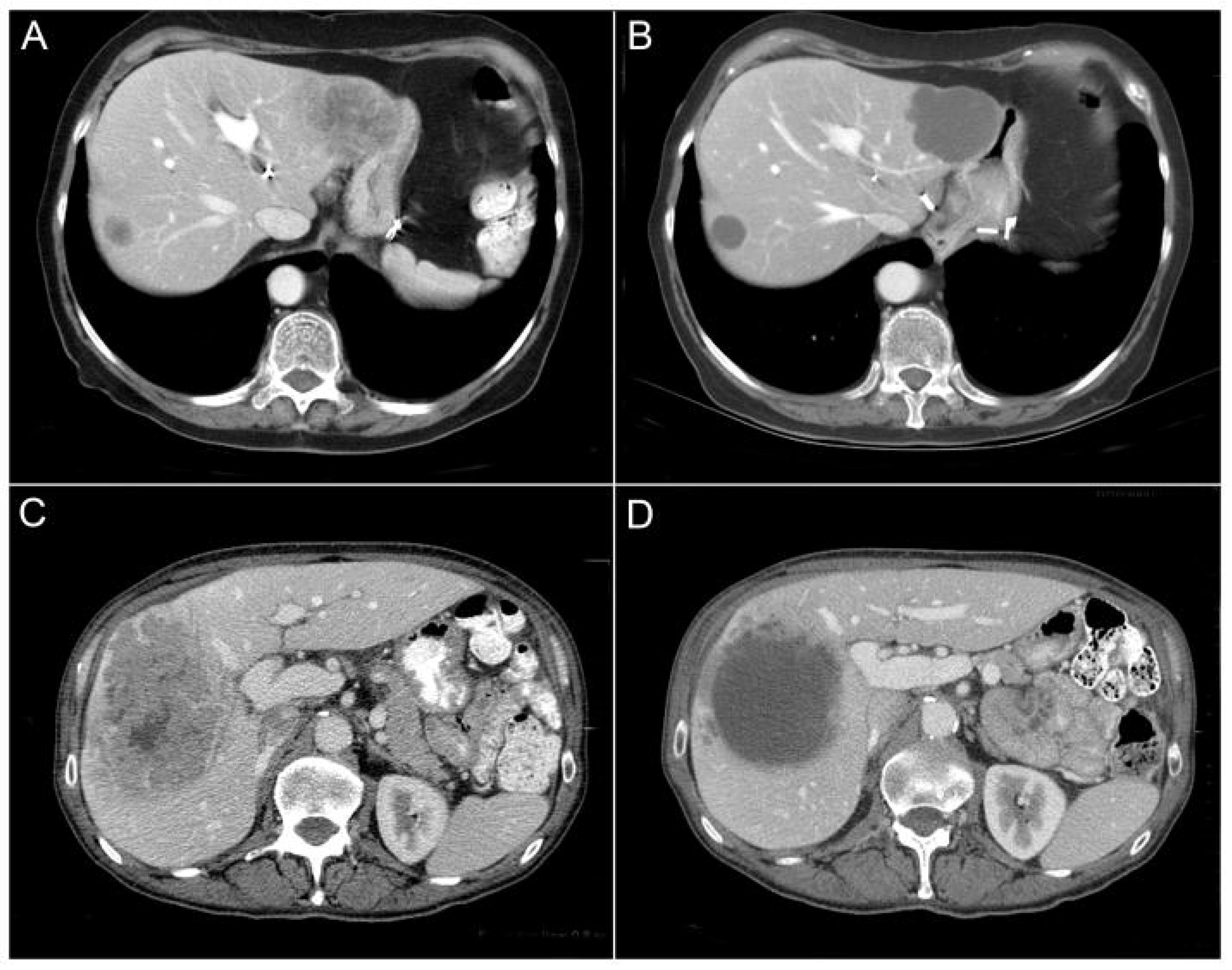
| Radiologic | WHO [11] | Decrease in tumor volume | Complete response: disappearance of all known disease Partial response: ≥50% decrease in total tumor load Progressive disease: ≥25% increase in the size of one lesion, emergence of new lesions No change: neither partial response nor progressive disease |
| RECIST [12] | Decrease in tumor largest diameter | Complete response: disappearance of all known disease Partial response: ≥30% decrease in the sum of diameters of all lesions Progressive disease: ≥20% increase, emergence of new lesions, or unequivocal increase in diameter of non-target lesions Stable disease: neither partial response nor progressive disease | |
| Morphologic [10] | Lower attenuation, More homogeneous content Sharper tumor–normal liver | Optimal response: a very pronounced change, like a hepatic cyst in the portal phase Incomplete response: subtle morphological changes, not reaching a pseudocyst appearance No response: no morphological changes between the pre-treatment and post-treatment imaging | |
| Pathologic | Tumor viability [16] | Percentage of residual cancer cells in the total tumor area | Complete response: no residual cancer cells Major response: 1% to 49% residual cancer cells Minor response: ≥50% residual cancer cells |
| Tumor regression grade [17] | Ratio of cancer cells to fibrosis | TRG1: no cancer cells and major fibrosis TRG2: rare cancer cells dispersed through pronounced fibrosis TRG3: more cancer cells in the setting of predominant fibrosis TRG4: residual cancer cells predominating fibrosis TRG5: major residual cancer cells with minimal to no fibrosis | |
| Tumor thickness [18] | Thickness of tumor–normal liver interface (TNI) | TNI < 0.5 mm TNI 0.5–5 mm TNI ≥ 5 mm |
| RECIST | Morphologic | |
|---|---|---|
| Assessment | Unidimensional tumor size measurements | Qualitative changes in tumor appearance including attenuation, enhancement, and tumor–liver interface |
| Advantages | - Objective quantitative measurements - Ease of implementation - Standardized and widely accepted indicator of response [19] - High interobserver agreement [22] | - Superior correlation with pathologic response [23] - Superior correlation with OS regardless of chemotherapy regimen [19] - Reflection of tumor biology (cell viability) [10] - High interobserver agreement [10] |
| Limitations | - Poor assessment of response to bevacizumab [10] - Inability to capture qualitative tumor changes - Inconsistent association with survival and pathologic response [21] - Strict cutoff values limiting discriminatory ability [24] | - Subjective assessment requiring experienced radiologists - Dependent on high-quality CT imaging and adequate enhancement - Challenging for small tumors (<1–1.5 cm) [19] - Limited validation with MRI [25] |
| Mutation | Pathway | Frequency | OS HR | RFS HR |
|---|---|---|---|---|
| RAS | RTK–RAS | 34.2% | 1.68 (1.54–1.84) a | 1.46 (1.33–1.61) b |
| BRAF | RTK–RAS | 4.8% | 2.62 (2.14–3.20) c | 1.89 (1.32–2.73) d |
| TP53 | p53 | 77.2% | 1.88 (1.3–2.74) e | - |
| SMAD4 | TGF-β | 11.5% | 1.93 (1.56–2.38) f | 1.95 (1.31–2.91) g |
| FBXW7 | Notch | 5.7% | 1.99 (1.15–3.45) h | - |
| APC | Wnt | 76.7% | 0.66 (0.49–0.89) i | - |
Disclaimer/Publisher’s Note: The statements, opinions and data contained in all publications are solely those of the individual author(s) and contributor(s) and not of MDPI and/or the editor(s). MDPI and/or the editor(s) disclaim responsibility for any injury to people or property resulting from any ideas, methods, instructions or products referred to in the content. |
© 2026 by the authors. Licensee MDPI, Basel, Switzerland. This article is an open access article distributed under the terms and conditions of the Creative Commons Attribution (CC BY) license.
Share and Cite
Madi, M.; Haddad, A.; Ito, K.; Bhutiani, N.; Vauthey, J.-N. Critical Evaluation of Treatment Response, Driver Mutations, and Circulating Tumor DNA as Markers of Tumor Biology in Colorectal Liver Metastasis. Cancers 2026, 18, 1111. https://doi.org/10.3390/cancers18071111
Madi M, Haddad A, Ito K, Bhutiani N, Vauthey J-N. Critical Evaluation of Treatment Response, Driver Mutations, and Circulating Tumor DNA as Markers of Tumor Biology in Colorectal Liver Metastasis. Cancers. 2026; 18(7):1111. https://doi.org/10.3390/cancers18071111
Chicago/Turabian StyleMadi, Mikel, Antony Haddad, Kyoji Ito, Neal Bhutiani, and Jean-Nicolas Vauthey. 2026. "Critical Evaluation of Treatment Response, Driver Mutations, and Circulating Tumor DNA as Markers of Tumor Biology in Colorectal Liver Metastasis" Cancers 18, no. 7: 1111. https://doi.org/10.3390/cancers18071111
APA StyleMadi, M., Haddad, A., Ito, K., Bhutiani, N., & Vauthey, J.-N. (2026). Critical Evaluation of Treatment Response, Driver Mutations, and Circulating Tumor DNA as Markers of Tumor Biology in Colorectal Liver Metastasis. Cancers, 18(7), 1111. https://doi.org/10.3390/cancers18071111