Therapeutic Potential of miR-4711-5p in Pancreatic Cancer: Antitumor Activity and Mechanistic Insights
 , ,
, ,
Simple Summary
Abstract
1. Introduction
2. Materials and Methods
2.1. Cell Culture
2.2. Transfection of miRNA
2.3. Western Blotting
2.4. Quantitative RT-PCR
2.5. Water-Soluble Tetrazolium (WST) Assay
2.6. Matrigel Invasion Assay
2.7. Bromodeoxyuridine (BrdU) Proliferation Assay
2.8. Annexin V Assay
2.9. Sphere Formation Assay
2.10. Production of Super Carbonate Apatite (sCA)
2.11. Non-Human Primate Safety Evaluation
2.12. RNA Sequencing
2.13. Data Analysis
2.14. Statistical Analysis
3. Results
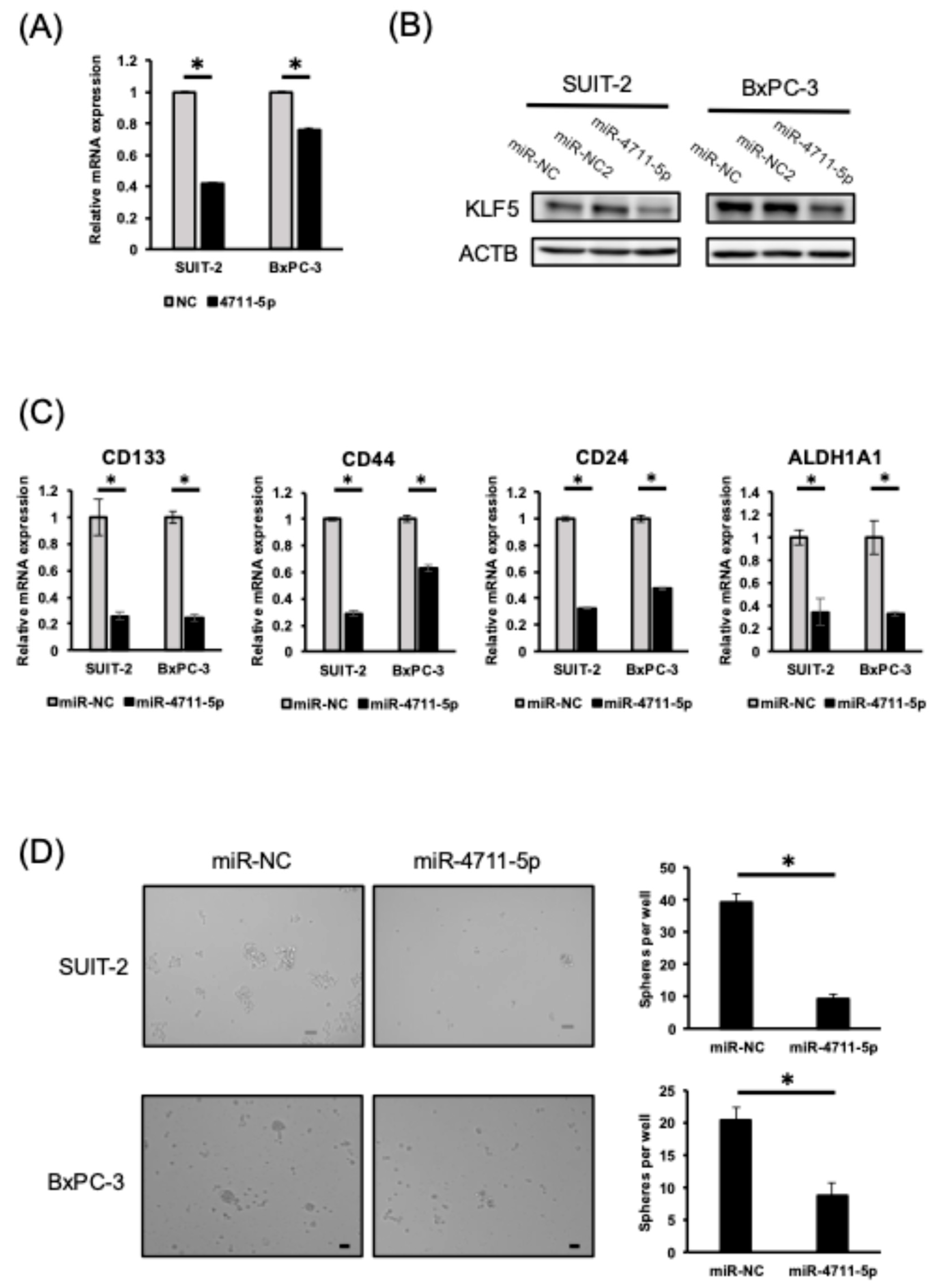
3.1. miR-4711-5p Suppresses KLF5 Expression and Cancer Stemness in Pancreatic Cancer Cells
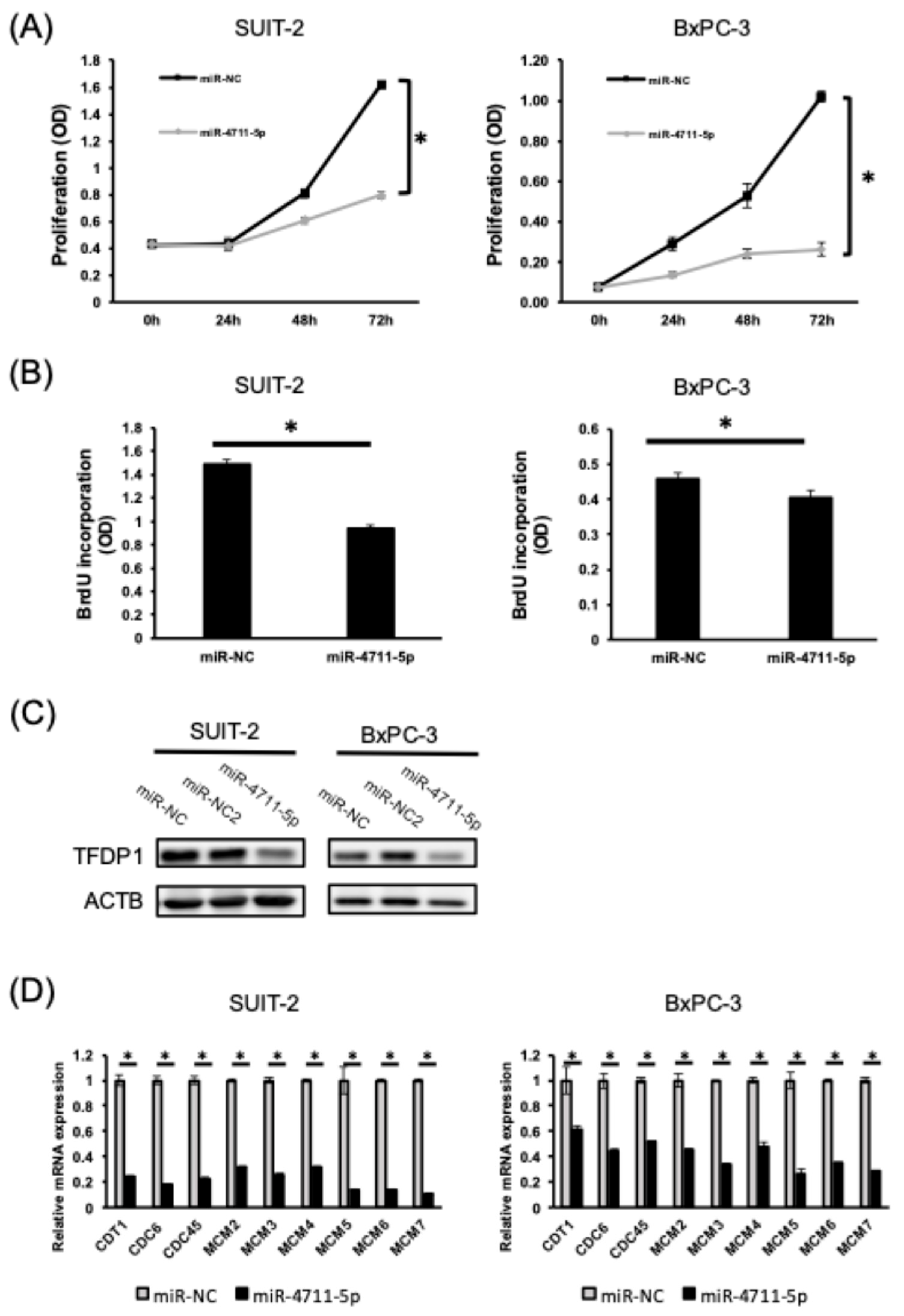
3.2. miR-4711-5p Suppresses Cell Proliferation and Cell Cycle Progression by Decreasing the Expression of TFDP1 and Pre-Replication Complex Genes in Pancreatic Cancer Cells
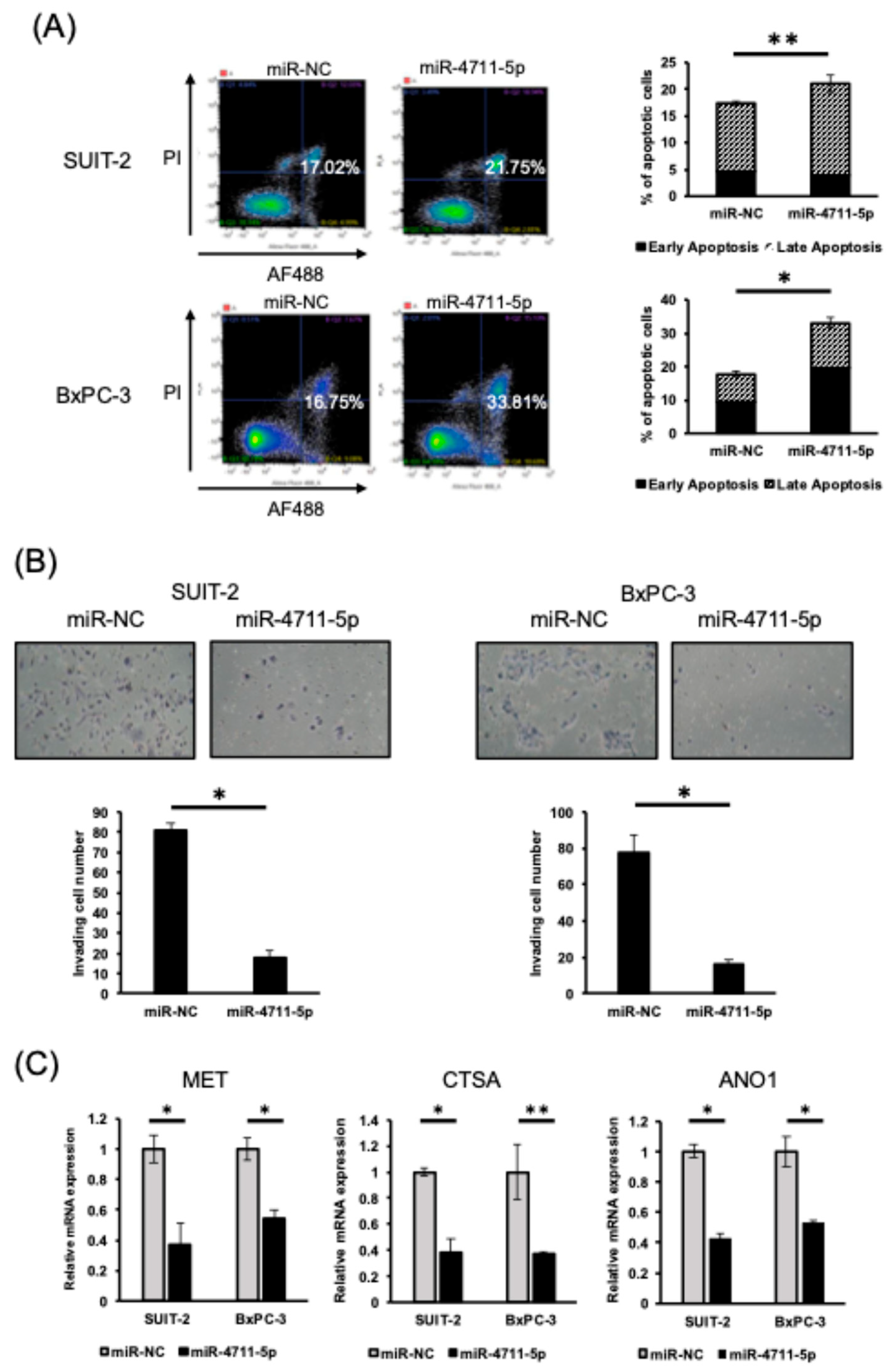
3.3. miR-4711-5p Induces Apoptosis and Suppresses Invasion of Pancreatic Cancer Cells
3.4. MET, CTSA, and ANO1 Genes Are Potential Targets of miR-4711-5p
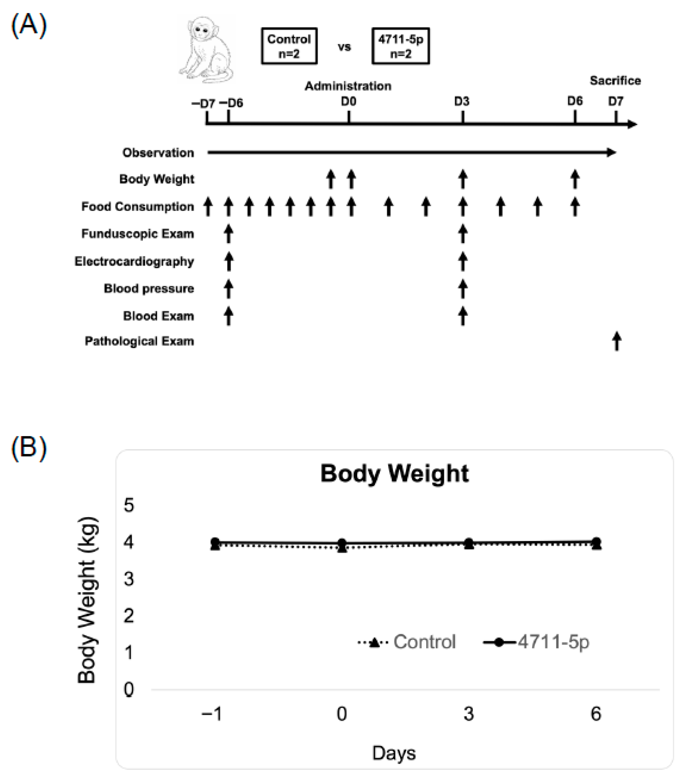
3.5. Safety Evaluation of miR-4711-5p in Cynomolgus Monkeys
4. Discussion
5. Conclusions
Supplementary Materials
Author Contributions
Funding
Institutional Review Board Statement
Informed Consent Statement
Data Availability Statement
Acknowledgments
Conflicts of Interest
References
- Bray, F.; Laversanne, M.; Sung, H.; Ferlay, J.; Siegel, R.L.; Soerjomataram, I.; Jemal, A. Global cancer statistics 2022: GLOBOCAN estimates of incidence and mortality worldwide for 36 cancers in 185 countries. CA Cancer J. Clin. 2024, 74, 229–263. [Google Scholar] [CrossRef]
- Barrambana, S.; Zamorano-Domínguez, E.; Liaki, V.; Guerra, C. Navigating the complexities of pancreatic ductal adenocarcinoma: A review on therapeutic models and RAS inhibitors. Adv. Drug Deliv. Rev. 2025, 224, 115652. [Google Scholar] [CrossRef] [PubMed]
- Dean, M.; Fojo, T.; Bates, S. Tumour stem cells and drug resistance. Nat. Rev. Cancer 2005, 5, 275–284. [Google Scholar] [CrossRef] [PubMed]
- Behbod, F.; Rosen, J.M. Will cancer stem cells provide new therapeutic targets? Carcinogenesis 2005, 26, 703–711. [Google Scholar] [CrossRef]
- Fitzgerald, T.L.; McCubrey, J.A. Pancreatic cancer stem cells: Association with cell surface markers, prognosis, resistance, metastasis and treatment. Adv. Biol. Regul. 2014, 56, 45–50. [Google Scholar] [CrossRef]
- McConnell, B.B.; Yang, V.W. Mammalian Krüppel-like factors in health and diseases. Physiol. Rev. 2010, 90, 1337–1381. [Google Scholar] [CrossRef] [PubMed]
- Ema, M.; Mori, D.; Niwa, H.; Hasegawa, Y.; Yamanaka, Y.; Hitoshi, S.; Mimura, J.; Kawabe, Y.; Hosoya, T.; Morita, M.; et al. Krüppel-like factor 5 is essential for blastocyst development and the normal self-renewal of mouse ESCs. Cell Stem Cell 2008, 3, 555–567. [Google Scholar] [CrossRef]
- McConnell, B.B.; Ghaleb, A.M.; Nandan, M.O.; Yang, V.W. The diverse functions of Krüppel-like factors 4 and 5 in epithelial biology and pathobiology. Bioessays 2007, 29, 549–557. [Google Scholar] [CrossRef]
- Takagi, Y.; Sakai, N.; Yoshitomi, H.; Furukawa, K.; Takayashiki, T.; Kuboki, S.; Takano, S.; Suzuki, D.; Kagawa, S.; Mishima, T.; et al. High expression of Krüppel-like factor 5 is associated with poor prognosis in patients with colorectal cancer. Cancer Sci. 2020, 111, 2078–2092. [Google Scholar] [CrossRef]
- Nakaya, T.; Ogawa, S.; Manabe, I.; Tanaka, M.; Sanada, M.; Sato, T.; Taketo, M.M.; Nakao, K.; Clevers, H.; Fukayama, M.; et al. KLF5 regulates the integrity and oncogenicity of intestinal stem cells. Cancer Res. 2014, 74, 2882–2891. [Google Scholar] [CrossRef]
- Li, Y.; Kong, R.; Chen, H.; Zhao, Z.; Li, L.; Li, J.; Hu, J.; Zhang, G.; Pan, S.; Wang, Y.; et al. Overexpression of KLF5 is associated with poor survival and G1/S progression in pancreatic cancer. Aging 2019, 11, 5035–5057. [Google Scholar] [CrossRef] [PubMed]
- He, P.; Yang, J.W.; Yang, V.W.; Bialkowska, A.B. Krüppel-like Factor 5, Increased in Pancreatic Ductal Adenocarcinoma, Promotes Proliferation, Acinar-to-Ductal Metaplasia, Pancreatic Intraepithelial Neoplasia, and Tumor Growth in Mice. Gastroenterology 2018, 154, 1494–1508.e13. [Google Scholar] [CrossRef]
- Morimoto, Y.; Mizushima, T.; Wu, X.; Okuzaki, D.; Yokoyama, Y.; Inoue, A.; Hata, T.; Hirose, H.; Qian, Y.; Wang, J.; et al. miR-4711-5p regulates cancer stemness and cell cycle progression via KLF5, MDM2 and TFDP1 in colon cancer cells. Br. J. Cancer 2020, 122, 1037–1049. [Google Scholar] [CrossRef] [PubMed]
- Yokoyama, Y.; Inoue, A.; Yamamoto, H.; Kouda, S.; Irie, Y.; Yang, R.; Zhang, Y.; Tsujimoto, M.; Nagata, N.; Morimoto, Y.; et al. The miR-29b Byproduct MIRTX Shows Superior Anti-tumor Activity, Compared to miR-29b-3p, in Pancreatic Cancer Cells. Anticancer Res. 2026, 46, 619–632. [Google Scholar] [CrossRef]
- Wang, J.; Yokoyama, Y.; Hirose, H.; Shimomura, Y.; Bonkobara, S.; Itakura, H.; Kouda, S.; Morimoto, Y.; Minami, K.; Takahashi, H.; et al. Functional assessment of miR-1291 in colon cancer cells. Int. J. Oncol. 2022, 60, 13. [Google Scholar] [CrossRef]
- Livak, K.J.; Schmittgen, T.D. Analysis of relative gene expression data using real-time quantitative PCR and the 2(Delta Delta C(T)) method. Methods 2001, 25, 402–408. [Google Scholar] [CrossRef]
- Nair, A.B.; Jacob, S. A simple practice guide for dose conversion between animals and human. J. Basic Clin. Pharm. 2016, 7, 27–31. [Google Scholar] [CrossRef]
- Fukata, T.; Mizushima, T.; Nishimura, J.; Okuzaki, D.; Wu, X.; Hirose, H.; Yokoyama, Y.; Kubota, Y.; Nagata, K.; Tsujimura, N.; et al. The Supercarbonate Apatite-MicroRNA Complex Inhibits Dextran Sodium Sulfate-Induced Colitis. Mol. Ther. Nucleic Acids 2018, 12, 658–671. [Google Scholar] [CrossRef]
- Wu, X.; Yamamoto, H.; Nakanishi, H.; Yamamoto, Y.; Inoue, A.; Tei, M.; Hirose, H.; Uemura, M.; Nishimura, J.; Hata, T.; et al. Innovative delivery of siRNA to solid tumours by super carbonate apatite. PLoS ONE 2015, 10, e0116022. [Google Scholar] [CrossRef]
- Carthew, R.W.; Sontheimer, E.J. Origins and mechanisms of miRNAs and siRNAs. Cell 2009, 136, 642–655. [Google Scholar] [CrossRef] [PubMed]
- Mohammadi, A.; Mansoori, B.; Baradaran, B. The role of microRNAs in colorectal cancer. Biomed. Pharmacother. 2016, 84, 705–713. [Google Scholar] [CrossRef]
- Rupaimoole, R.; Slack, F.J. MicroRNA therapeutics: Towards a new era for the management of cancer and other diseases. Nat. Rev. Drug Discov. 2017, 16, 203–222. [Google Scholar] [CrossRef]
- Li, Z.; Rana, T.M. Therapeutic targeting of microRNAs: Current status and future challenges. Nat. Rev. Drug Discov. 2014, 13, 622–638. [Google Scholar] [CrossRef]
- Rupaimoole, R.; Han, H.D.; Lopez-Berestein, G.; Sood, A.K. MicroRNA therapeutics: Principles, expectations, and challenges. Chin. J. Cancer 2011, 30, 368–370. [Google Scholar] [CrossRef][Green Version]
- Tamai, K.; Mizushima, T.; Wu, X.; Inoue, A.; Ota, M.; Yokoyama, Y.; Miyoshi, N.; Haraguchi, N.; Takahashi, H.; Nishimura, J.; et al. Photodynamic Therapy Using Indocyanine Green Loaded on Super Carbonate Apatite as Minimally Invasive Cancer Treatment. Mol. Cancer Ther. 2018, 17, 1613–1622. [Google Scholar] [CrossRef]
- Inoue, A.; Mizushima, T.; Wu, X.; Okuzaki, D.; Kambara, N.; Ishikawa, S.; Wang, J.; Qian, Y.; Hirose, H.; Yokoyama, Y.; et al. A miR-29b Byproduct Sequence Exhibits Potent Tumor-Suppressive Activities via Inhibition of NF-κB Signaling in KRAS-Mutant Colon Cancer Cells. Mol. Cancer Ther. 2018, 17, 977–987. [Google Scholar] [CrossRef] [PubMed]
- Forterre, A.; Komuro, H.; Aminova, S.; Harada, M. A comprehensive review of cancer microRNA therapeutic delivery strategies. Cancers 2020, 12, 1852. [Google Scholar] [CrossRef] [PubMed]
- Abd-Aziz, N.; Kamaruzman, N.I.; Poh, C.L. Development of microRNAs as potential therapeutics against cancer. J. Oncol. 2020, 2020, 8029721. [Google Scholar] [CrossRef] [PubMed]
- Merhautova, J.; Demlova, R.; Slaby, O. MicroRNA-based therapy in animal models of selected gastrointestinal cancers. Front. Pharmacol. 2016, 7, 329. [Google Scholar] [CrossRef]
- Takahashi, R.U.; Prieto-Vila, M.; Kohama, I.; Ochiya, T. Development of miRNA-based therapeutic approaches for cancer patients. Cancer Sci. 2019, 110, 1140–1147. [Google Scholar] [CrossRef]
- Moosavi, F.; Giovannetti, E.; Saso, L.; Firuzi, O. HGF/MET pathway aberrations as diagnostic, prognostic, and predictive biomarkers in human cancers. Crit. Rev. Clin. Lab. Sci. 2019, 56, 533–566. [Google Scholar] [CrossRef] [PubMed]
- Gherardi, E.; Birchmeier, W.; Birchmeier, C.; Vande Woude, G. Targeting MET in cancer: Rationale and progress. Nat. Rev. Cancer 2012, 12, 89–103. [Google Scholar] [CrossRef] [PubMed]
- Comoglio, P.M.; Trusolino, L.; Boccaccio, C. Known and novel roles of the MET oncogene in cancer. Nat. Rev. Cancer 2018, 18, 341–358. [Google Scholar] [CrossRef] [PubMed]
- Sauter, D.; Novak, I.; Pedersen, S.; Larsen, E.H.; Hoffmann, E.K. ANO1 (THEM16A) in pancreatic ductal adenocarcinoma (PDAC). Pflug. Arch. 2015, 467, 1495–1508. [Google Scholar] [CrossRef]
- Crottès, D.; Lin, Y.T.; Peters, C.J.; Gilchrist, J.M.; Wiita, A.P.; Jan, Y.N.; Jan, L.Y. THEM16A controls EGF-induced calcium signaling implicated in pancreatic cancer prognosis. Proc. Natl. Acad. Sci. USA 2019, 116, 13026–13035. [Google Scholar] [CrossRef]
- Guo, S.; Zhang, L.; Li, N. ANO1: More Than Just Calcium-Activated Chloride Channel in Cancer. Front. Oncol. 2022, 12, 922838. [Google Scholar] [CrossRef]
- Britschgi, A.; Bill, A.; Brinkhaus, H.; Rothwell, C.; Clay, I.; Duss, S.; Rebhan, M.; Raman, P.; Guy, C.T.; Wetzel, K.; et al. Calcium-activated chloride channel ANO1 promotes breast cancer progression by activating EGFR and CAMK signaling. Proc. Natl. Acad. Sci. USA 2013, 110, E1026–E1034. [Google Scholar] [CrossRef]
- Park, S.; Kwon, W.; Park, J.K.; Baek, S.M.; Lee, S.W.; Cho, G.J.; Ha, Y.S.; Lee, J.N.; Kwon, T.G.; Kim, M.O.; et al. Suppression of cathepsin a inhibits growth, migration, and invasion by inhibiting the p38 MAPK signaling pathway in prostate cancer. Arch. Biochem. Biophys. 2020, 688, 108407. [Google Scholar] [CrossRef]
- Nomura, T.; Katunuma, N. Involvement of cathepsins in the invasion, metastasis and proliferation of cancer cells. J. Med. Investig. 2005, 52, 1–9. [Google Scholar] [CrossRef]




| Pre-Treatment (Day −6) | Post-Treatment (Day 3) | |||||||||
|---|---|---|---|---|---|---|---|---|---|---|
| Control_1 | Control_2 | 4711_5p_1 | 4711_5p_2 | Control_1 | Control_2 | 4711_5p_1 | 4711_5p_2 | Control Student’s t-test p Value | 4711_5p Student’s t-test p Value | |
| Hematology | ||||||||||
| RBC (104/μL) | 557 | 545 | 558 | 604 | 505 | 499 | 546 | 552 | 0.039 | 0.356 |
| HGB (g/dL) | 13.1 | 14.2 | 14.4 | 14.1 | 11.9 | 12.8 | 14 | 12.8 | 0.049 | 0.310 |
| HCT (%) | 42.5 | 44.6 | 44 | 44.8 | 39.5 | 41.6 | 44 | 44.8 | NE | NE |
| MCV (fL) | 76.3 | 81.8 | 78.9 | 74.2 | 78.2 | 83.4 | 80.6 | 75 | 0.054 | 0.220 |
| MCH (pg) | 23.5 | 26.1 | 25.8 | 23.3 | 23.6 | 25.7 | 25.6 | 23.2 | 0.656 | 0.205 |
| MCHC (g/dL) | 30.8 | 31.8 | 32.7 | 31.5 | 30.1 | 30.8 | 31.8 | 30.9 | 0.111 | 0.126 |
| PLT (103/μL) | 392 | 426 | 452 | 362 | 324 | 364 | 320 | 350 | 0.029 | 0.442 |
| Reticulocyte (%) | 0.61 | 0.6 | 0.65 | 0.93 | 0.95 | 0.88 | 0.77 | 1.24 | 0.061 | 0.265 |
| WBC (103/μL) | 13.51 | 11.34 | 13.17 | 12.14 | 10.83 | 12.16 | 12.34 | 15.27 | 0.689 | 0.665 |
| NEUT (103/μL) | 3.88 | 3.7 | 3.31 | 3.13 | 3.48 | 3.88 | 3.61 | 5.22 | 0.769 | 0.409 |
| LYMP (103/μL) | 8.78 | 6.86 | 8.98 | 8.28 | 6.53 | 6.84 | 7.44 | 8.39 | 0.494 | 0.545 |
| MONO (103/μL) | 0.62 | 0.55 | 0.49 | 0.5 | 0.64 | 1.08 | 1.03 | 1.42 | 0.476 | 0.162 |
| EOS (103/μL) | 0.21 | 0.2 | 0.38 | 0.2 | 0.12 | 0.28 | 0.22 | 0.17 | 0.963 | 0.382 |
| BASO (103/μL) | 0.02 | 0.03 | 0.01 | 0.03 | 0.06 | 0.08 | 0.04 | 0.07 | 0.070 | 0.090 |
| Coagulation | ||||||||||
| PT (s) | 10.4 | 10.2 | 10.5 | 10.7 | 10.3 | 9.7 | 10.3 | 10.5 | 0.374 | NE |
| APTT (s) | 19.8 | 17.9 | 22.6 | 18.6 | 21.9 | 18 | 23.2 | 19.4 | 0.470 | 0.090 |
| Chemistry | ||||||||||
| AST (IU/L) | 29 | 31 | 27 | 24 | 34 | 47 | 35 | 28 | 0.307 | 0.205 |
| ALT (IU/L) | 23 | 39 | 25 | 33 | 39 | 77 | 41 | 47 | 0.246 | 0.042 |
| ALP (IU/L) | 981 | 1965 | 1305 | 1574 | 1140 | 1782 | 1220 | 1415 | 0.955 | 0.187 |
| CK (IU/L) | 128 | 150 | 191 | 122 | 245 | 198 | 230 | 173 | 0.252 | 0.084 |
| T_BIL (mg/dL) | 0.06 | 0.11 | 0.04 | 0.07 | 0.08 | 0.08 | 0.06 | 0.1 | 0.874 | 0.126 |
| TP (g/dL) | 6.7 | 7.7 | 6.8 | 7.5 | 6.9 | 7.6 | 6.9 | 7.5 | 0.795 | 0.500 |
| ALB (g/dL) | 3.8 | 3.9 | 3.8 | 4.2 | 3.7 | 3.7 | 3.8 | 4.1 | 0.205 | 0.500 |
| Globulin (g/dL) | 2.9 | 3.8 | 3 | 3.3 | 3.2 | 3.9 | 3.1 | 3.4 | 0.295 | NE |
| A/G | 1.31 | 1.03 | 1.27 | 1.27 | 1.16 | 0.95 | 1.23 | 1.21 | 0.188 | 0.126 |
| TG (mg/dL) | 34 | 44 | 26 | 60 | 54 | 100 | 51 | 100 | 0.282 | 0.144 |
| T_CHO (mg/dL) | 109 | 153 | 109 | 87 | 101 | 150 | 114 | 99 | 0.272 | 0.249 |
| GLU (mg/dL) | 65 | 87 | 81 | 83 | 76 | 74 | 74 | 90 | 0.947 | 1.000 |
| BUN (mg/dL) | 22.6 | 16.1 | 18.8 | 22.4 | 21.5 | 19.9 | 19.4 | 23.8 | 0.679 | 0.242 |
| CRNN (mg/dL) | 0.73 | 0.68 | 0.57 | 0.68 | 0.76 | 0.7 | 0.64 | 0.67 | 0.126 | 0.590 |
| P (mg/dL) | 5.93 | 6.35 | 6.58 | 4.77 | 4.69 | 5.02 | 4.96 | 3.96 | 0.022 | 0.205 |
| Ca (mg/dL) | 9.9 | 10.1 | 10 | 9.9 | 9.5 | 9.9 | 9.7 | 10.1 | 0.205 | 0.874 |
| Na (mmol/L) | 145 | 148 | 145 | 143 | 149 | 148 | 148 | 145 | 0.500 | 0.126 |
| K (mmol/L) | 4.6 | 4.4 | 4.8 | 4 | 4.4 | 5.2 | 4.9 | 4.7 | 0.656 | 0.410 |
| Cl (mmol/L) | 110 | 107 | 105 | 109 | 110 | 108 | 107 | 111 | 0.500 | NE |
| Animal No. | Body Weight (kg) | Pancreas (g/kg) | Spleen (g/kg) | Brain (g/kg) | Heart (g/kg) | Lung (g/kg) | Liver (g/kg) | Kidney_R (g/kg) | Kidney_L (g/kg) | Kidney_R + L (g/kg) |
|---|---|---|---|---|---|---|---|---|---|---|
| Control_1 | 3.87 | 1.19 | 1.5 | 18.22 | 2.84 | 4.63 | 16.15 | 1.63 | 1.6 | 3.23 |
| Control_2 | 3.91 | 1.25 | 1.28 | 16.5 | 3.58 | 4.65 | 17.8 | 1.94 | 1.97 | 3.91 |
| 4711-5p_1 | 3.74 | 1.93 | 1.12 | 18.29 | 3.32 | 5.27 | 18.8 | 2.01 | 1.98 | 3.98 |
| 4711-5p_2 | 4.04 | 1.78 | 1.01 | 16.68 | 3.49 | 4.98 | 19.33 | 1.71 | 1.91 | 3.61 |
| MU p value | 1.00 | 0.33 | 0.33 | 0.67 | 1.00 | 0.33 | 0.33 | 0.67 | 0.67 | 0.67 |
Disclaimer/Publisher’s Note: The statements, opinions and data contained in all publications are solely those of the individual author(s) and contributor(s) and not of MDPI and/or the editor(s). MDPI and/or the editor(s) disclaim responsibility for any injury to people or property resulting from any ideas, methods, instructions or products referred to in the content. |
© 2026 by the authors. Licensee MDPI, Basel, Switzerland. This article is an open access article distributed under the terms and conditions of the Creative Commons Attribution (CC BY) license.
Share and Cite
Yokoyama, Y.; Morimoto, Y.; Yamamoto, H.; Kouda, S.; Kawanami, S.; Yang, R.; Zhang, Y.; Tsujimoto, M.; Nagata, N.; Shimomura, Y.; et al. Therapeutic Potential of miR-4711-5p in Pancreatic Cancer: Antitumor Activity and Mechanistic Insights. Cancers 2026, 18, 1104. https://doi.org/10.3390/cancers18071104
Yokoyama Y, Morimoto Y, Yamamoto H, Kouda S, Kawanami S, Yang R, Zhang Y, Tsujimoto M, Nagata N, Shimomura Y, et al. Therapeutic Potential of miR-4711-5p in Pancreatic Cancer: Antitumor Activity and Mechanistic Insights. Cancers. 2026; 18(7):1104. https://doi.org/10.3390/cancers18071104
Chicago/Turabian StyleYokoyama, Yuhki, Yoshihiro Morimoto, Hiroyuki Yamamoto, Shihori Kouda, Shiho Kawanami, Ruijia Yang, Yingjue Zhang, Manami Tsujimoto, Nanami Nagata, Yuki Shimomura, and et al. 2026. "Therapeutic Potential of miR-4711-5p in Pancreatic Cancer: Antitumor Activity and Mechanistic Insights" Cancers 18, no. 7: 1104. https://doi.org/10.3390/cancers18071104
APA StyleYokoyama, Y., Morimoto, Y., Yamamoto, H., Kouda, S., Kawanami, S., Yang, R., Zhang, Y., Tsujimoto, M., Nagata, N., Shimomura, Y., Nishida, K., Hata, T., Inoue, A., Shibata, S., Yamamoto, H., & Mori, M. (2026). Therapeutic Potential of miR-4711-5p in Pancreatic Cancer: Antitumor Activity and Mechanistic Insights. Cancers, 18(7), 1104. https://doi.org/10.3390/cancers18071104