

Stimulated Raman Spectroscopy for Intraoperative Glioblastoma Diagnosis—A Complementary Tool to Frozen Section?

 , , , , , , and
Simple Summary
Abstract
1. Introduction
2. Methods
2.1. Patients
2.2. Study Design
2.3. HE Staining
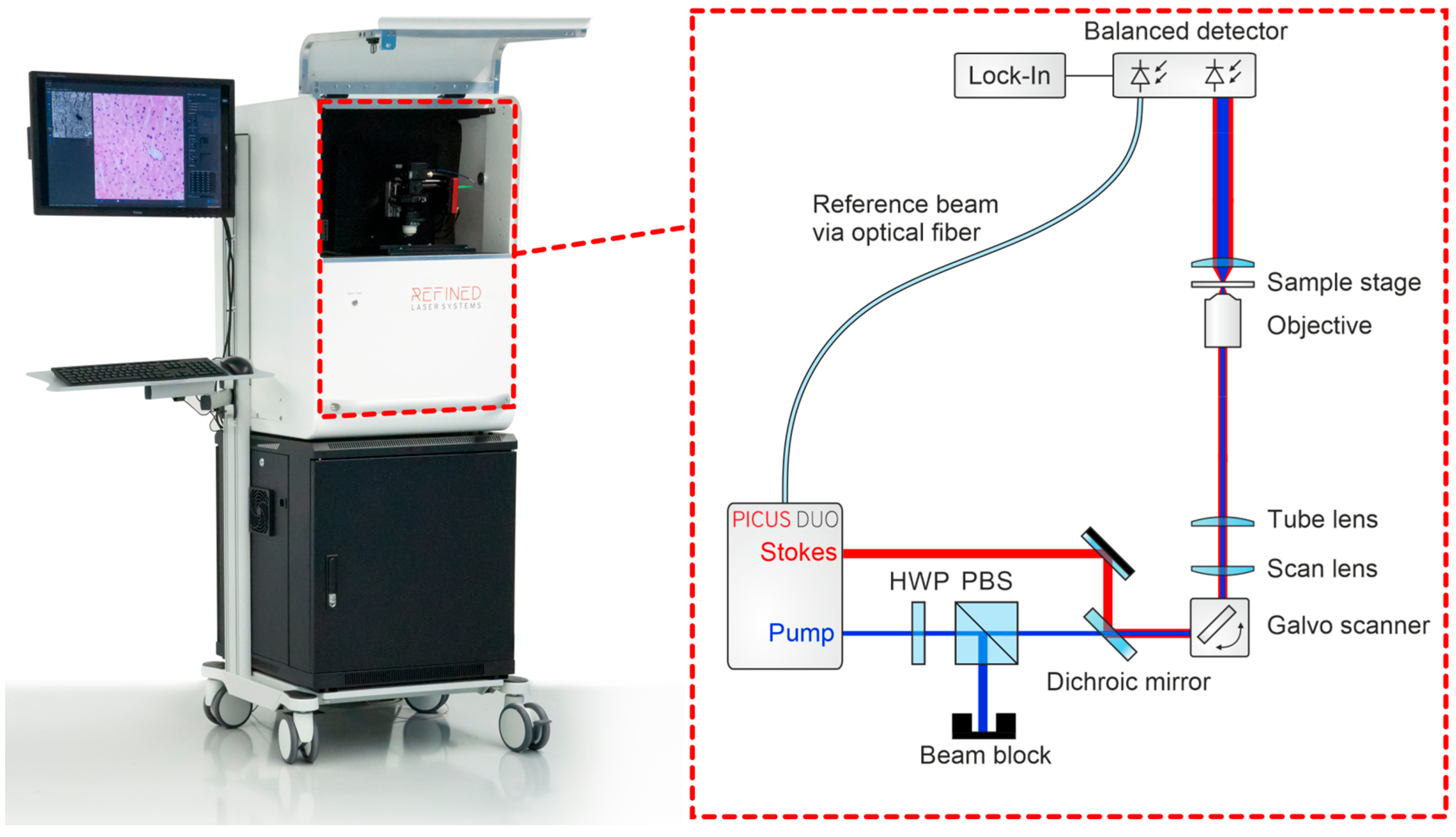
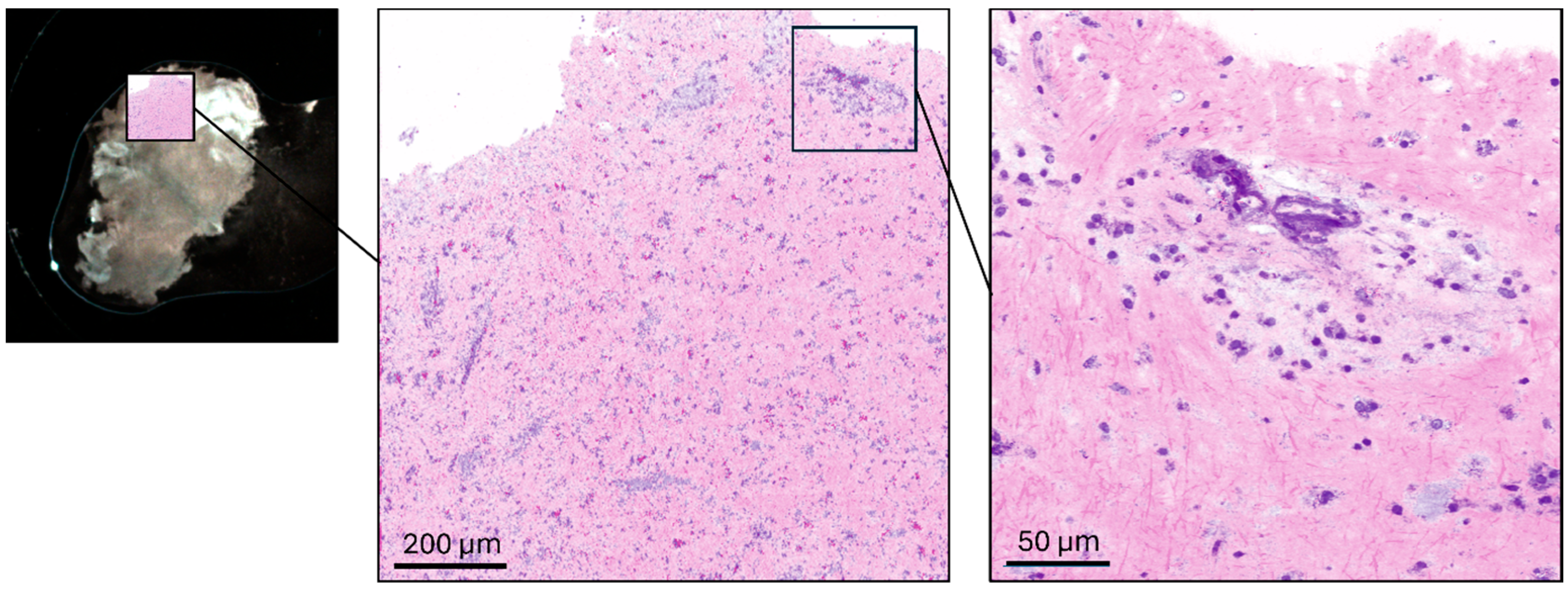
2.4. Raman Imaging
2.5. Statistics
3. Results
3.1. Descriptive Data
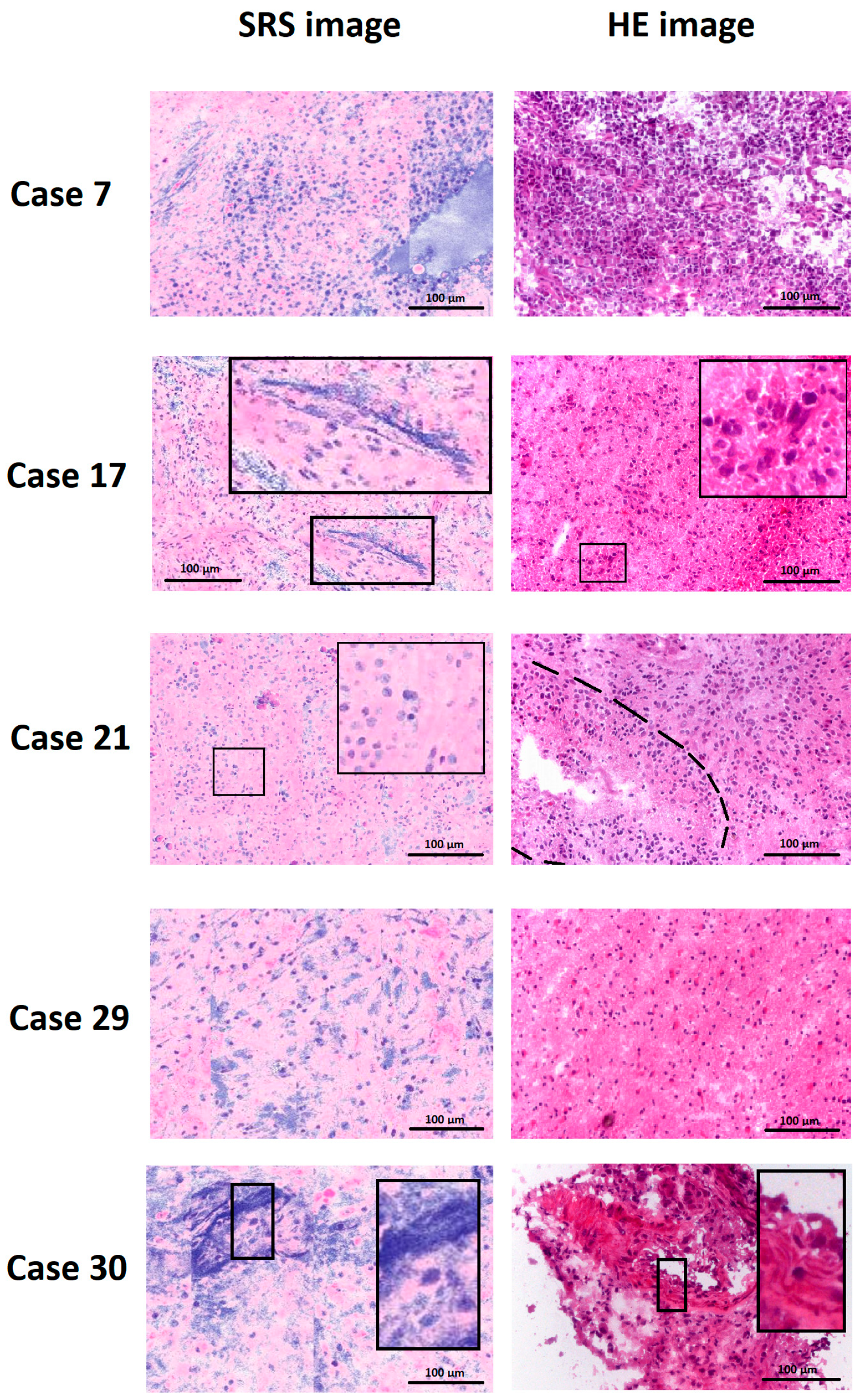
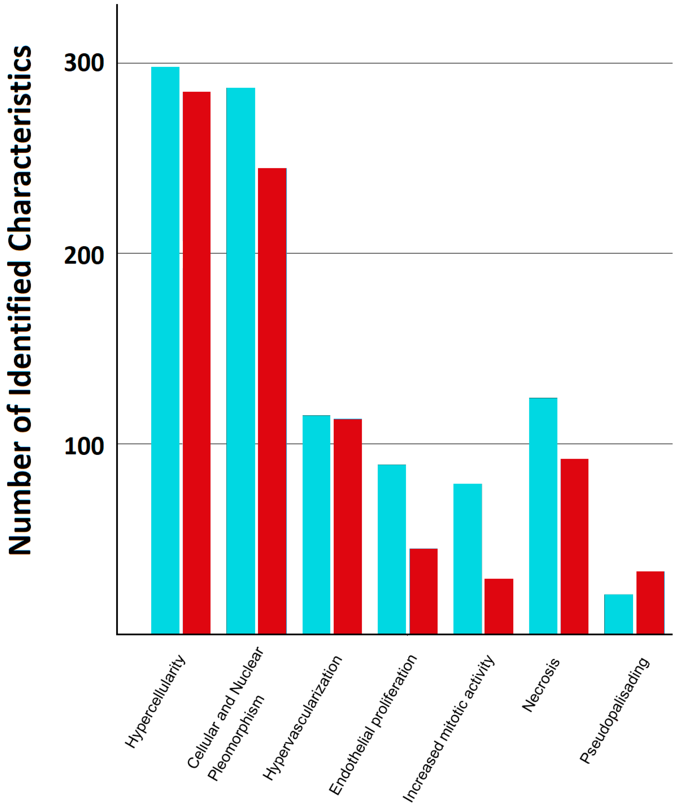
3.2. Exploratory Feature Visibility
3.3. Interobserver Agreement
3.4. Examiner Proficiency
4. Discussion
5. Conclusions
Supplementary Materials
Author Contributions
Funding
Institutional Review Board Statement
Informed Consent Statement
Data Availability Statement
Acknowledgments
Conflicts of Interest
References
- Gittleman, H.; Boscia, A.; Ostrom, Q.T.; Truitt, G.; Fritz, Y.; Kruchko, C.; Barnholtz-Sloan, J.S. Survivorship in Adults with Malignant Brain and Other Central Nervous System Tumor from 2000–2014. Neuro-Oncology 2018, 20, vii6–vii16. [Google Scholar] [CrossRef] [Scilit]
- Stupp, R.; Mason, W.P.; van den Bent, M.J.; Weller, M.; Fisher, B.; Taphoorn, M.J.B.; Belanger, K.; Brandes, A.A.; Marosi, C.; Bogdahn, U.; et al. Radiotherapy plus Concomitant and Adjuvant Temozolomide for Glioblastoma. N. Engl. J. Med. 2005, 352, 987–996. [Google Scholar] [CrossRef] [Scilit]
- Stupp, R.; Taillibert, S.; Kanner, A.; Read, W.; Steinberg, D.; Lhermitte, B.; Toms, S.; Idbaih, A.; Ahluwalia, M.S.; Fink, K.; et al. Effect of Tumor-Treating Fields Plus Maintenance Temozolomide vs. Maintenance Temozolomide Alone on Survival in Patients with Glioblastoma: A Randomized Clinical Trial. JAMA 2017, 318, 2306–2316. [Google Scholar] [CrossRef] [Scilit] [PubMed]
- Al-Ajmi, R.; Al-Kindi, H.; George, M.; Thomas, K. Correlation of Intraoperative Frozen Section Report and Histopathological Diagnosis of Central Nervous System Tumors—A Six-Year Retrospective Study. Oman Med. J. 2016, 31, 414–420. [Google Scholar] [CrossRef] [Scilit] [PubMed]
- Cicerone, M.T.; Camp, C.H. Histological Coherent Raman Imaging: A Prognostic Review. Analyst 2017, 143, 33–59. [Google Scholar] [CrossRef] [Scilit]
- Raman, C.V.; Krishnan, K.S. A New Type of Secondary Radiation. Nature 1928, 121, 501–502. [Google Scholar] [CrossRef] [Scilit]
- Orringer, D.A.; Pandian, B.; Niknafs, Y.S.; Hollon, T.C.; Boyle, J.; Lewis, S.; Garrard, M.; Hervey-Jumper, S.L.; Garton, H.J.L.; Maher, C.O.; et al. Rapid Intraoperative Histology of Unprocessed Surgical Specimens via Fibre-Laser-Based Stimulated Raman Scattering Microscopy. Nat. Biomed. Eng. 2017, 1, 0027. [Google Scholar] [CrossRef] [Scilit]
- Sarri, B.; Poizat, F.; Heuke, S.; Wojak, J.; Franchi, F.; Caillol, F.; Giovannini, M.; Rigneault, H. Stimulated Raman Histology: One to One Comparison with Standard Hematoxylin and Eosin Staining. Biomed. Opt. Express 2019, 10, 5378–5384. [Google Scholar] [CrossRef] [Scilit]
- Shin, K.S.; Laohajaratsang, M.; Men, S.; Figueroa, B.; Dintzis, S.M.; Fu, D. Quantitative Chemical Imaging of Breast Calcifications in Association with Neoplastic Processes. Theranostics 2020, 10, 5865–5878. [Google Scholar] [CrossRef] [Scilit]
- Brinkmann, M.; Neumann, F.; Droop, R.; Ullmann, S.; Würthwein, T.; Hellwig, T.; Stark, F.; Schneider, K.I.; Sippl, C.; Linsler, S. Towards Real-Time Molecular Profiling of Glioblastoma via Stimulated Raman Scattering Imaging. In Proceedings of the Optica Biophotonics Congress 2025; Optica Publishing Group: Coronado, CA, USA, 2025; p. TW1F.1. [Google Scholar]
- Di, L.; Eichberg, D.G.; Huang, K.; Shah, A.H.; Jamshidi, A.M.; Luther, E.M.; Lu, V.M.; Komotar, R.J.; Ivan, M.E.; Gultekin, S.H. Stimulated Raman Histology for Rapid Intraoperative Diagnosis of Gliomas. World Neurosurg. 2021, 150, e135–e143. [Google Scholar] [CrossRef] [Scilit]
- Bossuyt, P.M.; Reitsma, J.B.; Bruns, D.E.; Gatsonis, C.A.; Glasziou, P.P.; Irwig, L.; Lijmer, J.G.; Moher, D.; Rennie, D.; De Vet, H.C.W.; et al. STARD 2015: An Updated List of Essential Items for Reporting Diagnostic Accuracy Studies. BMJ 2015, 351, h5527. [Google Scholar] [CrossRef] [Scilit] [PubMed]
- Brinkmann, M.; Fast, A.; Hellwig, T.; Pence, I.; Evans, C.L.; Fallnich, C. Portable All-Fiber Dual-Output Widely Tunable Light Source for Coherent Raman Imaging. Biomed. Opt. Express 2019, 10, 4437. [Google Scholar] [CrossRef] [Scilit] [PubMed]
- Fitzgerald, C.W.R.; Dogan, S.; Bou-Nassif, R.; Mclean, T.; Woods, R.; Cracchiolo, J.R.; Ganly, I.; Tabar, V.; Cohen, M.A. Stimulated Raman Histology for Rapid Intra-Operative Diagnosis of Sinonasal and Skull Base Tumors. Laryngoscope 2022, 132, 2142–2147. [Google Scholar] [CrossRef] [Scilit]
- Jiao, C.; Chen, M.; Liao, J.; Li, J.; Zhang, R.; He, S. Rapid, Label-Free Detection of Colorectal Cancer with an Aberration-Free Line Scanning Confocal Raman Imager. Spectrochim. Acta A Mol. Biomol. Spectrosc. 2025, 339, 126182. [Google Scholar] [CrossRef] [Scilit]
- Stupak, E.V.; Glotov, V.M.; Askandaryan, A.S.; Clancy, S.E.; Hiana, J.C.; Cherkasova, O.P.; Stupak, V.V. Raman Spectroscopy in the Diagnosis of Brain Gliomas: A Literature Review. Cureus 2025, 17, e79165. [Google Scholar] [CrossRef] [Scilit] [PubMed]
- Beljebbar, A.; Dukic, S.; Amharref, N.; Manfait, M. Ex Vivo and in Vivo Diagnosis of C6 Glioblastoma Development by Raman Spectroscopy Coupled to a Microprobe. Anal. Bioanal. Chem. 2010, 398, 477–487. [Google Scholar] [CrossRef] [Scilit]
- Einstein, E.H.; Ablyazova, F.; Rosenberg, A.; Harshan, M.; Wahl, S.; Har-El, G.; Constantino, P.D.; Ellis, J.A.; Boockvar, J.A.; Langer, D.J.; et al. Stimulated Raman Histology Facilitates Accurate Diagnosis in Neurosurgical Patients: A One-to-One Noninferiority Study. J. Neurooncol. 2022, 159, 369–375. [Google Scholar] [CrossRef] [Scilit]
- Eichberg, D.G.; Shah, A.H.; Di, L.; Semonche, A.M.; Jimsheleishvili, G.; Luther, E.M.; Sarkiss, C.A.; Levi, A.D.; Gultekin, S.H.; Komotar, R.J.; et al. Stimulated Raman Histology for Rapid and Accurate Intraoperative Diagnosis of CNS Tumors: Prospective Blinded Study. J. Neurosurg. 2021, 134, 137–143. [Google Scholar] [CrossRef] [Scilit]
- Kowalska, A.A.; Berus, S.; Szleszkowski, Ł.; Kamińska, A.; Kmiecik, A.; Ratajczak-Wielgomas, K.; Jurek, T.; Zadka, Ł. Brain Tumour Homogenates Analysed by Surface-Enhanced Raman Spectroscopy: Discrimination among Healthy and Cancer Cells. Spectrochim. Acta A Mol. Biomol. Spectrosc. 2020, 231, 117769. [Google Scholar] [CrossRef] [Scilit]
- Hollon, T.C.; Pandian, B.; Adapa, A.R.; Urias, E.; Save, A.V.; Khalsa, S.S.S.; Eichberg, D.G.; D’Amico, R.S.; Farooq, Z.U.; Lewis, S.; et al. Near Real-Time Intraoperative Brain Tumor Diagnosis Using Stimulated Raman Histology and Deep Neural Networks. Nat. Med. 2020, 26, 52–58. [Google Scholar] [CrossRef] [Scilit]
- Redlich, J.-P.; Feuerhake, F.; Weis, J.; Schaadt, N.S.; Teuber-Hanselmann, S.; Buck, C.; Luttmann, S.; Eberle, A.; Nikolin, S.; Appenzeller, A.; et al. Applications of Artificial Intelligence in the Analysis of Histopathology Images of Gliomas: A Review. npj Imaging 2024, 2, 16. [Google Scholar] [CrossRef] [Scilit]
- Saar, B.G.; Freudiger, C.W.; Reichman, J.; Stanley, C.M.; Holtom, G.R.; Xie, X.S. Video-Rate Molecular Imaging in Vivo with Stimulated Raman Scattering. Science 2010, 330, 1368–1370. [Google Scholar] [CrossRef] [Scilit]
- Lee, M.; Herrington, C.S.; Ravindra, M.; Sepp, K.; Davies, A.; Hulme, A.N.; Brunton, V.G. Recent Advances in the Use of Stimulated Raman Scattering in Histopathology. Analyst 2021, 146, 789–802. [Google Scholar] [CrossRef] [Scilit]
- Klamminger, G.G.; Gérardy, J.-J.; Jelke, F.; Mirizzi, G.; Slimani, R.; Klein, K.; Husch, A.; Hertel, F.; Mittelbronn, M.; Kleine-Borgmann, F.B. Application of Raman Spectroscopy for Detection of Histologically Distinct Areas in Formalin-Fixed Paraffin-Embedded Glioblastoma. Neuro-Oncol. Adv. 2021, 3, vdab077. [Google Scholar] [CrossRef] [Scilit]
- Li, Q.; Shen, J.; Zhou, Y. Diagnosis of Glioma Using Raman Spectroscopy and the Entropy Weight Fuzzy-Rough Nearest Neighbor (EFRNN) Algorithm on Fresh Tissue. Anal. Lett. 2023, 56, 895–905. [Google Scholar] [CrossRef] [Scilit]
- Iturrioz-Rodríguez, N.; De Pasquale, D.; Fiaschi, P.; Ciofani, G. Discrimination of Glioma Patient-Derived Cells from Healthy Astrocytes by Exploiting Raman Spectroscopy. Spectrochim. Acta A Mol. Biomol. Spectrosc. 2022, 269, 120773. [Google Scholar] [CrossRef] [Scilit] [PubMed]
- Kopec, M.; Błaszczyk, M.; Radek, M.; Abramczyk, H. Raman Imaging and Statistical Methods for Analysis Various Type of Human Brain Tumors and Breast Cancers. Spectrochim. Acta A Mol. Biomol. Spectrosc. 2021, 262, 120091. [Google Scholar] [CrossRef] [Scilit]
- Becker, N.; Camelo-Piragua, S.; Conway, K.S. A Contemporary Approach to Intraoperative Evaluation in Neuropathology. Arch. Pathol. Lab. Med. 2024, 148, 649–658. [Google Scholar] [CrossRef] [Scilit] [PubMed]
- Van Den Bent, M.J. Interobserver Variation of the Histopathological Diagnosis in Clinical Trials on Glioma: A Clinician’s Perspective. Acta Neuropathol. 2010, 120, 297–304. [Google Scholar] [CrossRef] [Scilit] [PubMed]
- Erickson, L.C. The Role of O-6 Methylguanine DNA Methyltransferase (MGMT) in Drug Resistance and Strategies for Its Inhibition. Semin. Cancer Biol. 1991, 2, 257–265. [Google Scholar]
- Hartmann, C.; Meyer, J.; Balss, J.; Capper, D.; Mueller, W.; Christians, A.; Felsberg, J.; Wolter, M.; Mawrin, C.; Wick, W.; et al. Type and Frequency of IDH1 and IDH2 Mutations Are Related to Astrocytic and Oligodendroglial Differentiation and Age: A Study of 1,010 Diffuse Gliomas. Acta Neuropathol. 2009, 118, 469–474. [Google Scholar] [CrossRef] [Scilit] [PubMed]
- Liu, C.; Wang, J.; Shen, J.; Chen, X.; Ji, N.; Yue, S. Accurate and Rapid Molecular Subgrouping of High-Grade Glioma via Deep Learning-Assisted Label-Free Fiber-Optic Raman Spectroscopy. PNAS Nexus 2024, 3, pgae208. [Google Scholar] [CrossRef] [Scilit]

| Feature | Operational Definition |
|---|---|
| Hypercellularity | Increased density of tumor cells compared with normal brain parenchyma |
| Cellular and nuclear pleomorphism | Variation in cell and nuclear size and shape typical of high-grade glioma |
| Hypervascularization | Increased number of blood vessels within the tumor tissue |
| Endothelial proliferation | Multilayered endothelial cell growth within vascular structures |
| Increased mitotic activity | Presence of mitotic figures indicating active cell division |
| Necrosis | Areas of tissue breakdown with loss of cellular structure |
| Pseudopalisading | Radial arrangement of tumor cells surrounding necrotic areas |
| Feature | OR (SRS vs. HE) | 95% CI | FDR-Adjusted p |
|---|---|---|---|
| Hypercellularity | 0.10 | 0.01–0.69 | 0.035 |
| Cellular & nuclear pleomorphism | 0.40 | 0.20–0.80 | 0.023 |
| Hypervascularization | 1.00 | 0.62–1.61 | 0.998 |
| Endothelial proliferation | 0.43 | 0.26–0.73 | 0.006 |
| Increased mitotic activity | 0.31 | 0.20–0.48 | <0.001 |
| Necrosis | 0.58 | 0.32–1.04 | 0.079 |
| Pseudopalisading | 2.66 | 1.03–6.87 | 0.060 |
| Feature | HE κ | 95% CI (HE) | SRS κ | 95% CI (SRS) |
|---|---|---|---|---|
| Hypercellularity | 0.69 | 0.43–0.82 | 0.19 | 0.08–0.29 |
| Cellular & nuclear pleomorphism | 0.28 | 0.08–0.45 | 0.11 | 0.04–0.17 |
| Hypervascularization | 0.11 | 0.01–0.19 | 0.03 | −0.03–0.10 |
| Endothelial proliferation | 0.11 | 0.03–0.18 | 0.04 | −0.00–0.09 |
| Increased mitotic activity | 0.03 | −0.02–0.08 | −0.03 | −0.05–0.01 |
| Necrosis | 0.14 | 0.05–0.20 | 0.05 | −0.02–0.12 |
| Pseudopalisading | 0.18 | −0.03–0.29 | 0.03 | −0.02–0.08 |
| Recognition in 360 Assessments | ||
|---|---|---|
| Modality | N | |
| Hypercellularity | HE | 145/180 |
| SRS | 139/180 | |
| Cellular and Nuclear Pleomorphism | HE | 138/180 |
| SRS | 119/180 | |
| Hypervascularization | HE | 44/180 |
| SRS | 51/180 | |
| Endothelial Proliferation | HE | 25/180 |
| SRS | 11/180 | |
| Increased mitotic Activity | HE | 12/180 |
| SRS | 1/180 | |
| Necrosis | HE | 35/180 |
| SRS | 30/180 | |
| Pseudopalisading | HE | 8/180 |
| SRS | 15/180 |
Disclaimer/Publisher’s Note: The statements, opinions and data contained in all publications are solely those of the individual author(s) and contributor(s) and not of MDPI and/or the editor(s). MDPI and/or the editor(s) disclaim responsibility for any injury to people or property resulting from any ideas, methods, instructions or products referred to in the content. |
© 2026 by the authors. Licensee MDPI, Basel, Switzerland. This article is an open access article distributed under the terms and conditions of the Creative Commons Attribution (CC BY) license.
Share and Cite
Sippl, C.; Stark, F.; Schneider, K.I.; Reyes Medina, B.; Schulz-Schaeffer, W.; Brinkmann, M.; Neumann, F.; Droop, R.; Ullmann, S.; Würthwein, T.; et al. Stimulated Raman Spectroscopy for Intraoperative Glioblastoma Diagnosis—A Complementary Tool to Frozen Section? Cancers 2026, 18, 1053. https://doi.org/10.3390/cancers18071053
Sippl C, Stark F, Schneider KI, Reyes Medina B, Schulz-Schaeffer W, Brinkmann M, Neumann F, Droop R, Ullmann S, Würthwein T, et al. Stimulated Raman Spectroscopy for Intraoperative Glioblastoma Diagnosis—A Complementary Tool to Frozen Section? Cancers. 2026; 18(7):1053. https://doi.org/10.3390/cancers18071053
Chicago/Turabian StyleSippl, Christoph, Felix Stark, K. Isabel Schneider, Bernardo Reyes Medina, Walter Schulz-Schaeffer, Maximilian Brinkmann, Felix Neumann, Ramon Droop, Steffen Ullmann, Thomas Würthwein, and et al. 2026. "Stimulated Raman Spectroscopy for Intraoperative Glioblastoma Diagnosis—A Complementary Tool to Frozen Section?" Cancers 18, no. 7: 1053. https://doi.org/10.3390/cancers18071053
APA StyleSippl, C., Stark, F., Schneider, K. I., Reyes Medina, B., Schulz-Schaeffer, W., Brinkmann, M., Neumann, F., Droop, R., Ullmann, S., Würthwein, T., Hellwig, T., Hoffmann, L., Monfroy, N., Khafaji, F., Saffour, S., Gaber, K., & Linsler, S. (2026). Stimulated Raman Spectroscopy for Intraoperative Glioblastoma Diagnosis—A Complementary Tool to Frozen Section? Cancers, 18(7), 1053. https://doi.org/10.3390/cancers18071053

