Article

Utility of Circulating Tumor DNA to Assess Tumor Response in Patients with Locally Advanced Rectal Cancer Undergoing Neoadjuvant Therapy


by
Sakti Chakrabarti
1,*,†,
Stacey A. Cohen
2,†,
Antony Tin
3,
Autumn Dangl
3,
Ki Y. Chung
4,
Mohamedtaki A. Tejani
5,
Marwan G. Fakih
6,
Sreenivasa R. Chandana
7,
Colleen A. Donahue
8,
Virgilio George
8,
Midhun Malla
9,
Vasily N. Aushev
3,
Giby V. George
3,
J. Bryce Ortiz
3,
Whitney K. Herter
3,
Arun Nagarajan
10,
Benjamin A. Weinberg
11,
Vivek R. Sharma
12,
Gregory P. Botta
13,
May Cho
14,
Georges Azzi
15,
Anup Kasi
16,
Farshid Dayyani
17,
Diana L. Hanna
18,
Bradley G. Somer
19,
Meenakshi Malhotra
3,
Shruti Sharma
3,
Adham Jurdi
3,
Minetta C. Liu
3,
Ron G. Landmann
20 and
Arvind Dasari
21
add Show full author list remove Hide full author list
1
University Hospitals Seidman Cancer Center, Case Western Reserve University, Cleveland, OH 44106, USA
2
Fred Hutchinson Cancer Center, University of Washington School of Medicine, Seattle, WA 98109, USA
3
Natera, Inc., Austin, TX 78753, USA
4
Prisma Health Cancer Institute, Greenville, SC 29605, USA
5
AdventHealth, Altamonte Springs, FL 32701, USA
6
City of Hope Comprehensive Cancer Center, Duarte, CA 91010, USA
7
The Cancer & Hematology Centers, Grand Rapids, MI 49503, USA
8
Department of Surgery, Medical University of South Carolina, Charleston, SC 29425, USA
9
Division of Hematology and Oncology, University of Alabama, Birmingham, AL 35294, USA
10
Cleveland Clinic Florida, Weston, FL 33331, USA
11
Medstar Georgetown University Hospital, Washington, DC 20007, USA
12
University of Louisville, Louisville, KY 40202, USA
13
University of California San Diego Moores Cancer Center, La Jolla, CA 92093, USA
14
University of California, Irvine, CA 92697, USA
15
Holy Cross Health-Fort Lauderdale, Fort Lauderdale, FL 33308, USA
16
University of Kansas Medical Center, Kansas City, KS 66103, USA
17
Chao Family Comprehensive Cancer Center, University of California Irvine Health, Orange, CA 92868, USA
18
USC Norris Comprehensive Cancer Center, Los Angeles, CA 90033, USA
19
West Cancer Center & Research Institute, Memphis, TN 38138, USA
20
Baptist MD Anderson Cancer Center, Jacksonville, FL 32207, USA
21
University of Texas, MD Anderson Cancer Center, Houston, TX 77030, USA
*
Author to whom correspondence should be addressed.
These authors contributed equally to this work.
Cancers 2026, 18(4), 589; https://doi.org/10.3390/cancers18040589
Submission received: 8 January 2026 / Revised: 5 February 2026 / Accepted: 7 February 2026 / Published: 11 February 2026
(This article belongs to the Section Cancer Biomarkers)

Simple Summary

Here, we evaluated whether circulating tumor DNA (ctDNA) measured after neoadjuvant therapy (NAT) or surgery can help to assess tumor response and predict recurrence risk in patients with locally advanced rectal cancer, including those managed non-operatively (NOM). In this large real-world study of 220 patients, ctDNA positivity after NAT or surgery was strongly correlated with recurrence risk. Among the NOM patients, post-NAT ctDNA positivity identified individuals at nearly universal risk of local regrowth, while ctDNA negativity predicted durable responses. In the surgical patients, ctDNA clearance after resection was associated with favorable outcomes. ctDNA provides a molecular measure of treatment response that complements radiographic and endoscopic assessment. These findings suggest that ctDNA may help to tailor local therapy and surveillance strategies in rectal cancer and warrant validation in prospective ctDNA-guided trials.

Abstract

Background: Circulating tumor (ct)DNA is a prognostic biomarker in gastrointestinal malignancies. In rectal cancer, its utility to inform perioperative management and predict recurrence, particularly in patients undergoing non-operative management (NOM), remains unclear. Studies are needed to clarify how post-neoadjuvant therapy (NAT) and post-surgical ctDNA status correlate with clinical outcomes in localized rectal cancer. Methods: We retrospectively analyzed ctDNA data from 220 patients with rectal cancer using a personalized tumor-informed assay (Signatera™, Natera, Inc., Austin, TX, USA). Of these, 148 (67.3%) underwent NAT followed by surgery, and 72 (32.7%) underwent NAT followed by NOM. We assessed associations between post-NAT ctDNA status and survival outcomes. In the surgical cohort, we examined associations between post-operative ctDNA status and clinical response, pathological response, survival outcomes, and NAR scores. Results: In the surgical cohort, ctDNA positivity at the post-operative MRD timepoint was a strong predictor of recurrence, with an 88.3% relapse rate compared to 11.5% in ctDNA-negative patients (p < 0.001). Among the 64 NOM patients with post-NAT ctDNA, 21.9% (14/64) were ctDNA-positive, of whom 100% (14/14) relapsed (92.9% local-only), 13 relapsed by the time of data cut-off, and one relapsed 8 months after the cut-off. Only 10% (5/50) of the ctDNA-negative NOM patients experienced local recurrence (p < 0.0001). ctDNA positivity post-NAT was associated with inferior DFS (p = 0.003). Conclusion: ctDNA was a strong predictor of recurrence in rectal cancer, including in NOM settings. In NOM patients, ctDNA detected local recurrences, highlighting its potential to guide post-NAT surveillance and treatment.

1. Introduction

The management of locally advanced rectal cancer (LARC) is guided primarily by clinical stage [1,2,3]. Standard treatment for stages II–III disease includes neoadjuvant therapy (NAT), followed by surgery for those with residual disease, and selective use of adjuvant chemotherapy. For patients achieving a complete clinical response (cCR) after NAT, as assessed by radiographic and endoscopic evaluation, non-operative management (NOM) is considered [3,4,5]. The options for NAT include chemoradiation (CRT) alone or total neoadjuvant therapy (TNT), in which multiagent chemotherapy and CRT are delivered preoperatively, with TNT increasingly favored [6,7]. Phase III trials have demonstrated that TNT reduces disease-related treatment failure (RAPIDO) and improves overall survival (PRODIGE 23) [8,9,10,11,12]. TNT also increases cCR rates (20–40% vs. 10–20% with CRT), expands opportunities for NOM, and reduces distant metastases [1,8,13,14,15].
Approximately 10–30% of patients with localized rectal cancer achieve pathological complete response (pCR) [16,17]. Identifying patients with substantial tumor regression after NAT is critical for selecting candidates for organ-preserving strategies while avoiding post-operative morbidity [18]. However, clinical assessment remains imperfect: cCR does not reliably equate to pCR or absence of molecular residual disease (MRD) [19], and patients managed with NOM continue to face substantial risks of local regrowth or distant recurrence [20,21].
In recent years, circulating tumor DNA (ctDNA) assays have emerged as important investigational tools in localized and metastatic colorectal cancer [22,23,24,25]. In early-stage colon cancer, post-operative ctDNA detection is a strong predictor of recurrence [26,27]. Yet, the relevance of ctDNA detection after NAT or surgery in LARC, and its potential role in NOM, remain undefined.
To address this gap, we analyzed real-world data from patients with LARC treated with standard NAT, surgery, or NOM. Our goal was to evaluate the prognostic potential of ctDNA in LARC, including its ability to identify patients at increased risk of recurrence after NAT or surgery and those undergoing NOM.

2. Materials and Methods

2.1. Subjects and Study Design

A total of 283 patients with rectal cancer who underwent testing using a tumor-informed ctDNA assay, Signatera™ (Natera, Inc., Austin, TX, USA), from multiple institutions between April 2018 and July 2024 were eligible for analysis. Tests were ordered commercially according to the provider’s clinical practice, and patients (stages II–III) meeting the inclusion criteria (n = 220, 1572 plasma samples) were identified retrospectively. Criteria for patient inclusion were confirmed stages II–III LARC and receipt of NAT followed by surgical resection or NOM. Post-NAT clinical response used to stratify patients for surgery or for NOM was determined by the ordering physician based on post-NAT MRI, digital rectal exam (DRE), and proctoscopy results.
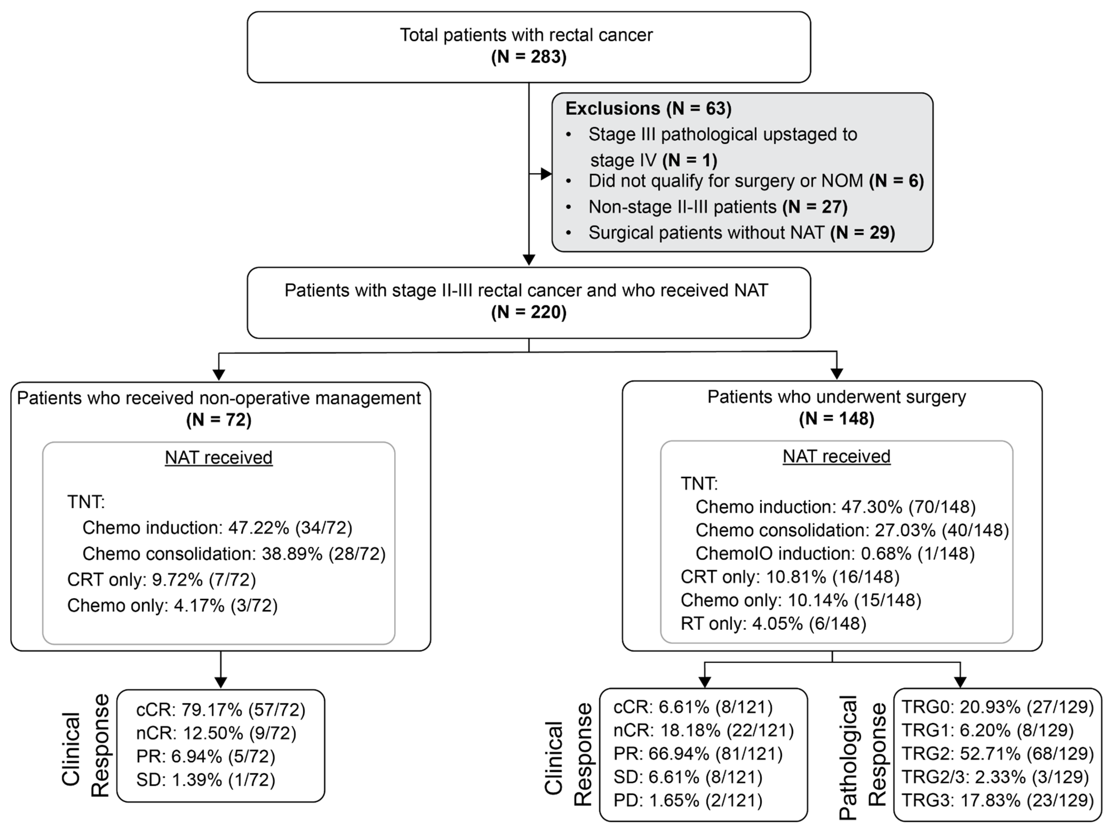
Patients were assigned to the surgical cohort if they underwent surgery following NAT. For patients in the surgical cohort, the MRD window was defined as 2–12 weeks after surgery prior to the initiation of NAT. Patients were assigned to the NOM cohort if, following NAT, they were initially managed without immediate surgery regardless of whether salvage TME surgery was later performed for local regrowth. Patients achieving complete clinical response (cCR) or near-complete response (nCR) were considered eligible for NOM. A small number of patients with either partial response (PR) or stable disease (SD) were included in the NOM cohort at the provider’s discretion or due to patient choice. Patients who underwent upfront surgery without NAT (N = 29); those who were not eligible for surgery or NOM, declined surgery, or were inoperable despite having gross residual disease post-NAT (N = 6); or who were not stages II–III (N = 27) were excluded (Figure 1).
This study was conducted in compliance with Natera’s Institutional Review Board (IRB)-approved protocol (Salus #21204-02B), the Declaration of Helsinki, Title 21 of the US Code of Federal Regulations (CFR) as applicable, Good Clinical Practice guidelines, and International Conference on Harmonization guidelines. A waiver of the consent process and of the requirement for documentation of informed consent was granted according to 45 CFR 46.116(d) and 45 CFR 46.117(c)(2), respectively.

2.2. Personalized ctDNA Assay Workflow

For all patients, blood specimens (two 10 mL Streck tubes) were collected at the discretion of the ordering physician, with an average of 7 samples per patient (range: 1–28). All biological specimens were processed according to a CLIA-validated standard operating procedure at Natera, Inc. ctDNA analysis was performed using a clinically validated, personalized, and tumor-informed 16-plex PCR next-generation sequencing (NGS) assay (Signatera™, Natera, Inc., Austin, TX, USA) in patients undergoing commercial ordering as previously described [28]. Briefly, whole-exome sequencing (WES) was performed on formalin-fixed and paraffin-embedded tumor tissue, along with matched normal DNA blood samples from each patient. Based on the WES results, a personalized assay consisting of multiplex PCR (mPCR) primers was designed for 16 high-ranked patient-specific somatic single-nucleotide variants (SNVs) for each patient. The mPCR assay was then utilized in the associated patients’ plasma-derived cfDNA to detect and track ctDNA. Plasma cfDNA samples with ≥2 SNVs above a predefined algorithmic confidence threshold were considered ctDNA-positive. ctDNA concentration (levels) was quantitatively reported as mean tumor molecules (MTMs) per mL of plasma.

2.3. Statistical Analyses

For surgical patients, the primary outcome was disease-free survival (DFS), defined as the time from surgery to endoscopic or radiological recurrence (locoregional or distant) or death, and censored at last follow-up or death. For NOM patients, the primary outcome was TME-free survival, measured as time from NAT completion to salvage surgery or death. Survival analysis was performed using the Kaplan–Meier method and R software (v4.3.1; RRID:SCR_000432). To account for immortal time bias, we applied a landmark analysis, restricting post-operative MRD assessment to the 2–12-week window after surgery and only including patients who were alive and event-free until ≥12 weeks after surgery. A multivariable Cox proportional hazards model was used to assess the most significant prognostic factor associated with DFS. To account for variability in the timing and frequency of ctDNA sampling during surveillance, ctDNA status was modeled as a time-varying covariate in a time-dependent Cox regression for some analyses. All p-values were based on two-sided testing and considered significant at p ≤ 0.05. Time-dependent Cox regression evaluated serial ctDNA analysis during surveillance.

3. Results

3.1. Patient Cohort

A total of 1572 plasma samples were collected from 220 patients with stages II–III rectal cancer (median age: 59 years [range: 32–89 years]). Specific details regarding tumor type, pathologic stage, and treatment are described in Table 1. In total, 32.9% (72/220) received non-operative management (NOM) after NAT, and 67.12% (148/220) of the patients underwent surgery following NAT (Figure 1). NAT included total neoadjuvant therapy (TNT), chemotherapy alone, and chemoradiation/radiation alone.
For the surgical cohort, ctDNA at the post-NAT timepoint was collected at a median of 1.4 months (range: 0.5–6.3 months) after the completion of planned NAT, prior to surgery, with a median length of time between completion of NAT and surgery of 68 days (range: 6–270 days). For the NOM cohort, the first timepoint collected was at a median of 2.9 months (range: 0–40 months) after completion of planned NAT. All serial post-NAT ctDNA timepoints from the completion of NAT up to the time of recurrence or last follow-up, if no relapse, were evaluated. In the NOM cohort, 32 patients had their first available ctDNA within 3 months post-NAT; 6.3% (2/32) were ctDNA-positive, both of whom experienced local recurrence. Of the remaining 30 ctDNA-negative patients, 20% (6/30) converted to ctDNA-positive during serial testing, and all subsequently developed local recurrence, and none developed distant metastatic recurrence.

3.2. NOM Cohort: Post-NAT Association of Clinical Response and ctDNA Status with DFS

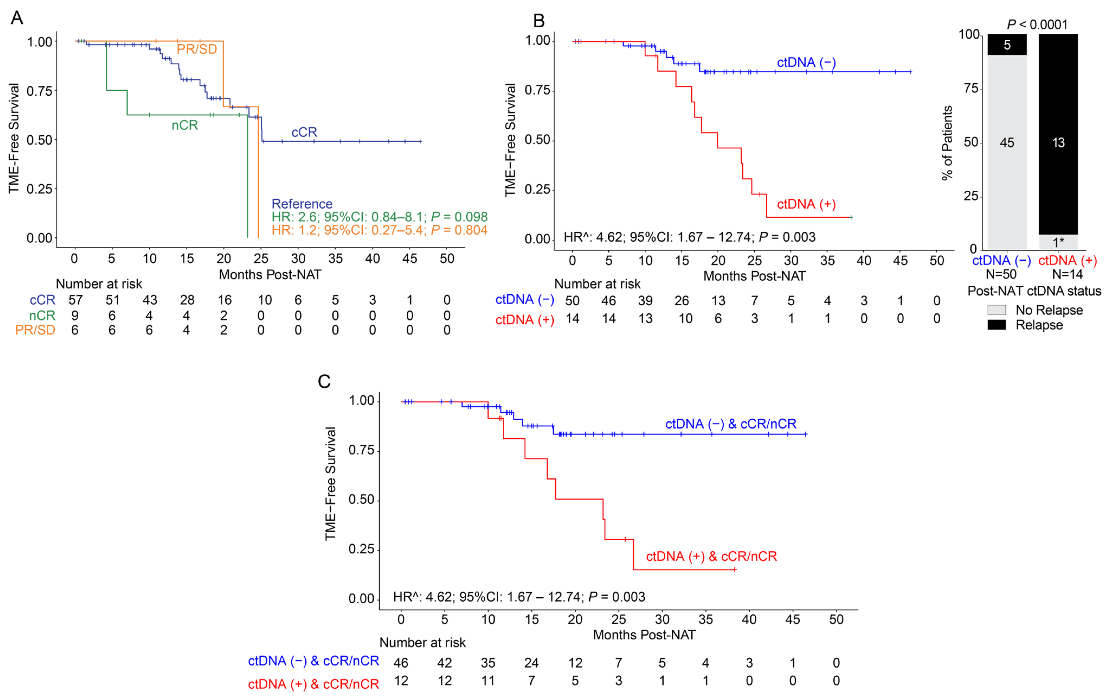
Of the 72 patients pursuing NOM after NAT, 79.1% (57/72) showed cCR, 12.5% (9/72) showed near-complete response (nCR), 6.9% (5/72) showed partial response (PR), and 1.4% (1/72) showed stable disease (SD). Of the six patients with PR or SD, three eventually achieved cCR without additional treatment intervention. The median follow-up post-NAT was 17 months (range: 1–48). When evaluating TME-free survival by clinical response, no statistically significant differences were observed between responses (Figure 2A).
By ctDNA status, 92.0% (46/50) of the ctDNA-negative patients showed cCR/nCR compared to 85.7% (12/14) of ctDNA-positive patients. Regardless of post-NAT ctDNA status, the clinical response status (cCR/nCR vs. PR/SD) between the ctDNA-negative or positive group was not statistically different, p = 1. However, when comparing relapse to non-relapse patients between the post-NAT ctDNA-negative and -positive groups, the difference was statistically significant, p < 0.0001 (Figure 2B), suggesting that, in NOM-eligible patients, post-NAT ctDNA can identify the patients who will eventually relapse.
On evaluating TME-free survival stratified by ctDNA status, the ctDNA-positive patients demonstrated significantly inferior outcomes compared to the ctDNA-negative patients (time-dependent HR: 4.62, 95% CI: 1.67–12.74, p = 0.003; Figure 2B). Due to the nature of serial testing and variable frequency of plasma collection for monitoring, the post-NAT analysis included ctDNA testing from the completion of NAT up to the time of recurrence or until the last clinical follow-up for patients who did not recur. Among the NOM patients with ctDNA available after completion of NAT (N = 64), 78.1% (50/64) were ctDNA-negative and 21.9% (14/64) were ctDNA-positive. Furthermore, 92.9% (13/14) of the ctDNA-positive post-NAT patients relapsed or progressed compared to only 10% (5/50) of the ctDNA-negative patients. The one remaining patient with ctDNA positivity post-NAT had multiple ctDNA-positive samples accompanied by a suspicious imaging 1 month after turning ctDNA-positive. This patient ultimately relapsed 8 months after the data cut-off. Among the five post-NAT ctDNA-negative patients who relapsed, one patient only had one post-NAT ctDNA draw that was taken 310 days prior to relapse, and another eventually experienced ctDNA positivity immediately after clinical recurrence 154 days later. We observed that the median time to molecular recurrence post-NAT was 8.9 months (range: 0.32–27.8) and the median time to clinical recurrence post-NAT was 13.5 months (range: 8.1–25.1) post-NAT.
Of note, among the 13 post-NAT ctDNA-positive patients who relapsed, all 13 (100%) had local regrowth or local recurrence, except one had both local and lung recurrence. Interestingly, when utilizing clinical response as an adjunct to ctDNA status post-NAT, among the patients who achieved cCR/nCR, ctDNA-positive status remained significantly associated with inferior TME-free survival (time-dependent HR: 4.62, 95% CI: 1.67–12.74, p = 0.003; Figure 2C).

3.3. Surgical Cohort: Association of ctDNA Status with Pathological Response and DFS

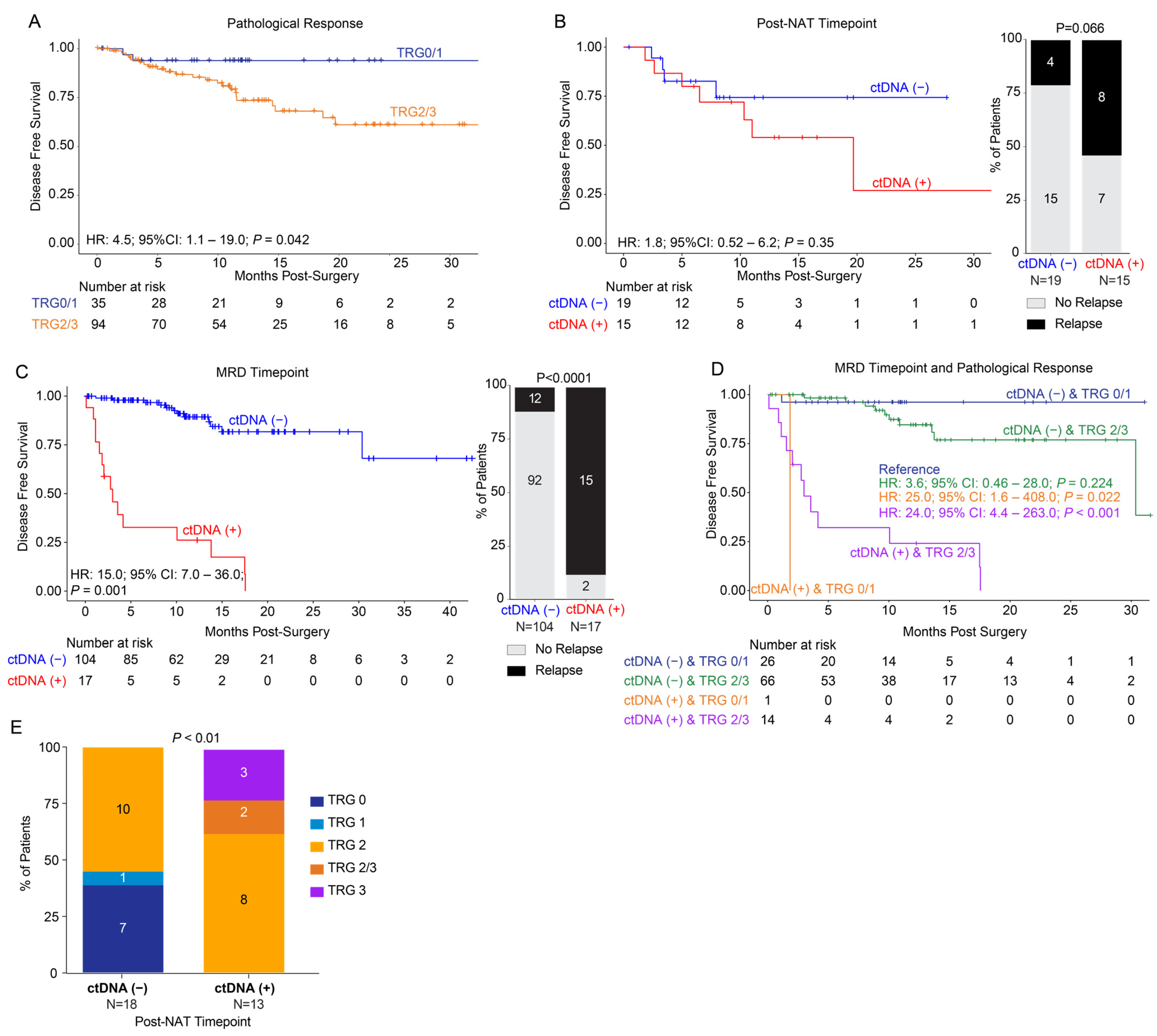
A total of 148 patients underwent surgical resection, and, of these, pathological response to neoadjuvant therapy was available for 129 patients. The median clinical follow-up post-surgery was 11 months (range: 0–44). For patients with pathological response data available, 20.9% (27/129) had a TRG 0 score, 6.2% (8/129) were TRG 1, 52.7% (68/129) were TRG 2, 2.3% (3/129) were TRG 2/3, and 17.8% (23/129) were TRG3. Upon evaluating DFS stratified by pathological response, patients with TRG scores 2–3 showed inferior outcomes when compared to patients with TRG scores 0–1 (pathological response: HR: 4.5, 95% CI: 1.1–19.0, p = 0.042; Figure 3A).
On stratifying patients with available ctDNA (N = 34) at the post-NAT timepoint, patients with ctDNA positivity showed worse DFS compared to ctDNA-negative patients (HR: 1.8, 95% CI: 0.52–6.2, p = 0.35), with relapse rates of 53% (8/15) and 21% (4/19) for the ctDNA-positive and -negative groups, respectively. However, this finding was not statistically significant (Figure 3B). The median follow-up for these 34 patients was 11 months (range: 0–27). Of note, at the post-operative MRD timepoint (N = 121), patients with ctDNA positivity showed a significantly inferior DFS (HR: 15.0; 95% CI: 7.0–36.0, p = 0.001. Figure 3C), with a relapse rate of 11.5% (12/104) for ctDNA-negative patients and 88% (15/17) for ctDNA-positive patients (p < 0.0001). Of the surgical patients who were ctDNA-positive at the post-NAT timepoint (n = 15), 80% (12/15) also had available ctDNA timepoints during the MRD window. Of these, three were ctDNA-positive in the MRD window, while nine were ctDNA-negative. All three patients with ctDNA positivity had distant recurrences. Of the nine patients who were ctDNA-negative, six remained relapse-free and three eventually relapsed (two distant and one local), with two of these turning ctDNA-positive prior to recurrence during surveillance.
Furthermore, upon assessing DFS based on ctDNA status in conjunction with pathological response, ctDNA-negative patients showed improved DFS regardless of TRG score. By contrast, ctDNA-positive patients with a TRG score of 0/1 (HR: 25.0; 95% CI: 1.6–408.0; p = 0.02) or a TRG score of 2/3 (HR: 24.0; 95% CI: 4.4–263.0, p < 0.001) displayed significantly inferior DFS (Figure 3D). Additionally, at the post-NAT timepoint, none of the ctDNA-negative patients showed TRG scores 2/3-3, while, among the ctDNA-positive patients, none had a TRG score 0/1. Each category of TRG score was statistically significantly different between the ctDNA-positive and -negative groups (p < 0.01; Figure 3E).

3.4. Surgical Cohort: NAR Score and Association with ctDNA Status and Outcomes

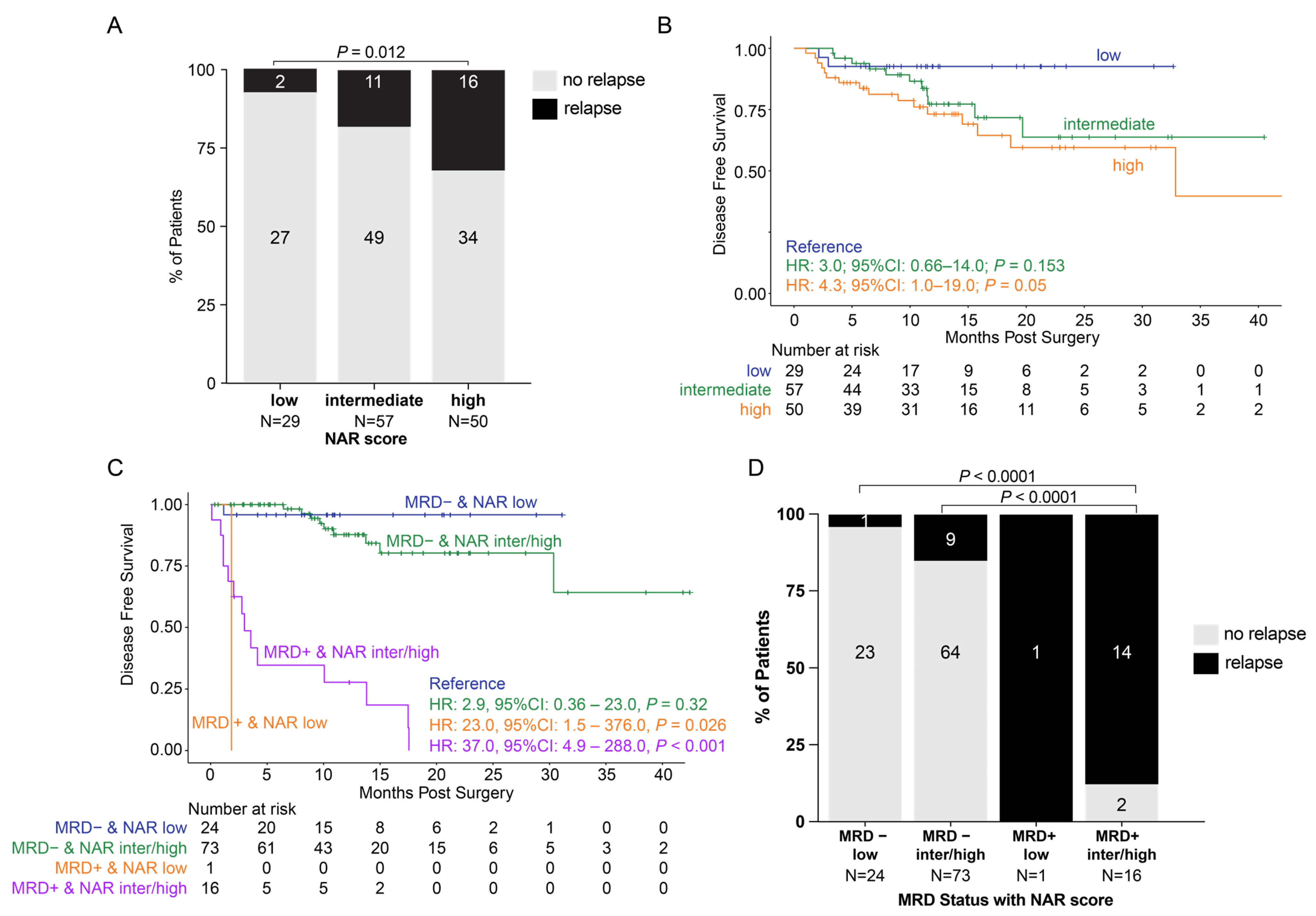
In our study, we observed that patients with high NAR scores exhibited a significantly higher recurrence rate of 32% (16/50) when compared to patients with low NAR scores (7% [2/29], p = 0.012. Figure 4A). Additionally, we observed that patients with high (HR: 4.3, 95% CI: 1.0–19.0, p = 0.05) and intermediate (HR: 3.0, 95% CI: 0.66–14.0, p = 0.153) NAR scores exhibited inferior DFS when compared to patients with low NAR scores (Figure 4B). When assessing ctDNA status at the post-op MRD timepoint in conjunction with the NAR score, we found that patients with high NAR scores were more frequently ctDNA-positive (27% [14/51]) than patients with intermediate (11% [6/54], p = 0.05) and low (4% [1/25], p = 0.02) NAR scores.
When evaluating DFS, we found that the use of ctDNA status at the MRD window enhances the predictive utility of the NAR score. While patients with intermediate or high NAR scores had a worse prognosis than those with low NAR scores, the majority of intermediate and high NAR scores did not recur (81% and 68%, respectively). When coupled with ctDNA status at the MRD window, ctDNA can further stratify the NAR low, intermediate, and high patients and predict relapse. As shown in Figure 4C, regardless of NAR score, patients who were ctDNA-positive in the MRD window had inferior outcomes compared to patients who were ctDNA-negative (ctDNA-positive with low NAR: HR: 23.0, 95% CI: 1.5–376.0, p = 0.026; ctDNA-positive with intermediate/high NAR: HR: 37.0, 95% CI: 4.9–288.0, p < 0.001). There was no statistically significant difference between ctDNA-negative patients with low and intermediate/high NAR scores. Finally, the relapse rates were higher in patients who were ctDNA-positive when compared to patients who were ctDNA-negative regardless of NAR score (p < 0.0001 for all comparisons. Figure 4D).

4. Discussion

ctDNA has emerged as a prognostic biomarker and shows the potential to transform response assessment and recurrence risk stratification in patients with LARC, particularly as treatment paradigms shift toward organ preservation. In this largest-to-date real-world study of ctDNA in LARC, we demonstrate the clinical relevance of ctDNA testing at both post-neoadjuvant and post-surgical timepoints across patients managed with surgery or NOM. Our findings reveal that ctDNA positivity robustly predicts recurrence, even among patients with radiographic or endoscopic complete response, highlighting its value in informing critical treatment decisions.
Importantly, this study offers real-world insights on the utility of ctDNA in the management of LARC. Among the surgical patients, particularly those achieving cCR, post-NAT ctDNA negativity may identify candidates for treatment de-escalation and less intensive surveillance. Conversely, in the NOM patients, post-NAT ctDNA positivity was strongly associated with local regrowth, suggesting a role for treatment escalation or earlier salvage surgery. Serial ctDNA testing during surveillance may refine risk stratification, distinguishing durable responders from patients at increased risk of recurrence. Additionally, ctDNA negativity may help to identify patients who are suitable for less intensive surveillance, while ctDNA positivity may inform risk stratification and consideration of closer monitoring or alternative management strategies. However, prospective interventional trials are underway and will be required before ctDNA-guided treatment escalation or de-escalation can be adopted in routine clinical practice. Collectively, these findings support the role of ctDNA as a complementary tool to imaging and endoscopy and provide a rationale for prospective trials evaluating ctDNA-guided management strategies in LARC.
A significant proportion of patients in the current study underwent NOM. Our study provides critical insight into the significance of post-NAT ctDNA status in this group. NOM, first popularized by Habr-Gama et al., is increasingly used for select patients with LARC [5]. A previous study reported baseline ctDNA combined with positive MRI extramural venous invasion to predict response [29]. Here, we found that post-NAT ctDNA status was strongly prognostic of outcomes among NOM patients (time-dependent HR: 4.62, 95% CI: 1.67–12.74, p = 0.003) independent of clinical response (cCR/nCR p = 0.003). Post-NAT ctDNA negativity was associated with remaining relapse-free, whereas ctDNA-positive patients had a high recurrence rate (13/14, 92.9%), suggesting that these patients may not be suitable for a non-operative approach. It is important to note that the one (7.1%; 1/14) remaining patient who was post-NAT ctDNA-positive recurred 8 months after the data cut-off. Therefore, even among patients with cCR/nCR, those who were ctDNA-positive had a 100% recurrence rate (PPV). Notably, all the ctDNA-positive patients had local recurrences, including the patient who recurred after data lock, with one patient having an additional lung recurrence.
Among the patients in the surgical cohort, we found that post-NAT ctDNA status correlated with pathological response, suggesting that it may serve as a surrogate for pathological response. An earlier phase 2 randomized trial reported improved 3-year DFS (71.6%) with adjuvant FOLFOX among patients with post-operative stages II–III disease and high-risk features following preoperative chemoradiotherapy [30]. Since then, several studies have demonstrated ctDNA positivity post-NAT to be associated with an increased risk of recurrence among patients with rectal cancer [25,31]. Here, we found that the prognostic performance of ctDNA differed depending on the timing of assessment in surgical patients. While ctDNA positivity at the post-NAT pre-surgical timepoint was associated with inferior DFS, this association did not reach statistical significance, likely reflecting the residual tumor heterogeneity prior to resection, variable timing of sampling, and limited sample size at this timepoint. In contrast, ctDNA status during the post-operative MRD window was a highly robust and statistically significant predictor of recurrence, consistent with the concept that definitive surgical clearance establishes the most informative biological context for MRD assessment. These findings suggest that ctDNA testing after surgical resection represents a reliable prognostic window for recurrence risk stratification in surgically managed patients with LARC.
Consistent with the published data [25,31,32,33], we found ctDNA positivity during the MRD window (2–12 weeks post-surgery) to be associated with poor outcomes (HR: 15.0, 95% CI: 7.0–36.0, p = 0.001). As previously described, the neoadjuvant rectal (NAR) score, calculated using the clinical tumor stage and pathological nodal and tumor stage, can be used as a composite short-term assessment of NAT response [34,35]. We observed that MRD ctDNA status was associated with NAR score, thereby substantiating our previous findings. When used as an adjunct to the NAR score, MRD ctDNA status was found to be the main driver of DFS when compared to the NAR score alone. These patients may benefit from ctDNA-specific clinical trials to address biochemical recurrence. Finally, it is worth noting that local relapse was more frequent among the NOM patients, whereas distant relapse predominated in the surgical cohort.
Our study has several limitations inherent to its real-world retrospective design. The treatment approaches varied across sites, including differences in NAT regimens and NOM implementation. ctDNA testing was performed at the discretion of the treating physicians, resulting in variability in timing and frequency of sampling. This may introduce selection bias as testing may have been preferentially performed in patients perceived to be at higher risk or with equivocal clinical responses. Moreover, while this heterogeneity may introduce confounding, it also reflects contemporary clinical practice and enhances the generalizability of our findings. In some instances, ctDNA results may have influenced clinical decisions in real time, which we acknowledge could affect outcome associations; however, this feature underscores the relevance of ctDNA in routine practice rather than diminishing its potential utility. The follow-up duration was relatively short, which may explain the predominance of local, rather than distant, recurrences observed. An additional limitation relates to the timing of post-NAT ctDNA assessments. Since this was not a prospectively designed study, only a small subset of patients underwent ctDNA testing at standardized timepoints post-NAT, precluding statistically meaningful subgroup analyses for clinical decision-making at the time of NOM consideration. Additionally, the median follow-up after surgery was relatively short, which may preferentially capture early local recurrences and underestimate the incidence of later distant metastases. Longer follow-up will be important to fully define the temporal relationship between the ctDNA dynamics and patterns of recurrence in this population. Despite these limitations, our study results are hypothesis-generating and represent the largest-to-date real-world cohort of patients with LARC undergoing NOM or surgery with serial ctDNA testing. The consistent associations we observed across multiple analyses support the robustness of our findings and highlight the potential of ctDNA to inform both surveillance and treatment decision-making.

5. Conclusions

In this large real-world cohort of patients with LARC, ctDNA status at both post-NAT and MRD timepoints correlated with clinical response, pathological response, and recurrence outcomes. ctDNA testing may offer significant value for real-time clinical decision-making, particularly in selecting patients for NOM or intensified post-operative surveillance. Future prospective studies are warranted to validate these findings and to evaluate ctDNA-guided management strategies in clinical trials.

Author Contributions

S.C. (Sakti Chakrabarti), S.A.C., A.T., A.J., M.C.L., and A.D. (Arvind Dasari) conceived and designed the study. S.C. (Sakti Chakrabarti), S.A.C., A.T., V.N.A., and S.S. developed the methodology. S.C. (Sakti Chakrabarti), S.A.C., K.Y.C., M.A.T., M.G.F., S.R.C., C.A.D., V.G., R.G.L., M.M. (Midhun Malla), A.N., B.A.W., V.R.S., G.P.B., M.C., G.A., A.K., F.D., D.L.H., B.G.S., and A.D. (Arvind Dasari) recruited patients and collected the data. S.C. (Sakti Chakrabarti), S.A.C., A.J., M.C.L., and A.D. (Arvind Dasari) supervised and aided in project administration. J.B.O., M.M. (Meenakshi Malhotra) and G.V.G. wrote the original draft. A.T. and J.B.O. performed visualization. A.T., A.D. (Autumn Dangl), V.N.A., W.K.H., S.S., A.J., and M.C.L. analyzed and interpreted the data. All authors reviewed, edited, and revised the manuscript. All authors verify that this study was conducted per protocol and vouch for data accuracy and completeness. All authors have read and agreed to the published version of the manuscript.

Funding

This research received no external funding.

Institutional Review Board Statement

This study was conducted in compliance with Natera’s Institutional Review Board (IRB)-approved protocol (Salus #21204-02B), the Declaration of Helsinki, Title 21 of the US Code of Federal Regulations (CFR) as applicable, Good Clinical Practice guidelines, and International Conference on Harmonization guidelines.

Informed Consent Statement

A waiver of the consent process and of the requirement for documentation of informed consent was granted according to 45 CFR 46.116(d) and 45 CFR 46.117(c)(2), respectively.

Data Availability Statement

The authors declare that all relevant non-proprietary data used to conduct the analyses are available within the article. To protect the privacy and confidentiality of the patients in this study, clinical data are not made publicly available in a repository but can be requested at any time from the corresponding author. All data shared will be de-identified.

Acknowledgments

The authors wish to acknowledge Seth Waller for their assistance in data collection.

Conflicts of Interest

Sakti Chakrabarti declares publication support from Natera, Inc. and Guardant Inc.; grants from Merck; consulting fees from HalioDx, QED Therapeutics, HistoSonics, Cancer Expert Now, Natera, Inc., Guardant, Neogenomics, Inc., Takeda, Athenium Analytics, BioMedical Insights.; honoraria and/or speaker bureau membership with Natera, Inc., Takeda, Merck, Bristol Myers Squibb, SeaGen/Pfizer, OmniHealth Media, GI Oncology Now, OncLive.; participation on data safety monitoring or advisory boards for HalioDx, QED Therapeutics, HistoSonics, Cancer Expert Now, Natera, Inc., Guardant, Neogenomics, Athenium Analytics, BioMedical Insights.; and co-chair of the Committee On Disparities in Clinical Research (Case Comprehensive Cancer Center), chair of the institutional GI Oncology Tumor Board (UH Seidman Cancer Center). Stacey Cohen declares consulting fees from Janssen, Incyte, Exact Sciences, Merck, Abbvie, Pfizer, Agenus, Guardant, Taiho, BNT, Regeneron; payment for expert testimony from Brewster & DeAngelis, The Cline Law Firm; participation on data safety monitoring or advisory board at GSK; and research (trial) funds to institution from Pfizer, BNT, Biomea, Tempus, BillionToOne. Antony Tin, Autumn Dangl, Vasily N. Aushev, Giby George, J. Bryce Ortiz, Whitney Herter, Meenakshi Malhotra, Shruti Sharma, Adham Jurdi, and Minetta C. Liu are employees of Natera, Inc. and receive salary and may own stock and/or stock options. Sreenivasa R. Chandana declares research funding to the institution from AbbVie, Adcentrx Therapeutics, Amgen, AstraZeneca, Cardiff Oncology, Dicephera, Elevation Oncology, Exact Sciences, Genentech/Roche, IDEAYA Biosciences, IGM Biosciences, Incyte, Ipsen, Janssen, Merck, Mirati Therapeutics, Novocure, Qualigen Therapeutics, Zymeworks; advisory board with Ipsen; and speaker bureau membership with Natera, Inc. Midhun Malla declares advisory board/honoraria/speakers bureau membership with: Ipsen, Exelexis, Merus, DoMOORE, Natera, Astrazenaca, Incyte, BMS; speaking/presentation: oral/poster: ASCO-GI, ESMO-GI, ASCO, Cholangiocarcinoma Summit, Cholangiocarcinoma Foundation; and research funding (to the institution): Tvardi, Seagen/Pfizer. Benjamin A. Weinberg declares publication support from Caris Life Sciences; grants or contracts from Ipsen, BioXcel, and Merck; speaker bureau membership with Natera, Inc., Jazz, Seagen, Sirtex, Taiho, and Merus; advisor for Foundation Medicine, Regeneron, Agenus, DoMore Diagnostics, Merus. Gregory P. Botta declares consulting fees from Calibr; honoraria from Natera, Inc.; and stock or stock options from TumorGen. Farshid Dayyani declares grants to their institution from Takeda, Taiho, Natera, Ipsen, Roche, Exelixis, Astrazeneca, Astellas, Amgen; consulting fees from Astrazeneca, DaiichiSankyo, Eisai, Jazz, Sirtex, Taiho; honoraria or speaker bureau membership from Takeda, Sirtex, Ipsen, Beigene, Astellas. MInetta C. Liu also reports grants/contracts: funding to institution (Mayo) from: Eisai, Exact Sciences, Genentech, Genomic Health, GRAIL, Menarini Silicon Biosystems, Merck, Novartis, Seattle Genetics, Tesaro; travel support reimbursement from AstraZeneca, Genomic Health, Ionis; ad hoc advisory board meetings. All funds to Mayo Clinic. No personal compensation from AstraZeneca, Celgene, Roche/Genentech, Genomic Health, GRAIL, Ionis, Merck, Pfizer, Seattle Genetics, or Syndax. Ron G. Landmann declares contributions to UpToDate; consulting fees from Johnson & Johnson MedTech; honoraria from Natera, Inc.; and membership on the Executive Council of National Accreditation Program Rectal Cancer. Arvind Dasari declares grants from Crinetics, Eisai, Enterome, Guardant Health, HutchMed, Natera, Neogenomics, Personalis, RayzeBio/BMS, Taiho, Xencor (to institution) and advisory board membership with Agenus, BMS, Exelixis, Illumina, Lantheus, Personalis, Sanofi, Taiho, and Takeda. Ki Y. Chung has nothing to disclose. Mohamedtaki A. Tejani has nothing to disclose. Marwan G. Fakih has nothing to disclose. Colleen A. Donahue has nothing to disclose. Virgilio George has nothing to disclose. Arun Nagarajan has nothing to disclose. Vivek R Sharma has nothing to disclose. May Cho has nothing to disclose. Diana L. Hanna has nothing to disclose. Bradley G. Somer has nothing to disclose.

Abbreviations

The following abbreviations are used in this manuscript:
ctDNACirculating tumor DNA
MRDMolecular residual disease
LARCLocally advanced rectal cancer
TMETotal mesorectal excision
NOMNon-operative management
NARNeoadjuvant rectal (score)
NATNeoadjuvant therapy
TNTTotal neoadjuvant therapy
CRTChemoradiation
TRGTumor regression grade
pCRPathological complete response
cCRComplete clinical response
nCRNear complete response
PRPartial response
SDStable disease
PDProgressive disease
DREDigital rectal exam
MTMMean tumor molecules
WESWhole exome sequencing
NGSNext generation sequencing
mPCRMultiplex polymerase chain reaction
SNVSingle nucleotide variants
DFSDisease-free survival
HRHazard ratio
CIConfidence Interval

References

  1. Liu, S.; Jiang, T.; Xiao, L.; Yang, S.; Liu, Q.; Gao, Y.; Chen, G.; Xiao, W. Total Neoadjuvant Therapy (TNT) versus Standard Neoadjuvant Chemoradiotherapy for Locally Advanced Rectal Cancer: A Systematic Review and Meta-Analysis. Oncologist 2021, 26, e1555–e1566. [Google Scholar] [CrossRef] [PubMed]
  2. Scott, A.J.; Kennedy, E.B.; Berlin, J.; Brown, G.; Chalabi, M.; Cho, M.T.; Cusnir, M.; Dorth, J.; George, M.; Kachnic, L.A.; et al. Management of locally advanced rectal cancer: ASCO guideline. J. Clin. Oncol. 2024, 42, 3355–3375. [Google Scholar] [CrossRef] [PubMed]
  3. Keller, D.S.; Berho, M.; Perez, R.O.; Wexner, S.D.; Chand, M. The multidisciplinary management of rectal cancer. Nat. Rev. Gastroenterol. Hepatol. 2020, 17, 414–429. [Google Scholar] [CrossRef] [PubMed]
  4. Yang, Y.; Huang, A.; Sun, Z.; Hong, H.-P.; Kim, N.K.; Gu, J. “Watch and wait” strategy after neoadjuvant chemoradiotherapy in rectal cancer: Opportunities and challenges. Holist. Integr. Oncol. 2023, 2, 4. [Google Scholar] [CrossRef]
  5. Habr-Gama, A.; São Julião, G.P.; Vailati, B.B.; Castro, I.; Raffaele, D. Management of the complete clinical response. Clin. Colon Rectal Surg. 2017, 30, 387–394. [Google Scholar] [CrossRef]
  6. Zwart, W.H.; Hotca, A.; Hospers, G.A.; Goodman, K.A.; Garcia-Aguilar, J. The multimodal management of locally advanced rectal cancer: Making sense of the new data. Am. Soc. Clin. Oncol. Educ. Book 2022, 42, 1–14. [Google Scholar] [CrossRef]
  7. Schrag, D.; Shi, Q.; Weiser, M.R.; Gollub, M.J.; Saltz, L.B.; Musher, B.L.; Goldberg, J.; Al Baghdadi, T.; Goodman, K.A.; McWilliams, R.R.; et al. Preoperative treatment of locally advanced rectal cancer. N. Engl. J. Med. 2023, 389, 322–334. [Google Scholar] [CrossRef]
  8. Verheij, F.S.; Omer, D.M.; Williams, H.; Lin, S.T.; Qin, L.X.; Buckley, J.T.; Thompson, H.M.; Yuval, J.B.; Kim, J.K.; Dunne, R.F.; et al. Long-term results of organ preservation in patients with rectal adenocarcinoma treated with total neoadjuvant therapy: The randomized phase II OPRA trial. J. Clin. Oncol. 2024, 42, 500–506. [Google Scholar] [CrossRef]
  9. Bahadoer, R.R.; Dijkstra, E.A.; van Etten, B.; Marijnen, C.A.; Putter, H.; Kranenbarg, E.M.; Roodvoets, A.G.; Nagtegaal, I.D.; Beets-Tan, R.G.; Blomqvist, L.K.; et al. Short-course radiotherapy followed by chemotherapy before total mesorectal excision (TME) versus preoperative chemoradiotherapy, TME, and optional adjuvant chemotherapy in locally advanced rectal cancer (RAPIDO): A randomised, open-label, phase 3 trial. Lancet Oncol. 2021, 22, 29–42. [Google Scholar] [CrossRef]
  10. Conroy, T.; Bosset, J.F.; Etienne, P.L.; Rio, E.; François, É.; Mesgouez-Nebout, N.; Vendrely, V.; Artignan, X.; Bouché, O.; Gargot, D.; et al. Neoadjuvant chemotherapy with FOLFIRINOX and preoperative chemoradiotherapy for patients with locally advanced rectal cancer (UNICANCER-PRODIGE 23): A multicentre, randomised, open-label, phase 3 trial. Lancet Oncol. 2021, 22, 702–715. [Google Scholar] [CrossRef]
  11. Dijkstra, E.A.; Nilsson, P.J.; Hospers, G.A.; Bahadoer, R.R.; Kranenbarg, E.M.; Roodvoets, A.G.; Putter, H.; Berglund, Å.; Cervantes, A.; Crolla, R.M.; et al. Locoregional failure during and after short-course radiotherapy followed by chemotherapy and surgery compared with long-course chemoradiotherapy and surgery: A 5-year follow-up of the RAPIDO trial. Ann. Surg. 2023, 278, e766–e772. [Google Scholar] [CrossRef]
  12. Conroy, T.; Etienne, P.L.; Rio, E.; Evesque, L.; Mesgouez-Nebout, N.; Vendrely, V.; Artignan, X.; Bouche, O.; Boileve, A.; Delaye, M.; et al. Total neoadjuvant therapy with mFOLFIRINOX versus preoperative chemoradiation in patients with locally advanced rectal cancer: 7-year results of PRODIGE 23 phase III trial, a UNICANCER GI trial. Am. Soc. Clin. Oncol. 2023, 47, LBA3504. [Google Scholar] [CrossRef]
  13. Garcia-Aguilar, J.; Patil, S.; Gollub, M.J.; Kim, J.K.; Yuval, J.B.; Thompson, H.M.; Verheij, F.S.; Omer, D.M.; Lee, M.; Dunne, R.F.; et al. Organ preservation in patients with rectal adenocarcinoma treated with total neoadjuvant therapy. J. Clin. Oncol. 2022, 40, 2546–2556. [Google Scholar] [CrossRef] [PubMed]
  14. van der Valk, M.J.; Hilling, D.E.; Bastiaannet, E.; Kranenbarg, E.M.; Beets, G.L.; Figueiredo, N.L.; Habr-Gama, A.; Perez, R.O.; Renehan, A.G.; van de Velde, C.J.; et al. Long-term outcomes of clinical complete responders after neoadjuvant treatment for rectal cancer in the International Watch & Wait Database (IWWD): An international multicentre registry study. Lancet 2018, 391, 2537–2545. [Google Scholar] [CrossRef] [PubMed]
  15. Glynne-Jones, R.; Wyrwicz, L.; Tiret, E.; Brown, G.; Rödel, C.D.; Cervantes, A.; Arnold, D.; ESMO Guidelines Committee. Rectal cancer: ESMO Clinical Practice Guidelines for diagnosis, treatment and follow-up. Ann. Oncol. 2017, 28, iv22–iv40. [Google Scholar] [CrossRef]
  16. Ludmir, E.B.; Palta, M.; Willett, C.G.; Czito, B.G. Total neoadjuvant therapy for rectal cancer: An emerging option. Cancer 2017, 123, 1497–1506. [Google Scholar] [CrossRef]
  17. Nahas, S.C.; Nahas, C.S.; Marques, C.F.; Ribeiro, U., Jr.; Cotti, G.C.; Imperiale, A.R.; Capareli, F.C.; Chen, A.T.; Hoff, P.M.; Cecconello, I. Pathologic complete response in rectal cancer: Can we detect it? Lessons learned from a proposed randomized trial of watch-and-wait treatment of rectal cancer. Dis. Colon Rectum 2016, 59, 255–263. [Google Scholar] [CrossRef]
  18. Sun, W.; Al-Rajabi, R.; Perez, R.O.; Abbasi, S.; Ash, R.; Habr-Gama, A. Controversies in rectal cancer treatment and management. In American Society of Clinical Oncology Educational Book; American Society of Clinical Oncology: Alexandria, VA, USA, 2020; Volume 40, pp. 136–146. [Google Scholar] [CrossRef]
  19. Thompson, H.M.; Omer, D.M.; Lin, S.; Kim, J.K.; Yuval, J.B.; Verheij, F.S.; Qin, L.X.; Gollub, M.J.; Wu, A.J.; Lee, M.; et al. Organ preservation and survival by clinical response grade in patients with rectal cancer treated with total neoadjuvant therapy: A secondary analysis of the OPRA randomized clinical trial. JAMA Netw. Open 2024, 7, e2350903. [Google Scholar] [CrossRef]
  20. Smith, J.J.; Strombom, P.; Chow, O.S.; Roxburgh, C.S.; Lynn, P.; Eaton, A.; Widmar, M.; Ganesh, K.; Yaeger, R.; Cercek, A.; et al. Assessment of a watch-and-wait strategy for rectal cancer in patients with a complete response after neoadjuvant therapy. JAMA Oncol. 2019, 5, e185896. [Google Scholar] [CrossRef]
  21. Wang, Y.; Yang, L.; Bao, H.; Fan, X.; Xia, F.; Wan, J.; Shen, L.; Guan, Y.; Bao, H.; Wu, X.; et al. Utility of ctDNA in predicting response to neoadjuvant chemoradiotherapy and prognosis assessment in locally advanced rectal cancer: A prospective cohort study. PLoS Med. 2021, 18, e1003741. [Google Scholar] [CrossRef] [PubMed]
  22. Cohen, S.A.; Liu, M.C.; Aleshin, A. Practical recommendations for using ctDNA in clinical decision making. Nature 2023, 619, 259–268. [Google Scholar] [CrossRef] [PubMed]
  23. Radomski, S.N.; Ali, S.; Lafaro, K.J.; Shubert, C.; Hidalgo, M.; Chung, H.; Christenson, E.S. The utilization of circulating tumor DNA to predict the risk and location of relapse after curative-intent local therapy in oligometastatic colorectal cancer. J. Gastrointest. Surg. 2024, 28, 534–537. [Google Scholar] [CrossRef] [PubMed]
  24. Kataoka, K.; Mori, K.; Nakamura, Y.; Watanabe, J.; Akazawa, N.; Hirata, K.; Yokota, M.; Kato, K.; Kotaka, M.; Yamazaki, K.; et al. Survival benefit of adjuvant chemotherapy based on molecular residual disease detection in resected colorectal liver metastases: Subgroup analysis from CIRCULATE-Japan GALAXY. Ann. Oncol. 2024, 35, 1015–1025. [Google Scholar] [CrossRef]
  25. Molinari, C.; Marisi, G.; Laliotis, G.; Spickard, E.; Rapposelli, I.G.; Petracci, E.; George, G.V.; Dutta, P.; Sharma, S.; Malhotra, M.; et al. Assessment of circulating tumor DNA in patients with locally advanced rectal cancer treated with neoadjuvant therapy. Sci. Rep. 2024, 14, 29536. [Google Scholar] [CrossRef]
  26. Chidharla, A.; Rapoport, E.; Agarwal, K.; Madala, S.; Linares, B.; Sun, W.; Chakrabarti, S.; Kasi, A. Circulating tumor DNA as a minimal residual disease assessment and recurrence risk in patients undergoing curative-intent resection with or without adjuvant chemotherapy in colorectal cancer: A systematic review and meta-analysis. Int. J. Mol. Sci. 2023, 24, 10230. [Google Scholar] [CrossRef]
  27. Shah, P.K.; Aushev, V.N.; Ensor, J.; Sanchez, S.A.; Wang, C.G.; Cannon, T.L.; Berim, L.D.; Feinstein, T.; Grothey, A.; McCollom, J.W.; et al. Circulating tumor DNA for detection of molecular residual disease (MRD) in patients (pts) with stage II/III colorectal cancer (CRC): Final analysis of the BESPOKE CRC sub-cohort. Am. Soc. Clin. Oncol. 2025, 43, 15. [Google Scholar] [CrossRef]
  28. Reinert, T.; Henriksen, T.V.; Christensen, E.; Sharma, S.; Salari, R.; Sethi, H.; Knudsen, M.; Nordentoft, I.; Wu, H.T.; Tin, A.S.; et al. Analysis of plasma cell-free DNA by ultradeep sequencing in patients with stages I to III colorectal cancer. JAMA Oncol. 2019, 5, 1124–1131. [Google Scholar] [CrossRef]
  29. Morais, M.; Fonseca, T.; Melo-Pinto, D.; Prieto, I.; Vilares, A.T.; Duarte, A.L.; Leitão, P.; Cirnes, L.; Machado, J.C.; Carneiro, S. Evaluation of ctDNA in the prediction of response to Neoadjuvant Therapy and Prognosis in locally advanced rectal Cancer patients: A prospective study. Pharmaceuticals 2023, 16, 427. [Google Scholar] [CrossRef]
  30. Hong, Y.S.; Nam, B.H.; Kim, K.P.; Kim, J.E.; Park, S.J.; Park, Y.S.; Park, J.O.; Kim, S.Y.; Kim, T.Y.; Kim, J.H.; et al. Oxaliplatin, fluorouracil, and leucovorin versus fluorouracil and leucovorin as adjuvant chemotherapy for locally advanced rectal cancer after preoperative chemoradiotherapy (ADORE): An open-label, multicentre, phase 2, randomised controlled trial. Lancet Oncol. 2014, 15, 1245–1253. [Google Scholar] [CrossRef]
  31. Tie, J.; Cohen, J.D.; Wang, Y.; Li, L.; Christie, M.; Simons, K.; Elsaleh, H.; Kosmider, S.; Wong, R.; Yip, D.; et al. Serial circulating tumour DNA analysis during multimodality treatment of locally advanced rectal cancer: A prospective biomarker study. Gut 2019, 68, 663–671. [Google Scholar] [CrossRef]
  32. Massihnia, D.; Pizzutilo, E.G.; Amatu, A.; Tosi, F.; Ghezzi, S.; Bencardino, K.; Di Masi, P.; Righetti, E.; Patelli, G.; Scaglione, F.; et al. Liquid biopsy for rectal cancer: A systematic review. Cancer Treat Rev. 2019, 79, 101893. [Google Scholar] [CrossRef]
  33. Dizdarevic, E.; Hansen, T.F.; Jakobsen, A. The prognostic importance of ctDNA in rectal cancer: A critical reappraisal. Cancers 2022, 14, 2252. [Google Scholar] [CrossRef]
  34. George, T.J.; Allegra, C.J.; Yothers, G. Neoadjuvant rectal (NAR) score: A new surrogate endpoint in rectal cancer clinical trials. Curr. Colorectal Cancer Rep. 2015, 11, 275–280. [Google Scholar] [CrossRef]
  35. Naffouje, S.A.; Manguso, N.; Imanirad, I.; Sahin, I.H.; Xie, H.; Hoffe, S.; Frakes, J.; Sanchez, J.; Dessureault, S.; Felder, S. Neoadjuvant rectal score is prognostic for survival: A population-based propensity-matched analysis. J. Surg. Oncol. 2022, 126, 1219–1231. [Google Scholar] [CrossRef]
Figure 1. Flow diagram of patient inclusion and exclusion criteria for analysis. NAT: neoadjuvant therapy; RT: radiation therapy; TNT: total neoadjuvant therapy; CRT: chemoradiation; cCR: clinical complete response; nCR: near-complete response; PR: partial response; SD: stable disease; PD: progressive disease; TRG: tumor regression grade.
Cancers 18 00589 g001
Figure 2. ctDNA and clinical response of patients with NOM management. (A) Kaplan–Meier estimates of patients who underwent non-operative management, representing TME-free survival stratified by clinical response post-NAT. (B) Kaplan–Meier estimates of TME-free survival stratified by ctDNA status (positive or negative) post-NAT (left panel) and the association of post-NAT ctDNA status with relapse (right panel). (C) Kaplan–Meier estimates of TME-free survival stratified by ctDNA status (positive or negative) post-NAT and clinical response (grouped by cCR/nCR only). ctDNA: circulating tumor DNA; CI: confidence interval; HR: hazard ratio; NAT: neoadjuvant therapy; cCR: complete clinical response; nCR: near-complete clinical response; SD: stable disease. * The remaining one patient who did not relapse by the time of data cut-off eventually relapsed 8 months after the end of the data cut-off. ^ Indicates time-dependent hazard ratio.
Cancers 18 00589 g002
Figure 3. Pathological and ctDNA status response in the surgical cohort post-NAT. (A) Kaplan–Meier estimates of patients who underwent surgical resection representing disease-free survival stratified by pathological response post-surgery (grouped by TRG0/1 and TRG2/3). (B) Kaplan–Meier estimates of disease-free survival at the post-NAT timepoint stratified by ctDNA status post-NAT, pre-surgery (positive or negative; left panel). Association of ctDNA status at the post-NAT timepoint with relapse status (right panel). (C) Kaplan–Meier estimates of disease-free survival at the MRD timepoint stratified by ctDNA status post-surgery (positive or negative; left panel). Association of ctDNA status at the MRD timepoint with relapse status (right panel). (D) Kaplan–Meier estimates of DFS stratified by ctDNA at the MRD timepoint in conjunction with pathological response. (E) Association of TRG scores with ctDNA negativity and ctDNA positivity at the post-NAT timepoint. ctDNA: circulating tumor DNA; CI: confidence interval; HR: hazard ratio; NAT: neoadjuvant therapy; TRG: tumor regression grade.
Cancers 18 00589 g003
Figure 4. NAR score and association with ctDNA status and outcomes in the NOM and surgical cohorts. (A) Association of NAR scores with relapse rates. (B) Kaplan–Meier estimates of patients in both the NOM and surgical cohorts representing disease-free survival stratified by NAR score. (C) Kaplan–Meier estimates of disease-free survival stratified by ctDNA status at the MRD timepoint (MRD positive or MRD negative) and NAR score (grouped by NAR low, NAR intermediate/high). (D) Association of ctDNA status at the MRD timepoint (MRD positive or MRD negative) and NAR score (grouped by NAR low, NAR intermediate/high) with relapse rates. CI: confidence interval; HR: hazard ratio; MRD: molecular residual disease; NAR: neoadjuvant rectal.
Cancers 18 00589 g004
Table 1. Cohort demographics, clinicopathologic features, and treatment lines.
CharacteristicsN = 220%
   Gender
       Male12355.9%
       Female9744.1%
   Median Age (range)59 (32–89)
   Cohort
       NOM7232.7%
       Surgical14867.3%
   Clinical Stage
       II5324.1%
           NOM219.5%
           Surgical3214.5%
       III16775.9%
           NOM5123.2%
           Surgical11652.7%
   Pathological Stage (surgical cohort)
       02416.2%
       I2919.6%
       II3423.0%
       III5235.1%
       Unknown96.1%
   Clinical → Pathological Staging (surgical cohort)
       Upstaged32.0%
       Downstaged7651.4%
       Unchanged6040.5%
       N/A96.1%
   MSI Status
       MSS21798.6%
       MSI31.4%
   Vital Status
       Alive21799.1%
       Deceased30.9%
   Median NAR Score15-
   Clinical Efficacy (months)
       Median DFS11-
       Median TME-free survival15-
       Median follow-up24-
   Pathological Response to NAT
       TRG 02712.3%
       TRG 183.6%
       TRG 26830.1%
       TRG 2/331.4%
       TRG 32310.5%
       N/A198.6%
   Clinical Response to NAT
       cCR6328.6%
             NOM5725.9%
             Surgical83.6%
       nCR3114.1%
             NOM94.1%
             Surgical2210.0%
       PR8639.1%
             NOM52.3%
             Surgical8136.8%
       SD94.1%
             NOM10.5%
             Surgical83.6%
       PD20.9%
             NOM0-
             Surgical20.9%
       Unknown2812.7%
             NOM10.5%
             Surgical2712.3%
   Neoadjuvant Treatment Regimen
   TNT19890.0%
       Chemo Induction10447.3%
             NOM3415.5%
             Surgical7031.8%
       Chemo Consolidation6830.9%
             NOM2812.7%
             Surgical4018.2%
       Chemoradiation Only2310.5%
             NOM73.2%
             Surgical167.3%
       ChemoIO Induction10.5%
             NOM0-
             Surgical10.5%
   Chemotherapy188.2%
             NOM31.4%
             Surgical156.8%
   Radiotherapy62.7%
             NOM0-
             Surgical62.7%
   Recurrence Site
       NOM24-
           Local2292%
           Lung + local28%
       Surgical34-
           Lung1338%
           Local515%
           Peritoneum515%
           Liver39%
           Pelvis39%
           Liver + bone13%
           Liver + lung13%
           Lung + local13%
           Lung + lymph node13%
           Vagina13%
Disclaimer/Publisher’s Note: The statements, opinions and data contained in all publications are solely those of the individual author(s) and contributor(s) and not of MDPI and/or the editor(s). MDPI and/or the editor(s) disclaim responsibility for any injury to people or property resulting from any ideas, methods, instructions or products referred to in the content.