Integrative Profiling of Tumor and Blood Microenvironments to Uncover Molecular and Immune Determinants of Prognosis and Treatment Efficacy in Metastatic Colorectal Cancer

 , ,
, ,  ,
,
Simple Summary
Abstract
1. Introduction
2. Tissue Biomarkers
2.1. Genomic Level
2.1.1. SMAD4
2.1.2. POLE/POLD1
2.1.3. Tumor Mutational Burden
2.1.4. The CpG Island Methylator Phenotype
2.1.5. Segment-Specific Molecular Heterogeneity Beyond Right/Left Classification
2.2. Transcriptomic Level
2.2.1. Immunoediting Score
2.2.2. CMS Classification
2.2.3. IMMETCOLS
2.3. Cellular Level
2.3.1. PD-1/PD-L1 Expressing Cells
2.3.2. Immune Cells Infiltration in the Tumor
Immunoscore
Biopsy-Adapted Immunoscore
Immunoscore-IC
Immunoactivation Score
2.3.3. Cancer-Associated Fibroblasts
2.3.4. Cancer-Associated Microbiome
3. Circulating Biomarkers
3.1. Genomic Level: Circulating Tumor DNA (ctDNA)
3.2. Genomic and Transcriptomic Level

| Biomarker | Biological Level | Type | Value | Key Finding | Validation Status | References |
|---|---|---|---|---|---|---|
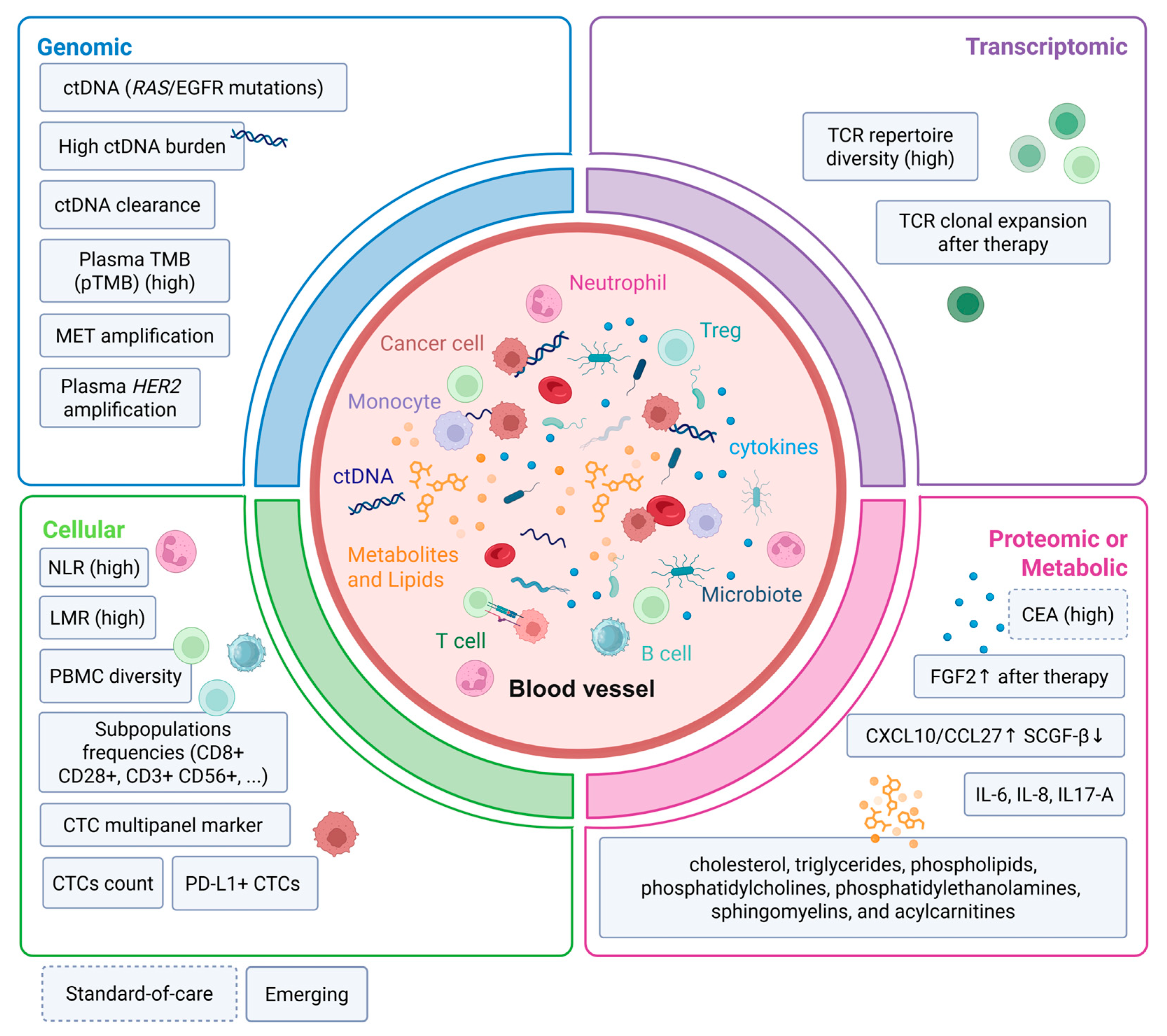
| ctDNA: RAS/EGFR pathway mutations | Genomic | Cell-free circulating tumour DNA | Predictive (anti-EGFR resistance); Prognostic | RAS mutations in blood associated with anti-EGFR resistance; ctDNA RAS burden correlates with radiological tumour burden (SLD) | Prospective trials; longitudinal monitoring validated (Valentino, REMARRY, C-PROWESS) | [127,128,129] |
| ctDNA: MSI-H status and plasma TMB (pTMB) | Genomic | Cell-free circulating tumour DNA | Predictive (ICI) | Plasma TMB (clonal and subclonal) predictive of improved outcomes with durvalumab + tremelimumab, whereas tissue TMB was not | Single prospective trial; larger validation needed (CO.26) | [130,131,132] |
| ctDNA kinetics (clearance/early dynamics) | Genomic | Cell-free circulating tumour DNA | Prognostic; Predictive (treatment monitoring) | ctDNA clearance associated with deeper tumour response (Valentino); ctDNA changes strongly associated with PFS and OS in immunotherapy arm (SAMCO-PRODIGE 54) | Multiple prospective trials; ongoing validation (Valentino, SAMCO-PRODIGE 54, COPERNIC) | [133,134,135,139,140,141] |
| Plasma HER2 amplification | Genomic | Cell-free circulating tumour DNA | Predictive (anti-HER2) | Higher plasma HER2 amplification associated with improved response and longer PFS with HER2-targeted therapy | Prospective; limited to HER2+ subgroup (DESTINY-CRC01, HERACLES) | [21,25,129] |
| MET amplification (ctDNA) | Genomic | Cell-free circulating tumour DNA | Predictive (investigational) | MET amplification associated with response to cabozantinib + panitumumab | Preliminary single case series/exploratory cohort; prospective validation absent | [138] |
| TCR repertoire diversity | Transcriptomic | T cell receptor sequencing (CDR3 clonotypes) | Prognostic; Predictive (ICI, investigational) | Decreased TCR diversity post-therapy, consistent with clonal expansion of tumour-specific T cells, associated with improved tumour response (OR = 5.29, p = 0.04); low baseline diversity linked to postoperative relapse | Retrospective longitudinal analyses/exploratory cohort; prospective validation lacking | [73,142,143,144,145,146] |
| IL-6 | Proteic | Cytokine | Prognostic | Independently prognostic in unresectable mCRC; elevated in patients with liver or lung metastases; CISIG-positive status predicts shorter OS (13.5 vs. 25.0 months) | Retrospective; not validated as standalone clinical biomarker (CISIG) | [147,148,149] |
| IL-8 | Proteic | Cytokine | Prognostic | Elevated IL-8 linked to poor prognosis (HR = 1.54) and stage IV disease (HR = 2.28); elevated pre-chemotherapy IL-8 correlates with disease progression | Retrospective/correlative; phase I targeting showed no ORR (Phase I (HuMax-IL8)) | [147,149,150,151,152] |
| CXCL10/CTACK (CCL27) | Proteic | Chemokine | Prognostic | Lower CXCL10 or higher CCL27 levels associated with improved DFS and OS | Retrospective; limited evidence | [148] |
| Neutrophil-to-lymphocyte ratio (NLR) | Cellular | Inflammation ratio | Predictive (limited); Prognostic | Fair predictive ability for early treatment response with pembrolizumab + chemotherapy; not significantly associated with PFS | Retrospective; widely studied but inconsistent cutoffs | [153] |
| Lymphocyte-to-monocyte ratio (LMR) | Cellular | Inflammation ratio | Prognostic (trend) | Higher LMR associated with trend toward improved PFS; slightly lower predictive performance than NLR for early response | Retrospective; limited prospective data | [153] |
| PBMC diversity | Cellular | Immune cell diversity (flow cytometry) | Predictive (investigational) | PBMC diversity correlated with response after 3 cycles of chemotherapy + avelumab (R2 = 0.14, p = 0.036); not significant at baseline | Single prospective trial (AVETUX); exploratory | [154] |
| Circulating tumour cells (CTCs) count | Cellular | Epithelial circulating tumour cells | Prognostic | ≥1 CTC/7.5 mL associated with shorter PFS and OS; independent prognostic factor for OS (HR = 3.14); high-volume CTCs (>3) associated with reduced OS (HR = 3.97) | Prospective; cutoff variability remains a limitation | [155,156,157] |
| PD-L1 expression on CTCs | Cellular | Immune checkpoint on circulating tumour cells | Predictive (regorafenib response) | PD-L1+ CTCs mainly in partial response/stable disease; PD-L1− CTCs associated with progressive disease; median PFS 6.3 vs. 2.1 months | Retrospective; single cohort; requires prospective validation | [158,159] |
| CTC marker panel (CD45, GAPDH, VIL1, TIMP1, CLU, SNAIL1/2, TWIST1, LOXL2/3, ZEB1/2, E47) | Cellular | Multimarker CTC signature | Prognostic; treatment monitoring | High baseline expression associated with shorter PFS (6.3 vs. 12.7 months) and OS (12.7 vs. 24.2 months); identified refractory patients missed by CT imaging | Retrospective; single cohort | [160] |
3.3. Protein Levels in Serum
3.4. Circulating Cells
3.4.1. Peripheral Blood Circulating Cells
3.4.2. Circulating Tumor Cells
4. Limits, Challenges and Perspectives
5. Conclusions
Author Contributions
Funding
Institutional Review Board Statement
Data Availability Statement
Acknowledgments
Conflicts of Interest
Abbreviations
| AUC | Area under the curve |
| BRAF | B-Raf proto-oncogene |
| CAF | Cancer-associated fibroblast |
| CDR3 | Complementarity-determining region 3 |
| CEA | Carcinoembryonic antigen |
| cfDNA | Cell-free DNA |
| CIMP | CpG island methylator phenotype |
| CISIG | Circulating inflammatory signature |
| CMS | Consensus molecular subtypes |
| CRC | Colorectal cancer |
| CT | Computed tomography |
| CTACK | Cutaneous T-cell-attracting chemokine |
| CTLA-4 | Cytotoxic T-lymphocyte-associated protein 4 |
| ctDNA | Circulating tumor DNA |
| CTC | Circulating tumor cell |
| CXCL10 | C-X-C motif chemokine ligand 10 |
| CCL27 | C-C motif chemokine ligand 27 |
| DC | Dendritic cell |
| DFS | Disease-free survival |
| dMMR | Deficient mismatch repair |
| ECM | Extracellular matrix |
| EGFR | Epidermal growth factor receptor |
| EMT | Epithelial–mesenchymal transition |
| FAP1 | Fibroblast activation protein 1 |
| FGF-2 | Fibroblast growth factor 2 |
| Fn | Fusobacterium nucleatum |
| Bf | Bacteroides fragilis |
| GIE | Genetic immunoediting |
| HER2 | Human epidermal growth factor receptor 2 |
| HR | Hazard ratio |
| ICI | Immune checkpoint inhibitor |
| ICR | Immunologic constant of rejection |
| IDO1 | Indoleamine-2,3-dioxygenase-1 |
| IES | Immunoediting score |
| IFN-γ | Interferon-gamma |
| IHC | Immunohistochemistry |
| IL | Interleukin |
| IMC | Immune-metabolic cluster |
| IMMETCOLS | Immune-metabolic colorectal signature |
| ISb | Biopsy-adapted Immunoscore |
| KRAS | Kirsten rat sarcoma viral proto-oncogene |
| LAG-3 | Lymphocyte-activation gene 3 |
| LDH | Lactate dehydrogenase |
| LMR | Lymphocyte-to-monocyte ratio |
| LOXL2/3 | Lysyl oxidase-like protein 2/3 |
| MCT1 | Monocarboxylate transporter 1 |
| MDSC | Myeloid-derived suppressor cell |
| MEK | Mitogen-activated protein kinase kinase |
| MET | Mesenchymal–epithelial transition factor |
| MFAP2 | Microfibril-associated protein 2 |
| MHC-I | Major histocompatibility complex class I |
| MMR | Mismatch repair |
| mCRC | Metastatic colorectal cancer |
| MSI-H | Microsatellite instability-high |
| MSS | Microsatellite stable |
| NGS | Next-generation sequencing |
| NK | Natural killer |
| NKT | Natural killer T cell |
| NLR | Neutrophil-to-lymphocyte ratio |
| NRAS | Neuroblastoma RAS viral proto-oncogene |
| NSCLC | Non-small cell lung cancer |
| ORR | Objective response rate |
| OS | Overall survival |
| OXPHOS | Oxidative phosphorylation |
| PBMC | Peripheral blood mononuclear cell |
| PD-1 | Programmed cell death protein 1 |
| PD-L1 | Programmed death-ligand 1 |
| PFS | Progression-free survival |
| pMMR | Proficient mismatch repair |
| POLE | DNA polymerase epsilon |
| POLD1 | DNA polymerase delta 1 |
| RAS | Rat sarcoma viral proto-oncogene |
| RFS | Recurrence-free survival |
| SCGF-β | Stem cell growth factor beta |
| SLD | Sum of longest diameters |
| SMAD4 | SMAD family member 4 |
| TCR | T cell receptor |
| TDO2 | Tryptophan-2,3-dioxygenase |
| TGF-β | Transforming growth factor beta |
| TH1 | Type 1 helper T cell |
| TH17 | Type 17 helper T cell |
| TIMP1 | Tissue inhibitor of metalloproteinases 1 |
| TMB | Tumor mutational burden |
| TME | Tumor microenvironment |
| TNM | Tumor-node-metastasis |
| Treg | Regulatory T cell |
| VEGF | Vascular endothelial growth factor |
| WES | Whole exome sequencing |
| WGS | Whole genome sequencing |
| WT | Wild-type |
References
- Bray, F.; Laversanne, M.; Sung, H.; Ferlay, J.; Siegel, R.L.; Soerjomataram, I.; Jemal, A. Global cancer statistics 2022: GLOBOCAN estimates of incidence and mortality worldwide for 36 cancers in 185 countries. CA Cancer J. Clin. 2024, 74, 229–263. [Google Scholar] [CrossRef] [Scilit] [PubMed]
- National Cancer Institute. SEER*Explorer: An Interactive Website for SEER Cancer Statistics. Surveillance Research Program. Data Source(s): SEER Incidence Data, November 2024 Submission (1975–2022), SEER 21 Registries (Excluding Illinois). 2025. Available online: https://seer.cancer.gov/statistics-network/explorer/ (accessed on 15 July 2026).
- Fearon, E.R.; Vogelstein, B. A genetic model for colorectal tumorigenesis. Cell 1990, 61, 759–767. [Google Scholar] [CrossRef] [Scilit] [PubMed]
- Wang, C.; Sandhu, J.; Ouyang, C.; Ye, J.; Lee, P.P.; Fakih, M. Clinical Response to Immunotherapy Targeting Programmed Cell Death Receptor 1/Programmed Cell Death Ligand 1 in Patients with Treatment-Resistant Microsatellite Stable Colorectal Cancer with and Without Liver Metastases. JAMA Netw. Open 2021, 4, e2118416. [Google Scholar] [CrossRef] [Scilit] [PubMed]
- Zeineddine, F.A.; Zeineddine, M.A.; Yousef, A.; Gu, Y.; Chowdhury, S.; Dasari, A.; Huey, R.W.; Johnson, B.; Kee, B.; Lee, M.S.; et al. Survival improvement for patients with metastatic colorectal cancer over twenty years. npj Precis Oncol. 2023, 7, 16. [Google Scholar] [CrossRef] [Scilit] [PubMed]
- Aparicio, J.; Esposito, F.; Serrano, S.; Falco, E.; Escudero, P.; Ruiz-Casado, A.; Manzano, H.; Fernandez-Montes, A. Metastatic Colorectal Cancer. First Line Therapy for Unresectable Disease. J. Clin. Med. 2020, 9, 3889. [Google Scholar] [CrossRef] [Scilit] [PubMed] [PubMed Central]
- Symonds, L.K.; Cohen, S.A. Use of perioperative chemotherapy in colorectal cancer metastatic to the liver. Gastroenterol. Rep. 2019, 7, 301–311. [Google Scholar] [CrossRef] [Scilit] [PubMed]
- Tiegs, G.; Lohse, A.W. Immune tolerance: What is unique about the liver. J. Autoimmun. 2010, 34, 1–6. [Google Scholar] [CrossRef] [Scilit] [PubMed]
- Doherty, D.G. Immunity, tolerance and autoimmunity in the liver: A comprehensive review. J. Autoimmun. 2016, 66, 60–75. [Google Scholar] [CrossRef] [Scilit] [PubMed]
- Doherty, D.G. Antigen-specific immune tolerance in the liver. Nat. BioMed Eng. 2019, 3, 763–765. [Google Scholar] [CrossRef] [Scilit] [PubMed]
- Yu, J.; Green, M.D.; Li, S.; Sun, Y.; Journey, S.N.; Choi, J.E.; Rizvi, S.M.; Qin, A.; Waninger, J.J.; Lang, X.; et al. Liver metastasis restrains immunotherapy efficacy via macrophage-mediated T cell elimination. Nat. Med. 2021, 27, 152–164. [Google Scholar] [CrossRef] [Scilit] [PubMed]
- Cremolini, C.; Chalabi, M.; Elez, E.; Fassan, M.; Gelli, M.; Goéré, D.; Modest, D.; Montagut, C.; Osterlund, P.; Ricke, J.; et al. Metastatic colorectal cancer: ESMO Clinical Practice Guideline for diagnosis, treatment and follow-up. Ann. Oncol. 2026, 37, 759–776. [Google Scholar] [CrossRef] [Scilit] [PubMed]
- Prior, I.A.; Lewis, P.D.; Mattos, C. A Comprehensive Survey of Ras Mutations in Cancer. Cancer Res. 2012, 72, 2457–2467. [Google Scholar] [CrossRef] [Scilit] [PubMed]
- Yaeger, R.; Weiss, J.; Pelster, M.S.; Spira, A.I.; Barve, M.; Ou, S.H.I.; Leal, T.A.; Bekaii-Saab, T.S.; Paweletz, C.P.; Heavey, G.A.; et al. Adagrasib with or without Cetuximab in Colorectal Cancer with Mutated KRAS G12C. N. Engl. J. Med. 2023, 388, 44–54. [Google Scholar] [CrossRef] [Scilit] [PubMed]
- Fakih, M.G.; Salvatore, L.; Esaki, T.; Modest, D.P.; Lopez-Bravo, D.P.; Taieb, J.; Karamouzis, M.V.; Ruiz-Garcia, E.; Kim, T.-W.; Kuboki, Y.; et al. Sotorasib plus Panitumumab in Refractory Colorectal Cancer with Mutated KRAS G12C. N. Engl. J. Med. 2023, 389, 2125–2139. [Google Scholar] [CrossRef] [Scilit] [PubMed]
- Rowland, A.; Dias, M.M.; Wiese, M.D.; Kichenadasse, G.; McKinnon, R.A.; Karapetis, C.S.; Sorich, M.J. Meta-analysis of BRAF mutation as a predictive biomarker of benefit from anti-EGFR monoclonal antibody therapy for RAS wild-type metastatic colorectal cancer. Br. J. Cancer 2015, 112, 1888–1894. [Google Scholar] [CrossRef] [Scilit] [PubMed]
- Kopetz, S.; Grothey, A.; Yaeger, R.; Van Cutsem, E.; Desai, J.; Yoshino, T.; Wasan, H.; Ciardiello, F.; Loupakis, F.; Hong, Y.S.; et al. Encorafenib, Binimetinib, and Cetuximab in BRAF V600E–Mutated Colorectal Cancer. N. Engl. J. Med. 2019, 381, 1632–1643. [Google Scholar] [CrossRef] [Scilit] [PubMed]
- Tabernero, J.; Grothey, A.; Van Cutsem, E.; Yaeger, R.; Wasan, H.; Yoshino, T.; Desai, J.; Ciardiello, F.; Loupakis, F.; Hong, Y.S.; et al. Encorafenib Plus Cetuximab as a New Standard of Care for Previously Treated BRAF V600E–Mutant Metastatic Colorectal Cancer: Updated Survival Results and Subgroup Analyses from the BEACON Study. J. Clin. Oncol. 2021, 39, 273–284. [Google Scholar] [CrossRef] [Scilit] [PubMed]
- Elez, E.; Yoshino, T.; Shen, L.; Lonardi, S.; Cutsem, E.V.; Eng, C.; Kim, T.W.; Wasan, H.S.; Desai, J.; Ciardiello, F.; et al. Encorafenib, Cetuximab, and mFOLFOX6 in BRAF-Mutated Colorectal Cancer. N. Engl. J. Med. 2025, 392, 2425–2437. [Google Scholar] [CrossRef] [Scilit] [PubMed]
- Rossini, D.; Boccaccino, A.; Carullo, M.; Antoniotti, C.; Dima, G.; Ciracì, P.; Marmorino, F.; Moretto, R.; Masi, G.; Cremolini, C. Primary tumour side as a driver for treatment choice in RAS wild-type metastatic colorectal cancer patients: A systematic review and pooled analysis of randomised trials. Eur. J. Cancer 2023, 184, 106–116. [Google Scholar] [CrossRef] [Scilit] [PubMed]
- Sartore-Bianchi, A.; Trusolino, L.; Martino, C.; Bencardino, K.; Lonardi, S.; Bergamo, F.; Zagonel, V.; Leone, F.; Depetris, I.; Martinelli, E.; et al. Dual-targeted therapy with trastuzumab and lapatinib in treatment-refractory, KRAS codon 12/13 wild-type, HER2-positive metastatic colorectal cancer (HERACLES): A proof-of-concept, multicentre, open-label, phase 2 trial. Lancet Oncol. 2016, 17, 738–746. [Google Scholar] [CrossRef] [Scilit] [PubMed]
- Richman, S.D.; Southward, K.; Chambers, P.; Cross, D.; Barrett, J.; Hemmings, G.; Taylor, M.; Wood, H.; Hutchins, G.; Foster, J.M.; et al. HER2 overexpression and amplification as a potential therapeutic target in colorectal cancer: Analysis of 3256 patients enrolled in the QUASAR, FOCUS and PICCOLO colorectal cancer trials. J. Pathol. 2016, 238, 562–570. [Google Scholar] [CrossRef] [Scilit] [PubMed]
- Strickler, J.H.; Cercek, A.; Siena, S.; André, T.; Ng, K.; Van Cutsem, E.; Wu, C.; Paulson, A.S.; Hubbard, J.M.; Coveler, A.L.; et al. Tucatinib plus trastuzumab for chemotherapy-refractory, HER2+, RAS wild-type metastatic colorectal cancer (MOUNTAINEER): Final analysis. Nat. Commun. 2026, 17, 1068. [Google Scholar] [CrossRef] [Scilit] [PubMed]
- Siena, S.; Bartolomeo, M.D.; Raghav, K.; Masuishi, T.; Loupakis, F.; Kawakami, H.; Yamaguchi, K.; Nishina, T.; Fakih, M.; Elez, E.; et al. Trastuzumab deruxtecan (DS-8201) in patients with HER2-expressing metastatic colorectal cancer (DESTINY-CRC01): A multicentre, open-label, phase 2 trial. Lancet Oncol. 2021, 22, 779–789. [Google Scholar] [CrossRef] [Scilit] [PubMed]
- Raghav, K.; Siena, S.; Takashima, A.; Kato, T.; den Eynde, M.V.; Pietrantonio, F.; Komatsu, Y.; Kawakami, H.; Peeters, M.; Andre, T.; et al. Trastuzumab deruxtecan in patients with HER2-positive advanced colorectal cancer (DESTINY-CRC02): Primary results from a multicentre, randomised, phase 2 trial. Lancet Oncol. 2024, 25, 1147–1162. [Google Scholar] [CrossRef] [Scilit] [PubMed]
- Friedman, C.F.; Hainsworth, J.D.; Kurzrock, R.; Spigel, D.R.; Burris, H.A.; Sweeney, C.J.; Meric-Bernstam, F.; Wang, Y.; Levy, J.; Grindheim, J.; et al. Atezolizumab Treatment of Tumors with High Tumor Mutational Burden from MyPathway, a Multicenter, Open-Label, Phase IIa Multiple Basket Study. Cancer Discov. 2022, 12, 654–669. [Google Scholar] [CrossRef] [Scilit] [PubMed]
- Konishi, T.; Shimada, Y.; Hsu, M.; Tufts, L.; Jimenez-Rodriguez, R.; Cercek, A.; Yaeger, R.; Saltz, L.; Smith, J.J.; Nash, G.M.; et al. Association of Preoperative and Postoperative Serum Carcinoembryonic Antigen and Colon Cancer Outcome. JAMA Oncol. 2018, 4, 309. [Google Scholar] [CrossRef] [Scilit] [PubMed]
- Peters, N.A.; Heinz, M.C.; Küçükköse, E.; Viergever, B.J.; Baars, M.; Amini, M.; Huismans, M.A.; Andel, D.; Verheem, A.; Vercoulen, Y.; et al. Analysis of experimental colorectal liver metastases identifies distinct proliferation-independent transcriptional programs in micro- versus macrometastases. J. Transl. Med. 2025, 24, 37. [Google Scholar] [CrossRef] [Scilit] [PubMed]
- Liu, Y.; Jadhav, A.S.; Pan, Y.; Liao, J.; Khanduri, I.; Liu, Y.; Katkhuda, R.; Lu, W.; Cho, K.S.; Tong, Z.; et al. Spatial multi-omics landscape of colorectal cancer macro- and micrometastases. Cancer Cell 2026, 44, 1568–1586.e11. [Google Scholar] [CrossRef] [Scilit] [PubMed]
- Fang, T.; Liang, T.; Wang, Y.; Wu, H.; Liu, S.; Xie, L.; Liang, J.; Wang, C.; Tan, Y. Prognostic role and clinicopathological features of SMAD4 gene mutation in colorectal cancer: A systematic review and meta-analysis. BMC Gastroenterol. 2021, 21, 297. [Google Scholar] [CrossRef] [Scilit] [PubMed]
- Yan, P.; Klingbiel, D.; Saridaki, Z.; Ceppa, P.; Curto, M.; McKee, T.A.; Roth, A.; Tejpar, S.; Delorenzi, M.; Bosman, F.T.; et al. Reduced Expression of SMAD4 Is Associated with Poor Survival in Colon Cancer. Clin. Cancer Res. 2016, 22, 3037–3047. [Google Scholar] [CrossRef] [Scilit] [PubMed]
- Wasserman, I.; Lee, L.H.; Ogino, S.; Marco, M.R.; Wu, C.; Chen, X.; Datta, J.; Sadot, E.; Szeglin, B.; Guillem, J.G.; et al. SMAD4 Loss in Colorectal Cancer Patients Correlates with Recurrence, Loss of Immune Infiltrate, and Chemoresistance. Clin. Cancer Res. 2019, 25, 1948–1956. [Google Scholar] [CrossRef] [Scilit] [PubMed]
- Yao, Y.C.; He, M.M.; Wang, M.; Mo, Y.X.; Zhao, Q.; Jin, Y.; Hou, T.; Chen, Y.-X.; Wang, F. Exploratory biomarker findings from regorafenib plus toripalimab in patients with refractory metastatic colorectal cancer (REGOTORI study). Gastroenterol. Rep. 2026, 14, goag009. [Google Scholar] [CrossRef] [Scilit] [PubMed]
- Lee, M.S.; Cho, H.J.; Hong, J.Y.; Lee, J.; Park, S.H.; Park, J.O.; Park, Y.S.; Lim, H.Y.; Kang, W.K.; Cho, Y.B.; et al. Clinical and molecular distinctions in patients with refractory colon cancer who benefit from regorafenib treatment. Ther. Adv. Med. Oncol. 2020, 12, 1758835920965842. [Google Scholar] [CrossRef] [Scilit] [PubMed]
- Palles, C.; Cazier, J.B.; Howarth, K.M.; Domingo, E.; Jones, A.M.; Broderick, P.; Kemp, Z.; Spain, S.L.; Guarino, E.; Salguero, I.; et al. Germline mutations affecting the proofreading domains of POLE and POLD1 predispose to colorectal adenomas and carcinomas. Nat. Genet. 2013, 45, 136–144. [Google Scholar] [CrossRef] [Scilit] [PubMed]
- Shlien, A.; Campbell, B.B.; De Borja, R.; Alexandrov, L.B.; Merico, D.; Wedge, D.; Van Loo, P.; Tarpey, P.S.; Coupland, P.; Behjati, S.; et al. Combined hereditary and somatic mutations of replication error repair genes result in rapid onset of ultra-hypermutated cancers. Nat. Genet. 2015, 47, 257–262. [Google Scholar] [CrossRef] [Scilit] [PubMed]
- Keshinro, A.; Vanderbilt, C.; Kim, J.K.; Firat, C.; Chen, C.T.; Yaeger, R.; Ganesh, K.; Segal, N.H.; Gonen, M.; Shia, J.; et al. Tumor-Infiltrating Lymphocytes, Tumor Mutational Burden, and Genetic Alterations in Microsatellite Unstable, Microsatellite Stable, or Mutant POLE/POLD1 Colon Cancer. JCO Precis Oncol. 2021, 5, 817–826. [Google Scholar] [CrossRef] [Scilit] [PubMed]
- Garmezy, B.; Gheeya, J.; Lin, H.Y.; Huang, Y.; Kim, T.; Jiang, X.; Thein, K.Z.; Pilié, P.G.; Zeineddine, F.; Wang, W.; et al. Clinical and Molecular Characterization of POLE Mutations as Predictive Biomarkers of Response to Immune Checkpoint Inhibitors in Advanced Cancers. JCO Precis Oncol. 2022, 6, e2100267. [Google Scholar] [CrossRef] [Scilit] [PubMed]
- Rousseau, B.; Bieche, I.; Pasmant, E.; Hamzaoui, N.; Leulliot, N.; Michon, L.; de Reynies, A.; Attignon, V.; Foote, M.B.; Masliah-Planchon, J.; et al. PD-1 Blockade in Solid Tumors with Defects in Polymerase Epsilon. Cancer Discov. 2022, 12, 1435–1448. [Google Scholar] [CrossRef] [Scilit] [PubMed]
- Oh, C.R.; Kim, J.E.; Hong, Y.S.; Kim, S.Y.; Ahn, J.B.; Baek, J.Y.; Lee, M.; Kang, M.J.; Cho, S.H.; Beom, S.; et al. Phase II study of durvalumab monotherapy in patients with previously treated microsatellite instability-high/mismatch repair-deficient or POLE -mutated metastatic or unresectable colorectal cancer. Int. J. Cancer 2022, 150, 2038–2045. [Google Scholar] [CrossRef] [Scilit] [PubMed]
- Ambrosini, M.; Rousseau, B.; Manca, P.; Artz, O.; Marabelle, A.; André, T.; Maddalena, G.; Mazzoli, G.; Intini, R.; Cohen, R.; et al. Immune checkpoint inhibitors for POLE or POLD1 proofreading-deficient metastatic colorectal cancer. Ann. Oncol. 2024, 35, 643–655. [Google Scholar] [CrossRef] [Scilit] [PubMed]
- Addeo, A.; Friedlaender, A.; Banna, G.L.; Weiss, G.J. TMB or not TMB as a biomarker: That is the question. Crit. Rev. Oncol. Hematol. 2021, 163, 103374. [Google Scholar] [CrossRef] [Scilit] [PubMed]
- Chalmers, Z.R.; Connelly, C.F.; Fabrizio, D.; Gay, L.; Ali, S.M.; Ennis, R.; Schrock, A.; Campbell, B.; Shlien, A.; Chmielecki, J.; et al. Analysis of 100,000 human cancer genomes reveals the landscape of tumor mutational burden. Genome Med. 2017, 9, 34. [Google Scholar] [CrossRef] [Scilit] [PubMed]
- Fabrizio, D.A.; George, T.J., Jr.; Dunne, R.F.; Frampton, G.; Sun, J.; Gowen, K.; Kennedy, M.; Greenbowe, J.; Schrock, A.B.; Hezel, A.F.; et al. Beyond microsatellite testing: Assessment of tumor mutational burden identifies subsets of colorectal cancer who may respond to immune checkpoint inhibition. J. Gastrointest. Oncol. 2018, 9, 610–617. [Google Scholar] [CrossRef] [Scilit] [PubMed]
- Goodman, A.M.; Sokol, E.S.; Frampton, G.M.; Lippman, S.M.; Kurzrock, R. Microsatellite-Stable Tumors with High Mutational Burden Benefit from Immunotherapy. Cancer Immunol. Res. 2019, 7, 1570–1573. [Google Scholar] [CrossRef] [Scilit] [PubMed]
- Marabelle, A.; Fakih, M.; Lopez, J.; Shah, M.; Shapira-Frommer, R.; Nakagawa, K.; Chung, H.C.; Kindler, H.L.; Lopez-Martin, J.A.; Miller, W.H., Jr.; et al. Association of tumour mutational burden with outcomes in patients with advanced solid tumours treated with pembrolizumab: Prospective biomarker analysis of the multicohort, open-label, phase 2 KEYNOTE-158 study. Lancet Oncol. 2020, 21, 1353–1365. [Google Scholar] [CrossRef] [Scilit] [PubMed]
- Yamaguchi, K.; Tsuchihashi, K.; Ueno, S.; Uehara, K.; Taguchi, R.; Ito, M.; Isobe, T.; Imajima, T.; Kitazono, T.; Tanoue, K.; et al. Efficacy of pembrolizumab in microsatellite-stable, tumor mutational burden-high metastatic colorectal cancer: Genomic signatures and clinical outcomes. ESMO Open 2025, 10, 104108. [Google Scholar] [CrossRef] [Scilit] [PubMed]
- Gaber, O.; Karan, C.; Walko, C.M.; Knepper, T.C.; Kim, R.D.; Sahin, I.H. Effect of immunotherapy on the survival outcomes in tumor mutational burden-high (TMB-H) microsatellite stable (MSS) metastatic colorectal cancer (mCRC): A single-institution experience. J. Clin. Oncol. 2023, 41, 239. [Google Scholar] [CrossRef] [Scilit]
- Fukuoka, S.; Hara, H.; Takahashi, N.; Kojima, T.; Kawazoe, A.; Asayama, M.; Yoshii, T.; Kotani, D.; Tamura, H.; Mikamoto, Y.; et al. Regorafenib Plus Nivolumab in Patients with Advanced Gastric or Colorectal Cancer: An Open-Label, Dose-Escalation, and Dose-Expansion Phase Ib Trial (REGONIVO, EPOC1603). J. Clin. Oncol. 2020, 38, 2053–2061. [Google Scholar] [CrossRef] [Scilit] [PubMed]
- Li, Y.; Ma, Y.; Wu, Z.; Zeng, F.; Song, B.; Zhang, Y.; Li, J.; Lui, S.; Wu, M. Tumor Mutational Burden Predicting the Efficacy of Immune Checkpoint Inhibitors in Colorectal Cancer: A Systematic Review and Meta-Analysis. Front. Immunol. 2021, 12, 751407. [Google Scholar] [CrossRef] [Scilit] [PubMed]
- Schrock, A.B.; Ouyang, C.; Sandhu, J.; Sokol, E.; Jin, D.; Ross, J.S.; Miller, V.A.; Lim, D.; Amanam, I.; Chao, J.; et al. Tumor mutational burden is predictive of response to immune checkpoint inhibitors in MSI-high metastatic colorectal cancer. Ann. Oncol. 2019, 30, 1096–1103. [Google Scholar] [CrossRef] [Scilit] [PubMed]
- Budczies, J.; Allgäuer, M.; Litchfield, K.; Rempel, E.; Christopoulos, P.; Kazdal, D.; Endris, V.; Thomas, M.; Fröhling, S.; Peters, S.; et al. Optimizing panel-based tumor mutational burden (TMB) measurement. Ann. Oncol. 2019, 30, 1496–1506. [Google Scholar] [CrossRef] [Scilit] [PubMed]
- Advani, S.M.; Swartz, M.D.; Loree, J.; Davis, J.S.; Sarsashek, A.M.; Lam, M.; Lee, M.S.; Bressler, J.; Lopez, D.S.; Daniel, C.R.; et al. Epidemiology and Molecular-Pathologic Characteristics of CpG Island Methylator Phenotype (CIMP) in Colorectal Cancer. Clin. Colorectal Cancer 2021, 20, 137–147.e1. [Google Scholar] [CrossRef] [Scilit] [PubMed]
- Qian, F.; Li, Q.; Chang, H.; Wei, K.; Chen, X.; Huang, T.; Li, Y. Identification of DNA methylation characteristics associated with metastasis and prognosis in colorectal cancer. BMC Med. Genom. 2024, 17, 127. [Google Scholar] [CrossRef] [Scilit] [PubMed]
- Kim, J.H.; Hong, J.; Lee, J.A.; Jung, M.; Choi, E.; Cho, N.Y.; Kang, G.H.; Kim, S. Immune microenvironmental heterogeneity according to tumor DNA methylation phenotypes in microsatellite instability-high colorectal cancers. Cancer Immunol. Immunother. 2024, 73, 215. [Google Scholar] [CrossRef] [Scilit] [PubMed]
- Cha, Y.; Kim, K.J.; Han, S.W.; Rhee, Y.Y.; Bae, J.M.; Wen, X.; Cho, N.-Y.; Lee, D.-W.; Lee, K.-H.; Kim, T.-Y.; et al. Adverse prognostic impact of the CpG island methylator phenotype in metastatic colorectal cancer. Br. J. Cancer 2016, 115, 164–171. [Google Scholar] [CrossRef] [Scilit] [PubMed]
- Zhang, X.; Shimodaira, H.; Soeda, H.; Komine, K.; Takahashi, H.; Ouchi, K.; Inoue, M.; Takahashi, M.; Takahashi, S.; Ishioka, C. CpG island methylator phenotype is associated with the efficacy of sequential oxaliplatin- and irinotecan-based chemotherapy and EGFR-related gene mutation in Japanese patients with metastatic colorectal cancer. Int. J. Clin. Oncol. 2016, 21, 1091–1101. [Google Scholar] [CrossRef] [Scilit] [PubMed]
- Shen, L.; Catalano, P.J.; Benson, A.B.; O’Dwyer, P.; Hamilton, S.R.; Issa, J.P.J. Association between DNA Methylation and Shortened Survival in Patients with Advanced Colorectal Cancer Treated with 5-Fluorouracil–Based Chemotherapy. Clin. Cancer Res. 2007, 13, 6093–6098. [Google Scholar] [CrossRef] [Scilit] [PubMed]
- Roelands, J.; Kuppen, P.J.K.; Ahmed, E.I.; Mall, R.; Masoodi, T.; Singh, P.; Monaco, G.; Raynaud, C.; de Miranda, N.F.; Ferraro, L.; et al. An integrated tumor, immune and microbiome atlas of colon cancer. Nat. Med. 2023, 29, 1273–1286. [Google Scholar] [CrossRef] [Scilit] [PubMed]
- Angelova, M.; Mlecnik, B.; Vasaturo, A.; Bindea, G.; Fredriksen, T.; Lafontaine, L.; Buttard, B.; Morgand, E.; Bruni, D.; Jouret-Mourin, A.; et al. Evolution of Metastases in Space and Time under Immune Selection. Cell 2018, 175, 751–765.e16. [Google Scholar] [CrossRef] [Scilit] [PubMed]
- Wang, E.; Worschech, A.; Marincola, F.M. The immunologic constant of rejection. Trends Immunol. 2008, 29, 256–262. [Google Scholar] [CrossRef] [Scilit] [PubMed]
- Bertucci, F.; Finetti, P.; Simeone, I.; Hendrickx, W.; Wang, E.; Marincola, F.M.; Viens, P.; Mamessier, E.; Ceccarelli, M.; Birnbaum, D.; et al. The immunologic constant of rejection classification refines the prognostic value of conventional prognostic signatures in breast cancer. Br. J. Cancer 2018, 119, 1383–1391. [Google Scholar] [CrossRef] [Scilit] [PubMed] [PubMed Central]
- Huyghe, N.; Benidovskaya, E.; Masoodi, T.; Sinapi, I.; De Cuyper, A.; Vempalli, F.; Beyaert, S.; Bouzin, C.; Osorio, F.M.; Ferraro, L.; et al. Impact of the tumor immune contexture in microsatellite-stable metastatic colorectal cancer treated with avelumab, cetuximab, and irinotecan. Cell Rep. Med. 2025, 6, 102201. [Google Scholar] [CrossRef] [Scilit] [PubMed] [PubMed Central]
- Guinney, J.; Dienstmann, R.; Wang, X.; De Reyniès, A.; Schlicker, A.; Soneson, C.; Marisa, L.; Roepman, P.; Nyamundanda, G.; Angelino, P.; et al. The consensus molecular subtypes of colorectal cancer. Nat. Med. 2015, 21, 1350–1356. [Google Scholar] [CrossRef] [Scilit] [PubMed]
- Picard, E.; Verschoor, C.P.; Ma, G.W.; Pawelec, G. Relationships Between Immune Landscapes, Genetic Subtypes and Responses to Immunotherapy in Colorectal Cancer. Front Immunol. 2020, 11, 369. [Google Scholar] [CrossRef] [Scilit] [PubMed]
- Karpinski, P.; Rossowska, J.; Sasiadek, M.M. Immunological landscape of consensus clusters in colorectal cancer. Oncotarget 2017, 8, 105299–105311. [Google Scholar] [CrossRef] [Scilit] [PubMed]
- Loupakis, F.; Biason, P.; Prete, A.A.; Cremolini, C.; Pietrantonio, F.; Pella, N.; Dell’aQuila, E.; Sperti, E.; Zichi, C.; Intini, R.; et al. CK7 and consensus molecular subtypes as major prognosticators in V600EBRAF mutated metastatic colorectal cancer. Br. J. Cancer 2019, 121, 593–599. [Google Scholar] [CrossRef] [Scilit] [PubMed]
- Chowdhury, S.; Xiu, J.; Ribeiro, J.R.; Nicolaides, T.; Zhang, J.; Korn, W.M.; Poorman, K.A.; Lenz, H.-J.; Marshall, J.L.; Oberley, M.J.; et al. Consensus molecular subtyping of metastatic colorectal cancer expands biomarker-directed therapeutic benefit for patients with CMS1 and CMS2 tumors. Br. J. Cancer 2024, 131, 1328–1339. [Google Scholar] [CrossRef] [Scilit] [PubMed]
- Stahler, A.; Modest, D.P.; Stintzing, S.; Borelli, B.; Keller, T.; Held, S.; von Weikersthal, L.F.; Müller, L.; Graeven, U.; Decker, T.; et al. Individual Patient Data Meta-Analysis of Consensus Molecular Subtypes as Biomarkers of First-Line Treatment in RAS Wild-Type Metastatic Colorectal Cancer. J. Clin. Oncol. 2026, 44, 31–41. [Google Scholar] [CrossRef] [Scilit] [PubMed]
- Takei, S.; Tanaka, Y.; Lin, Y.T.; Koyama, S.; Fukuoka, S.; Hara, H.; Nakamura, Y.; Kuboki, Y.; Kotani, D.; Kojima, T.; et al. Multiomic molecular characterization of the response to combination immunotherapy in MSS/pMMR metastatic colorectal cancer. J. Immunother. Cancer 2024, 12, e008210. [Google Scholar] [CrossRef] [Scilit] [PubMed]
- Rejali, L.; Seifollahi Asl, R.; Sanjabi, F.; Fatemi, N.; Asadzadeh Aghdaei, H.; Saeedi Niasar, M.; Moghadam, P.K.; Mojarad, E.N.; Mini, E.; Nobili, S. Principles of Molecular Utility for CMS Classification in Colorectal Cancer Management. Cancers 2023, 15, 2746. [Google Scholar] [CrossRef] [Scilit] [PubMed]
- Langerud, J.; Eilertsen, I.A.; Moosavi, S.H.; Klokkerud, S.M.K.; Reims, H.M.; Backe, I.F.; Hektoen, M.; Sjo, O.H.; Jeanmougin, M.; Tejpar, S.; et al. Multiregional transcriptomics identifies congruent consensus subtypes with prognostic value beyond tumor heterogeneity of colorectal cancer. Nat. Commun. 2024, 15, 4342. [Google Scholar] [CrossRef] [Scilit] [PubMed]
- Sakimura, S.; Nagayama, S.; Fukunaga, M.; Hu, Q.; Kitagawa, A.; Kobayashi, Y.; Hasegawa, T.; Noda, M.; Kouyama, Y.; Shimizu, D.; et al. Impaired tumor immune response in metastatic tumors is a selective pressure for neutral evolution in CRC cases. PLoS Genet. 2021, 17, e1009113. [Google Scholar] [CrossRef] [Scilit] [PubMed]
- Pedrosa, L.; Foguet, C.; Oliveres, H.; Archilla, I.; de Herreros, M.G.; Rodríguez, A.; Postigo, A.; Benítez-Ribas, D.; Camps, J.; Cuatrecasas, M.; et al. A novel gene signature unveils three distinct immune-metabolic rewiring patterns conserved across diverse tumor types and associated with outcomes. Front Immunol. 2022, 13, 926304. [Google Scholar] [CrossRef] [Scilit] [PubMed]
- Renner, K.; Bruss, C.; Schnell, A.; Koehl, G.; Becker, H.M.; Fante, M.; Menevse, A.-N.; Kauer, N.; Blazquez, R.; Hacker, L.; et al. Restricting Glycolysis Preserves T Cell Effector Functions and Augments Checkpoint Therapy. Cell Rep. 2019, 29, 135–150.e9. [Google Scholar] [CrossRef] [Scilit] [PubMed]
- Gandini, S.; Massi, D.; Mandalà, M. PD-L1 expression in cancer patients receiving anti PD-1/PD-L1 antibodies: A systematic review and meta-analysis. Crit. Rev. Oncol. Hematol. 2016, 100, 88–98. [Google Scholar] [CrossRef] [Scilit] [PubMed]
- Garon, E.B.; Rizvi, N.A.; Hui, R.; Leighl, N.; Balmanoukian, A.S.; Eder, J.P.; Patnaik, A.; Aggarwal, C.; Gubens, M.; Horn, L.; et al. Pembrolizumab for the Treatment of Non–Small-Cell Lung Cancer. N. Engl. J. Med. 2015, 372, 2018–2028. [Google Scholar] [CrossRef] [Scilit] [PubMed]
- Patel, S.P.; Kurzrock, R. PD-L1 Expression as a Predictive Biomarker in Cancer Immunotherapy. Mol. Cancer Ther. 2015, 14, 847–856. [Google Scholar] [CrossRef] [Scilit] [PubMed]
- Aguiar, P.N.; Santoro, I.L.; Tadokoro, H.; de Lima Lopes, G.; Filardi, B.A.; Oliveira, P.; Mountzios, G.; de Mello, R.A. The role of PD-L1 expression as a predictive biomarker in advanced non-small-cell lung cancer: A network meta-analysis. Immunotherapy 2016, 8, 479–488. [Google Scholar] [CrossRef] [Scilit] [PubMed]
- Yu, J.; Wang, X.; Teng, F.; Kong, L. PD-L1 expression in human cancers and its association with clinical outcomes. OncoTargets Ther. 2016, 9, 5023–5039. [Google Scholar] [CrossRef] [Scilit] [PubMed]
- André, T.; Overman, M.; Lonardi, S.; Aglietta, M.; McDermott, R.; Wong, K.Y.; Morse, M.; Hendlisz, A.; Moss, R.; Ledeine, J.-M.; et al. Analysis of tumor PD-L1 expression and biomarkers in relation to clinical activity in patients (pts) with deficient DNA mismatch repair (dMMR)/high microsatellite instability (MSI-H) metastatic colorectal cancer (mCRC) treated with nivolumab (NIVO) + ipilimumab (IPI): CheckMate 142. Ann. Oncol. 2017, 28, v163. [Google Scholar] [CrossRef] [Scilit]
- Morse, M.A.; Hochster, H.; Benson, A. Perspectives on Treatment of Metastatic Colorectal Cancer with Immune Checkpoint Inhibitor Therapy. Oncologist 2020, 25, 33–45. [Google Scholar] [CrossRef] [Scilit] [PubMed] [PubMed Central]
- Yang, L.; Xue, R.; Pan, C. Prognostic and clinicopathological value of PD-L1 in colorectal cancer: A systematic review and meta-analysis. OncoTargets Ther. 2019, 12, 3671–3682. [Google Scholar] [CrossRef] [Scilit] [PubMed]
- Alexander, P.G.; McMillan, D.C.; Park, J.H. A meta-analysis of CD274 (PD-L1) assessment and prognosis in colorectal cancer and its role in predicting response to anti-PD-1 therapy. Crit. Rev. Oncol. Hematol. 2021, 157, 103147. [Google Scholar] [CrossRef] [Scilit] [PubMed]
- Eng, C.; Kim, T.W.; Bendell, J.; Argilés, G.; Tebbutt, N.C.; Di Bartolomeo, M.; Falcone, A.; Fakih, M.; Kozloff, M.; Segal, N.H.; et al. Atezolizumab with or without cobimetinib versus regorafenib in previously treated metastatic colorectal cancer (IMblaze370): A multicentre, open-label, phase 3, randomised, controlled trial. Lancet Oncol. 2019, 20, 849–861. [Google Scholar] [CrossRef] [Scilit] [PubMed]
- Llosa, N.J.; Luber, B.; Tam, A.J.; Smith, K.N.; Siegel, N.; Awan, A.H.; Fan, H.; Oke, T.; Zhang, J.; Domingue, J.; et al. Intratumoral Adaptive Immunosuppression and Type 17 Immunity in Mismatch Repair Proficient Colorectal Tumors. Clin. Cancer Res. 2019, 25, 5250–5259. [Google Scholar] [CrossRef] [Scilit] [PubMed]
- Gros, A.; Robbins, P.F.; Yao, X.; Li, Y.F.; Turcotte, S.; Tran, E.; Wunderlich, J.R.; Mixon, A.; Farid, S.; Dudley, M.E.; et al. PD-1 identifies the patient-specific CD8+ tumor-reactive repertoire infiltrating human tumors. J. Clin. Investig. 2014, 124, 2246–2259. [Google Scholar] [CrossRef] [Scilit] [PubMed]
- Thommen, D.S.; Koelzer, V.H.; Herzig, P.; Roller, A.; Trefny, M.; Dimeloe, S.; Kiialainen, A.; Hanhart, J.; Schill, C.; Hess, C.; et al. A transcriptionally and functionally distinct PD-1+ CD8+ T cell pool with predictive potential in non-small-cell lung cancer treated with PD-1 blockade. Nat. Med. 2018, 24, 994–1004. [Google Scholar] [CrossRef] [Scilit] [PubMed]
- Al-Jussani, G.N.; Alsughayer, A.; Yousuf, M.S.; Mullahuwash, Y.; Dabbagh, T.; Sughayer, M.A. The clinicopathological features of programmed death ligand-1 expression in colorectal carcinoma. Int. J. Biol. Markers 2022, 37, 322–327. [Google Scholar] [CrossRef] [Scilit] [PubMed]
- Garralda, E.; Sukari, A.; Lakhani, N.J.; Patnaik, A.; Lou, Y.; Im, S.A.; Golan, T.; Geva, R.; Wermke, M.; de Miguel, M.; et al. A first-in-human study of the anti-LAG-3 antibody favezelimab plus pembrolizumab in previously treated, advanced microsatellite stable colorectal cancer. ESMO Open 2022, 7, 100639. [Google Scholar] [CrossRef] [Scilit] [PubMed]
- Galon, J.; Costes, A.; Sanchez-Cabo, F.; Kirilovsky, A.; Mlecnik, B.; Lagorce-Pagès, C.; Tosolini, M.; Camus, M.; Berger, A.; Wind, P.; et al. Type, Density, and Location of Immune Cells Within Human Colorectal Tumors Predict Clinical Outcome. Science 2006, 313, 1960–1964. [Google Scholar] [CrossRef] [Scilit] [PubMed]
- Pagès, F.; Mlecnik, B.; Marliot, F.; Bindea, G.; Ou, F.S.; Bifulco, C.; Lugli, A.; Zlobec, I.; Rau, T.T.; Berger, M.D.; et al. International validation of the consensus Immunoscore for the classification of colon cancer: A prognostic and accuracy study. Lancet 2018, 391, 2128–2139. [Google Scholar] [CrossRef] [Scilit] [PubMed]
- Van Den Eynde, M.; Mlecnik, B.; Bindea, G.; Fredriksen, T.; Church, S.E.; Lafontaine, L.; Haicheur, N.; Marliot, F.; Angelova, M.; Vasaturo, A.; et al. The Link between the Multiverse of Immune Microenvironments in Metastases and the Survival of Colorectal Cancer Patients. Cancer Cell 2018, 34, 1012–1026.e3. [Google Scholar] [CrossRef] [Scilit] [PubMed]
- Angell, H.K.; Bruni, D.; Barrett, J.C.; Herbst, R.; Galon, J. The Immunoscore: Colon Cancer and Beyond. Clin. Cancer Res. 2020, 26, 332–339. [Google Scholar] [CrossRef] [Scilit] [PubMed]
- Emile, J.F.; Julié, C.; Le Malicot, K.; Lepage, C.; Tabernero, J.; Mini, E.; Folprecht, G.; Van Laethem, J.-L.; Dimet, S.; Boulagnon-Rombi, C.; et al. Prospective validation of a lymphocyte infiltration prognostic test in stage III colon cancer patients treated with adjuvant FOLFOX. Eur. J. Cancer 2017, 82, 16–24. [Google Scholar] [CrossRef] [Scilit] [PubMed]
- Tougeron, D.; Emile, J.F.; Kim, S.; Monterymard, C.; Gilabert, M.; Jérémie, B.; Lievre, A.; Dahan, L.; Laurent-Puig, P.; Mineur, L.; et al. 337 Pembrolizumab in combination with xelox bevacizumab in patients with microsatellite stable (MSS) metastatic colorectal cancer and a high immune infiltrate: A proof of concept study. FFCD 1703 POCHI. J. Immunother. Cancer 2020, 8, A206. [Google Scholar] [CrossRef] [Scilit]
- El Sissy, C.; Kirilovsky, A.; Van Den Eynde, M.; Muşină, A.M.; Anitei, M.G.; Romero, A.; Marliot, F.; Junca, A.; Doyen, J.; Mlecnik, B.; et al. A Diagnostic Biopsy-Adapted Immunoscore Predicts Response to Neoadjuvant Treatment and Selects Patients with Rectal Cancer Eligible for a Watch-and-Wait Strategy. Clin. Cancer Res. 2020, 26, 5198–5207. [Google Scholar] [CrossRef] [Scilit] [PubMed]
- El Sissy, C.; Kirilovsky, A.; Lagorce Pagès, C.; Marliot, F.; Custers, P.A.; Dizdarevic, E.; Sroussi, M.; Castillo-Martin, M.; Haicheur, N.; Dermani, M.; et al. International Validation of the Immunoscore Biopsy in Patients with Rectal Cancer Managed by a Watch-and-Wait Strategy. J. Clin. Oncol. 2024, 42, 70–80. [Google Scholar] [CrossRef] [Scilit] [PubMed]
- Hijazi, A.; Antoniotti, C.; Cremolini, C.; Galon, J. Light on life: Immunoscore immune-checkpoint, a predictor of immunotherapy response. OncoImmunology 2023, 12, 2243169. [Google Scholar] [CrossRef] [Scilit] [PubMed]
- Antoniotti, C.; Carullo, M.; Rossini, D.; Pietrantonio, F.; Salvatore, L.; Lonardi, S.; Tamberi, S.; Sciortino, C.; Conca, V.; Calegari, M.; et al. Liver metastases do not predict resistance to the addition of atezolizumab to first-line FOLFOXIRI plus bevacizumab in proficient MMR metastatic colorectal cancer: A secondary analysis of the AtezoTRIBE study. ESMO Open 2025, 10, 104135. [Google Scholar] [CrossRef] [Scilit] [PubMed]
- Middleton, G.; Gaskell, C.; Savage, J.; Bridgewater, J.; Ross, P.; Saunders, M.; Palmer, D.; Plummer, R.; Clive, S.; Coyle, V.; et al. Final results of ANICCA-Class II, a single arm, open-label phase II trial assessing nivolumab in tissue-specific class II expressing metastatic microsatellite stable colorectal cancer, with a parallel assessment of the immunoscore-immune checkpoint as a predictive biomarker for single-agent anti-PD-1. J. Immunother. Cancer 2025, 13, e012749. [Google Scholar] [CrossRef] [Scilit] [PubMed]
- Ballhausen, A.; Swoboda, S.; Horst, D.; Fruehauf, S.; Graeven, U.; Müller, L.; Trarbach, T.; von Weikersthal, L.F.; Goekkurt, E.; Heinemann, V.; et al. Spatial Tumor Immune Microenvironment as a Prognostic and Predictive Biomarker in Anti–EGFR-Based Maintenance for RAS wt Metastatic CRC—The PanaMa (AIO KRK0212) Trial. Clin. Cancer Res. 2025, 31, 4049–4058. [Google Scholar] [CrossRef] [Scilit] [PubMed]
- Sugai, T.; Yamada, N.; Osakabe, M.; Hashimoto, M.; Uesugi, N.; Eizuka, M.; Tanaka, Y.; Sugimoto, R.; Yanagawa, N.; Matsumoto, T. Microenvironmental markers are correlated with lymph node metastasis in invasive submucosal colorectal cancer. Histopathology 2021, 79, 584–598. [Google Scholar] [CrossRef] [Scilit] [PubMed]
- Hjazi, A.; Saleh, R.O.; Al-Khafaji, Z.A.; Alkhathami, A.G.; Kundlas, M.H.; Kundlas, M.; Maharana, L.; Chauhan, A.S.; Mustafa, Y.F.; Hamzah, H.F. Exosomal Dialogue: The Emerging Role of Cancer-Associated Fibroblast-Derived Exosomes in Colorectal Cancer Progression and Therapy Resistance. J. Biochem Mol. Toxicol. 2026, 40, e70780. [Google Scholar] [CrossRef] [Scilit] [PubMed]
- Tang, Y.A.; Chen, Y.; Bao, Y.; Mahara, S.; Yatim, S.M.J.M.; Oguz, G.; Lee, P.L.; Feng, M.; Cai, Y.; Tan, E.Y.; et al. Hypoxic tumor microenvironment activates GLI2 via HIF-1α and TGF-β2 to promote chemoresistance in colorectal cancer. Proc. Natl. Acad. Sci. USA 2018, 115, E5990–E5999. [Google Scholar] [CrossRef] [Scilit] [PubMed]
- Nicolas, A.M.; Pesic, M.; Engel, E.; Ziegler, P.K.; Diefenhardt, M.; Kennel, K.B.; Buettner, F.; Conche, C.; Petrocelli, V.; Elwakeel, E.; et al. Inflammatory fibroblasts mediate resistance to neoadjuvant therapy in rectal cancer. Cancer Cell 2022, 40, 168–184.e13. [Google Scholar] [CrossRef] [Scilit] [PubMed]
- Tan, Y.; Yang, Y.; Tian, F.; Li, N.; Hu, L.; Deng, M.; Wang, Y.; Xia, Z.; Sun, R. Single-Cell Sequencing Data Revealed Mechanisms of Interactions Between Tumor Cells and Cancer-Associated Fibroblasts in Metastatic Colorectal Cancer. BioMed Res. Int. 2026, 2026, 8830690. [Google Scholar] [CrossRef] [Scilit] [PubMed]
- Zhang, X.; Fei, Y.; Xie, C.; Li, T.; Niu, B.; Yang, Z.; Song, M.; Meng, F.; Diao, H.; Ji, J.; et al. Cancer-associated fibroblasts (CAFs) derived from MFAP2 promote CRC proliferation and metastasis while suppressing CD8+ T cell-mediated antitumor immunity. Cell Death Dis. 2026, 17, 159. [Google Scholar] [CrossRef] [Scilit] [PubMed]
- Chu, Z.; Pan, P.; Qi, S.; Fang, S.; Zhang, Z.; Cheng, Z.; Feng, B. MFAP2 promotes the progress of esophageal squamous cell carcinoma by enhancing PTGS2 signaling. J. Cancer 2025, 16, 2846–2856. [Google Scholar] [CrossRef] [Scilit] [PubMed] [PubMed Central]
- Wei, R.; Song, J.; Liu, X.; Huo, S.; Liu, C.; Liu, X. Immunosuppressive MFAP2+ cancer associated fibroblasts conferred unfavorable prognosis and therapeutic resistance in gastric cancer. Cell Oncol. 2024, 47, 55–68. [Google Scholar] [CrossRef] [Scilit] [PubMed]
- Nejman, D.; Livyatan, I.; Fuks, G.; Gavert, N.; Zwang, Y.; Geller, L.T.; Rotter-Maskowitz, A.; Weiser, R.; Mallel, G.; Gigi, E.; et al. The human tumor microbiome is composed of tumor type–specific intracellular bacteria. Science 2020, 368, 973–980. [Google Scholar] [CrossRef] [Scilit] [PubMed]
- Hamada, T.; Zhang, X.; Mima, K.; Bullman, S.; Sukawa, Y.; Nowak, J.A.; Kosumi, K.; Masugi, Y.; Twombly, T.S.; Cao, Y.; et al. Fusobacterium nucleatum in Colorectal Cancer Relates to Immune Response Differentially by Tumor Microsatellite Instability Status. Cancer Immunol. Res. 2018, 6, 1327–1336. [Google Scholar] [CrossRef] [Scilit] [PubMed]
- Stevens, P.; Benidovskaya, E.; Llorens-Rico, V.; Raes, J.; Van Den Eynde, M. Bacteria in metastatic sites: Unveiling hidden players in cancer progression. Cancer Cell 2024, 42, 1142–1146. [Google Scholar] [CrossRef] [Scilit] [PubMed]
- Bullman, S.; Pedamallu, C.S.; Sicinska, E.; Clancy, T.E.; Zhang, X.; Cai, D.; Neuberg, D.; Huang, K.; Guevara, F.; Nelson, T.; et al. Analysis of Fusobacterium persistence and antibiotic response in colorectal cancer. Science 2017, 358, 1443–1448. [Google Scholar] [CrossRef] [Scilit] [PubMed]
- Serna, G.; Ruiz-Pace, F.; Hernando, J.; Alonso, L.; Fasani, R.; Landolfi, S.; Comas, R.; Jimenez, J.; Elez, E.; Bullman, S.; et al. Fusobacterium nucleatum persistence and risk of recurrence after preoperative treatment in locally advanced rectal cancer. Ann. Oncol. 2020, 31, 1366–1375. [Google Scholar] [CrossRef] [Scilit] [PubMed]
- Borowsky, J.; Haruki, K.; Lau, M.C.; Dias Costa, A.; Väyrynen, J.P.; Ugai, T.; Arima, K.; da Silva, A.; Felt, K.D.; Zhao, M.; et al. Association of Fusobacterium nucleatum with Specific T-cell Subsets in the Colorectal Carcinoma Microenvironment. Clin. Cancer Res. 2021, 27, 2816–2826. [Google Scholar] [CrossRef] [Scilit] [PubMed]
- Gao, Y.; Bi, D.; Xie, R.; Li, M.; Guo, J.; Liu, H.; Guo, X.; Fang, J.; Ding, T.; Zhu, H.; et al. Fusobacterium nucleatum enhances the efficacy of PD-L1 blockade in colorectal cancer. Signal Transduct. Target Ther. 2021, 6, 398. [Google Scholar] [CrossRef] [Scilit] [PubMed]
- Abed, J.; Emgård, J.E.M.; Zamir, G.; Faroja, M.; Almogy, G.; Grenov, A.; Sol, A.; Naor, R.; Pikarsky, E.; Atlan, K.A.; et al. Fap2 Mediates Fusobacterium nucleatum Colorectal Adenocarcinoma Enrichment by Binding to Tumor-Expressed Gal-GalNAc. Cell Host Microbe 2016, 20, 215–225. [Google Scholar] [CrossRef] [Scilit] [PubMed]
- Li, M.; Li, Y.; Li, C.; Liu, A.; Liu, Y.; Li, Y.; Xiao, J.; Zhang, D.; Jin, Y.; Wang, G.; et al. Dynamic reorganisation of intratumoural bacterial florae during colorectal cancer progression. Br. J. Cancer 2026, 134, 1261–1275. [Google Scholar] [CrossRef] [Scilit] [PubMed]
- Oliveira, D.M.; Laudanna, C.; Migliozzi, S.; Zoppoli, P.; Santamaria, G.; Grillone, K.; Elia, L.; Mignogna, C.; Biamonte, F.; Sacco, R.; et al. Identification of different mutational profiles in cancers arising in specific colon segments by next generation sequencing. Oncotarget 2018, 9, 23960–23974. [Google Scholar] [CrossRef] [Scilit] [PubMed]
- Loree, J.M.; Pereira, A.A.L.; Lam, M.; Willauer, A.N.; Raghav, K.; Dasari, A.; Morris, V.K.; Advani, S.; Menter, D.G.; Eng, C.; et al. Classifying Colorectal Cancer by Tumor Location Rather than Sidedness Highlights a Continuum in Mutation Profiles and Consensus Molecular Subtypes. Clin. Cancer Res. 2018, 24, 1062–1072. [Google Scholar] [CrossRef] [Scilit] [PubMed]
- Camus, M.; Tosolini, M.; Mlecnik, B.; Pagès, F.; Kirilovsky, A.; Berger, A.; Costes, A.; Bindea, G.; Charoentong, P.; Bruneval, P.; et al. Coordination of Intratumoral Immune Reaction and Human Colorectal Cancer Recurrence. Cancer Res. 2009, 69, 2685–2693. [Google Scholar] [CrossRef] [Scilit] [PubMed]
- Tosolini, M.; Kirilovsky, A.; Mlecnik, B.; Fredriksen, T.; Mauger, S.; Bindea, G.; Berger, A.; Bruneval, P.; Fridman, W.-H.; Pagès, F.; et al. Clinical Impact of Different Classes of Infiltrating T Cytotoxic and Helper Cells (Th1, Th2, Treg, Th17) in Patients with Colorectal Cancer. Cancer Res. 2011, 71, 1263–1271. [Google Scholar] [CrossRef] [Scilit] [PubMed]
- Ge, P.; Wang, W.; Li, L.; Zhang, G.; Gao, Z.; Tang, Z.; Dang, X.; Wu, Y. Profiles of immune cell infiltration and immune-related genes in the tumor microenvironment of colorectal cancer. BioMed Pharmacother. 2019, 118, 109228. [Google Scholar] [CrossRef] [Scilit] [PubMed]
- Ye, L.; Zhang, T.; Kang, Z.; Guo, G.; Sun, Y.; Lin, K.; Huang, Q.; Shi, X.; Ni, Z.; Ding, N.; et al. Tumor-Infiltrating Immune Cells Act as a Marker for Prognosis in Colorectal Cancer. Front. Immunol. 2019, 10, 2368. [Google Scholar] [CrossRef] [Scilit] [PubMed]
- Kuang, C.; Park, Y.; Augustin, R.C.; Lin, Y.; Hartman, D.J.; Seigh, L.; Pai, R.K.; Sun, W.; Bahary, N.; Ohr, J.; et al. Pembrolizumab plus azacitidine in patients with chemotherapy refractory metastatic colorectal cancer: A single-arm phase 2 trial and correlative biomarker analysis. Clin. Epigenet. 2022, 14, 3. [Google Scholar] [CrossRef] [Scilit] [PubMed]
- Schmiegel, W.; Scott, R.J.; Dooley, S.; Lewis, W.; Meldrum, C.J.; Pockney, P.; Draganic, B.; Smith, S.; Hewitt, C.; Philimore, H.; et al. Blood-based detection of RAS mutations to guide anti-EGFR therapy in colorectal cancer patients: Concordance of results from circulating tumor DNA and tissue-based RAS testing. Mol. Oncol. 2017, 11, 208–219. [Google Scholar] [CrossRef] [Scilit] [PubMed] [PubMed Central]
- Internò, V.; Tucci, M.; Pezzicoli, G.; Silvestris, F.; Porta, C.; Mannavola, F. Liquid Biopsy as a Tool Exploring in Real-Time Both Genomic Perturbation and Resistance to EGFR Antagonists in Colorectal Cancer. Front. Oncol. 2020, 10, 581130. [Google Scholar] [CrossRef] [Scilit] [PubMed] [PubMed Central]
- Siena, S.; Sartore-Bianchi, A.; Garcia-Carbonero, R.; Karthaus, M.; Smith, D.; Tabernero, J.; Van Cutsem, E.; Guan, X.; Boedigheimer, M.; Ang, A.; et al. Dynamic molecular analysis and clinical correlates of tumor evolution within a phase II trial of panitumumab-based therapy in metastatic colorectal cancer. Ann. Oncol. 2018, 29, 119–126. [Google Scholar] [CrossRef] [Scilit] [PubMed]
- Nakajima, H.; Kotani, D.; Bando, H.; Kato, T.; Oki, E.; Shinozaki, E.; Sunakawa, Y.; Yamazaki, K.; Yuki, S.; Nakamura, Y.; et al. REMARRY and PURSUIT trials: Liquid biopsy-guided rechallenge with anti-epidermal growth factor receptor (EGFR) therapy with panitumumab plus irinotecan for patients with plasma RAS wild-type metastatic colorectal cancer. BMC Cancer 2021, 21, 674. [Google Scholar] [CrossRef] [Scilit] [PubMed]
- Osumi, H.; Ishizuka, N.; Takashima, A.; Kumekawa, Y.; Nakano, D.; Shiozawa, M.; Denda, T.; Sawada, R.; Ouchi, K.; Wakatsuki, T.; et al. Multicentre single-arm phase II trial evaluating the safety and effiCacy of Panitumumab and iRinOtecan in NeoRAS Wild-type mEtaStatic colorectal cancer patientS (C-PROWESS trial): Study protocol. BMJ Open 2022, 12, e063071. [Google Scholar] [CrossRef] [Scilit] [PubMed]
- Masuishi, T.; Taniguchi, H.; Kotani, D.; Bando, H.; Komatsu, Y.; Shinozaki, E.; Nakajima, T.E.; Satoh, T.; Nishina, T.; Esaki, T.; et al. Rationale and design of the BRAVERY study (EPOC1701): A multicentre phase II study of eribulin in patients with BRAF V600E mutant metastatic colorectal cancer. ESMO Open 2019, 4, e000590. [Google Scholar] [CrossRef] [Scilit] [PubMed]
- Manca, P.; Paoli, M.; Galardi, F.; Morano, F.; Di Donato, S.; Biganzoli, L.; Malorni, L.; Nardone, A.; Ambrosini, M.; Sciortino, C.; et al. CtDNA Detection with Low-Pass Whole-Genome Bisulfite Sequencing in RAS Wild-Type Metastatic Colorectal Cancer: An Exploratory Objective of the VALENTINO Trial. Clin. Cancer Res. 2026, 32, 905–915. [Google Scholar] [CrossRef] [Scilit] [PubMed]
- Martelli, V.; Vidal, J.; Gibert, J.; Fernández-Rodríguez, M.C.; Linares, J.; García-Alfonso, P.; Páez, D.; Alonso, V.; Gomez-España, M.; Guix, M.; et al. Clinical validation of liquid biopsy-RECIST (LB-RECIST) in metastatic colorectal cancer (mCRC) patients: Findings from the PLATFORM-B study. ESMO Open 2025, 10, 105760. [Google Scholar] [CrossRef] [Scilit] [PubMed] [PubMed Central]
- Taïeb, J.; Sullo, F.G.; Lecanu, A.; Bourreau, C.; Barbier, E.; Gandini, A.; Bez, J.; Mulot, C.; Di Fiore, F.; Elhajbi, F.; et al. Early ctDNA and Survival in Metastatic Colorectal Cancer Treated with Immune Checkpoint Inhibitors: A Secondary Analysis of the SAMCO-PRODIGE 54 Randomized Clinical Trial. JAMA Oncol. 2025, 11, 874–882. [Google Scholar] [CrossRef] [Scilit] [PubMed]
- Pastor, B.; André, T.; Henriques, J.; Trouilloud, I.; Tournigand, C.; Jary, M.; Mazard, T.; Louvet, C.; Azan, S.; Bauer, A.; et al. Monitoring levels of circulating cell-free DNA in patients with metastatic colorectal cancer as a potential biomarker of responses to regorafenib treatment. Mol. Oncol. 2021, 15, 2401–2411. [Google Scholar] [CrossRef] [Scilit] [PubMed]
- Hamfjord, J.; Guren, T.K.; Dajani, O.; Johansen, J.S.; Glimelius, B.; Sorbye, H.; Pfeiffer, P.; Lingjærde, O.; Tveit, K.; Kure, E.; et al. Total circulating cell-free DNA as a prognostic biomarker in metastatic colorectal cancer before first-line oxaliplatin-based chemotherapy. Ann. Oncol. 2019, 30, 1088–1095. [Google Scholar] [CrossRef] [Scilit] [PubMed]
- Jia, J.; Morse, M.A.; Nagy, R.J.; Lanman, R.B.; Strickler, J.H. Cell-Free DNA Profiling to Discover Mechanisms of Exceptional Response to Cabozantinib Plus Panitumumab in a Patient with Treatment Refractory Metastatic Colorectal Cancer. Front Oncol. 2018, 8, 305. [Google Scholar] [CrossRef] [Scilit] [PubMed]
- Bregni, G.; Pretta, A.; Senti, C.; Acedo Reina, E.; Vandeputte, C.; Trevisi, E.; Gkolfakis, P.; Kehagias, P.; Deleporte, A.; Van Laethem, J.-L.; et al. Circulating DNA in the neoadjuvant setting of early stage colon cancer. Acta Oncol. 2022, 61, 1223–1229. [Google Scholar] [CrossRef] [Scilit] [PubMed]
- Tie, J.; Wang, Y.; Lo, S.N.; Lahouel, K.; Cohen, J.D.; Wong, R.; Shapiro, J.D.; Harris, S.J.; Khattak, A.; Burge, M.E.; et al. Circulating tumor DNA analysis guiding adjuvant therapy in stage II colon cancer: 5-year outcomes of the randomized DYNAMIC trial. Nat. Med. 2025, 31, 1509–1518. [Google Scholar] [CrossRef] [Scilit] [PubMed]
- Assaf, I.; Bregni, G.; Anthoine, G.; Aparicio, T.; Artru, P.; Abdelghani, M.B.; Buyse, M.; Chibaudel, B.; Coart, E.; Diaz, M.; et al. Rationale and Design of the COPERNIC Trial: A Study of On-treatment ctDNA Changes in Chemo-refractory Colorectal Cancer Patients. Clin. Colorectal Cancer 2025, 24, 101–105. [Google Scholar] [CrossRef] [Scilit] [PubMed]
- Kidman, J.; Principe, N.; Watson, M.; Lassmann, T.; Holt, R.A.; Nowak, A.K.; Lesterhuis, W.J.; Lake, R.A.; Chee, J. Characteristics of TCR Repertoire Associated with Successful Immune Checkpoint Therapy Responses. Front. Immunol. 2020, 11, 587014. [Google Scholar] [CrossRef] [Scilit] [PubMed] [PubMed Central]
- Chen, Y.T.; Hsu, H.C.; Lee, Y.S.; Liu, H.; Tan, B.C.M.; Chin, C.Y.; Chang, I.Y.-F.; Yang, C.-Y. Longitudinal High-Throughput Sequencing of the T-Cell Receptor Repertoire Reveals Dynamic Change and Prognostic Significance of Peripheral Blood TCR Diversity in Metastatic Colorectal Cancer During Chemotherapy. Front Immunol. 2021, 12, 743448. [Google Scholar] [CrossRef] [Scilit] [PubMed] [PubMed Central]
- Ribot, J.C.; Lopes, N.; Silva-Santos, B. γδ T cells in tissue physiology and surveillance. Nat. Rev. Immunol. 2021, 21, 221–232. [Google Scholar] [CrossRef] [Scilit] [PubMed]
- Kabelitz, D.; Zeissig, S. γδ T cells in colorectal and liver cancer. Nat. Rev. Gastroenterol. Hepatol. 2026, 1–15. [Google Scholar] [CrossRef] [Scilit] [PubMed]
- Monjazeb, A.M.; Giobbie-Hurder, A.; Lako, A.; Thrash, E.M.; Brennick, R.C.; Kao, K.Z.; Manuszak, C.; Gentzler, R.D.; Tesfaye, A.; Jabbour, S.K.; et al. A Randomized Trial of Combined PD-L1 and CTLA-4 Inhibition with Targeted Low-Dose or Hypofractionated Radiation for Patients with Metastatic Colorectal Cancer. Clin. Cancer Res. 2021, 27, 2470–2480. [Google Scholar] [CrossRef] [Scilit] [PubMed]
- Ueda, T.; Shimada, E.; Urakawa, T. Serum levels of cytokines in patients with colorectal cancer: Possible involvement of interleukin-6 and interleukin-8 in hematogenous metastasis. J. Gastroenterol. 1994, 29, 423–429. [Google Scholar] [CrossRef] [Scilit] [PubMed]
- Costantini, S.; Di Gennaro, E.; Capone, F.; De Stefano, A.; Nasti, G.; Vitagliano, C.; Setola, S.V.; Tatangelo, F.; Delrio, P.; Izzo, F.; et al. Plasma metabolomics, lipidomics and cytokinomics profiling predict disease recurrence in metastatic colorectal cancer patients undergoing liver resection. Front Oncol. 2023, 12, 1110104. [Google Scholar] [CrossRef] [Scilit] [PubMed]
- Varkaris, A.; Katsiampoura, A.; Davis, J.S.; Shah, N.; Lam, M.; Frias, R.L.; Ivan, C.; Shimizu, M.; Morris, J.; Menter, D.; et al. Circulating inflammation signature predicts overall survival and relapse-free survival in metastatic colorectal cancer. Br. J. Cancer 2019, 120, 340–345. [Google Scholar] [CrossRef] [Scilit] [PubMed] [PubMed Central]
- Xia, W.; Chen, W.; Zhang, Z.; Wu, D.; Wu, P.; Chen, Z.; Li, C.; Huang, J. Prognostic Value, Clinicopathologic Features and Diagnostic Accuracy of Interleukin-8 in Colorectal Cancer: A Meta-Analysis. PLoS ONE 2015, 10, e0123484. [Google Scholar] [CrossRef] [Scilit] [PubMed]
- Burz, C.; Bojan, A.; Balacescu, L.; Pop, V.V.; Silaghi, C.; Lupan, I.; Aldea, C.; Sur, D.; Samasca, G.; Cainap, C.; et al. Interleukin 8 as predictive factor for response to chemotherapy in colorectal cancer patients. Acta Clin. Belg. 2021, 76, 113–118. [Google Scholar] [CrossRef] [Scilit] [PubMed]
- Bilusic, M.; Heery, C.R.; Collins, J.M.; Donahue, R.N.; Palena, C.; Madan, R.A.; Karzai, F.; Marté, J.L.; Strauss, J.; Gatti-Mays, M.E.; et al. Phase I trial of HuMax-IL8 (BMS-986253), an anti-IL-8 monoclonal antibody, in patients with metastatic or unresectable solid tumors. J. Immunother. Cancer. 2019, 7, 240. [Google Scholar] [CrossRef] [Scilit] [PubMed]
- Lasagna, A.; Muzzana, M.; Ferretti, V.V.; Klersy, C.; Pagani, A.; Cicognini, D.; Pedrazzoli, P.; Brugnatelli, S.G. The Role of Pre-treatment Inflammatory Biomarkers in the Prediction of an Early Response to Panitu mumab in Metastatic Colorectal Cancer. Cureus 2022, 14, e24347. [Google Scholar] [CrossRef] [Scilit] [PubMed] [PubMed Central]
- Tintelnot, J.; Ristow, I.; Sauer, M.; Simnica, D.; Schultheiß, C.; Scholz, R.; Goekkurt, E.; von Wenserski, L.; Willscher, E.; Paschold, L.; et al. Translational analysis and final efficacy of the AVETUX trial—Avelumab, cetuximab and FOLFOX in metastatic colorectal cancer. Front. Oncol. 2022, 12, 993611. [Google Scholar] [CrossRef] [Scilit] [PubMed] [PubMed Central]
- Abdalla, T.S.A.; Meiners, J.; Riethdorf, S.; König, A.; Melling, N.; Gorges, T.; Karstens, K.-F.; Izbicki, J.R.; Pantel, K.; Reeh, M. Prognostic value of preoperative circulating tumor cells counts in patients with UICC stage I-IV colorectal cancer. PLoS ONE 2021, 16, e0252897. [Google Scholar] [CrossRef] [Scilit] [PubMed] [PubMed Central]
- Connor, A.A.; McNamara, K.; Al-Sukhni, E.; Diskin, J.; Chan, D.; Ash, C.; Lowes, L.E.; Allan, A.L.; Zogopoulos, G.; Moulton, C.-A.; et al. Central, But Not Peripheral, Circulating Tumor Cells are Prognostic in Patients Undergoing Resection of Colorectal Cancer Liver Metastases. Ann. Surg. Oncol. 2016, 23, 2168–2175. [Google Scholar] [CrossRef] [Scilit] [PubMed]
- Pan, R.J.; Hong, H.J.; Sun, J.; Yu, C.R.; Liu, H.S.; Li, P.Y.; Zheng, M.H. Detection and Clinical Value of Circulating Tumor Cells as an Assisted Prognostic Marker in Colorectal Cancer Patients. Cancer Manag. Res. 2021, 13, 4567–4578. [Google Scholar] [CrossRef] [Scilit] [PubMed]
- Raimondi, L.; Raimondi, F.M.; Benedetto, L.D.; Cimino, G.; Spinelli, G.P. PD-L1 Expression on Circulating Tumour Cells May Be Predictive of Response to Regorafenib in Patients Diagnosed with Chemorefractory Metastatic Colorectal Cancer. Int. J. Mol. Sci. 2020, 21, 6907. [Google Scholar] [CrossRef] [Scilit] [PubMed]
- Satelli, A.; Batth, I.S.; Brownlee, Z.; Rojas, C.; Meng, Q.H.; Kopetz, S.; Li, S. Potential role of nuclear PD-L1 expression in cell-surface vimentin positive circulating tumor cells as a prognostic marker in cancer patients. Sci. Rep. 2016, 6, 28910. [Google Scholar] [CrossRef] [Scilit] [PubMed]
- Barbazán, J.; Muinelo-Romay, L.; Vieito, M.; Candamio, S.; Díaz-López, A.; Cano, A.; Gómez-Tato, A.; Cal, M.d.L.Á.C.d.; Abal, M.; López-López, R. A multimarker panel for circulating tumor cells detection predicts patient outcome and therapy response in metastatic colorectal cancer. Int. J. Cancer 2014, 135, 2633–2643. [Google Scholar] [CrossRef] [Scilit] [PubMed]
- Liu, C.; Liu, R.; Wang, B.; Lian, J.; Yao, Y.; Sun, H.; Zhang, C.; Fang, L.; Guan, X.; Shi, J.; et al. Blocking IL-17A enhances tumor response to anti-PD-1 immunotherapy in microsatellite stable colorectal cancer. J. Immunother. Cancer 2021, 9, e001895. [Google Scholar] [CrossRef] [Scilit] [PubMed] [PubMed Central]
- Giampieri, R.; Ziranu, P.; Daniele, B.; Zizzi, A.; Ferrari, D.; Lonardi, S.; Zaniboni, A.; Cavanna, L.; Rosati, G.; Casagrande, M.; et al. From CENTRAL to SENTRAL (SErum aNgiogenesis cenTRAL): Circulating Predictive Biomarkers to Anti-VEGFR Therapy. Cancers 2020, 12, 1330. [Google Scholar] [CrossRef] [Scilit] [PubMed]
- Kagamu, H.; Kitano, S.; Yamaguchi, O.; Yoshimura, K.; Horimoto, K.; Kitazawa, M.; Fukui, K.; Shiono, A.; Mouri, A.; Nishihara, F.; et al. CD4+ T-cell Immunity in the Peripheral Blood Correlates with Response to Anti-PD-1 Therapy. Cancer Immunol. Res. 2020, 8, 334–344. [Google Scholar] [CrossRef] [Scilit] [PubMed]
- del Prado-Montero, J.; Cantero-Cid, R.; Guevara-Martínez, J.; Lozano-Rodríguez, R.; Cueto, F.J.; Sáenz de Santa María-Diez, G.; Terrón-Arcos, V.; Abad-Moret, R.; Díaz-Serrano, E.; Córdoba-García, L.; et al. Peripheral blood immune cell subsets as non-invasive biomarkers of colorectal cancer stage, laterality, metastasis and survival. Cell Oncol. 2026, 49, 49. [Google Scholar] [CrossRef] [Scilit] [PubMed]
- Fei, F.; Lu, P.; Ni, J. Peripheral blood CD8 + CD28+ T cells as predictive biomarkers for treatment response in metastatic colorectal cancer. Biomarkers 2025, 30, 10–22. [Google Scholar] [CrossRef] [Scilit] [PubMed]
- Benidovskaya, E.; Huyghe, N.; Carrasco, J.; De Cuyper, A.; Sinapi, I.; Verstraelen, E.; Goffette, P.; Ghaye, B.; Papier, M.; van Maanen, A.; et al. 120P Avelumab (AVE), cetuximab (CET) and irinotecan (IRI) for treatment refractory microsatellite stable (MSS) metastatic colorectal cancer (mCRC): Characterization of the circulating immune response in the AVETUXIRI phase II trial. Ann. Oncol. 2024, 35, S56. [Google Scholar] [CrossRef] [Scilit]
- Gerlinger, M.; Rowan, A.J.; Horswell, S.; Larkin, J.; Endesfelder, D.; Gronroos, E.; Martinez, P.; Matthews, N.; Stewart, A.; Tarpey, P.; et al. Intratumor Heterogeneity and Branched Evolution Revealed by Multiregion Sequencing. N. Engl. J. Med. 2012, 366, 883–892. [Google Scholar] [CrossRef] [Scilit] [PubMed]
- Campbell, P.J.; Yachida, S.; Mudie, L.J.; Stephens, P.J.; Pleasance, E.D.; Stebbings, L.A.; Morsberger, L.A.; Latimer, C.; McLaren, S.; Lin, M.-L.; et al. The patterns and dynamics of genomic instability in metastatic pancreatic cancer. Nature 2010, 467, 1109–1113. [Google Scholar] [CrossRef] [Scilit] [PubMed]
- Jamal-Hanjani, M.; Quezada, S.A.; Larkin, J.; Swanton, C. Translational Implications of Tumor Heterogeneity. Clin. Cancer Res. 2015, 21, 1258–1266. [Google Scholar] [CrossRef] [Scilit] [PubMed]
- Huyghe, N.; Benidovskaya, E.; Stevens, P.; Van den Eynde, M. Biomarkers of Response and Resistance to Immunotherapy in Microsatellite Stable Colorectal Cancer: Toward a New Personalized Medicine. Cancers 2022, 14, 2241. [Google Scholar] [CrossRef] [Scilit] [PubMed] [PubMed Central]
- Tian, Y.; Xu, T.; Huang, J.; Zhang, L.; Xu, S.; Xiong, B.; Wang, Y.; Tang, H. Tissue Metabonomic Phenotyping for Diagnosis and Prognosis of Human Colorectal Cancer. Sci. Rep. 2016, 6, 20790. [Google Scholar] [CrossRef] [Scilit] [PubMed]
- Wang, H.; Wang, L.; Zhang, H.; Deng, P.; Chen, J.; Zhou, B.; Hu, J.; Zou, J.; Lu, W.; Xiang, P.; et al. 1H NMR-based metabolic profiling of human rectal cancer tissue. Mol. Cancer 2013, 12, 121. [Google Scholar] [CrossRef] [Scilit] [PubMed]
- Zhang, H.; Qiao, L.; Li, X.; Wan, Y.; Yang, L.; Wang, H. Tissue metabolic profiling of lymph node metastasis of colorectal cancer assessed by 1H NMR. Oncol. Rep. 2016, 36, 3436–3448. [Google Scholar] [CrossRef] [Scilit] [PubMed]
- Farshidfar, F.; Weljie, A.M.; Kopciuk, K.A.; Hilsden, R.; McGregor, S.E.; Buie, W.D.; MacLean, A.; Vogel, H.J.; Bathe, O.F. A validated metabolomic signature for colorectal cancer: Exploration of the clinical value of metabolomics. Br. J. Cancer 2016, 115, 848–857. [Google Scholar] [CrossRef] [Scilit] [PubMed]
- Williams, M.D.; Zhang, X.; Park, J.J.; Siems, W.F.; Gang, D.R.; Resar, L.M.S.; Reeves, R.; Hill, H.H. Characterizing metabolic changes in human colorectal cancer. Anal. Bioanal. Chem. 2015, 407, 4581–4595. [Google Scholar] [CrossRef] [Scilit] [PubMed]
- Qiu, Y.; Cai, G.; Zhou, B.; Li, D.; Zhao, A.; Xie, G.; Li, H.; Cai, S.; Xie, D.; Huang, C.; et al. A Distinct Metabolic Signature of Human Colorectal Cancer with Prognostic Potential. Clin. Cancer Res. 2014, 20, 2136–2146. [Google Scholar] [CrossRef] [Scilit] [PubMed]
- Zhang, H.; Zhang, J.; Yan, M.; Liu, X.; Yang, S.; Wu, J.; Huang, S.; Guo, X.; Zhu, W.; Wang, J.; et al. Primary tissue metabolic fingerprinting for efficient diagnosis of lymph node metastasis and metabolic reprogramming mechanisms in colorectal cancer. Mater. Today Bio 2026, 36, 102712. [Google Scholar] [CrossRef] [Scilit] [PubMed]
- Liu, T.; Peng, F.; Yu, J.; Tan, Z.; Rao, T.; Chen, Y.; Wang, Y.; Liu, Z.; Zhou, H.; Peng, J. LC-MS-based lipid profile in colorectal cancer patients: TAGs are the main disturbed lipid markers of colorectal cancer progression. Anal. Bioanal. Chem. 2019, 411, 5079–5088. [Google Scholar] [CrossRef] [Scilit] [PubMed]
- Kim, M.; Park, K. Dietary Fat Intake and Risk of Colorectal Cancer: A Systematic Review and Meta-Analysis of Prospective Studies. Nutrients 2018, 10, 1963. [Google Scholar] [CrossRef] [Scilit] [PubMed]
- Yang, J.; Wei, H.; Zhou, Y.; Szeto, C.H.; Li, C.; Lin, Y.; Coker, O.O.; Lau, H.C.H.; Chan, A.W.; Sung, J.J.; et al. High-Fat Diet Promotes Colorectal Tumorigenesis Through Modulating Gut Microbiota and Metabolites. Gastroenterology 2022, 162, 135–149.e2. [Google Scholar] [CrossRef] [Scilit] [PubMed]
- Janssen, K.P.; Basic, M.; Bolsega, S.; Metwaly, A.; Jokisch, F.; Von Gamm, S.; Scheiber, J.; Burkhardt, R.; Liebisch, G.; Neuhaus, K.; et al. Extrinsic lipids are absorbed and accumulate in colorectal cancer. Gut 2026. [Google Scholar] [CrossRef] [Scilit] [PubMed]
- Dai, Z.; Coker, O.O.; Nakatsu, G.; Wu, W.K.K.; Zhao, L.; Chen, Z.; Chan, F.K.L.; Kristiansen, K.; Sung, J.J.Y.; Wong, S.H.; et al. Multi-cohort analysis of colorectal cancer metagenome identified altered bacteria across populations and universal bacterial markers. Microbiome 2018, 6, 70. [Google Scholar] [CrossRef] [Scilit] [PubMed]
- Alvandi, E.; Wong, W.K.M.; Joglekar, M.V.; Spring, K.J.; Hardikar, A.A. Short-chain fatty acid concentrations in the incidence and risk-stratification of colorectal cancer: A systematic review and meta-analysis. BMC Med. 2022, 20, 323. [Google Scholar] [CrossRef] [Scilit] [PubMed]
- Pot, G.K.; Geelen, A.; Van Heijningen, E.B.; Siezen, C.L.E.; Van Kranen, H.J.; Kampman, E. Opposing associations of serum n-3 and n-6 polyunsaturated fatty acids with colorectal adenoma risk: An endoscopy-based case–control study. Int. J. Cancer 2008, 123, 1974–1977. [Google Scholar] [CrossRef] [Scilit] [PubMed]
- Luu, M.; Riester, Z.; Baldrich, A.; Reichardt, N.; Yuille, S.; Busetti, A.; Klein, M.; Wempe, A.; Leister, H.; Raifer, H.; et al. Microbial short-chain fatty acids modulate CD8+ T cell responses and improve adoptive immunotherapy for cancer. Nat. Commun. 2021, 12, 4077. [Google Scholar] [CrossRef] [Scilit] [PubMed]
- Hu, Z.; Pan, Z.; Dai, Z.; Zhao, W.; Li, C.; Yu, H.; Qin, X. Ω-6 polyunsaturated fatty acids derived lipid mediators promote colorectal cancer growth by providing an immunosuppressive microenvironment. Int. Immunopharmacol. 2023, 116, 109818. [Google Scholar] [CrossRef] [Scilit]
- Donato, S.D.; Vignoli, A.; Biagioni, C.; Malorni, L.; Mori, E.; Tenori, L.; Calamai, V.; Parnofiello, A.; Di Pierro, G.; Migliaccio, I.; et al. A Serum Metabolomics Classifier Derived from Elderly Patients with Metastatic Colorectal Cancer Predicts Relapse in the Adjuvant Setting. Cancers 2021, 13, 2762. [Google Scholar] [CrossRef] [Scilit] [PubMed]
- Tristán, A.I.; González-Flores, E.; Salmerón, A.d.M.; Abreu, A.C.; Caba, O.; Jiménez-Luna, C.; Melguizo, C.; Prados, J.; Fernández, I. Serum nuclear magnetic resonance metabolomics analysis of human metastatic colorectal cancer: Biomarkers and pathway analysis. NMR Biomed. 2023, 36, e4935. [Google Scholar] [CrossRef] [Scilit] [PubMed]
- Bertini, I.; Cacciatore, S.; Jensen, B.V.; Schou, J.V.; Johansen, J.S.; Kruhøffer, M.; Luchinat, C.; Nielsen, D.L.; Turano, P. Metabolomic NMR Fingerprinting to Identify and Predict Survival of Patients with Metastatic Colorectal Cancer. Cancer Res. 2012, 72, 356–364. [Google Scholar] [CrossRef] [Scilit] [PubMed]
- Geijsen, A.J.M.R.; van Roekel, E.H.; van Duijnhoven, F.J.B.; Achaintre, D.; Bachleitner-Hofmann, T.; Baierl, A.; Bergmann, M.M.; Boehm, J.; Bours, M.J.; Brenner, H.; et al. Plasma metabolites associated with colorectal cancer stage: Findings from an international consortium. Int. J. Cancer 2020, 146, 3256–3266. [Google Scholar] [CrossRef] [Scilit] [PubMed]
- Zaimenko, I.; Jaeger, C.; Brenner, H.; Chang-Claude, J.; Hoffmeister, M.; Grötzinger, C.; Detjen, K.; Burock, S.; Schmitt, C.A.; Stein, U.; et al. Non-invasive metastasis prognosis from plasma metabolites in stage II colorectal cancer patients: The DACHS study. Int. J. Cancer 2019, 145, 221–231. [Google Scholar] [CrossRef] [Scilit] [PubMed]
- Liu, J.; Wang, J.; Ma, X.; Feng, Y.; Chen, Y.; Wang, Y.; Xue, D.; Qiao, S. Study of the Relationship Between Serum Amino Acid Metabolism and Lymph Node Metastasis in Patients with Colorectal Cancer. OncoTargets Ther. 2020, 13, 10287–10296. [Google Scholar] [CrossRef] [Scilit] [PubMed]
- Li, Y.; Wu, X.; Fang, D.; Luo, Y. Informing immunotherapy with multi-omics driven machine learning. npj Digit Med. 2024, 7, 67. [Google Scholar] [CrossRef] [Scilit] [PubMed]
- Benabbou, N.; Abik, M.; Baichoo, S. Integrative multi-omics and machine learning/deep learning approaches in cancer knowledge discovery: A scoping review. Cancer Treat. Res. Commun. 2026, 47, 101136. [Google Scholar] [CrossRef] [Scilit] [PubMed]
- Uyar, B.; Savchyn, T.; Naghsh Nilchi, A.; Sarigun, A.; Wurmus, R.; Shaik, M.M.; Grüning, B.; Franke, V.; Akalin, A. Flexynesis: A deep learning toolkit for bulk multi-omics data integration for precision oncology and beyond. Nat. Commun. 2025, 16, 8261. [Google Scholar] [CrossRef] [Scilit] [PubMed]
- Chai, H.; Zhou, X.; Zhang, Z.; Rao, J.; Zhao, H.; Yang, Y. Integrating multi-omics data through deep learning for accurate cancer prognosis prediction. Comput Biol. Med. 2021, 134, 104481. [Google Scholar] [CrossRef] [Scilit] [PubMed]
- Ouhmouk, M.; Baichoo, S.; Abik, M. Challenges in AI-driven multi-omics data analysis for Oncology: Addressing dimensionality, sparsity, transparency and ethical considerations. Inf. Med. Unlocked 2025, 57, 101679. [Google Scholar] [CrossRef] [Scilit]
- Tao, W.; Sun, Q.; Xu, B.; Wang, R. Towards the Prediction of Responses to Cancer Immunotherapy: A Multi-Omics Review. Life 2025, 15, 283. [Google Scholar] [CrossRef] [Scilit] [PubMed]
- Hsu, C.Y.; Askar, S.; Alshkarchy, S.S.; Nayak, P.P.; Attabi, K.A.L.; Khan, M.A.; Mayan, J.A.; Sharma, M.K.; Islomov, S.; Samarkhazan, H.S. AI-driven multi-omics integration in precision oncology: Bridging the data deluge to clinical decisions. Clin. Exp. Med. 2025, 26, 29. [Google Scholar] [CrossRef] [Scilit] [PubMed]
- Tuerhong, X.; Zheng, Y.; Chen, S.; Zhang, Z.; Aihaiti, Y.; Sun, L. Molecular subtyping and prognostic modeling of colon adenocarcinoma based on programmed cell death features: A multi-omics and machine learning study. Front. Immunol. 2026, 17, 1736554. [Google Scholar] [CrossRef] [Scilit] [PubMed]
- Zhang, J.; Zhao, X.; Wang, Y. Integrative multi-omics analysis identifies a CMA-associated heterogeneity risk score and a cDCs-based immune score for robust prognostic stratification in colon cancer with single-center and experimental validation. Front. Immunol. 2026, 17, 1838405. [Google Scholar] [CrossRef] [Scilit] [PubMed]
- Deng, Z.; Lu, C.; Wang, Z. Multiomics Identification of Radioresistance-Associated Biomarkers and Prognostic Model Construction in Rectal Cancer. Hum. Mutat. 2026, 2026, 2759900. [Google Scholar] [CrossRef] [Scilit] [PubMed]
- Xinmo, F.; Minghua, B.; Xu, Q.; Weiwei, L.; Lin, W.; Qinghui, D.; Rui, W.; Ji, Z. Integrative Multiomics and Single-Cell Analyses Identify FKBP10 as a Predictor of Radiotherapy Outcome in Colorectal Cancer. Hum. Mutat. 2026, 2026, 3905052. [Google Scholar] [CrossRef] [Scilit] [PubMed]
- Gu, Z.; Chen, J.; Wu, J.; Liu, J.; Hu, Y.; He, J.; Liu, Y. Combining machine learning and multi-omics analysis to explore the role of CPT1C in colorectal tumor cancer transformation. Sci. Rep. 2026, 16, 19074. [Google Scholar] [CrossRef] [Scilit] [PubMed]
- Hosseini, S.T.; AminianToosi, K.; Nemati, F.; BishehKolaei, R.; Deng, Y. Deciphering immune and cellular reprogramming during the progression from inflammatory bowel disease to colorectal cancer using multi-omics single-cell and spatial transcriptomics. J. Transl. Med. 2026, 24, 591. [Google Scholar] [CrossRef] [Scilit] [PubMed]
- Strickland, K.C.; Wallen, Z.D.; Pabla, S.; Ko, H.C.; Previs, R.A.; Green, M.F.; Hastings, S.; Dillard, A.; Sathyan, P.; Saini, K.S.; et al. Machine Learning Algorithm for the Detection of Tumor Microsatellite Instability Based on Multiomics Biomarkers. JCO Clin. Cancer Inf. 2026, 10, e2500367. [Google Scholar] [CrossRef] [Scilit] [PubMed]

| Biomarker | Biological Level | Type | Value | Key Finding | Clinical Context | Validation Status | References |
|---|---|---|---|---|---|---|---|
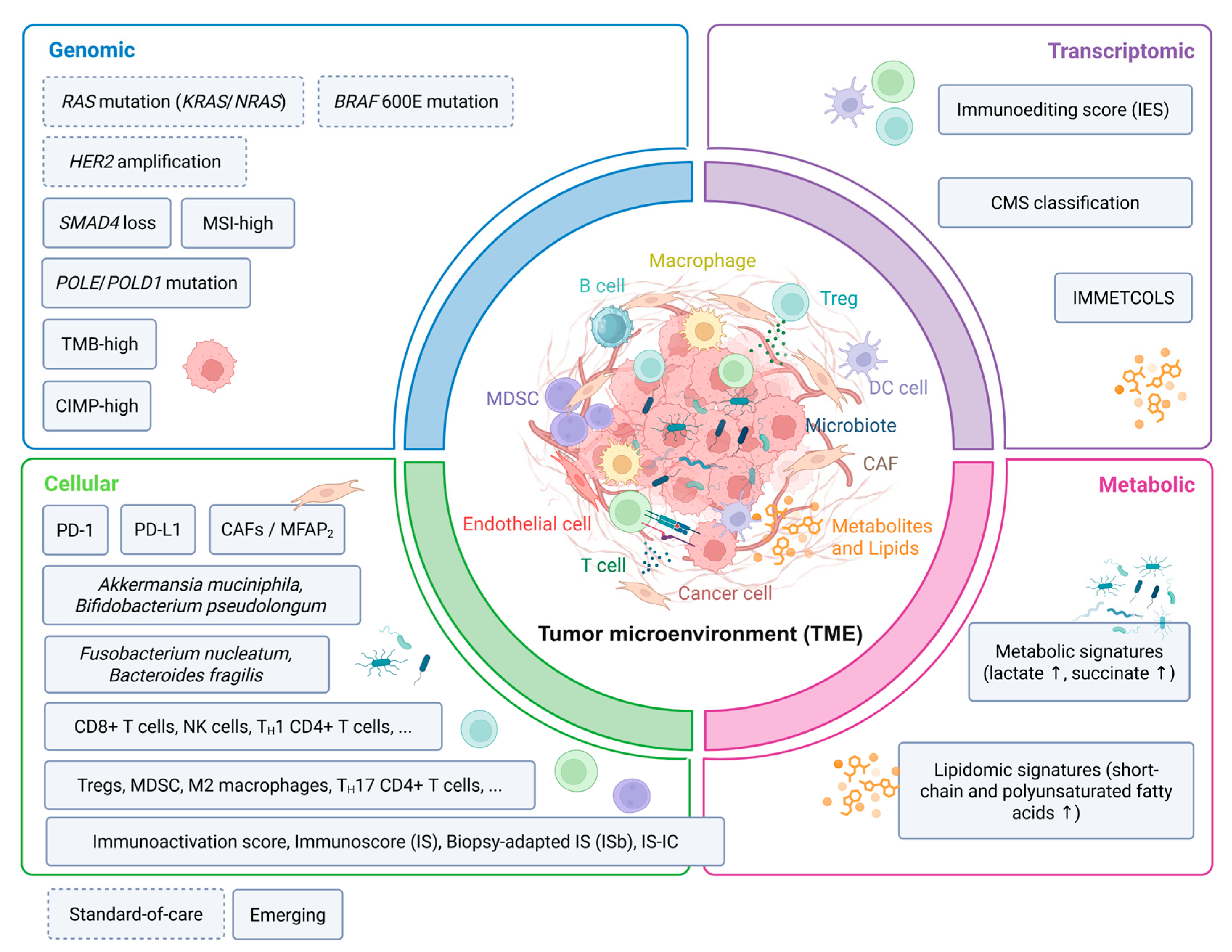
| SMAD4 loss | Genomic | Tumor suppressor inactivation | Prognostic (negative); Predictive (negative, ICI combinations) | Somatic SMAD4 alterations associated with shorter PFS (2.4 vs. 1.6 months) and OS (not reached vs. 5.1 months) with regorafenib + toripalimab | Refractory mCRC | Retrospective analysis of prospective trial (REGOTORI) | [30,31,32,33,34] |
| POLE/POLD1 mutation | Genomic | DNA polymerase proofreading deficiency | Predictive (positive, ICI) | ORR 89% with anti-PD-L1 ± anti-CTLA-4 in proofreading-deficient tumors vs. 54% in dMMR/MSI-H mCRC | Refractory mCRC; independent of MMR status | Retrospective multicentre study | [35,36,37,38,39,40,41] |
| TMB-high (≥10–16 mut/Mb) | Genomic | High somatic mutation load; increased neoantigen burden | Predictive (positive, ICI) | ORR 38.1% in TMB ≥ 16 mut/Mb vs. 2.1% in TMB < 16 mut/Mb with atezolizumab (MyPathway) | Refractory mCRC; optimal cutoff remains debated | Prospective basket trial; CRC-specific validation ongoing | [26,38,42,43,44,45,46,47,48,49,50,51,52] |
| CIMP-high | Epigenomic | CpG island promoter hypermethylation; gene silencing | Prognostic (negative) | Associated with reduced OS and PFS with standard chemotherapy or anti-EGFR therapy; limited added value beyond MSI status for ICI prediction | mCRC with proximal tumors and peritoneal metastases | Retrospective cohort studies | [53,54,55,56,57,58] |
| Immunoediting Score (IES) | Transcriptomic | Integration of neoantigen depletion (GIE) and immune activity (ICR) | Prognostic | High IES associated with immune control; low IES associated with immune evasion and disease progression | mCRC | Retrospective; predictive value for ICI not yet validated | [59,60,61,62,63] |
| CMS classification | Transcriptomic | Gene expression-based molecular subtyping (CMS1-4) | Prognostic and Predictive | CMS1: benefit from pembrolizumab and bevacizumab; CMS2: benefit from anti-EGFR; CMS4: benefit from regorafenib + nivolumab (REGONIVO) | All lines of mCRC treatment | Retrospective analyses of prospective trials; limited by spatial heterogeneity | [63,64,65,66,67,68,69,70,71,72,73] |
| IMMETCOLS (IMC1-3) | Transcriptomic | Immunometabolic subtyping of mCRC | Predictive (investigational) | IMC1 (mesenchymal): inflamed TME, potential benefit from ICI + MCT1 inhibitors; IMC3 (epithelial): high OXPHOS, potential benefit from OXPHOS inhibitors | mCRC specifically | Single-cohort transcriptomic study; prospective validation absent | [74,75] |
| PD-L1/PD-1 expression | Cellular | Immune checkpoint expression on tumor and immune cells | Predictive (limited in MSS CRC) | Low ORRs with anti-PD-1/PD-L1 in MSS mCRC regardless of PD-L1 expression; high PD-1+ T cells without TH17 infiltration may identify MSI-H-like MSS tumors | MSS mCRC | Meta-analyses; no prospective validation as standalone biomarker | [76,77,78,79,80,81,82,83,84,85,86,87,88,89,90] |
| Immunoscore (IS) | Cellular | Density of CD3+ and CD8+ T cells in tumor centre and invasive margin | Prognostic (strong); Predictive (emerging) | High IS associated with reduced metastatic burden and improved survival; predictive benefit from ICI suggested in MSS mCRC (POCHI trial) | All stages; mCRC across lesions | Internationally validated (prognostic); prospective predictive validation ongoing | [63,91,92,93,94,95,96] |
| Biopsy-adapted Immunoscore (ISb) | Cellular | CD3+ and CD8+ T cell density in biopsy core; spatial ISb_20 variant | Prognostic and Predictive | ISb-high associated with improved PFS (HR = 0.24) and OS in AVETUXIRI; ISb_20 further refined stratification | Metastatic setting; pre-treatment biopsies | Prospective (AVETUXIRI); validation in larger cohorts warranted | [63,97,98] |
| Immunoscore-IC | Cellular | CD8+ T cell density, spatial organization, and PD-L1+ cell density | Predictive (emerging, ICI) | Greater benefit from atezolizumab in IS-IC-high MSS tumors in AtezoTRIBE; not confirmed in ANICCA-Class II | MSS mCRC; 1st-line combination immunotherapy | Phase II exploratory analyses; conflicting results across trials | [33,99,100,101] |
| Immunoactivation score | Cellular | Integration of lymphocyte, checkpoint, and monocyte markers (12 parameters) | Predictive (anti-EGFR maintenance) | Immunoactivation-high patients showed improved PFS (HR = 0.50) and OS (HR = 0.54) with panitumumab maintenance | RAS wild-type mCRC maintenance | Prospective (PanaMa trial); single-trial evidence | [102] |
| Cancer-associated fibroblasts (CAFs)/MFAP2 | Cellular | Stromal remodelling; CD8+ T cell suppression via MFAP2 | Prognostic (negative) | High FAP1+ CAF infiltration associated with lymph node metastasis, chemoresistance, and immunotherapy resistance; MFAP2 linked to advanced disease and poor outcome | mCRC | Retrospective; mechanistic evidence from preclinical models | [74,103,104,105,106,107,108,109,110] |
| Tumor microbiome (Fn, Bf, Prevotella) | Cellular/Microbial | Intratumoral bacteria modulating immune cytotoxicity and treatment response | Prognostic (negative, Fn/Bf); Predictive (investigational) | Fn associated with poor chemotherapy response, lower CD8+ T cell infiltration, and potential modulation of PD-L1; shared between primary tumors and metastases | mCRC; rectal cancer post-chemoradiotherapy | Retrospective/correlative; no prospective ICI biomarker validation | [111,112,113,114,115,116,117,118,119] |
Disclaimer/Publisher’s Note: The statements, opinions and data contained in all publications are solely those of the individual author(s) and contributor(s) and not of MDPI and/or the editor(s). MDPI and/or the editor(s) disclaim responsibility for any injury to people or property resulting from any ideas, methods, instructions or products referred to in the content. |
© 2026 by the authors. Licensee MDPI, Basel, Switzerland. This article is an open access article distributed under the terms and conditions of the Creative Commons Attribution (CC BY) license.
Share and Cite
Benidovskaya, E.; Huyghe, N.; Giolito, M.V.; Coulie, P.; Van den Eynde, M. Integrative Profiling of Tumor and Blood Microenvironments to Uncover Molecular and Immune Determinants of Prognosis and Treatment Efficacy in Metastatic Colorectal Cancer. Cancers 2026, 18, 2651. https://doi.org/10.3390/cancers18162651
Benidovskaya E, Huyghe N, Giolito MV, Coulie P, Van den Eynde M. Integrative Profiling of Tumor and Blood Microenvironments to Uncover Molecular and Immune Determinants of Prognosis and Treatment Efficacy in Metastatic Colorectal Cancer. Cancers. 2026; 18(16):2651. https://doi.org/10.3390/cancers18162651
Chicago/Turabian StyleBenidovskaya, Elena, Nicolas Huyghe, Maria Virginia Giolito, Pierre Coulie, and Marc Van den Eynde. 2026. "Integrative Profiling of Tumor and Blood Microenvironments to Uncover Molecular and Immune Determinants of Prognosis and Treatment Efficacy in Metastatic Colorectal Cancer" Cancers 18, no. 16: 2651. https://doi.org/10.3390/cancers18162651
APA StyleBenidovskaya, E., Huyghe, N., Giolito, M. V., Coulie, P., & Van den Eynde, M. (2026). Integrative Profiling of Tumor and Blood Microenvironments to Uncover Molecular and Immune Determinants of Prognosis and Treatment Efficacy in Metastatic Colorectal Cancer. Cancers, 18(16), 2651. https://doi.org/10.3390/cancers18162651