1. Introduction
Neutrophils are increasingly recognized as active regulators of tumor progression rather than passive bystanders within the tumor immune microenvironment (TIME) [
1,
2]. Beyond their conventional roles in antimicrobial defense and acute inflammation, neutrophils can acquire tumor-promoting properties through the release of neutrophil extracellular traps (NETs), a process referred to as NET formation or NETosis [
3,
4]. NETs are web-like extracellular structures composed of decondensed chromatin decorated with granular and cytoplasmic proteins, including myeloperoxidase (MPO), neutrophil elastase, and citrullinated histone H3 (citH3) [
5]. Accumulating evidence indicates that NETs facilitate tumor cell invasion, metastasis, vascular adhesion, thrombosis, and immune evasion in multiple cancer types [
6,
7]. Mechanistically, NETs may promote tumor progression by trapping circulating tumor cells, remodeling the extracellular matrix, amplifying inflammatory signaling, and impairing antitumor immune responses [
8,
9,
10]. Despite these advances, the upstream cellular and molecular mechanisms sustaining NETosis within the TIME remain incompletely understood.
Neutrophils within the tumor microenvironment do not function in isolation [
11]. Their recruitment, activation, and phenotypic remodeling depend on dynamic interactions with tumor cells, tumor-associated macrophages (TAMs), stromal cells, and other immune populations [
12,
13,
14]. In particular, TAMs serve as major regulators of local inflammatory networks within the TIME. Through soluble mediators and cell–cell communication, TAMs can influence neutrophil polarization toward N1 or N2 phenotypes, thereby modulating their biological behavior [
15]. Previous studies have shown that TAMs can induce NETosis; however, the specific TAM subtypes responsible for this process, along with their defining molecular markers and functional characteristics, remain poorly characterized.
Our previous work demonstrated that P2RY13 is significantly downregulated in lung adenocarcinoma (LUAD) and associated with poor prognosis [
16]. Its expression has also been linked to the pro-tumor activity of neutrophils, suggesting a potential role in regulating myeloid cell interactions within the tumor microenvironment. P2RY13 is a G protein-coupled purinergic receptor responsive to extracellular adenosine diphosphate (ADP) signaling that has been implicated in immune regulation, inflammatory responses, and cellular metabolic homeostasis [
17,
18]. Purinergic signaling constitutes an important component of the inflammatory microenvironment, in which extracellular nucleotides function as danger-associated molecular signals that regulate immune cell activation, migration, and effector functions [
19,
20]. Accordingly, P2RY13 may act not only as a prognosis-associated molecule in cancer but also as an immune regulatory factor within the tumor microenvironment.
Emerging evidence further supports a role for P2RY13 in tumor immune regulation. For example, Lan et al. reported that P2RY13-positive dendritic cells exhibit enhanced antigen-presenting capacity and stronger interactions with lymphocytes, particularly T cells, suggesting a potential role for P2RY13 in promoting antitumor immune responses [
21]. However, the expression pattern and functional significance of P2RY13 in TAMs remain insufficiently characterized. In particular, whether P2RY13 regulates TAM–neutrophil interactions and contributes to NETosis remains unclear.
Accordingly, the present study was conducted to better understand the mechanisms governing NETosis within the TIME. By using single-cell transcriptomic analysis, we identified minimal P2RY13 expression in tumor cells and predominant enrichment within TAM populations. Moreover, by integrating bioinformatic analyses, in vitro functional validation, and tissue-level validation using tissue microarrays from three common cancer types, a broad pro-NETosis role of P2RY13-negative TAMs in multiple tumors was preliminarily identified and validated. These findings provide new insights into the TAM-mediated regulation of NETosis and highlight potential therapeutic targets for pan-cancer treatment.
2. Materials and Methods
2.1. Pan-Cancer Transcriptomic Data Acquisition and Processing
Pan-cancer transcriptomic and clinical data were obtained from the SangerBox platform (
http://sangerbox.com/login.html, accessed on 13 April 2026) [
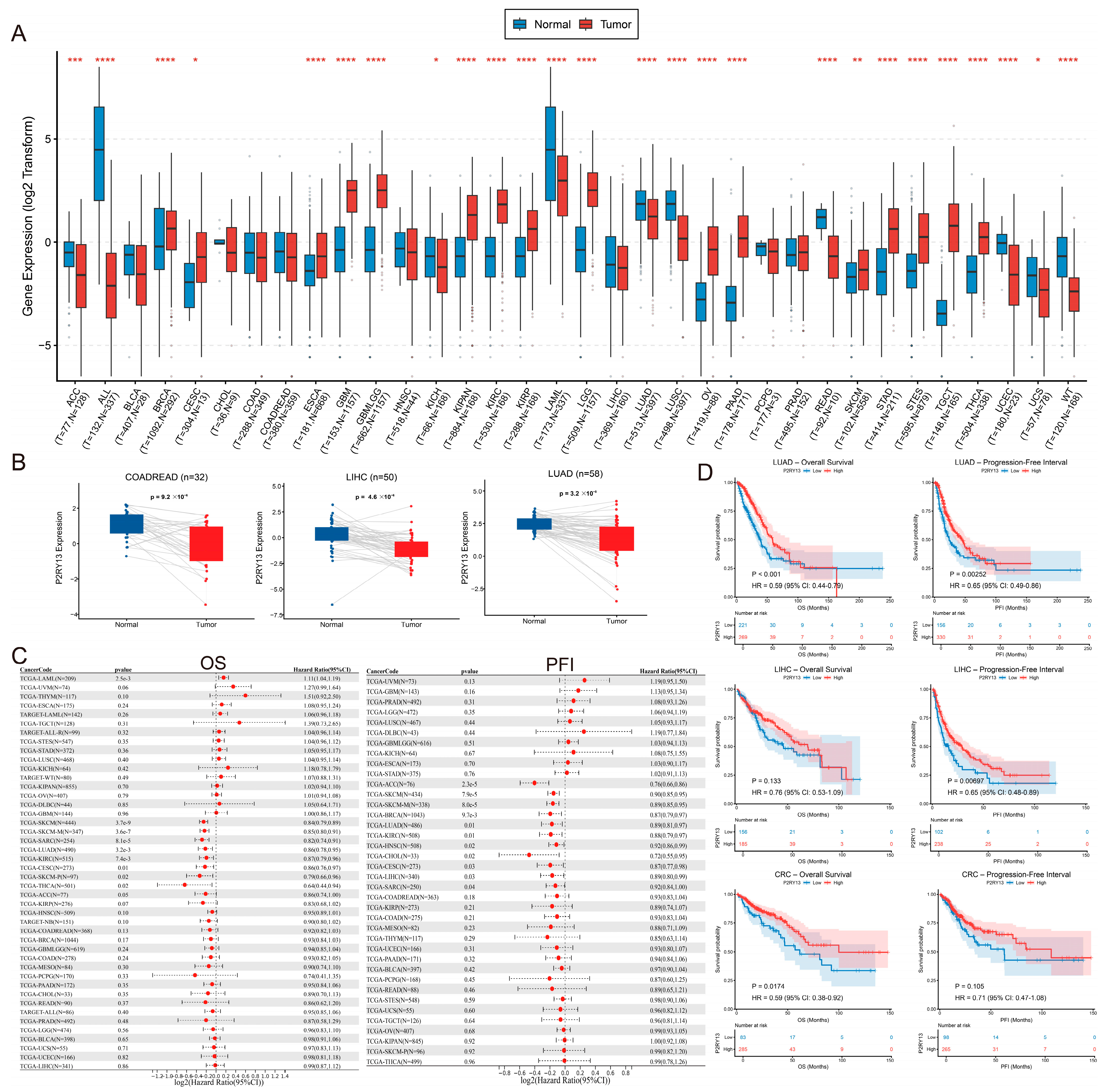
22], which integrates uniformly processed datasets from The Cancer Genome Atlas (TCGA) and the Genotype-Tissue Expression (GTEx) project. TCGA tumor RNA-sequencing data and TCGA/GTEx normal tissue data were used to compare P2RY13 expression between tumor and normal tissues among cancer types. For paired analyses, only TCGA samples with matched tumor and adjacent normal tissues were included.
Expression values were log2-transformed according to the SangerBox data-processing pipeline when appropriate. Cancer type abbreviations followed TCGA nomenclature. LUAD, liver hepatocellular carcinoma (LIHC), and colorectal cancer (CRC)-related cohorts, represented by the combined TCGA COADREAD cohort, were selected for subsequent analyses because P2RY13 exhibited consistent downregulation in these tumor types.
2.2. Differential Expression Analysis of P2RY13
Differential expression analysis of P2RY13 between tumor and normal tissues was performed using SangerBox (
http://sangerbox.com/login.html, accessed on 13 April 2026). For pan-cancer unpaired analyses, TCGA tumor samples were compared with corresponding normal tissues from TCGA and/or GTEx. For paired analyses, matched tumor and adjacent normal samples from TCGA were evaluated. Statistical significance was assessed using the default methods implemented in SangerBox, and results were visualized as box plots or paired comparison plots.
2.3. Survival Analysis
The prognostic significance of P2RY13 expression was evaluated using TCGA clinical data through SangerBox (
http://sangerbox.com/login.html, accessed on 13 April 2026). Survival endpoints included overall survival (OS), progression-free interval (PFI), disease-specific survival, and disease-free interval. Pan-cancer Cox proportional hazards regression analysis was performed to estimate hazard ratios (HRs) and 95% confidence intervals (CIs). An HR < 1 indicated that higher P2RY13 expression was associated with a lower risk of death or disease progression.
For Kaplan–Meier survival analyses, patients were stratified into high- and low-expression groups according to the P2RY13 expression cutoff implemented in SangerBox. Survival differences between groups were assessed using the log-rank test. Representative Kaplan–Meier survival curves were generated for LUAD, LIHC, and COADREAD.
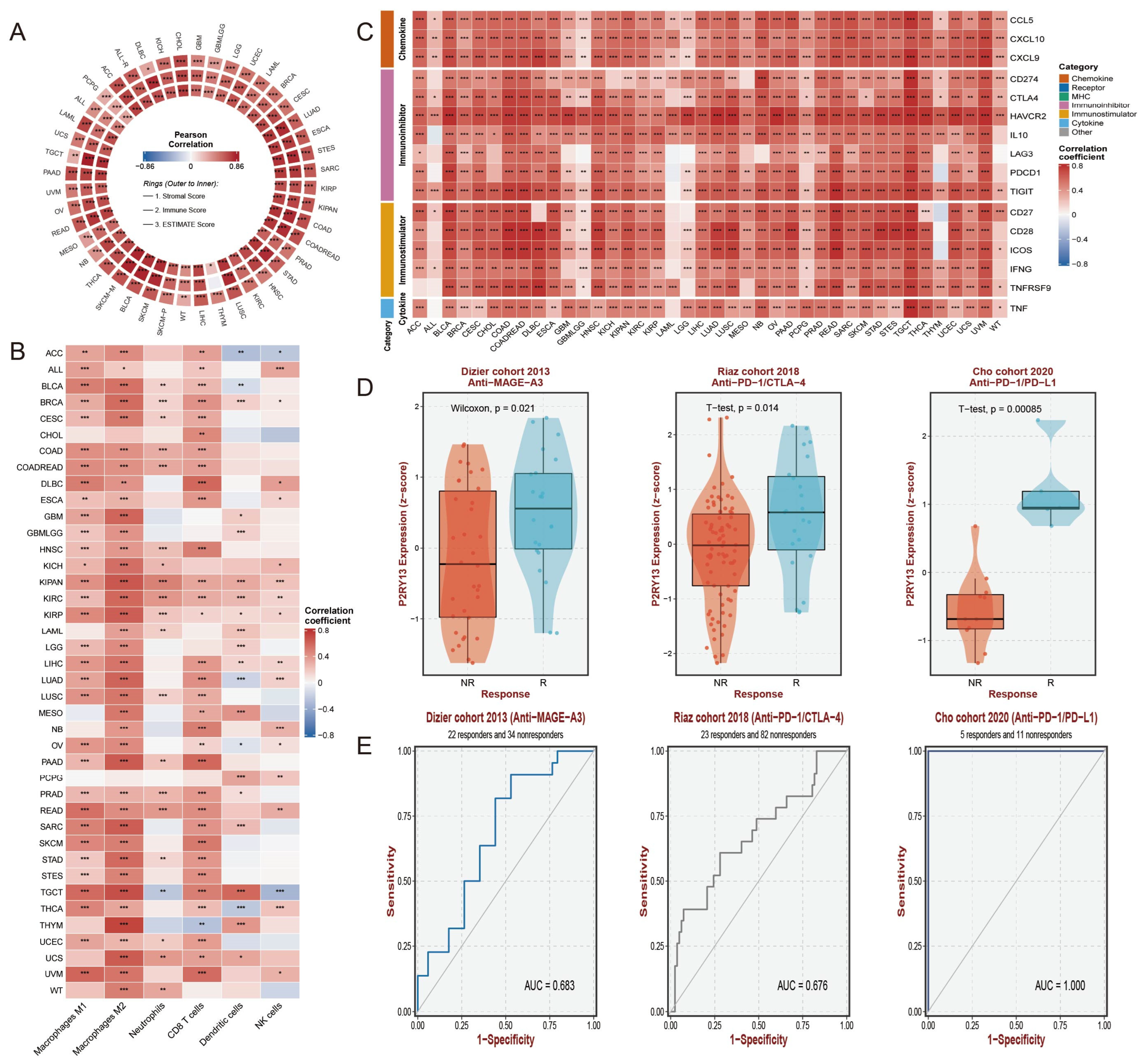
2.4. Immune Microenvironment and Immune Infiltration Analysis
Precomputed immune microenvironment-related data were downloaded from the SangerBox platform (
http://sangerbox.com/login.html, accessed on 13 April 2026), including ESTIMATE-derived Stromal Score, Immune Score, and ESTIMATE Score, as well as immune cell infiltration profiles generated using quanTIseq, MCPcounter, and EPIC based on TCGA transcriptomic data. Following data export, correlations between P2RY13 expression and immune scores or immune cell infiltration levels were analyzed locally using R (version 3.6.4). Correlation coefficients and
p values were calculated among cancer types, and results were visualized as heatmaps.
2.5. Analysis of Immunomodulatory Molecules and Immunophenoscore
Immunomodulatory gene expression data and immunophenoscore-related metrics were downloaded from the SangerBox platform (
http://sangerbox.com/login.html, accessed on 13 April 2026). Correlations between P2RY13 expression and immune regulatory genes or immunophenoscore metrics were analyzed using R (version 3.6.4). Immune regulatory genes included chemokines, chemokine receptors, major histocompatibility complex-related genes, immunostimulatory molecules, immunoinhibitory molecules, cytokines, and immune checkpoint-related genes. Correlation results were visualized using R-generated heatmaps.
2.6. Immunotherapy Response Analysis
The association between P2RY13 expression and immunotherapy response was evaluated using publicly available immunotherapy-treated cohorts accessed through the ROC Plotter platform (
https://rocplot.com/, accessed on 15 April 2026). Three independent cohorts were included, comprising patients treated with anti-MAGE-A3, anti-PD-1/CTLA-4, or anti-PD-1/PD-L1 therapies. Patients were classified as responders or non-responders according to the response annotations provided in the original datasets.
P2RY13 expression levels were compared between responders and non-responders. Receiver operating characteristic (ROC) curve analysis was performed to evaluate the predictive performance of P2RY13 expression for immunotherapy response. The area under the curve (AUC) was calculated for each cohort.
2.7. Single-Cell RNA-Sequencing Data Acquisition, Processing, and Macrophage Subset Definition
Single-cell RNA-sequencing (scRNA-seq) datasets for LUAD, LIHC, and CRC were obtained from the Tumor Immune Single-cell Hub 2 [
23] (TISCH2;
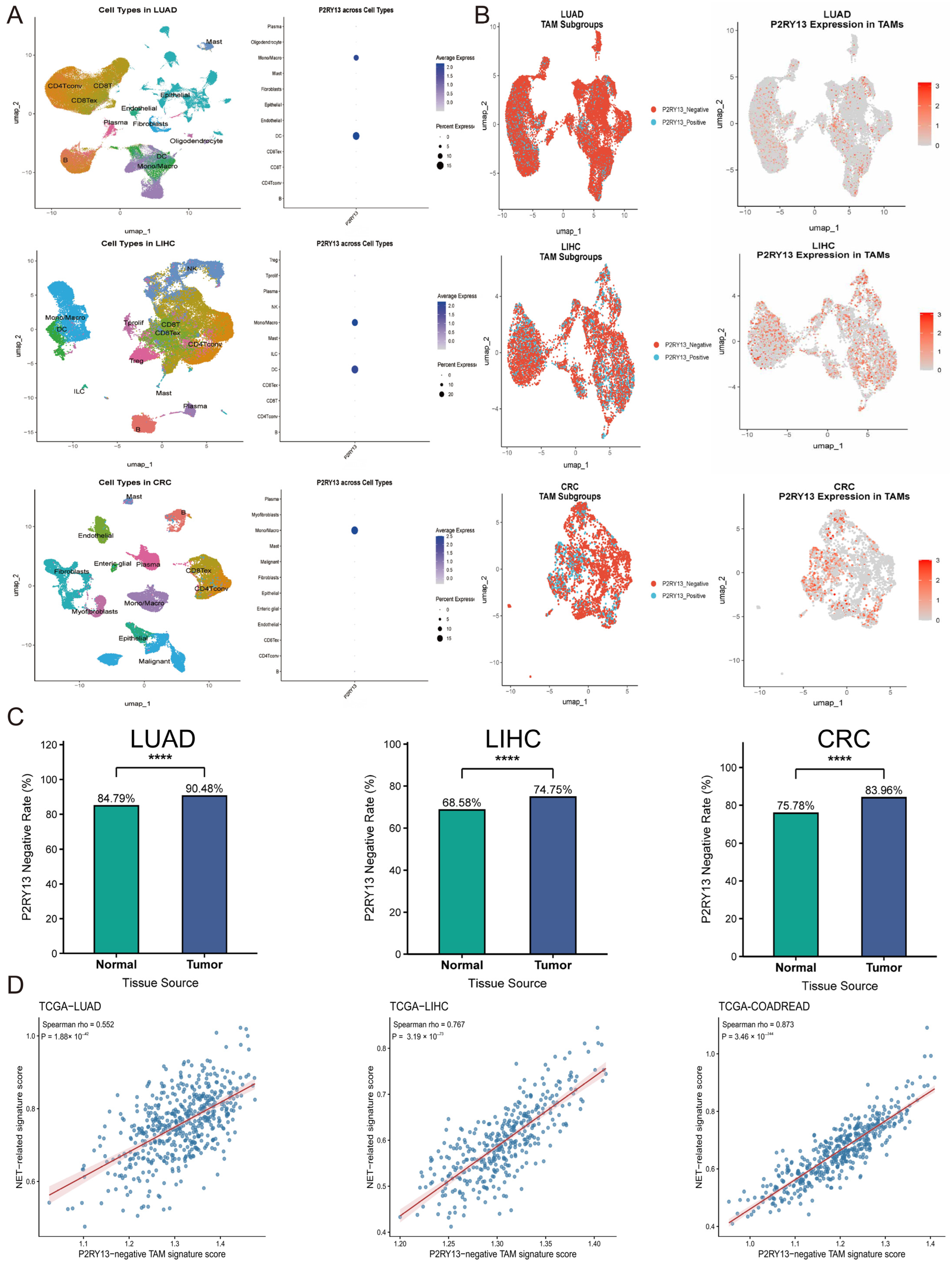
https://tisch.compbio.cn/home/, accessed on 15 April 2026), with priority given to datasets containing both tumor and matched normal tissues when available. Processed expression matrices and cell-type annotations were downloaded for downstream analyses. P2RY13 expression was evaluated in major cell populations, including epithelial/tumor cells, T cells, B cells, myeloid cells, endothelial cells, and fibroblasts. Given the association between P2RY13 expression and myeloid infiltration identified in bulk analyses, annotated macrophages were extracted for focused analysis and visualized using uniform manifold approximation and projection (UMAP). Macrophages were classified as P2RY13-positive or P2RY13-negative according to the presence or absence of detectable P2RY13 transcript expression, respectively. In tumor tissues, these subsets were considered TAM-related populations. The proportions of P2RY13- negative macrophages were calculated in tumor and normal tissues for each cancer type to evaluate relative changes during tumorigenesis.
Nevertheless, this detection-based classification has an inherent methodological limitation. Because scRNA-seq is characterized by sparse and incomplete transcript capture, failure to detect P2RY13 in an individual macrophage does not necessarily indicate the true absence of its expression. Therefore, we used the term “P2RY13-negative” as an operational, detection-based designation for macrophages with no detectable P2RY13 transcripts under the applied analytical conditions. Accordingly, the resulting proportions were interpreted as relative estimates of the transcript-undetected population rather than precise measurements of the true prevalence of biologically P2RY13-null macrophages.
2.8. Single-Cell-Derived P2RY13-Negative TAM and NET-Related Signature Analysis
Single-cell RNA-seq datasets from LUAD, LIHC, and CRC were analyzed separately to identify P2RY13-negative TAMs and derive transcriptional signatures. Within each cancer type, TAMs without detectable P2RY13 transcripts were compared with P2RY13-positive TAMs. Genes significantly upregulated in the P2RY13-negative subset were used to construct the corresponding P2RY13-negative TAM signature. The gene sets derived from the LUAD, LIHC, and CRC datasets are provided in
Tables S1–S3, respectively.
The single-cell-derived signatures were subsequently projected onto bulk transcriptomic data from the TCGA-LUAD, TCGA-LIHC, and TCGA-COADREAD cohorts. For each patient, a P2RY13-negative TAM signature score was calculated using single-sample gene set enrichment analysis. NET-related signature scores were calculated independently using a previously reported gene set and scoring method [
24]. Within each cancer cohort, the association between P2RY13-negative TAM and NET-related signature scores was evaluated using Spearman correlation analysis. These analyses were performed to assess the transcriptomic association between the relative enrichment of the P2RY13-negative TAM state and NET-related activity in bulk tumor tissues.
2.9. Tissue Microarray Acquisition
Tissue microarrays (TMAs) containing paired tumor and adjacent normal tissues from LUAD, LIHC, and CRC were purchased from Shanghai Outdo Biotech Co., Ltd. (Shanghai, China). The CRC TMA (Lot XT15-008, HCol-Ade060Lym-01) included 20 patients (60 cores), the LIHC TMA (Lot I11-003, HLiv-HCC050PG-01) included 25 patients (50 cores), and the LUAD TMA (Lot XT24-015, HLugA060PG03) included 30 patients (60 cores). Baseline clinicopathological characteristics are summarized in
Tables S4–S6.
2.10. Immunohistochemistry
TMA sections were subjected to immunohistochemical staining. After deparaffinization, antigen retrieval was performed by microwave heating in 5 mM Tris-HCl buffer for 10 min. Endogenous peroxidase activity was blocked with 3% hydrogen peroxide, followed by serum blocking to minimize non-specific antibody binding. Sections were then incubated overnight at 4 °C with antibodies against citrullinated histone H3 (histone H3 [citrullinated at Arg2, Arg8, and Arg17]; 1:200; Novus Biologicals, Centennial, CO, USA) or P2RY13 (1:250; Proteintech, Wuhan, China). After incubation with horseradish peroxidase-conjugated secondary antibodies for 30 min at room temperature, immunoreactivity was visualized using diaminobenzidine, and sections were counterstained with hematoxylin.
2.11. Multiplex Immunofluorescence Staining and Quantitative Analysis
Multiplex immunofluorescence (mIF) staining was performed on TMA sections to identify and quantify P2RY13-negative TAMs and NETs. After deparaffinization and rehydration, sections underwent antigen retrieval, followed by blocking of endogenous peroxidase activity and non-specific antibody binding. Sections were sequentially incubated with antibodies against MPO (1:250; Proteintech, Wuhan, China), citrullinated histone H3 (histone H3 [citrullinated at Arg2, Arg8, and Arg17]; 1:200; Novus Biologicals, Centennial, CO, USA), P2RY13 (1:250; Proteintech, Wuhan, China), and CD68 (1:250; Proteintech, Wuhan, China). Multiplex detection was performed using a sequential tyramide signal amplification protocol, and nuclei were counterstained with DAPI to generate a four-marker, five-color mIF panel.
P2RY13
−CD68
+ cells were defined as P2RY13-negative macrophages, whereas NETs were identified according to established criteria as extracellular web-like structures exhibiting colocalized MPO, CitH3, and DAPI signals [
25]. For quantitative analysis, three randomly selected, non-overlapping high-power fields (×400) within the tumor region of each TMA core were evaluated. P2RY13-negative TAMs were counted in each field, and the mean count per high-power field was used to represent the infiltration level. NET-associated areas were quantified, and NET expression was calculated as the percentage of the analyzed tumor area occupied by MPO
+/CitH3
+ extracellular web-like structures. The mean percentage from the three fields was recorded as the NET expression level for each sample.
2.12. Cell Culture and Macrophage-like Differentiation
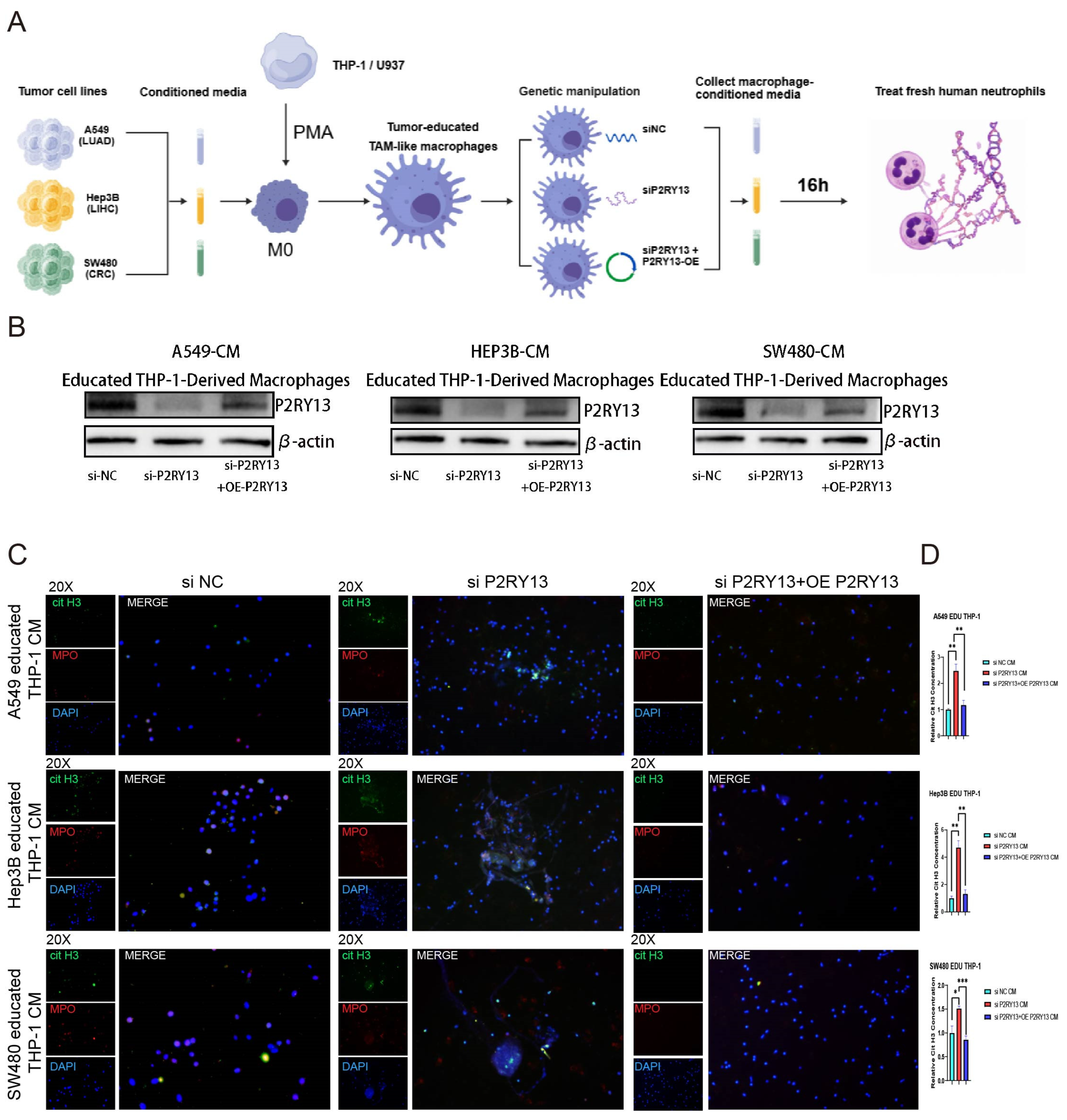
THP-1 and U937 cells were maintained in RPMI 1640 medium supplemented with 10% fetal bovine serum, 100 IU/mL penicillin, and 100 μg/mL streptomycin at 37 °C in a humidified atmosphere containing 5% CO2. To generate M0-like macrophages, THP-1 and U937 cells were treated with phorbol 12-myristate 13-acetate (PMA; 100 ng/mL) for 48 h. The PMA-containing medium was then removed, and cells were washed with phosphate-buffered saline (PBS) and allowed to rest in fresh complete medium for an additional 24 h. Acquisition of an adherent macrophage-like morphology was used to confirm successful differentiation before treatment with tumor-cell-conditioned medium.
2.13. Generation of Tumor-Educated TAM-like Macrophages
A549, Hep3B, and SW480 cells were used to generate tumor-cell-conditioned medium (TCM) representative of lung cancer, hepatocellular carcinoma, and colorectal cancer, respectively. All cell lines were maintained in DMEM supplemented with 10% fetal bovine serum, 100 IU/mL penicillin, and 100 μg/mL streptomycin at 37 °C. When cultures reached approximately 80% confluence, cells were washed twice with PBS and incubated for an additional 24 h in DMEM containing 1% fetal bovine serum. Supernatants were then collected, centrifuged at 1000× g for 10 min at 4 °C, and passed through 0.22-μm filters to remove residual cells and debris. The resulting A549-, Hep3B-, and SW480-derived TCM was used immediately or stored in aliquots at −80 °C, avoiding repeated freeze–thaw cycles. To generate tumor-educated TAM-like macrophages, PMA-differentiated THP-1- and U937-derived M0-like macrophages were exposed for 48 h to the corresponding TCM mixed 1:1 (v/v) with fresh complete RPMI 1640 medium.
2.14. P2RY13 Knockdown and Rescue
After tumor education, THP-1- and U937-derived TAM-like macrophages were washed with PBS and assigned to one of three groups: negative-control siRNA (siNC), siRNA targeting P2RY13 (siP2RY13), or siP2RY13 plus a P2RY13 overexpression construct (OE-P2RY13) for rescue. Cells were transfected using Lipofectamine 2000(Thermo Fisher Scientific, Waltham, MA, USA) according to the manufacturer’s instructions, with siRNAs used at a final concentration of 50 nM. P2RY13-targeting and negative-control siRNAs were purchased from RiboBio Co., Ltd. (Guangzhou, China), whereas the P2RY13 overexpression construct was generated by GeneChem Co., Ltd. (Shanghai, China). P2RY13 protein expression was assessed by western blotting to confirm knockdown and rescue efficiencies.
2.15. Neutrophil Isolation and Conditioned-Medium Treatment
Primary human neutrophils were isolated from peripheral blood samples obtained from healthy donors using Polymorphprep density-gradient medium (Axis-Shield, Dundee, UK) according to the manufacturer’s instructions. Neutrophil purity was assessed using a Fast Giemsa Staining Kit (Yeasen Biotechnology, Shanghai, China), after which freshly isolated cells were resuspended in RPMI 1640 medium (Gibco, Grand Island, NY, USA). Conditioned medium was collected separately from each experimental group of tumor-educated TAM-like macrophages and applied to freshly isolated neutrophils for 16 h. This conditioned-medium culture system was used to evaluate the effects of soluble factors released by tumor-educated TAM-like macrophages on neutrophils.
2.16. In Vitro Assessment of NET Formation
Following conditioned-medium treatment, neutrophils cultured on coverslips were processed for immunofluorescence staining. Briefly, cells were fixed with 4% paraformaldehyde for 30 min, permeabilized with 0.5% Triton X-100 for 15 min, and blocked with 1% bovine serum albumin in PBS for 1 h. Primary antibodies against citrullinated histone H3 (histone H3 [citrullinated at Arg2, Arg8, and Arg17]; 1:200; Novus Biologicals) and MPO (5 μg/mL; R&D Systems) were applied overnight at 4 °C. After incubation with the corresponding Alexa Fluor-conjugated secondary antibodies for 1 h at room temperature, nuclei and extracellular DNA were stained with DAPI (Sigma-Aldrich, USA) for 5 min. Fluorescence images were subsequently acquired using a fluorescence microscope.
In parallel, culture supernatants were collected after conditioned-medium treatment and analyzed for CitH3 using a commercially available ELISA kit (Cayman Chemical, Ann Arbor, MI, USA) according to the manufacturer’s instructions. CitH3 concentrations were determined from standard curves fitted using a four-parameter logistic regression model in GraphPad Prism (version 11.0.1).
2.17. Western Blotting
Western blotting was performed to determine P2RY13 protein expression and verify the efficiencies of P2RY13 knockdown and subsequent re-expression. Protein lysates from the indicated experimental groups were subjected to immunoblotting using antibodies against P2RY13 (1:2000; Proteintech, Wuhan, China) and β-actin (1:20,000; Proteintech, Wuhan, China). β-Actin served as the loading control, and P2RY13 protein expression was normalized to β-actin.
2.18. Quantification of Neutrophil Infiltration in TMA
Neutrophil infiltration was evaluated in hematoxylin and eosin-stained TMA sections from LUAD, LIHC, and CRC tissues. Each tissue core was initially examined at low magnification to identify viable tumor regions suitable for evaluation, and areas containing necrosis, hemorrhage, tissue folds, uneven staining, or other processing artifacts were excluded. Three non-overlapping high-power fields (×400) were subsequently selected from each evaluable core. Neutrophils were identified based on their characteristic histomorphological features, including deeply stained segmented or multilobed nuclei and scant, lightly eosinophilic granular cytoplasm. Only morphologically intact neutrophils within the tumor parenchyma or stroma were counted. Cells within vascular lumina, nuclear debris in necrotic regions, and inflammatory cells that could not be reliably identified morphologically were excluded. Neutrophil infiltration was expressed as the mean number of neutrophils per high-power field and subsequently correlated with the infiltration level of P2RY13−CD68+ TAMs in the corresponding tumor tissue.
2.19. Statistical Analysis
Statistical analyses were performed using R (version 3.6.4) and GraphPad Prism (version 11.0.1) according to the characteristics of the data. Student’s t-test was used for data with equal variance, whereas the Mann–Whitney U test was applied to data with unequal variance. For comparisons involving small sample sizes, differences between two independent groups were assessed using a two-sided Welch’s t-test without assuming equalvariances. Pearson correlation analysis was used to evaluate correlations, and the log-rank test was applied for survival analysis. A p value < 0.05 was considered statistically significant. Schematic diagrams were generated using BioGDP.com.
4. Discussion
This study characterizes P2RY13-negative TAMs as a tumor-enriched macrophage state associated with neutrophil infiltration and NET-related features and provides experimental evidence that P2RY13-dependent alterations in macrophage-conditioned medium modulate NET formation in vitro. At the pan-cancer level, P2RY13 was frequently downregulated in solid tumors and showed marked reductions in LUAD, LIHC, and COADREAD/CRC, where low expression was associated with poorer clinical outcomes (
Figure 1 and
Figures S1–S3). P2RY13 expression was also associated with an immune-inflamed tumor microenvironment and was higher in responders across three exploratory immunotherapy cohorts, although its predictive value requires validation in larger prospective cohorts (
Figure 2 and
Figures S4–S7). Single-cell analyses further revealed an increased proportion of macrophages lacking detectable P2RY13 transcripts in tumor tissues. Cancer-specific signatures derived from these cells were positively correlated with NET-related signatures after projection onto the corresponding TCGA bulk transcriptomic cohorts (
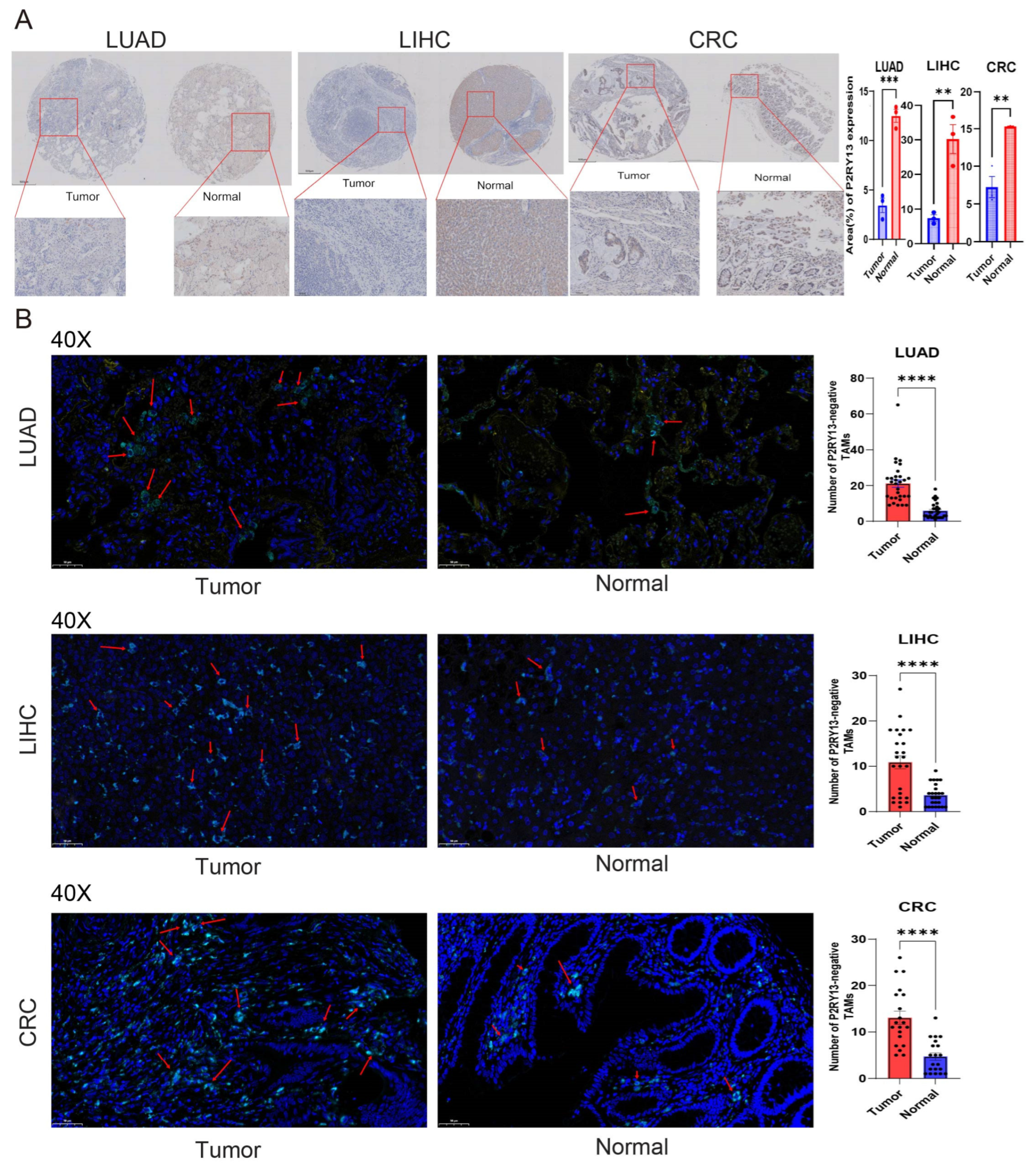
Figure 3). Protein-level analyses demonstrated reduced P2RY13 expression and increased infiltration of P2RY13
−CD68
+ macrophages in LUAD, LIHC, and CRC tumors (
Figure 4). Conditioned medium from P2RY13-silenced, tumor-educated TAM-like macrophages enhanced NET formation, whereas P2RY13 re-expression attenuated this effect (
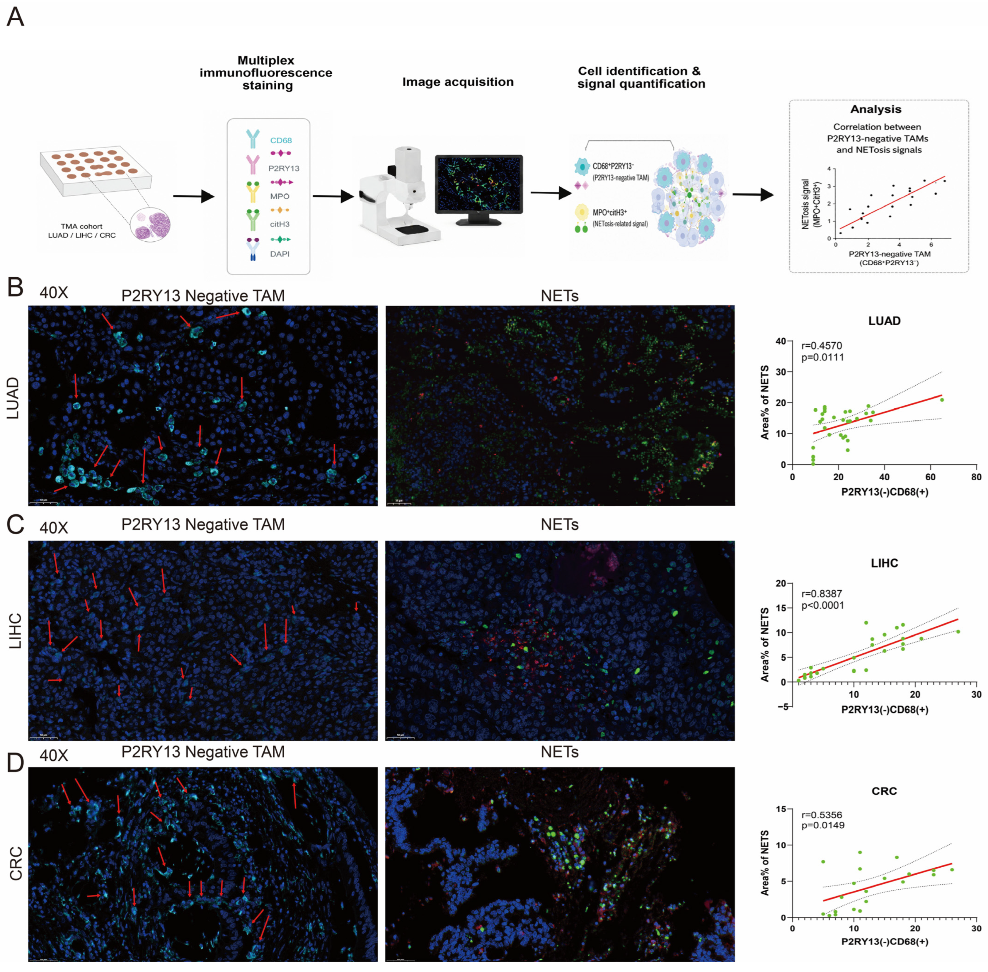
Figure 5). In parallel, tissue analyses demonstrated positive associations between P2RY13-negative TAM infiltration and both NET expression and neutrophil infiltration (
Figure 6 and
Figure S11). Collectively, these complementary findings support an association between the P2RY13-negative TAM state and a neutrophil- and NET-enriched tumor microenvironment but do not, by themselves, establish a direct causal mechanism in vivo.
P2RY13 belongs to the purinergic receptor family, which is involved in extracellular nucleotide sensing, inflammatory regulation, and immune cell communication [
27,
28]. Purinergic signaling is increasingly recognized as an important regulatory axis in cancer, particularly within the tumor microenvironment, where ATP, ADP, adenosine, and related metabolites shape immune cell recruitment, activation, and suppression. Consistent with these observations, pan-cancer and single-cell analyses demonstrated strong associations between P2RY13 expression and immune components of the tumor microenvironment. Notably, single-cell transcriptomic analyses showed minimal P2RY13 expression in malignant epithelial cells but predominant localization within macrophage populations (
Figure 3A). This distribution suggests that the tumor-associated functions of P2RY13 may be mediated primarily through immune stromal components, particularly macrophages, rather than through tumor-intrinsic mechanisms. Importantly, the positive association between bulk tumor P2RY13 expression and immune infiltration does not necessarily conflict with the enrichment of P2RY13-negative macrophages in tumors. Bulk P2RY13 expression reflects both immune cell abundance and gene expression across multiple cellular populations, whereas single-cell analysis specifically captures P2RY13 heterogeneity within the macrophage compartment. Accordingly, bulk-tissue P2RY13 expression should not be interpreted as a direct measure of macrophage-intrinsic P2RY13 activity or the abundance of P2RY13-negative TAMs.
Macrophages are highly plastic components of the tumor microenvironment and can adopt diverse functional states depending on tissue context [
29,
30,
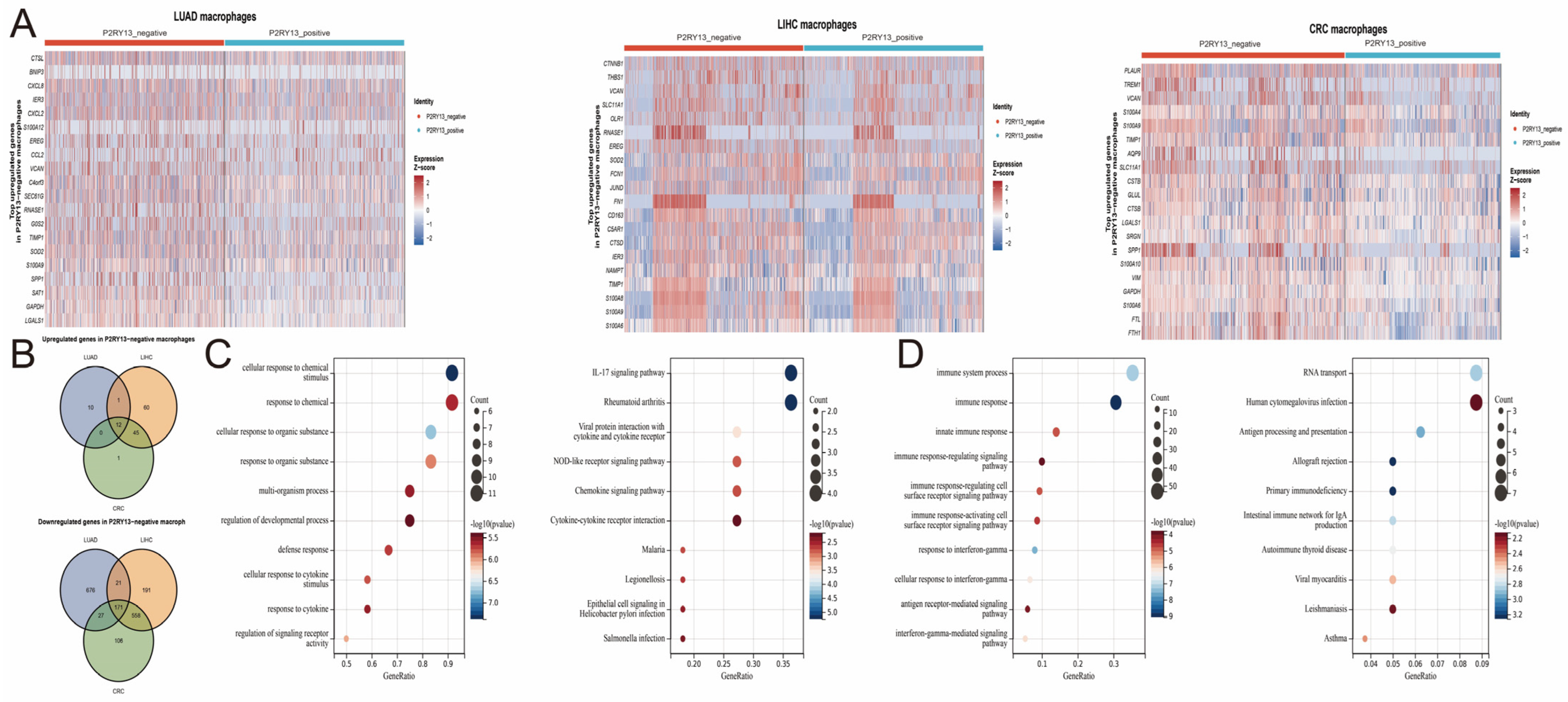
31]. The transcriptional profile identified in
Figure 7 did not conform to the conventional M1/M2 polarization paradigm. P2RY13-negative TAMs exhibited enhanced inflammatory and chemotactic programs, including IL-17, NOD-like receptor, cytokine, and chemokine signaling, while showing relative attenuation of interferon-γ-response and antigen presentation pathways. This combination suggests a context-dependent, transcriptionally remodeled macrophage state rather than a uniformly M1- or M2-polarized phenotype. This interpretation is consistent with transcriptomic and single-cell studies showing that macrophage activation occurs along a continuum and that TAMs may simultaneously express features traditionally assigned to different polarization states [
32,
33]. Accordingly, tumor-cell-conditioned medium was used to generate tumor-educated TAM-like macrophages rather than imposing a defined IL-4/IL-13- or IFN-γ/LPS-induced polarization state. This approach exposes macrophages to soluble factors derived from three tumor-cell contexts and may better reflect mixed tumor-driven activation. Nevertheless, it does not fully recapitulate the cellular, extracellular matrix, metabolic, and spatial cues encountered by bona fide TAMs in vivo.
The association between P2RY13-negative TAMs and NET-related activity was supported at multiple analytical levels, although each has distinct interpretive limitations. Projection of the single-cell-derived signatures onto TCGA data demonstrated coordinated enrichment of P2RY13-negative TAM and NET-related transcriptional programs (
Figure 3D), but these bulk-tissue scores do not directly quantify either cell abundance or NET formation. In the tissue microarrays, P2RY13-negative TAM infiltration was positively correlated with mIF-defined NET expression (
Figure 6) and with histomorphologically quantified neutrophil infiltration (
Figure S11). These findings suggest two non-mutually exclusive mechanisms. P2RY13-negative TAM-associated signals may promote neutrophil recruitment or retention within tumors, thereby increasing the cellular substrate available for NET formation, and/or enhance the propensity of infiltrating neutrophils to undergo NETosis. The current data do not demonstrate that macrophage P2RY13 directly suppresses NETosis. Rather, they suggest that preserved P2RY13 expression restrains a macrophage state whose paracrine activity promotes neutrophil accumulation and NET formation.
More importantly, the conditioned-medium experiments provide evidence of a P2RY13-dependent paracrine effect. Conditioned medium from P2RY13-silenced, tumor-educated TAM-like macrophages increased MPO
+/CitH3
+ extracellular DNA structures and CitH3 release by neutrophils, whereas P2RY13 re-expression attenuated these effects (
Figure 5 and
Figures S8–S10). Because macrophages and neutrophils were physically separated in this system, the observed phenotype is attributable to alterations in macrophage-conditioned medium rather than direct cell–cell contact. However, knockdown of a single receptor may affect multiple downstream pathways and secreted factors, and the present experiments do not identify the mediator responsible for the NET-promoting effect. The enrichment of CXCL2, CXCL8, S100A8, and S100A9 transcripts in P2RY13-negative TAMs, together with the chemokine-related enrichment observed in
Figure 7, identifies candidate mechanisms for future investigation but does not demonstrate that these molecules were secreted or mediated the observed phenotype. Future studies should combine secretome profiling or cytokine arrays with targeted ELISA, neutralizing antibodies, receptor blockade, and recombinant-protein experiments to determine whether specific chemokines, cytokines, metabolites, or extracellular vesicles account for the effect. Validation using primary human TAMs and in vivo models will also be required to determine whether this paracrine interaction occurs within native tumor tissues.
Despite the potential significance of these findings, several limitations should be acknowledged. First, the designation “P2RY13-negative” was based on the absence of detectable P2RY13 transcripts in scRNA-seq data. Because single-cell transcript detection is sparse and low-abundance transcripts may escape detection, a zero value does not confirm complete biological absence of P2RY13 and may overestimate the P2RY13-negative fraction. Although a binary presence/absence criterion was adopted as a transparent operational threshold, the robustness of the findings to alternative expression thresholds was not assessed. Accordingly, the reported population should be interpreted as macrophages lacking detectable P2RY13 transcripts under the applied analytical conditions. Furthermore, transcript-defined P2RY13-negative macrophages and protein-defined P2RY13−CD68+ macrophages represent related, but not necessarily identical, populations. Second, the P2RY13-negative TAM and NET-related scores derived from bulk TCGA transcriptomes reflect the relative enrichment of their respective transcriptional programs rather than absolute P2RY13-negative TAM abundance or direct NET formation. Their correlation may also be influenced by differences in tumor cellular composition or by overlapping genes between the two signatures. Third, THP-1- and U937-derived tumor-educated macrophages are reductionist cell-line models and were not systematically benchmarked against the transcriptomic or proteomic profiles of primary human TAMs. Accordingly, their designation as TAM-like macrophages should be regarded as operational. In addition, canonical M1- and M2-associated markers were not evaluated in the TMA specimens. Therefore, although single-cell analysis suggests a mixed inflammatory and immune-remodeled phenotype, the relationship between P2RY13-negative TAMs and conventional macrophage polarization states remains to be validated at the protein and spatial levels. Fourth, the positive tissue correlation between P2RY13-negative TAMs and neutrophil counts does not demonstrate enhanced neutrophil recruitment because macrophage-conditioned neutrophil migration assays and in vivo trafficking studies were not performed. Moreover, hematoxylin and eosin-based neutrophil identification relies on histomorphological assessment. Fifth, the cytokines, chemokines, metabolites, extracellular vesicles, or other soluble mediators responsible for the NET-promoting activity of macrophage-conditioned medium remain undefined. Finally, the relatively small TMA cohorts, lack of comprehensive clinical follow-up data, and limited, heterogeneous immunotherapy cohorts constrain the prognostic and translational interpretation of these findings. Further validation using primary human TAMs, larger independent clinical cohorts, targeted secretome analyses, and in vivo models is warranted.
Despite these limitations, the findings identify a reproducible association between a P2RY13-low/undetectable macrophage state and neutrophil- and NET-related features across LUAD, LIHC, and CRC. The knockdown–rescue experiments further support a P2RY13-dependent alteration in the NET-promoting paracrine activity of tumor-educated TAM-like macrophages. These findings establish a testable macrophage–neutrophil interaction model that warrants mechanistic investigation rather than demonstrating a definitive causal pathway in patients.



 ,
,