Article

Factors Associated with the Timing of Liver Metastasis After Colorectal Cancer Surgery: A Retrospective Multicenter Study

1
Department of Surgical Oncology and General Surgery, The First Affiliated Hospital of China Medical University, Shenyang 110001, China
2
Department of Colorectal Surgery, National Cancer Center/National Clinical Research Center for Cancer/Cancer Hospital, Chinese Academy of Medical Sciences and Peking Union Medical College, Beijing 100021, China
*
Author to whom correspondence should be addressed.
Cancers 2026, 18(15), 2445; https://doi.org/10.3390/cancers18152445
Submission received: 19 May 2026 / Revised: 24 July 2026 / Accepted: 25 July 2026 / Published: 29 July 2026
(This article belongs to the Special Issue Colorectal Cancer Liver Metastases)

Simple Summary

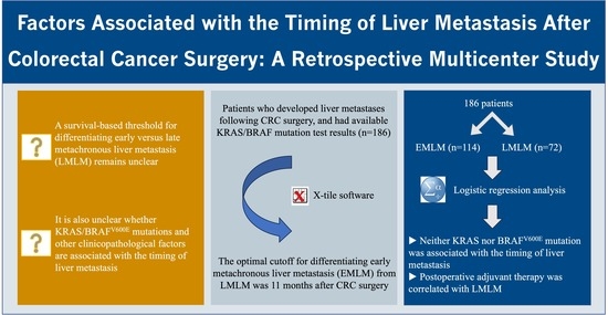
Approximately 25% of patients with colorectal cancer (CRC) develop metachronous liver metastasis (MLM) after curative surgery, with earlier onset portending a worse prognosis. Current definitions distinguishing “early” from “late” MLM, however, are largely based on empirical convention or fixed follow-up intervals rather than on objective prognosis-derived inference. To address this gap, this study aimed to define a survival-based threshold for differentiating early versus late MLM, and to investigate whether KRAS/BRAFV600E mutations and other clinicopathological factors are associated with the time to onset of liver metastasis after CRC surgery. In a cohort of 186 CRC patients who develop liver metastasis after radical surgery, we identified 11 months post-surgery as the optimal temporal cutoff, and found that KRAS/BRAFV600E mutations were not significantly associated with the timing of liver metastasis. In contrast, postoperative adjuvant therapy was significantly correlated with it. Collectively, this temporal cutoff identified in this study facilitates postoperative risk stratification and, when combined with routine clinicopathological features, may serve as a pragmatic benchmark for guiding intensified surveillance protocols. Furthermore, the associated factors discussed in this study offer valuable insights for future mechanistic investigations into the temporal heterogeneity of post-surgical liver metastasis.

Abstract

Purpose: This study aimed to determine the optimal temporal threshold for distinguishing “early” from “late” liver metastasis in patients who developed liver metastasis after colorectal cancer (CRC) surgery, and to evaluate whether KRAS and BRAFV600E mutations, along with other clinicopathological factors, are associated with the timing of liver metastasis. Methods: This retrospective study utilized clinical and pathological data from patients who developed liver metastasis after radical CRC surgery at two centers from 2019 to 2023. X-tile software was used to identify the optimal temporal threshold. Logistic regression analysis was applied to determine if KRAS/BRAFV600E mutations and other potential factors are independently associated with the time to onset of liver metastasis. Results: X-tile analysis identified 11 months post-surgery as the optimal cutoff for distinguishing early metachronous liver metastasis (EMLM) from late metachronous liver metastasis (LMLM), classifying 114 cases into the EMLM group and 72 into the LMLM group. Comparative analysis indicated statistically significant differences between the two groups in lymphovascular tumor emboli, perineural invasion, and postoperative adjuvant therapy (p < 0.05). Logistic regression analysis revealed that neither KRAS mutation (OR, 1.185; 95% CI: 0.641–2.190; p = 0.587) nor BRAFV600E mutation (OR, 2.836; 95% CI: 0.302–26.642; p = 0.363) was independently associated with the timing of liver metastasis. In contrast, postoperative adjuvant therapy showed a statistical association with a likelihood of LMLM (OR, 0.253; 95% CI: 0.105–0.611; p = 0.002). Conclusions: This study identified 11 months post-CRC surgery as the optimal cutoff for differentiating EMLM versus LMLM. In this cohort, no statistically significant association was observed between KRAS/BRAFV600E mutations and the timing of liver metastasis, whereas postoperative adjuvant therapy was statistically correlated with the likelihood of LMLM. This stratification may guide personalized surveillance strategies and provide valuable insights for future mechanistic investigations into the temporal heterogeneity of post-surgical liver metastasis. However, the interpretation and generalization of the findings require external validation in prospective cohorts.

Graphical Abstract

1. Introduction

Cancer remains a formidable challenge to global public health systems [1], with colorectal cancer (CRC) constituting a particularly critical subset due to its high incidence and mortality rates [2]. Liver metastasis is the leading cause of death among CRC patients [3]. Clinically, approximately 45% of CRC patients eventually develop liver metastasis, among whom about 20% present with synchronous liver metastasis (SLM) at the time of initial diagnosis, and an additional 25% develop metachronous liver metastasis (MLM) following curative surgery [4].
Importantly, emerging evidence suggests that survival outcomes for MLM may not be inferior to those for SLM [5,6]. Meanwhile, early metachronous liver metastasis (EMLM) is consistently associated with worse outcomes, characterized by rapid disease progression and resistance to standard therapies [7]. Nevertheless, no unified consensus has yet been reached within the academic and clinical communities regarding the optimal time point for defining “early” versus “late” liver metastasis [8]. Current classifications largely rely on arbitrary cutoffs based on empirical convention or fixed follow-up intervals, rather than on objective statistical inference derived from patient survival data [9].
Furthermore, the pathophysiological mechanisms governing the timing of metastatic seeding remain poorly delineated [10]. Conventional wisdom posits that specific driver mutations (e.g., KRAS, BRAF, TP53, etc.) confer an enhanced intrinsic ability for hematogenous dissemination [11,12]. RAS is one of the most frequently mutated oncogenes in CRC, with approximately 40% of patients harboring activating missense mutations in KRAS [13]. BRAF mutations, predominantly the V600E variant, are present in up to 10% of CRC patients [14]. Prior studies have revealed that RAS and BRAF mutations are associated with poor clinical outcomes in patients with colorectal liver metastasis (CRLM), with the BRAFV600E mutation in particular serving as a poorer prognostic factor for overall survival (OS) [15,16]. Accordingly, a critical unresolved question is whether RAS and BRAF mutations are associated with the timing of liver metastasis and thereby contribute to the observed prognostic disparity.
To address this knowledge gap, this study aimed to: (1) determine the optimal cutoff for differentiating early from late metachronous liver metastasis (LMLM) based on objective survival data, and (2) evaluate the association of KRAS/BRAFV600E mutations and other potential clinicopathological factors with the timing of liver metastasis in patients who developed liver metastasis after CRC surgery. These findings are expected to provide novel clinical insights for risk-adapted surveillance and future mechanistic investigations into the temporal heterogeneity of post-surgical liver metastasis.

2. Materials and Methods

2.1. Patients

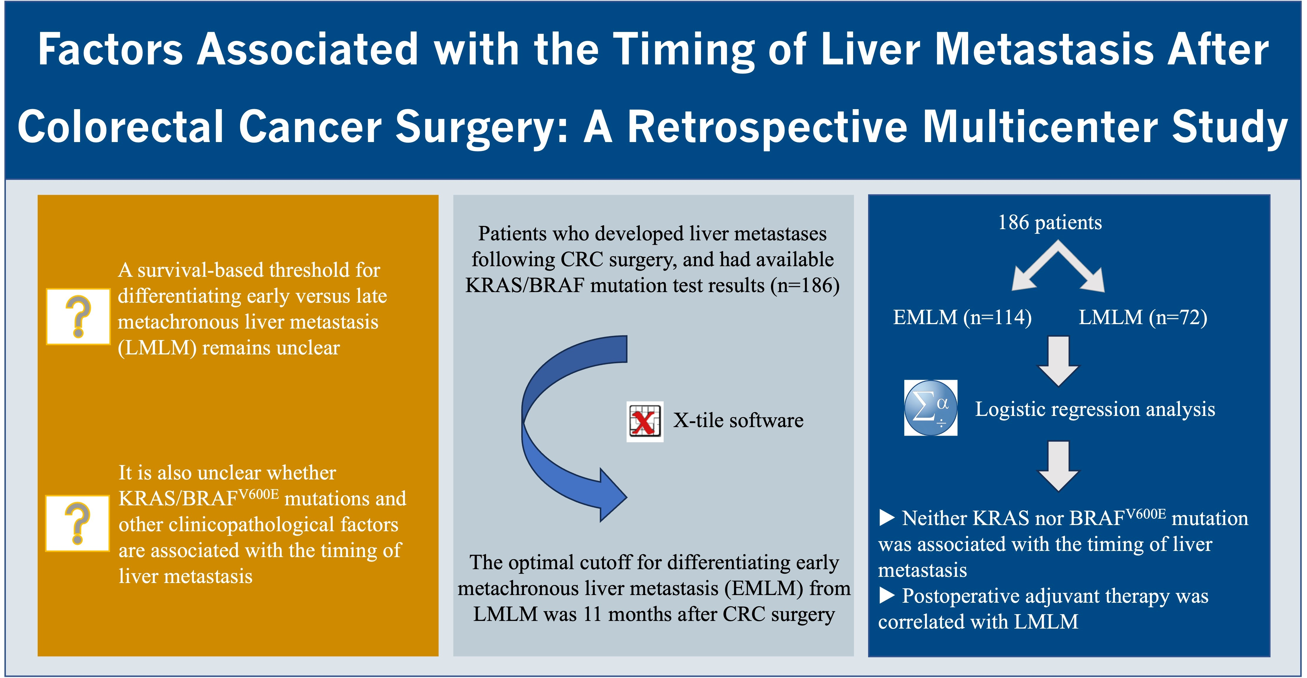
This multicenter retrospective study was conducted at two institutions: the National Cancer Center/Cancer Hospital, Chinese Academy of Medical Sciences (Beijing, China), and the First Affiliated Hospital of China Medical University (Shenyang, China). The study cohort consisted of CRC patients who developed liver metastasis after curative surgery between 2019 and 2023, and for whom complete KRAS/BRAFV600E mutation testing results were available. All enrolled patients met the following inclusion criteria:
(1)
Pathologically confirmed diagnosis of CRC;
(2)
Underwent radical CRC surgery at either participating institution;
(3)
Postoperative liver metastasis confirmed by pathological examination or imaging studies.
The exclusion criteria were:
(1)
Primary CRC was pathologically confirmed as non-adenocarcinoma, such as neuroendocrine carcinoma;
(2)
Presence of distant metastasis prior to surgery;
(3)
Non-radical surgery;
(4)
Indeterminate liver metastasis status;
(5)
Development of extrahepatic distant metastasis after CRC surgery;
(6)
Incomplete clinicopathological data.

2.2. Sample Collection and Molecular Profiling

This study utilized CRC tumor samples obtained from surgical specimens. All samples were fixed with formalin and embedded in paraffin by experienced pathologists at two medical centers. All CRC tumors were tested for deficient mismatch repair (dMMR) via immunohistochemistry or microsatellite instability-high (MSI-H) via polymerase chain reaction (PCR). RAS/BRAF mutation profiling was performed using PCR or next-generation sequencing (NGS) assays. Given the considerable time span and the medical disparities between the two centers, the detection platforms and the panel of mutations assessed were not uniform. Therefore, the analysis was restricted to the clinically prevalent KRAS mutation and the BRAFV600E variant. The molecular-testing details are provided in Table S1.

2.3. Definitions

Anatomically, radical surgery for colon cancer is defined as complete colectomy with systematic regional lymphadenectomy [17]. For rectal cancer, it refers to either total mesorectal excision or extended mesocolic excision [18]. MLM was defined as liver metastasis detected after CRC surgery [5]. All cases were staged per the 8th edition of the American Joint Committee on Cancer (AJCC) Cancer Staging Manual TNM classification system. OS was defined as time interval from surgical resection to date of mortality or last clinical follow-up. Metastasis-free survival (MFS) was defined as time interval from surgical resection to the time of liver metastasis, death, or the last follow-up. The last follow-up date was 1 January 2025.

2.4. Statistical Analyses

X-tile software (Robert L. Camp, M.D., Ph.D., Yale University, version 3.6.1) was used to identify the optimal cutoff for differentiating EMLM from LMLM based on OS differences. Comprehensive statistical analyses were performed using SPSS 26.0 software (Armonk, NY, USA) and GraphPad Prism 8 (La Jolla, CA, USA).
Continuous variables with normal distribution were described as mean ± standard deviation (SD) and compared using the Student t-test. Continuous variables with a skewed distribution were described as median with interquartile range (IQR) and compared using the Mann–Whitney U test. Categorical variables with unordered categories were described as frequency with percentage and compared using the chi-square test or Fisher’s exact probability test. All statistical tests were two-sided, and a p-value lower than 0.05 was considered statistically significant. To prevent the omission of potentially significant risk factors, the significance level for univariable analysis was set at 0.1 [19], and for multivariable analysis it was set at 0.05. Variables with p < 0.1 in the univariable analysis were entered into the multivariable logistic regression model. However, based on a priori clinical and biological knowledge, the primary exposure variables—KRAS and BRAFV600E mutations—were forced into the multivariable model irrespective of their univariable significance. Collinearity among variables was assessed using variance inflation factors (VIFs), with VIF > 10 indicating significant collinearity.

3. Results

3.1. Patient Characteristics

A total of 186 MLM patients with complete KRAS/BRAFV600E mutational profiling were ultimately enrolled for further analysis (Figure 1). The clinicopathological baseline characteristics of the 186 included patients are shown in Table 1. The median age was 63 years (SD, ±10); 10 patients (5.4%) had a family history of CRC; 87 patients (46.8%) had concomitant diseases (liver cirrhosis, diabetes, cardiovascular and cerebrovascular diseases, hypertension, and chronic obstructive pulmonary disease); primary tumors were located in the right colon in 35 patients (18.82%), left colon in 49 patients (26.34%), and rectum in 102 patients (54.84%); 95 patients (51.1%) received neoadjuvant therapy prior to surgery; 112 patients (60.2%) had lymphovascular tumor emboli; 120 patients (64.5%) exhibited perineural invasion; regional lymph node metastasis was present in 139 patients (74.7%); dMMR/MSI-H was observed in 5 patients (2.7%); KRAS mutation was detected in 81 patients (43.5%); the BRAFV600E mutation was identified in 6 patients (3.2%); concurrent wild-type status of RAS and BRAF was present in 99 patients (53.2%); postoperative adjuvant therapy was administered to 140 patients (75.3%); preoperative carcinoembryonic antigen (CEA) levels had a median value of 4.57 (IQR, 2.52–16.41) ng/mL; and preoperative carbohydrate antigen 19-9 (CA19-9) levels had a median value of 15.94 (IQR, 8.44–27.31) U/mL. Median follow-up was 37 (IQR, 21–52) months. During follow-up, 66 deaths (35.5%) were recorded.

3.2. The Optimal Cutoff for Differentiating EMLM from LMLM

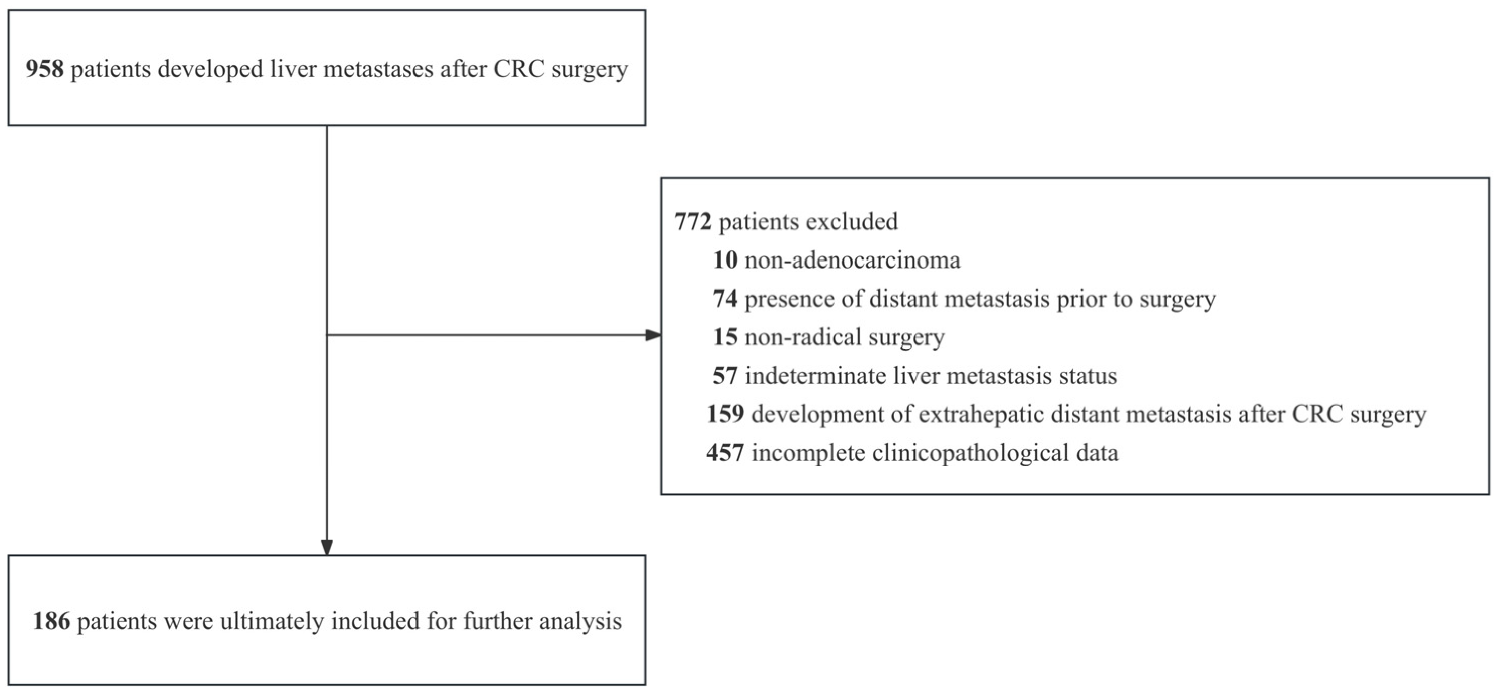
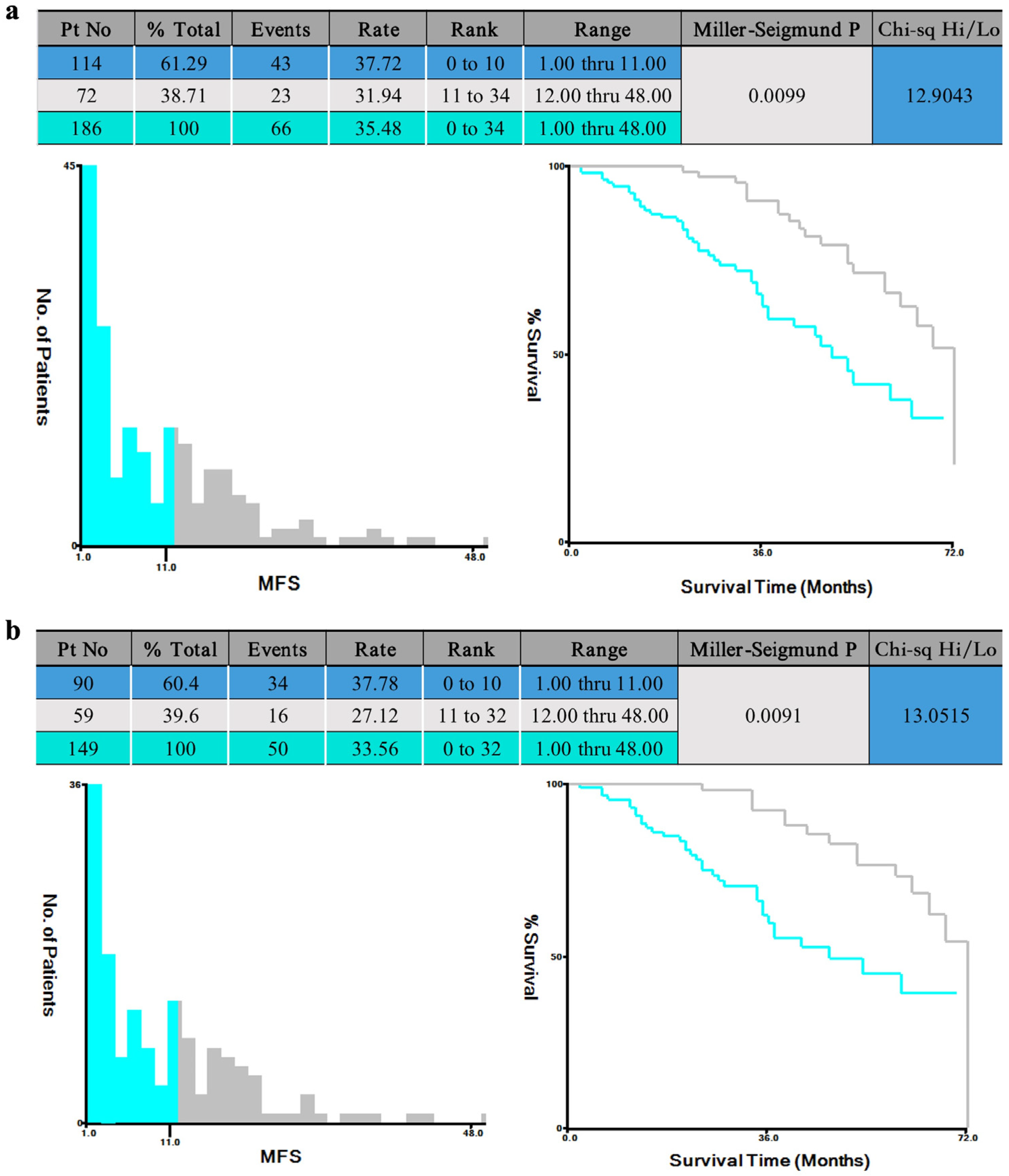
When OS was used as the dependent variable and MFS as the independent variable in X-tile software, the optimal cutoff calculated by the software was 11 months (Figure 2a). This indicates that when defining 11 months post-surgery as the cutoff, the OS difference between EMLM and LMLM is maximized, with a p-value of less than 0.01. As no external independent validation cohort was available, 80% (149 of 186) of the total patient population were randomly allocated without replacement to form the internal sensitivity analysis subset. X-tile software again yielded an optimal cutoff of 11 months (p < 0.01) (Figure 2b). Therefore, the optimal temporal threshold for distinguishing EMLM from LMLM was identified as 11 months post-CRC surgery in this study.

3.3. Comparative Analysis of EMLM and LMLM

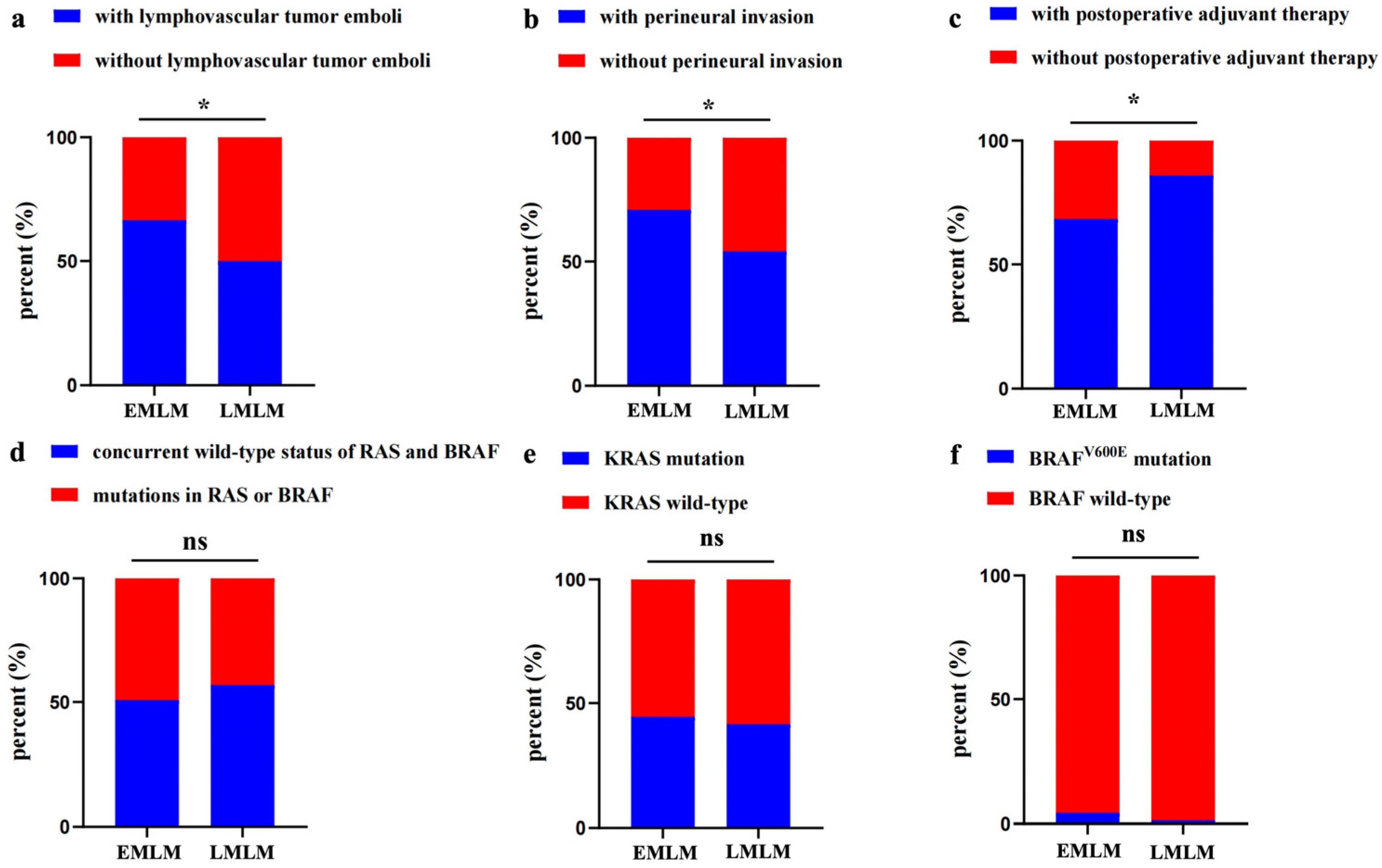
Using the 11-month cutoff, the 186 patients were divided into the EMLM group (n = 114) and the LMLM group (n = 72). Comparative analysis between the two groups revealed statistically significant differences in lymphovascular tumor emboli (p = 0.024), perineural invasion (p = 0.019), and postoperative adjuvant therapy (p = 0.006) (Figure 3a–c). The difference in the concurrent wild-type status of RAS and BRAF, KRAS mutation, and the BRAFV600E mutation between the two groups was not statistically significant (p > 0.05) (Figure 3d–f). A comprehensive comparison of clinicopathological data between the EMLM and LMLM groups is provided in Table 2.

3.4. Factors Associated with the Time to Onset of Liver Metastasis

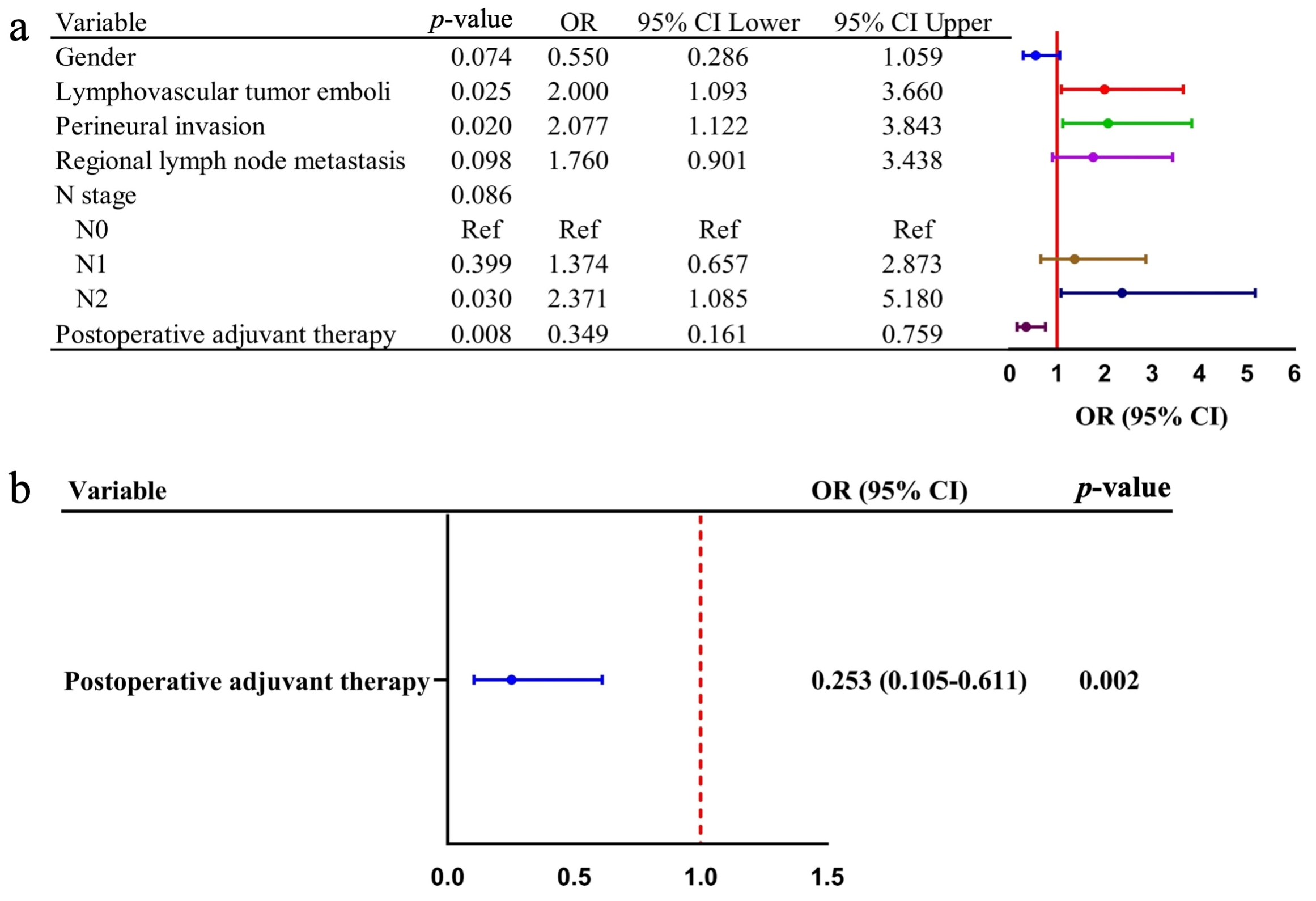
Univariable logistic regression analysis was conducted, and the results showed that the variables with statistically significant differences (p < 0.1) included gender (OR = 0.550, 95% CI: 0.286–1.059, p = 0.074), lymphovascular tumor emboli (OR = 2.000, 95% CI: 1.093–3.660, p = 0.025), perineural invasion (OR = 2.077, 95% CI: 1.122–3.843, p = 0.020), regional lymph node metastasis (OR = 1.760, 95% CI: 0.901–3.438, p = 0.098), N stage (p = 0.086), and postoperative adjuvant therapy (OR = 0.349, 95% CI: 0.161–0.759, p = 0.008) (Figure 4a). To evaluate the association between KRAS/BRAFV600E mutations and the timing of liver metastasis, we additionally performed a clinically prespecified forced-entry multivariable model in which the two mutations were retained irrespective of their univariable significance. Other variables with p < 0.1 in the univariable analysis were also entered into the multivariable logistic regression model. Collinearity assessment using VIF showed no significant collinearity (all VIF < 2). In the final multivariable analysis, neither KRAS mutation (OR, 1.185; 95% CI: 0.641–2.190; p = 0.587) nor BRAFV600E mutation (OR, 2.836; 95% CI: 0.302–26.642; p = 0.363) was independently associated with the timing of liver metastasis. However, postoperative adjuvant therapy (OR = 0.253, 95% CI: 0.105–0.611, p = 0.002) demonstrated a statistical association with the likelihood of LMLM (Figure 4b). The results of univariable and multivariable analyses are detailed in Table 3.

4. Discussion

In this study, 11 months post-CRC surgery was identified as the optimal cutoff for differentiating EMLM versus LMLM. No statistically significant association was observed between KRAS/BRAFV600E mutations and the timing of liver metastasis, whereas postoperative adjuvant therapy was statistically associated with the likelihood of LMLM in this cohort. To the best of our knowledge, this is the first study to define a survival-derived optimal temporal cutoff for distinguishing EMLM from LMLM, and to systematically evaluate whether KRAS/BRAFV600E mutations alongside other potential factors are associated with the time to onset of liver metastasis.
The 11-month threshold closely aligns with the 12-month threshold established by the Multisocietal European Consensus on CRLM [22]. It should be noted, however, that the consensus defines the 12-month cutoff based on the date of primary tumor diagnosis rather than the surgical date. Since most patients who develop MLM present with advanced-stage CRC and may undergo varying durations of neoadjuvant therapy—often up to three months—between diagnosis and surgery [23], a one-month discrepancy between these two definitions is entirely expected and can be regarded as equivalent. However, because our cutoff is exploratory and has not undergone external validation, the interpretation and generalization of this cutoff value should be cautious.
In our study, KRAS mutations were detected in 81 patients (43.5%) and BRAFV600E mutations in 6 patients (3.2%), with prevalence rates generally consistent with those reported in previous studies [15,24,25]. Previous studies have revealed that the KRAS mutation is associated with a poor prognosis in CRC patients, particularly in the metastatic setting [13,15,26]. Subsequent research further suggested that the KRAS mutation serves as an independent prognostic factor in CRLM patients [24]. Some studies have even confirmed that the KRAS mutation is an important risk factor affecting the development of liver metastasis after CRC surgery [27,28]. In addition, the BRAF mutation has also been identified in prior work as an independent risk factor for unfavorable outcomes [16,29,30]. A pooled analysis of multiple clinical trials indicated that patients harboring the BRAFV600E mutation exhibit shorter OS and progression-free survival (PFS) compared to those with wild-type BRAF [31]. Some research further revealed that the BRAFV600E mutation emerged as a powerful, independent risk factor for the occurrence of MLM [27,32]. Importantly, our study adds to this understanding by demonstrating that KRAS/BRAFV600E mutations are not significantly associated with the timing of liver metastasis, thereby substantially addressing a gap in this area of research. Nevertheless, the association between the BRAFV600E mutation and the timing of liver metastasis must be interpreted with caution, given the small number of BRAF-mutated cases (n = 6) and the wide confidence interval (OR, 2.836; 95% CI: 0.302–26.642).
Because this study included only patients who had already developed liver metastasis after CRC surgery, our findings pertain to factors associated with the timing of metastasis rather than to risk factors for developing liver metastasis. A clear appreciation of this distinction is essential for the appropriate interpretation of the results. Given that most patients with MLM present with advanced CRC, postoperative adjuvant therapy—including chemotherapy, radiotherapy, immunotherapy, and targeted therapy, used alone or in combination—is routinely recommended in clinical practice [23,33]. With the ongoing evolution of drug regimens, technological advancements, and refinements in multidisciplinary treatment (MDT) concepts, it is reasonable to expect that postoperative adjuvant therapy would play a significant role in delaying or preventing the development of liver metastasis after CRC surgery [23,34]. Paradoxically, however, our study found a statistically significant association between postoperative adjuvant therapy and the likelihood of LMLM, a finding that appears contradictory to this expectation. In fact, this finding precisely reveals the multifaceted roles that adjuvant therapy may exert in the postoperative setting.
First, the primary role of postoperative adjuvant therapy is to destroy the microscopic metastases that remain in the body after surgery. For microscopic foci that are sensitive to postoperative adjuvant therapy, complete eradication can be achieved, thereby reducing the overall incidence of postoperative liver metastasis. For microscopic foci that are partially sensitive, although complete elimination is not possible, their proliferation can be significantly inhibited [35]. Consequently, postoperative adjuvant therapy induces a rightward shift in the temporal distribution curve, such that patients who still develop liver metastasis after postoperative adjuvant treatment are statistically predisposed to late-onset metastasis [36].
Second, owing to inherent tumor heterogeneity, not all CRC cells are equally responsive to postoperative adjuvant therapy [37]. While postoperative adjuvant therapy effectively eliminates the majority of actively proliferating CRC cells—the principal contributors to early metastasis—a small subset of drug-resistant or dormant (G0-phase) tumor stem cells may survive therapeutic assault [38]. These cells resemble “dormant seeds,” residing quiescently in the liver microenvironment [39]. Unaffected by postoperative adjuvant therapy, they may be reactivated years after surgery, triggered by declining immune function or local microenvironmental changes (e.g., inflammation), ultimately giving rise to late liver metastasis [40]. Thus, postoperative adjuvant therapy inadvertently “selects” for cells with inherent latent and drug-tolerant characteristics, and the biological attributes of these cells predispose them to cause metastasis at a later rather than an earlier time point.
Third, interference from non-tumor factors should also be considered. Long-term or intensive postoperative adjuvant therapy may cause sinus damage or liver fibrosis. Although tumors are controlled in the short term, the damaged liver microenvironment may produce inflammatory factors during prolonged repair, thereby providing a fertile ground for residual, dormant tumor cells to be reactivated years later [41]. Furthermore, postoperative adjuvant therapy may reshape the immune system, but over time, immune depletion or immunosenescence may become exacerbated, such that the immune system is no longer capable of suppressing residual lesions in the late stage [42].
However, the definition of postoperative adjuvant therapy in this study was relatively broad, and therefore this finding should also be interpreted with caution. Based on the understanding of the dual dimensions of KRAS/BRAFV600E mutations and postoperative adjuvant therapy, future strategies for the prevention and management of MLM are expected to evolve toward deeper precision and dynamic monitoring. At the basic research level, it is necessary to deeply analyze how driver genes synergistically shape the “metastatic potential” and “treatment sensitivity” of tumors, and to explore the key role of the liver immune microenvironment in drug resistance and tumor dormancy [43,44]. At the clinical translational level, circulating tumor DNA (ctDNA)-based monitoring of microscopic residual disease (MRD) represents a promising breakthrough [45]. Such monitoring could capture the amplification of drug-resistant clones after postoperative adjuvant therapy screening in real time, enabling dynamic risk stratification and guiding individualized adjuvant therapy up-staging or down-staging [46]. The ultimate goal is to realize precise decision-making on “when and how to treat”, thereby shifting the paradigm of MLM management from “treating the disease” to “preventing the disease”.
In addition, regardless of tumor type, multidisciplinary integrated-treatment thinking remains a cornerstone in the management of oncological diseases. Recently, the role of Toll-like receptors (TLRs) in liver regeneration after partial hepatectomy has attracted considerable attention, as TLR signaling regulates inflammatory and reparative signals that influence liver recovery [47]. In this context, TLR-mediated modulation of the hepatic microenvironment may indirectly affect metastatic outgrowth and immune responses, an area that merits dedicated exploration in MLM models [48]. Meanwhile, artificial intelligence (AI) and image processing have shown great potential in the diagnosis, treatment, and prognosis of CRLM, particularly in radiomics and deep learning-assisted decision-making, which similarly holds promise for improving the detection of small metastases, predicting treatment responses, and optimizing individualized surveillance strategies [49,50]. Nevertheless, current studies remain largely retrospective, lack prospective validation, and face challenges in model interpretability [51]. Future efforts should focus on integrating multicenter, multimodal data to facilitate the clinical translation of AI in CRLM management.
Several limitations should be considered in this study. First, because our cutoff was derived from the same cohort using an outcome-driven approach, there is a risk of overfitting. External validation in independent cohorts is needed to confirm the generalizability of this threshold. Second, the sample size of the BRAFV600E mutation subgroup was limited (n = 6), attributable both to the low natural prevalence of this mutation and to our deliberate exclusion of patients with extrahepatic metastasis to ensure the clarity of survival endpoint assessment. Moreover, restricted accessibility to genetic testing further constrained patient enrollment. The resulting lack of statistical power reduces the robustness of subgroup estimates and raises the possibility of selection bias. Therefore, while our preliminary findings are informative, they warrant cautious interpretation and require validation in independent, larger patient cohorts. Third, given the wide time span, heterogeneous medical practices across centers, and the complexity and volume of follow-up data, postoperative adjuvant therapy was analyzed as a composite categorical variable in this study. This analytical approach inherently introduces substantial risks of treatment-selection bias, confounding by indication, and immortal-time bias. Furthermore, each treatment modality has distinct clinical indications and mechanisms of action, which may introduce bias and compromise the scientific validity and reliability of our conclusions. Therefore, future investigations should prioritize either single-center cohorts with standardized treatment protocols or prospective trials specifically designed to dissect the independent contribution of each treatment to the timing of metastasis. Finally, owing to time constraints, a more comprehensive investigation was not feasible in the current study; however, further in-depth investigations utilizing single-cell sequencing and spatial transcriptomics would be both valuable and promising in this context.

5. Conclusions

In conclusion, this study identifies 11 months post-surgery as an optimal survival-based cutoff for distinguishing EMLM from LMLM, provides a quantifiable metric for refining postoperative risk assessment, and holds promise for personalizing postoperative surveillance protocols—enabling intensified imaging follow-up during the initial high-risk window for patients with early-onset disease. Multivariable analysis demonstrated that KRAS/BRAFV600E mutations were not associated with the timing of MLM, implying that the chronobiology of metastasis may be more heavily influenced by therapeutic interventions and host-related factors than by genomic characteristics of the primary tumor alone. Conversely, postoperative adjuvant therapy was statistically associated with the likelihood of LMLM, underscoring the pivotal role of timely and complete postoperative adjuvant therapy not only in controlling disease progression but also in reshaping the temporal dynamics of hematogenous hepatic dissemination.
Nevertheless, given the retrospective nature of the cohort and the sample size limitations, our findings should be interpreted as exploratory. Prospective, multicenter, and better-designed validation studies with larger sample sizes are needed to confirm the clinical utility of this cutoff and to further elucidate the biological mechanisms that determine the temporal heterogeneity of metastatic occurrence.

Supplementary Materials

The following supporting information can be downloaded at: https://www.mdpi.com/article/10.3390/cancers18152445/s1, Table S1: The molecular-testing details.

Author Contributions

Conceptualization, X.L. and Q.L.; methodology, X.L. and S.M.; software, X.L., C.Z., F.W., S.M., and F.H.; validation, Q.L.; formal analysis, T.X., F.H., and F.Z.; investigation, W.Q. and Z.L.; data curation, X.L., C.Z., Z.L., and F.Z.; writing—original draft preparation, C.Z.; writing—review and editing, F.W. and T.X.; visualization, W.Q.; supervision, W.Q. and Q.L.; project administration, Q.L. All authors have read and agreed to the published version of the manuscript.

Funding

This work was supported by the Chinese Academy of Medical Sciences Innovation Fund for Medical Sciences (CIFMS; No.2022-I2M-C&T-B-057), Beijing Municipal Natural Science Foundation (7252121), Beijing Municipal Science and Technology Program (Z251100004625089), and National High Level Hospital Clinical Research Funding, Cooperation Fund of Cancer Hospital of Chinese Academy of Medical Sciences (CFA202502001).

Institutional Review Board Statement

The study was conducted in accordance with the Declaration of Helsinki, and approved by the Ethics Committee of National Cancer Center/Cancer Hospital, Chinese Academy of Medical Sciences and Peking Union Medical College (NCC2025C-902; 8 August 2025).

Informed Consent Statement

Informed consent was obtained from all subjects involved in the study. Written informed consent has been obtained from the patients to publish this paper.

Data Availability Statement

Access to the datasets utilized and/or analyzed during the current investigation can be obtained by contacting the corresponding authors, subject to reasonable request.

Acknowledgments

We thank all the colleagues who assisted in laboratory analyses and clinical information collection. The authors have reviewed and edited the output and take full responsibility for the content of this publication.

Conflicts of Interest

The authors declare no conflicts of interest.

Abbreviations

The following abbreviations are used in this manuscript:
AJCCAmerican Joint Committee on Cancer
AIArtificial intelligence
CA19-9Carbohydrate antigen 19-9
CEACarcinoembryonic antigen
CIConfidence interval
CRCColorectal cancer
CRLMColorectal liver metastasis
ctDNACirculating tumor DNA
dMMRDeficient mismatch repair
EGFREpidermal growth factor receptor
EMLMEarly metachronous liver metastasis
IQRInterquartile range
LMLMLate metachronous liver metastasis
MDTMultidisciplinary treatment
MFSMetastasis-free survival
MLMMetachronous liver metastasis
MRDMicroscopic residual disease
MSI-HMicrosatellite instability-high
NGSNext-generation sequencing
OROdds ratio
OSOverall survival
PCRPolymerase chain reaction
PFSProgression-free survival
pMMRProficient mismatch repair
SDStandard deviation
SLMSynchronous liver metastasis
TLRToll-like receptor
VEGFVascular endothelial growth factor
VIFVariance inflation factor

References

  1. GBD 2023 Disease and Injury and Risk Factor Collaborators. Burden of 375 diseases and injuries, risk-attributable burden of 88 risk factors, and healthy life expectancy in 204 countries and territories, including 660 subnational locations, 1990–2023: A systematic analysis for the Global Burden of Disease Study 2023. Lancet 2025, 406, 1873–1922. [Google Scholar] [CrossRef] [PubMed]
  2. Diao, X.; Guo, C.; Jin, Y.; Li, B.; Gao, X.; Du, X.; Chen, Z.; Jo, M.; Zeng, Y.; Ding, C.; et al. Cancer situation in China: An analysis based on the global epidemiological data released in 2024. Cancer Commun. 2025, 45, 178–197. [Google Scholar] [CrossRef] [PubMed]
  3. Sung, H.; Ferlay, J.; Siegel, R.L.; Laversanne, M.; Soerjomataram, I.; Jemal, A.; Bray, F. Global Cancer Statistics 2020: GLOBOCAN Estimates of Incidence and Mortality Worldwide for 36 Cancers in 185 Countries. CA Cancer J. Clin. 2021, 71, 209–249. [Google Scholar] [CrossRef] [PubMed]
  4. Xu, X.; Gan, J.; Gao, Z.; Li, R.; Huang, D.; Lin, L.; Luo, Y.; Yang, Q.; Xu, J.; Li, Y.; et al. 3D genome landscape of primary and metastatic colorectal carcinoma reveals the regulatory mechanism of tumorigenic and metastatic gene expression. Commun. Biol. 2025, 8, 365. [Google Scholar] [CrossRef] [PubMed]
  5. Reboux, N.; Jooste, V.; Goungounga, J.; Robaszkiewicz, M.; Nousbaum, J.B.; Bouvier, A.M. Incidence and Survival in Synchronous and Metachronous Liver Metastases From Colorectal Cancer. JAMA Netw. Open 2022, 5, e2236666. [Google Scholar] [CrossRef] [PubMed]
  6. Wisneski, A.D.; Jin, C.; Huang, C.Y.; Warren, R.; Hirose, K.; Nakakura, E.K.; Corvera, C.U. Synchronous Versus Metachronous Colorectal Liver Metastasis Yields Similar Survival in Modern Era. J. Surg. Res. 2020, 256, 476–485. [Google Scholar] [CrossRef] [PubMed]
  7. Kemna, R.; Schulz, H.; Ali, M.; Verpalen, I.; Buffart, T.; Swijnenburg, R.J.; Meijerink, M.; Kazemier, G. Prognostic Value of Categorizing Colorectal Cancer Liver Metastases Based on Detection Time Interval: Survival Outcomes from the Amsterdam Colorectal Liver Met Registry. HPB 2025, 27, S397. [Google Scholar] [CrossRef]
  8. Oliveira, C.V.C.; Santana, R.C.; Coimbra, F.J.F.; Kow, A.; Pawlik, T.M.; Adam, R.; Soubrane, O.; Herman, P.; Cotta-Pereira, R.L. Metachronous Colorectal Liver Metastasis. Arq. Bras. Cir. Dig. 2025, 38, e1874. [Google Scholar] [CrossRef] [PubMed]
  9. Johannsen, I.R.; Boysen, A.K.; Nielsen, M.F.; Mortensen, F.V.; Kirkegård, J. Liver Metastases and Survival Among Patients with Colorectal Cancer. JAMA Netw. Open 2025, 8, e2550467. [Google Scholar] [CrossRef] [PubMed]
  10. Hu, Z.; Ding, J.; Ma, Z.; Sun, R.; Seoane, J.A.; Scott Shaffer, J.; Suarez, C.J.; Berghoff, A.S.; Cremolini, C.; Falcone, A.; et al. Quantitative evidence for early metastatic seeding in colorectal cancer. Nat. Genet. 2019, 51, 1113–1122. [Google Scholar] [CrossRef] [PubMed]
  11. Lu, Y.T.; Huang, X.X.; Chen, W.C.; Xiao, Q.Y.; Duan, J.L.; Li, Z.J.; Pat Fong, W.; Wang, F.H.; Li, Y.H.; Wang, D.S. A clinical-molecular nomogram for predicting early recurrence following resection of initially unresectable colorectal liver metastases. Ther. Adv. Med. Oncol. 2026, 18, 17588359251411660. [Google Scholar] [CrossRef] [PubMed]
  12. Lipsyc, M.; Yaeger, R. Impact of somatic mutations on patterns of metastasis in colorectal cancer. J. Gastrointest. Oncol. 2015, 6, 645–649. [Google Scholar] [CrossRef] [PubMed]
  13. Dienstmann, R.; Connor, K.; Byrne, A.T. Precision Therapy in RAS Mutant Colorectal Cancer. Gastroenterology 2020, 158, 806–811. [Google Scholar] [CrossRef] [PubMed]
  14. Tabernero, J.; Ros, J.; Élez, E. The Evolving Treatment Landscape in BRAF-V600E-Mutated Metastatic Colorectal Cancer. Am. Soc. Clin. Oncol. Educ. Book. 2022, 42, 1–10. [Google Scholar] [CrossRef] [PubMed]
  15. Tang, W.; Liu, Y.; Ji, M.; Liu, T.; Chen, Y.; Zhuang, A.; Mao, Y.; Chang, W.; Wei, Y.; Ren, L.; et al. Association of RAS/BRAF Status and Prognosis of Metastatic Colorectal Cancer: Analysis of 1002 Consecutive Cases. Ann. Surg. Oncol. 2022, 29, 3593–3603. [Google Scholar] [CrossRef] [PubMed]
  16. Chu, J.E.; Johnson, B.; Kugathasan, L.; Morris, V.K.; Raghav, K.; Swanson, L.; Lim, H.J.; Renouf, D.J.; Gill, S.; Wolber, R.; et al. Population-based Screening for BRAF (V600E) in Metastatic Colorectal Cancer Reveals Increased Prevalence and Poor Prognosis. Clin. Cancer Res. 2020, 26, 4599–4605. [Google Scholar] [CrossRef] [PubMed]
  17. Benson, A.B.; Venook, A.P.; Adam, M.; Chang, G.; Chen, Y.J.; Ciombor, K.K.; Cohen, S.A.; Cooper, H.S.; Deming, D.; Garrido-Laguna, I.; et al. Colon Cancer, Version 3.2024, NCCN Clinical Practice Guidelines in Oncology. J. Natl. Compr. Cancer Netw. 2024, 22, e240029. [Google Scholar] [CrossRef] [PubMed]
  18. Benson, A.B.; Venook, A.P.; Adam, M.; Chang, G.; Chen, Y.J.; Ciombor, K.K.; Cohen, S.A.; Cooper, H.S.; Deming, D.; Garrido-Laguna, I.; et al. NCCN Guidelines® Insights: Rectal Cancer, Version 3.2024. J. Natl. Compr. Cancer Netw. 2024, 22, 366–375. [Google Scholar] [CrossRef] [PubMed]
  19. Tenny, S.; Abdelgawad, I. Statistical Significance. In StatPearls; StatPearls Publishing LLC.: Treasure Island, FL, USA, 2026. [Google Scholar]
  20. Firth, D. Bias reduction of maximum likelihood estimates. Biometrika 1993, 80, 27–38. [Google Scholar] [CrossRef]
  21. Heinze, G.; Schemper, M. A solution to the problem of separation in logistic regression. Stat. Med. 2002, 21, 2409–2419. [Google Scholar] [CrossRef] [PubMed]
  22. Siriwardena, A.K.; Serrablo, A.; Fretland, Å.A.; Wigmore, S.J.; Ramia-Angel, J.M.; Malik, H.Z.; Stättner, S.; Søreide, K.; Zmora, O.; Meijerink, M.; et al. Multisocietal European consensus on the terminology, diagnosis, and management of patients with synchronous colorectal cancer and liver metastases: An E-AHPBA consensus in partnership with ESSO, ESCP, ESGAR, and CIRSE. Br. J. Surg. 2023, 110, 1161–1170. [Google Scholar] [CrossRef] [PubMed]
  23. Guideline for diagnosis and comprehensive treatment of colorectal liver metastases (version 2025). Zhonghua Wei Chang Wai Ke Za Zhi 2025, 28, 815–831. [CrossRef] [PubMed]
  24. Sakai, N.; Furukawa, K.; Takayashiki, T.; Kuboki, S.; Takano, S.; Ohtsuka, M. Differential effects of KRAS mutational status on long-term survival according to the timing of colorectal liver metastases. BMC Cancer 2021, 21, 412. [Google Scholar] [CrossRef] [PubMed]
  25. Grothey, A.; Fakih, M.; Tabernero, J. Management of BRAF-mutant metastatic colorectal cancer: A review of treatment options and evidence-based guidelines. Ann. Oncol. 2021, 32, 959–967. [Google Scholar] [CrossRef] [PubMed]
  26. Zhu, G.; Pei, L.; Xia, H.; Tang, Q.; Bi, F. Role of oncogenic KRAS in the prognosis, diagnosis and treatment of colorectal cancer. Mol. Cancer 2021, 20, 143. [Google Scholar] [CrossRef] [PubMed]
  27. Wang, N.; Yuan, D.; Zhu, Y.; Ding, L. Development and validation of a multicenter nomogram predicting the risk of liver metastasis after curative resection of colorectal cancer. J. Peking Univ. (Health Sci.) 2026, 58, 290–300. [Google Scholar] [CrossRef] [PubMed]
  28. Liu, G.; Zhang, J.; Li, Q.; Xue, X.; Guo, X.; Han, J.; Wang, G.; Shi, F.; Wang, K.; Ding, Y.; et al. Methyltransferase MGMT upregulation drives metastasis by activating epithelial-mesenchymal transition in KRAS mutant colon cancer. Cell Death Dis. 2026, 17, 628. [Google Scholar] [CrossRef] [PubMed]
  29. Djanani, A.; Eller, S.; Öfner, D.; Troppmair, J.; Maglione, M. The Role of BRAF in Metastatic Colorectal Carcinoma-Past, Present, and Future. Int. J. Mol. Sci. 2020, 21, 9001. [Google Scholar] [CrossRef] [PubMed]
  30. Prasanna, T.; Wong, R.; Price, T.; Shapiro, J.; Tie, J.; Wong, H.L.; Nott, L.; Roder, D.; Lee, M.; Kosmider, S.; et al. Metastasectomy and BRAF mutation; an analysis of survival outcome in metastatic colorectal cancer. Curr. Probl. Cancer 2021, 45, 100637. [Google Scholar] [CrossRef] [PubMed]
  31. García-Alfonso, P.; Lièvre, A.; Loupakis, F.; Tadmouri, A.; Khan, S.; Barcena, L.; Stintzing, S. Systematic review of randomised clinical trials and observational studies for patients with RAS wild-type or BRAF(V600E)-mutant metastatic and/or unresectable colorectal cancer. Crit. Rev. Oncol. Hematol. 2022, 173, 103646. [Google Scholar] [CrossRef] [PubMed]
  32. Akabane, M.; Baldo, A.; Kawashima, J.; Aucejo, F.; Popescu, I.; Kitago, M.; Poultsides, G.A.; Ruzzenente, A.; Endo, I.; Pawlik, T.M. BRAF Mutation Drives Early Multifocal Intrahepatic Recurrence after Curative Resection of Liver-Limited Colorectal Metastases. J. Am. Coll. Surg. 2026, 242, 751–758. [Google Scholar] [CrossRef] [PubMed]
  33. Dantas, A.A.G.; de Oliveira, N.P.D.; Costa, G.A.B.; Martins, L.F.L.; Dos Santos, J.E.M.; Migowski, A.; de Camargo Cancela, M.; de Souza, D.L.B. Multilevel analysis of social determinants of advanced stage colorectal cancer diagnosis. Sci. Rep. 2024, 14, 9667. [Google Scholar] [CrossRef] [PubMed]
  34. Liu, X.; Liu, Q.; Wu, X.; Yu, W.; Bao, X. Efficacy of various adjuvant chemotherapy methods in preventing liver metastasis from potentially curative colorectal cancer: A systematic review network meta-analysis of randomized clinical trials. Cancer Med. 2023, 12, 2238–2247. [Google Scholar] [CrossRef] [PubMed]
  35. Yang, B.; Wei, X.; Wang, L.; Yuan, W.; Ma, Y.; Ma, J. Orosomucoid 2 upregulation mediates liver injury-induced colorectal cancer liver metastasis by promoting EMT and cell migration. Cancer Sci. 2025, 116, 44–55. [Google Scholar] [CrossRef] [PubMed]
  36. Kokudo, T.; Saiura, A.; Takayama, T.; Miyagawa, S.; Yamamoto, J.; Ijichi, M.; Teruya, M.; Yoshimi, F.; Kawasaki, S.; Koyama, H.; et al. Adjuvant chemotherapy can prolong recurrence-free survival but did not influence the type of recurrence or subsequent treatment in patients with colorectal liver metastases. Surgery 2021, 170, 1151–1154. [Google Scholar] [CrossRef] [PubMed]
  37. Selvaggi, F.; Catalano, T.; Lattanzio, R.; Cotellese, R.; Aceto, G.M. Wingless/It/β-catenin signaling in liver metastasis from colorectal cancer: A focus on biological mechanisms and therapeutic opportunities. World J. Gastroenterol. 2023, 29, 2764–2783. [Google Scholar] [CrossRef] [PubMed]
  38. Zhou, Y.; Geng, H.; Xu, Y.; Hong, Y.; Mei, B.; Wu, H.; Jin, X.; Ye, M.; Wang, Y.; Shen, Z.; et al. From paradox to target: IFN-β hijacks MEK signaling to drive a cell death-evading dormant phenotype in colorectal cancer. J. Exp. Clin. Cancer Res. 2026. [Google Scholar] [CrossRef] [PubMed]
  39. Zhu, X.; Wang, F.; Wu, X.; Li, Z.; Wang, Z.; Ren, X.; Zhou, Y.; Song, F.; Liang, Y.; Zeng, Z.; et al. FBX8 promotes metastatic dormancy of colorectal cancer in liver. Cell Death Dis. 2020, 11, 622. [Google Scholar] [CrossRef] [PubMed]
  40. Touil, Y.; Igoudjil, W.; Corvaisier, M.; Dessein, A.F.; Vandomme, J.; Monté, D.; Stechly, L.; Skrypek, N.; Langlois, C.; Grard, G.; et al. Colon cancer cells escape 5FU chemotherapy-induced cell death by entering stemness and quiescence associated with the c-Yes/YAP axis. Clin. Cancer Res. 2014, 20, 837–846. [Google Scholar] [CrossRef] [PubMed]
  41. Hu, X.; Marietta, A.; Dai, W.X.; Li, Y.Q.; Ma, X.J.; Zhang, L.; Cai, S.J.; Peng, J.J. Prediction of hepatic metastasis and relapse in colorectal cancers based on concordance analyses with liver fibrosis scores. Clin. Transl. Med. 2020, 9, 13. [Google Scholar] [CrossRef] [PubMed]
  42. Ding, K.; Mao, A.; Wang, Y.; Ni, Z.; Shi, Y.; Liu, K.; Zhang, J.; Zheng, W.; Liu, L.; Liu, Q.; et al. MARCO+ macrophages drive immunosuppressive remodeling and metastasis in chemotherapy-associated steatohepatitis. J. Hepatol. 2026, 84, 859–873. [Google Scholar] [CrossRef] [PubMed]
  43. Schwantes, I.R.; Mayo, S.C. The genomics of liver metastases from colon and rectal cancer: An Invited Commentary (invitation from Lee Ocuin, MD and Kevin Behrns, MD) for the special issue of “Management of Advanced Primary and Secondary Liver Malignancies”. Surgery 2023, 174, 422–424. [Google Scholar] [CrossRef] [PubMed]
  44. Kuang, J.; Li, J.; Zhou, S.; Li, Y.; Lin, J.; Huang, W.; Yuan, X. Genomic and micro-environmental insights into drug resistance in colorectal cancer liver metastases. Discov. Oncol. 2025, 16, 241. [Google Scholar] [CrossRef] [PubMed]
  45. Kawashima, M.; Yamada, T.; Miyasaka, T.; Kanaka, S.; Kuriyama, S.; Uehara, K.; Matsuda, A.; Ohta, R.; Sonoda, H.; Taniai, N.; et al. Impact of Minimal Residual Disease on Early Recurrence of Liver Metastatic Colorectal Cancer. Cancer Sci. 2025, 116, 1366–1374. [Google Scholar] [CrossRef] [PubMed]
  46. Conticchio, M.; Uldry, E.; Hübner, M.; Digklia, A.; Fraga, M.; Sempoux, C.; Raisaro, J.L.; Fuks, D. Prognostic Factors in Colorectal Liver Metastases: An Exhaustive Review of the Literature and Future Prospectives. Cancers 2025, 17, 2539. [Google Scholar] [CrossRef] [PubMed]
  47. Dritsas, S.; Frountzas, M.; Schizas, D.; Mulita, F.; Katsimpoulas, M.; Gkolfakis, P.; Lidoriki, I.; Karavokyros, I.; Liolis, E.; Missiakos, E.; et al. The role of Toll-like receptors in liver regeneration after partial hepatectomy. Prz. Gastroenterol. 2025, 20, 294–304. [Google Scholar] [CrossRef] [PubMed]
  48. Seki, E.; Park, E.; Fujimoto, J. Toll-like receptor signaling in liver regeneration, fibrosis and carcinogenesis. Hepatol. Res. 2011, 41, 597–610. [Google Scholar] [CrossRef] [PubMed]
  49. Weyman, K.M.; Kothari, A.N. Artificial Intelligence for the Treatment and Management of Colorectal Liver Metastases. Clin. Colon Rectal Surg. 2026, 39, 235–241. [Google Scholar] [CrossRef] [PubMed]
  50. Chen, Y.; Zhang, Y.; Wei, Y.; Jiang, H.; Zhang, L.; Long, L.; Song, B.; Peng, T. Advances in magnetic resonance imaging for the evaluation of colorectal liver metastases in context of individualized precision medicine. Iliver 2025, 4, 100168. [Google Scholar] [CrossRef] [PubMed]
  51. Mian, A.; Young, R.; Lakha, A.S.; Leptidis, I.; Jones, R.P.; Gordon-Weeks, A. CT radiomics for survival risk stratification in resectable colorectal liver metastases: A multi-centre study. Sci. Rep. 2026, 16, 19580. [Google Scholar] [CrossRef] [PubMed]
Figure 1. Flowchart of included patients. Abbreviations: CRC, colorectal cancer.
Cancers 18 02445 g001
Figure 2. X-tile analysis determined 11 months as the optimal cutoff. (a) In the derivation cohort of 186 patients, X-tile analysis calculated an optimal MFS of 11 months. Based on this cutoff, patients were divided into EMLM and LMLM groups. The patient distribution and the survival curves for the two groups are presented (blue legend for EMLM; gray legend for LMLM). (b) In the validation cohort of 149 patients, the software again yielded an optimal cutoff of 11 months. After grouping, the patient distribution and the survival curves are also shown (blue legend for EMLM; gray legend for MLM). Abbreviations: MFS, metastasis-free survival; EMLM, early metachronous liver metastasis; LMLM, late metachronous liver metastasis.
Cancers 18 02445 g002
Figure 3. Comparisons between the two groups when patients were grouped according to the 11-month cutoff. (ac) Three clinicopathological variables showing statistically significant differences between the two groups. (df) Comparison of RAS/BRAF gene status between the two groups. (*: p < 0.05; ns: no significant). Abbreviations: EMLM, early metachronous liver metastasis; LMLM, late metachronous liver metastasis.
Cancers 18 02445 g003
Figure 4. Forest plot of variables with statistical significance. (a) The results of the univariable analysis indicated that six variables were statistically significant. (b) Further multivariable analysis revealed that one variable remained statistically significant. Abbreviations: OR, odds ratio; CI, confidence interval.
Cancers 18 02445 g004
Table 1. Clinicopathological baseline characteristics of MLM patients.
VariablesMLM (n = 186)
Age (years), mean (SD)63 (10)
Gender (female:male)61: 125
Family history, n (%)10 (5.4)
Concomitant diseases, n (%)87 (46.8)
CRC location, n (%)
 Right-sided colon35 (18.82)
 Left-sided colon49 (26.34)
 Rectum102 (54.84)
Preoperative neoadjuvant therapy, n (%)95 (51.1)
Lymphovascular tumor emboli, n (%)112 (60.2)
Perineural invasion, n (%)120 (64.5)
T stage, n (%)
 T12 (1.1)
 T214 (7.5)
 T388 (47.3)
 T482 (44.1)
Regional lymph node metastasis, n (%)139 (74.7)
N stage, n (%)
 N047 (25.3)
 N173 (39.2)
 N266 (35.5)
General types, n (%)
 Proliferative type72 (38.7)
 Ulcerative type70 (37.6)
 Infiltrative type44 (23.7)
Histological types, n (%)
 Adenocarcinoma178 (95.7)
 Mucinous adenocarcinoma8 (4.3)
Differentiation types, n (%)
 Well-differentiated3 (1.6)
 Moderately differentiated159 (85.5)
 Poorly differentiated24 (12.9)
dMMR/MSI-H, n (%)5 (2.7)
KRAS mutation, n (%)81 (43.5)
BRAFV600E mutation, n (%)6 (3.2)
Concurrent wild-type RAS and BRAF, n (%)99 (53.2)
Preoperative CEA (ng/mL), median (IQR)4.57 (2.52–16.41)
Preoperative CA19-9 (U/mL), median (IQR)15.94 (8.44–27.31)
Postoperative adjuvant therapy, n (%)140 (75.3)
Abbreviations: CRC, colorectal cancer; MLM, metachronous liver metastasis; SD, standard deviation; IQR, interquartile range; dMMR, deficient mismatch repair; MSI-H, microsatellite instability-high; CEA, carcinoembryonic antigen; CA19-9, carbohydrate antigen 19-9.
Table 2. Comparison of clinicopathological data between the two groups.
VariablesEMLM (n = 114)LMLM (n = 72)p-Value
Age (years), mean (SD)62.49 (9.574)63.11 (10.544)0.680
Gender (female:male)71: 4354: 180.072
Family history, n (%)8 (7.0)2 (2.8)0.360
Concomitant diseases, n (%)56 (49.1)31 (43.1)0.419
CRC location, n (%)
 Right-sided colon20 (17.5)15 (20.8)0.124
 Left-sided colon25 (21.9)24 (33.3)
 Rectum69 (60.5)33 (45.8)
Preoperative neoadjuvant therapy, n (%)62 (54.4)33 (45.8)0.256
Lymphovascular tumor emboli, n (%)76 (66.7)36 (50.0)0.024
Perineural invasion, n (%)81 (71.1)39 (54.2)0.019
T stage, n (%)
 T12 (1.8)0 (0.0)0.200
 T29 (7.0)5 (6.9)
 T348 (42.1)40 (55.6)
 T455 (48.2)27 (37.5)
Regional lymph node metastasis, n (%)90 (78.9)49 (68.1)0.096
N stage, n (%)
 N024 (21.1)23 (31.9)0.083
 N143 (37.7)30 (41.7)
 N247 (41.2)19 (26.4)
General types, n (%)
 Proliferative type41 (36.0)31 (43.1)0.483
 Ulcerative type43 (37.7)27 (37.5)
 Infiltrative type30 (26.3)14 (19.4)
Histological types, n (%)
 Adenocarcinoma109 (95.6)69 (95.8)0.943
 Mucinous adenocarcinoma5 (4.4)3 (4.2)
Differentiation types, n (%)
 Well-differentiated0 (0.0)3 (4.2)0.140
 Moderately differentiated96 (84.2)63 (87.5)
 Poorly differentiated18 (15.8)6 (8.3)
dMMR/MSI-H, n (%)3 (2.6)2 (2.8)0.952
KRAS mutation, n (%)51 (44.7)30 (41.7)0.681
BRAFV600E mutation, n (%)5 (4.4)1 (1.4)0.483
Concurrent wild-type RAS and BRAF, n (%)58 (50.9)41 (56.9)0.419
Preoperative CEA (ng/mL), median (IQR)4.61 (2.38–19.48)4.42 (2.58–12.25)0.465
Preoperative CA19-9 (U/mL), median (IQR)16.11 (9.16–31.89)15.94 (7.75–23.35)0.235
Postoperative adjuvant therapy, n (%)78 (68.4)62 (86.1)0.006
Abbreviations: EMLM, early metachronous liver metastasis; LMLM, late metachronous liver metastasis; SD, standard deviation; CRC, colorectal cancer; IQR, interquartile range; dMMR, deficient mismatch repair; MSI-H, microsatellite instability-high; CEA, carcinoembryonic antigen; CA19-9, carbohydrate antigen 19-9. Bold p-values indicate statistical significance (p < 0.05).
Table 3. Logistic regression analysis of factors associated with the timing of liver metastasis.
VariablesUnivariableMultivariable
OR (95% CI)p-ValueOR (95% CI)p-Value
Age (years)0.994 (0.964–1.024)0.678
Gender (female:male)0.550 (0.286–1.059)0.074
Family history (yes:no)2.624 (0.545–12.807)0.228
Concomitant diseases (yes:no)1.277 (0.705–2.312)0.420
CRC location 0.127
 Right-sided colonRef
 Left-sided colon0.781 (0.326–1.870)0.579
 Rectum1.568 (0.713–3.447)0.263
Preoperative neoadjuvant therapy
(yes:no)
1.409 (0.779–2.548)0.257
Lymphovascular tumor emboli
(yes:no)
2.000 (1.093–3.660)0.025
Perineural invasion (yes:no)2.077 (1.122–3.843)0.020
T stage 0.202
 T1, T2 1*Ref
 T31.080 (0.341–3.421)0.896
 T40.589 (0.316–1.098)0.096
Regional lymph node metastasis
(yes:no)
1.760 (0.901–3.438)0.098
N stage 0.086
 N0Ref
 N11.374 (0.657–2.873)0.399
 N22.371 (1.085–5.180)0.030
General types 0.485
 Proliferative typeRef
 Ulcerative type1.204 (0.616–2.354)0.587
 Infiltrative type1.620 (0.737–3.561)0.230
Histological types
(mucinous adenocarcinoma:adenocarcinoma)
1.055 (0.244–4.556)0.943
Differentiation types
(well-differentiated and moderate:poor) 2*
0.485 (0.183–1.286)0.146
MMR status (dMMR:pMMR)0.946 (0.154–5.803)0.952
KRAS mutation (yes:no)1.133 (0.624–2.058)0.6811.185 (0.641–2.190)0.587
BRAFV600E mutation (yes:no)3.257 (0.373–28.460)0.2862.836 (0.302–26.642)0.363
Concurrent wild-type RAS and BRAF (yes:no)0.783 (0.433–1.418)0.420
Preoperative CEA (ng/mL)1.009 (0.997–1.021)0.132
Preoperative CA19-9 (U/mL)1.002 (0.997–1.007)0.442
Postoperative adjuvant therapy
(yes:no)
0.349 (0.161–0.759)0.0080.253 (0.105–0.611)0.002
1* The number of cases with T1 stage in the LMLM group being zero. 2* The number of well-differentiated cases in the EMLM group being zero. Both 1* and 2* experienced complete separation, meaning that a particular category of the independent variable perfectly predicts the outcome of the dependent variable. Under such conditions, an interpretable OR cannot be calculated [20]. Therefore, this study merged the categories causing complete separation with adjacent categories to form a new variable and reanalyzed the data [21]. Abbreviations: EMLM, early metachronous liver metastasis; LMLM, late metachronous liver metastasis; OR, odds ratio; CI, confidence interval; CRC, colorectal cancer; MMR, mismatch repair; dMMR, deficient mismatch repair; pMMR, proficient mismatch repair; CEA, carcinoembryonic antigen; CA19-9, carbohydrate antigen 19-9. Bold p-values indicate statistical significance (p < 0.1 in univariable analysis; p < 0.05 in multivariable analysis).
Disclaimer/Publisher’s Note: The statements, opinions and data contained in all publications are solely those of the individual author(s) and contributor(s) and not of MDPI and/or the editor(s). MDPI and/or the editor(s) disclaim responsibility for any injury to people or property resulting from any ideas, methods, instructions or products referred to in the content.