Metastases to the Male Breast: A Systematic Review

 , ,
, , 

Simple Summary
Abstract
1. Introduction
Breast Metastases from Extramammary Malignancies in Men
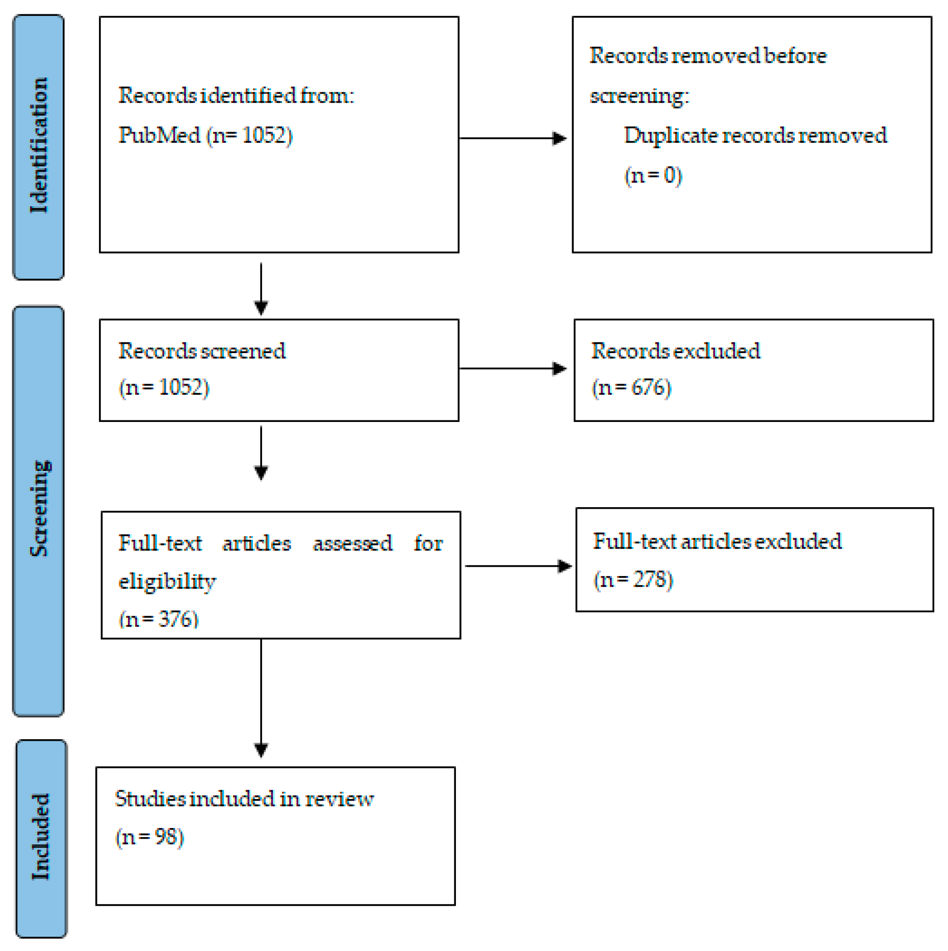
2. Materials and Methods
3. Results
3.1. Demographic and Clinical Characteristics of Patients
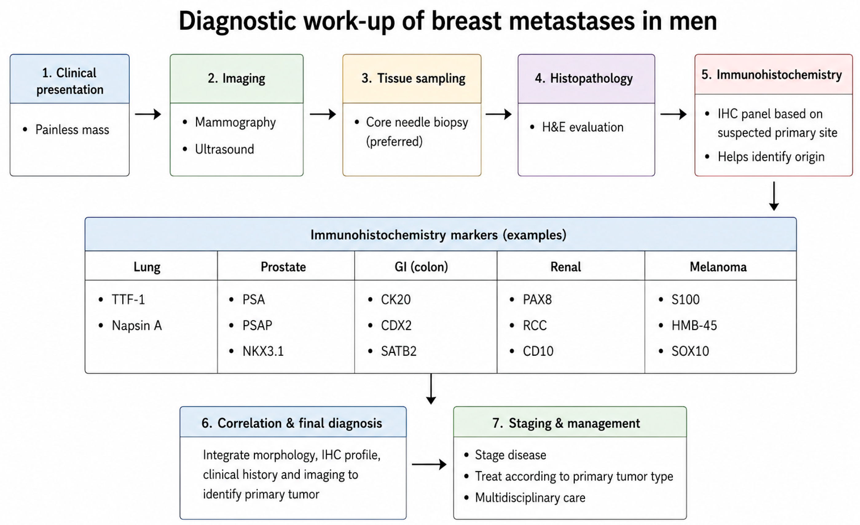
3.2. Diagnostic Approach
3.3. Management
4. Discussion
4.1. Breast Metastases from Prostate Cancer
4.2. Breast Metastases from Lung Cancer
4.3. Breast Metastases from Melanoma
4.4. Extranodal Involvement of Lymphomas in the Male Breast
4.5. Gastrointestinal Malignancies
4.6. Urinary Tract Malignancies
4.7. Immunohistochemical Markers in the Differential Diagnosis
4.8. Limitations
5. Conclusions
Supplementary Materials
Author Contributions
Funding
Institutional Review Board Statement
Informed Consent Statement
Data Availability Statement
Conflicts of Interest
References
- Khattab, A.; Kashyap, S.; Monga, D.K. Male Breast Cancer. In StatPearls; StatPearls Publishing: Treasure Island, FL, USA, 2025. [Google Scholar] [PubMed]
- Yalaza, M.; İnan, A.; Bozer, M. Male Breast Cancer. J. Breast Health 2016, 12, 1–8. [Google Scholar] [CrossRef] [Scilit] [PubMed] [PubMed Central]
- Giordano, S.H. Breast Cancer in Men. N. Engl. J. Med. 2018, 378, 2311–2320. [Google Scholar] [CrossRef] [Scilit] [PubMed]
- Bitencourt, A.G.V.; Gama, R.R.M.; Graziano, L.; Negrão, E.M.S.; Sabino, S.M.P.S.; Watanabe, A.H.U.; Guatelli, C.S.; Souza, J.A.; Mauad, E.C.; Marques, E.F. Breast metastases from extramammary malignancies: Multimodality imaging aspects. Br. J. Radiol. 2017, 90, 20170197. [Google Scholar] [CrossRef] [Scilit] [PubMed] [PubMed Central]
- Zhang, S.; Peng, W.; Cheng, L.; Pei, B.; Zhou, X.; Wang, M.; Jiang, L.; Lu, C.; Xu, L. Metastasis of prostate cancer to breast: A case report. Front. Oncol. 2025, 15, 1580441. [Google Scholar] [CrossRef] [Scilit] [PubMed] [PubMed Central]
- Moreno-Astudillo, L.; Villaseñor-Navarro, Y.; Sánchez-Goytia, V.; Porras-Reyes, F.; Lara-Mercado, A.; Sollozo-Dupont, I. A Case Series of Breast Metastases from Different Extramammary Malignancies and Their Literature Review. Case Rep. Radiol. 2019, 2019, 9454201. [Google Scholar] [CrossRef] [Scilit] [PubMed] [PubMed Central]
- Picasso, R.; Pistoia, F.; Zaottini, F.; Sanguinetti, S.; Calabrese, M.; Martinoli, C.; Derchi, L. Breast Metastases: Updates on Epidemiology and Radiologic Findings. Cureus 2020, 12, e12258. [Google Scholar] [CrossRef] [Scilit] [PubMed] [PubMed Central]
- Yeh, C.N.; Lin, C.H.; Chen, M.F. Clinical and ultrasonographic characteristics of breast metastases from extramammary malignancies. Am. Surg. 2004, 70, 287–290. [Google Scholar] [CrossRef] [Scilit] [PubMed]
- Mariano, L.; Nicosia, L.; Latronico, A.; Pesapane, F.; Grimaldi, E.; Borella, M.; Quercioli, G.; Mazzarol, G.; Bozzini, A.C.; Cassano, E. Metastatic breast involvement from extramammary malignancies: A review of dissemination pathways, imaging features, and management strategies. Radiol. Med. 2025, 130, 1767–1776. [Google Scholar] [CrossRef] [Scilit] [PubMed]
- Nakata, J.; Whittall, I.; Russo, M.G.; Su, S.; Friedberg, J. Systematic review of lung cancer with breast metastasis. J. Thorac. Dis. 2025, 17, 7430–7439. [Google Scholar] [CrossRef] [Scilit] [PubMed] [PubMed Central]
- Surov, A.; Fiedler, E.; Holzhausen, H.J.; Ruschke, K.; Schmoll, H.J.; Spielmann, R.P. Metastases to the breast from non-mammary malignancies: Primary tumors, prevalence, clinical signs, and radiological features. Acad. Radiol. 2011, 18, 565–574. [Google Scholar] [CrossRef] [Scilit] [PubMed]
- Lee, A.H.S. The histological diagnosis of metastases to the breast from extramammary malignancies. J. Clin. Pathol. 2007, 60, 1333–1341. [Google Scholar] [CrossRef] [Scilit] [PubMed] [PubMed Central]
- Bombonati, A.; Lerwill, M.F. Metastases to and from the Breast. Surg. Pathol. Clin. 2012, 5, 719–747. [Google Scholar] [CrossRef] [Scilit] [PubMed]
- Mokoala, K.; Lawal, I.O.; Lengana, T.; Vorster, M.; Sathekge, M.M. Symmetric breasts metastatic prostate cancer shown by (68)Ga-PSMA PET/CT scan. Hell. J. Nucl. Med. 2019, 22, 76. [Google Scholar] [PubMed]
- Shah, P.; Mustafa, F.; Aiman, A.; Shah, O.J.; Shafi, S.; Charak, A.; Mateen, D.; Feroz, I. Metastases to breast—A 29 year experience in a tertiary care hospital. Gulf J. Oncol. 2014, 1, 40–45. [Google Scholar] [CrossRef] [Scilit] [PubMed]
- Njiaju, U.O.; Truica, C.I. Metastatic prostatic adenocarcinoma mimicking inflammatory breast carcinoma: A case report. Clin. Breast Cancer. 2010, 10, E3–E5. [Google Scholar] [CrossRef] [Scilit] [PubMed]
- Monaco, S.E.; Navolotskaia, O.; Cai, G. Fine-needle aspiration biopsy diagnosis of prostate carcinoma metastatic to the breast. Diagn. Cytopathol. 2009, 37, 388–390. [Google Scholar] [CrossRef] [Scilit] [PubMed]
- Fulciniti, F.; Losito, S.; Botti, G.; Di Mattia, D.; La Mura, A.; Pisano, C.; Pignata, S. Metastases to the breast: Role of fine needle cytology samples. Our experience with nine cases in 2 years. Ann. Oncol. 2008, 19, 682–687. [Google Scholar] [CrossRef] [Scilit] [PubMed]
- Cheng, C.W.; Chan, L.W.; Ng, C.F.; Chan, C.K.; Tse, M.K.G.; Lai, M.M.F. Breast metastasis from prostate cancer and interpretation of immunoreactivity to prostate-specific antigen. Int. J. Urol. 2006, 13, 463–465. [Google Scholar] [CrossRef] [Scilit] [PubMed]
- Hsieh, C.M.; Chen, M.Y.; Chen, F.H.; Chiu, J.F. Breast metastasis of prostate cancer. Breast J. 2005, 11, 78. [Google Scholar] [CrossRef] [Scilit] [PubMed]
- Sahoo, S.; Smith, R.E.; Potz, J.L.; Rosen, P.P. Metastatic prostatic adenocarcinoma within a primary solid papillary carcinoma of the male breast. Arch. Pathol. Lab. Med. 2001, 125, 1101–1103. [Google Scholar] [CrossRef] [Scilit] [PubMed]
- Yan, Z.; Hummel, P.; Waisman, J.; Newstead, G.M.; Chachoua, A.; Chhieng, D.; Cohen, J.M.; Cangiarella, J. Prostatic adenocarcinoma metastatic to the breasts: Report of a case with diagnosis by fine needle aspiration biopsy. Urology 2000, 55, 590. [Google Scholar] [CrossRef] [Scilit] [PubMed]
- Ramamurthy, L.; Cooper, R.A. Metastatic carcinoma to the male breast. Br. J. Radiol. 1991, 64, 277–278. [Google Scholar] [CrossRef] [Scilit] [PubMed]
- Moldwin, R.M.; Orihuela, E. Breast masses associated with adenocarcinoma of the prostate. Cancer 1989, 63, 2229–2233. [Google Scholar] [CrossRef] [Scilit] [PubMed]
- Toombs, B.D.; Kalisher, L. Metastatic disease to the breast: Clinical, pathologic, and radiographic features. AJR Am. J. Roentgenol. 1977, 129, 673–676. [Google Scholar] [CrossRef] [Scilit] [PubMed]
- Green, L.K.; Klima, M. The use of immunohistochemistry in metastatic prostatic adenocarcinoma to the breast. Hum. Pathol. 1991, 22, 242–246. [Google Scholar] [CrossRef] [Scilit] [PubMed]
- Salyer, W.R.; Salyer, D.C. Metastases of prostatic carcinoma to the breast. J. Urol. 1973, 109, 671–675. [Google Scholar] [CrossRef] [Scilit] [PubMed]
- Allen, F.J.; Van Velden, D.J. Prostate carcinoma metastatic to the male breast. Br. J. Urol. 1991, 67, 434–435. [Google Scholar] [CrossRef] [Scilit] [PubMed]
- Drelichman, A.; Amer, M.; Pontes, E.; Al-Sarraf, M.; Vaitkevicius, V.K. Carcinoma of prostate metastatic to breast. Urology 1980, 16, 250–255. [Google Scholar] [CrossRef] [Scilit] [PubMed]
- Naritoku, W.Y.; Taylor, C.R. Immunohistologic diagnosis of 2 cases of metastatic prostate cancer to breast. J. Urol. 1983, 130, 365–367. [Google Scholar] [CrossRef] [Scilit] [PubMed]
- Kitano, T.; Nakahara, M.; Yasukawa, A.; Usui, T.; Aoki, Y.; Miyachi, Y.; Nihira, H. Metastatic breast carcinoma of prostatic origin. Urol. Int. 1983, 38, 182–184. [Google Scholar] [CrossRef] [Scilit] [PubMed]
- Kumar, P.P.; Good, R.R.; Newland, J.R. Immunodiagnosis by prostatic acid phosphatase to differentiate primary male breast cancer from metastatic prostate cancer. J. Natl. Med. Assoc. 1986, 78, 782–784, 786. [Google Scholar] [PubMed] [PubMed Central]
- Hesdorffer, C.; Bezwoda, W.R. Metastases of prostate cancer to breast. A case report. S. Afr. Med. J. 1987, 72, 504–505. [Google Scholar] [PubMed]
- Benson, W.R. Carcinoma of the prostate with metastases to breasts and testis; critical review of the literature and report of a case. Cancer 1957, 10, 1235–1245. [Google Scholar] [CrossRef] [Scilit] [PubMed]
- Campbell, J.H.; Cummins, S.D.; Kirk, D.L. MATHEWS WR. Secondary breast cancer of prostatic origin. JAMA 1962, 179, 458–460. [Google Scholar] [CrossRef] [Scilit] [PubMed]
- Deeley, T.J. Secondary deposits in the breast. Br. J. Cancer 1965, 19, 738–743. [Google Scholar] [CrossRef] [Scilit] [PubMed] [PubMed Central]
- Hartley, L.C.; Little, J.H. Bilateral mammary metastases from carcinoma of the prostate during oestrogen therapy. Med. J. Aust. 1971, 1, 434–436. [Google Scholar] [CrossRef] [Scilit] [PubMed]
- Lome, L.G.; Austen, G.J. Metastatic breast carcinoma of prostatic origin. Am. J. Surg. 1970, 120, 113–115. [Google Scholar] [CrossRef] [Scilit] [PubMed]
- Pribe, W.A.; Ockuly, E.A. PROSTATIC METASTASIS TO THE BREAST AND THE ROLE OF ESTROGENS: CASE REPORT AND REVIEW. J. Am. Geriatr. Soc. 1963, 11, 891–901. [Google Scholar] [CrossRef] [Scilit] [PubMed]
- Scott, J.; Robb-Smith, A.H.; Burns, I. Bilateral breast metastases from carcinoma of the prostate. Br. J. Urol. 1974, 46, 209–214. [Google Scholar] [CrossRef] [Scilit] [PubMed]
- Seipel, W.D.; Malament, M. Carcinoma of the prostate with bilateral breast metastases. J. Am. Geriatr. Soc. 1972, 20, 510–513. [Google Scholar] [CrossRef] [Scilit] [PubMed]
- Kidwai, N.; Gong, Y.; Sun, X.; Deshpande, C.G.; Yeldandi, A.V.; Rao, M.S.; Badve, S. Expression of androgen receptor and prostate-specific antigen in male breast carcinoma. Breast Cancer Res. 2004, 6, R18–R23. [Google Scholar] [CrossRef] [Scilit] [PubMed] [PubMed Central]
- Gurel, B.; Ali, T.Z.; Montgomery, E.A.; Begum, S.; Hicks, J.; Goggins, M.; Eberhart, C.G.; Clark, D.P.; Bieberich, C.J.; Epstein, J.I.; et al. NKX3.1 as a marker of prostatic origin in metastatic tumors. Am. J. Surg. Pathol. 2010, 34, 1097–1105. [Google Scholar] [CrossRef] [Scilit] [PubMed] [PubMed Central]
- Yin, M.; Dhir, R.; Parwani, A.V. Diagnostic utility of p501s (prostein) in comparison to prostate specific antigen (PSA) for the detection of metastatic prostatic adenocarcinoma. Diagn. Pathol. 2007, 2, 41. [Google Scholar] [CrossRef] [Scilit] [PubMed] [PubMed Central]
- Sheridan, T.; Herawi, M.; Epstein, J.I.; Illei, P.B. The role of P501S and PSA in the diagnosis of metastatic adenocarcinoma of the prostate. Am. J. Surg. Pathol. 2007, 31, 1351–1355. [Google Scholar] [CrossRef] [Scilit] [PubMed]
- Hashmi, A.A.; Vougiouklakis, T.; Gazzo, A.; Wen, H.Y.; Ross, D.; Pareja, F.; Brogi, E. Lung Carcinoma Metastatic to Breast: A Comprehensive Analysis of Clinical Presentation, Morphologic and Molecular Features, with Emphasis on Diagnostic Pitfalls. Mod. Pathol. 2025, 39, 100936. [Google Scholar] [CrossRef] [Scilit] [PubMed]
- Lahham, E.E.; Masri, H.M.; Farhoud, Z.A.; Neiroukh, A.N.; Ramahi, M.; Awad, F.; Qubaja, M. From lung to breast: A rare case of metastatic lung adenocarcinoma presenting as a breast lump in a male patient. J. Surg. Case Rep. 2025, 2025, rjaf148. [Google Scholar] [CrossRef] [Scilit] [PubMed]
- Koh, S.; Koi, Y.; Tajiri, W.; Kawasaki, J.; Akiyoshi, S.; Nakamura, Y.; Koga, C.; Okamoto, T.; Taguchi, K.; Tokunaga, E. Bilateral breast metastases from anaplastic lymphoma kinase-positive lung cancer in a male: A case report. J. Med. Case Rep. 2024, 18, 402. [Google Scholar] [CrossRef] [Scilit] [PubMed]
- Klingen, T.A.; Klaasen, H.; Aas, H.; Chen, Y.; Akslen, L.A. Secondary breast cancer: A 5-year population-based study with review of the literature. APMIS 2009, 117, 762–767. [Google Scholar] [CrossRef] [Scilit] [PubMed]
- Lin, Q.; Cai, G.P.; Yang, K.Y.; Yang, L.; Chen, C.S.; Li, Y.P. Case report: Small cell transformation and metastasis to the breast in a patient with lung adenocarcinoma following maintenance treatment with epidermal growth factor receptor tyrosine kinase inhibitors. BMC Cancer 2016, 16, 593. [Google Scholar] [CrossRef] [Scilit] [PubMed] [PubMed Central]
- Wan, X.; Zhang, H.; Zhang, Y.; Peng, Y. Metastases to the Breast from Extramammary Nonhematological Malignancies: Case Series. Int. J. Gen. Med. 2020, 13, 1105–1114. [Google Scholar] [CrossRef] [Scilit] [PubMed] [PubMed Central]
- Grigoropoulos, P.; Mariolis-Sapsakos, T.; Karantonis, I.; Kaklamanos, I.; Karanasiou, V.; Zografos, C.G.; Chrysikos, D. Non small cell lung cancer metastasized to the breast and treated with modified radical mastectomy: A case report. G. Chir. 2019, 40, 304–307. [Google Scholar] [PubMed]
- Erhamamci, S.; Reyhan, M.; Canpolat, T.; Nursal, G.N.; Yapar, A.F. A Case of a Man with Isolated Breast Metastasis from Lung Adenocarcinoma Incidentally Detected by FDG PET/CT. Clin. Nucl. Med. 2016, 41, e146–e148. [Google Scholar] [CrossRef] [Scilit] [PubMed]
- Hachisuka, A.; Takahashi, R.; Nakagawa, S.; Takahashi, H.; Inoue, Y.; Akashi, M.; Ichiki, M.; Momosaki, S.; Kawahara, A.; Shirouzu, K.; et al. Lung adenocarcinoma metastasis to the male breast: A case report. Kurume Med. J. 2014, 61, 35–41. [Google Scholar] [CrossRef] [Scilit] [PubMed]
- Altintoprak, F.; Baytekin, H.F.; Tasdemir, C. Primary small cell carcinoma of the lung presenting with breast and skin metastases. Korean J. Intern. Med. 2011, 26, 207–209. [Google Scholar] [CrossRef] [Scilit] [PubMed] [PubMed Central]
- Ucar, N.; Kurt, O.K.; Alpar, S.; Orsel, O.; Demirag, F.; Kurt, B. Breast metastasis in a male patient with nonsmall cell lung carcinoma. South. Med. J. 2007, 100, 850–851. [Google Scholar] [CrossRef] [Scilit] [PubMed]
- Wood, B.; Sterrett, G.; Frost, F.; Swarbrick, N. Diagnosis of extramammary malignancy metastatic to the breast by fine needle biopsy. Pathology 2008, 40, 345–351. [Google Scholar] [CrossRef] [Scilit] [PubMed]
- Gómez-Caro, A.; Piñero, A.; Roca, M.J.; Torres, J.; Ferri, B.; Galindo, P.J.; Parrilla, P. Surgical treatment of solitary metastasis in the male breast from non-small cell lung cancer. Breast J. 2006, 12, 366–367. [Google Scholar] [CrossRef] [Scilit] [PubMed]
- Hejmadi, R.K.; Day, L.J.; Young, J.A. Extramammary metastatic neoplasms in the breast: A cytomorphological study of 11 cases. Cytopathology 2003, 14, 191–194. [Google Scholar] [CrossRef] [Scilit] [PubMed]
- Ramar, K.; Pervez, H.; Potti, A.; Mehdi, S. Breast metastasis from non-small-cell lung carcinoma. Med. Oncol. 2003, 20, 181–184. [Google Scholar] [CrossRef] [Scilit] [PubMed]
- David, O.; Gattuso, P.; Razan, W.; Moroz, K.; Dhurandhar, N. Unusual cases of metastases to the breast. A report of 17 cases diagnosed by fine needle aspiration. Acta Cytol. 2002, 46, 377–385. [Google Scholar] [CrossRef] [Scilit] [PubMed]
- Siddiqui, M.T.; Zakowski, M.F.; Ashfaq, R.; Ali, S.Z. Breast masses in males: Multi-institutional experience on fine-needle aspiration. Diagn. Cytopathol. 2002, 26, 87–91. [Google Scholar] [CrossRef] [PubMed]
- Muttarak, M.; Nimmonrat, A.; Chaiwun, B. Metastatic carcinoma to the male and female breast. Australas. Radiol. 1998, 42, 16–19. [Google Scholar] [CrossRef] [Scilit] [PubMed]
- Domanski, H.A. Metastases to the breast from extramammary neoplasms. A report of six cases with diagnosis by fine needle aspiration cytology. Acta Cytol. 1996, 40, 1293–1300. [Google Scholar] [CrossRef] [Scilit] [PubMed]
- Cooper, R.A.; Gunter, B.A.; Ramamurthy, L. Mammography in men. Radiology 1994, 191, 651–656. [Google Scholar] [CrossRef] [Scilit] [PubMed]
- Verger, E.; Conill, C.; Velasco, M.; Sole, M. Metastasis in the male breast from a lung adenocarcinoma. Acta Oncol. 1992, 31, 479. [Google Scholar] [CrossRef] [Scilit] [PubMed]
- Sneige, N.; Zachariah, S.; Fanning, T.V.; Dekmezian, R.H.; Ordóñez, N.G. Fine-needle aspiration cytology of metastatic neoplasms in the breast. Am. J. Clin. Pathol. 1989, 92, 27–35. [Google Scholar] [CrossRef] [Scilit] [PubMed]
- Ye, J.; Findeis-Hosey, J.J.; Yang, Q.; McMahon, L.A.; Yao, J.L.; Li, F.; Xu, H. Combination of napsin A and TTF-1 immunohistochemistry helps in differentiating primary lung adenocarcinoma from metastatic carcinoma in the lung. Appl. Immunohistochem. Mol. Morphol. 2011, 19, 313–317. [Google Scholar] [CrossRef] [Scilit] [PubMed]
- Yang, M.; Nonaka, D. A study of immunohistochemical differential expression in pulmonary and mammary carcinomas. Mod. Pathol. 2010, 23, 654–661. [Google Scholar] [CrossRef] [Scilit] [PubMed]
- Zhao, W.; Wang, H.; Peng, Y.; Tian, B.; Peng, L.; Zhang, D.C. ΔNp63, CK5/6, TTF-1 and napsin A, a reliable panel to subtype non-small cell lung cancer in biopsy specimens. Int. J. Clin. Exp. Pathol. 2014, 7, 4247–4253. [Google Scholar] [PubMed] [PubMed Central]
- Gurda, G.T.; Zhang, L.; Wang, Y.; Chen, L.; Geddes, S.; Cho, W.C.; Askin, F.; Gabrielson, E.; Li, Q.K. Utility of five commonly used immunohistochemical markers TTF-1, Napsin A, CK7, CK5/6 and P63 in primary and metastatic adenocarcinoma and squamous cell carcinoma of the lung: A retrospective study of 246 fine needle aspiration cases. Clin. Transl. Med. 2015, 4, 16. [Google Scholar] [CrossRef] [Scilit] [PubMed] [PubMed Central]
- Santana Valenciano, A.; Juez Sáez, L.; Pérez Mies, B.; Moreno, S.; Fidalgo, S.; Montero, J. Breast metastases from non-primary breast malignancies: What should we know? Breast Dis. 2023, 42, 223–228. [Google Scholar] [CrossRef] [Scilit] [PubMed]
- Kang, B.S.; Kim, S.K. Malignant melanoma with metastasis to the male breast. Indian. J. Dermatol. Venereol. Leprol. 2014, 80, 566–568. [Google Scholar] [CrossRef] [Scilit] [PubMed]
- Bacchi, C.E.; Wludarski, S.C.; Ambaye, A.B.; Lamovec, J.; Salviato, T.; Falconieri, G. Metastatic melanoma presenting as an isolated breast tumor: A study of 20 cases with emphasis on several primary mimickers. Arch. Pathol. Lab. Med. 2013, 137, 41–49. [Google Scholar] [CrossRef] [Scilit] [PubMed]
- Garza-Guajardo, R.; Mendez-Olvera, N.; Flores-Gutierrez, J.P.; Hernandez-Martinez, S.; Candanosa-Mc Cann, M.; Ancer-Rodriguez, J.; Barboza-Quintana, O. Fine needle aspiration biopsy diagnosis of metastatic neoplasms of the breast. A three-case report. Cytojournal 2005, 2, 17. [Google Scholar] [CrossRef] [Scilit] [PubMed] [PubMed Central]
- Shukla, R.; Pooja, B.; Radhika, S.; Nijhawan, R.; Rajwanshi, A. Fine-needle aspiration cytology of extramammary neoplasms metastatic to the breast. Diagn. Cytopathol. 2005, 32, 193–197. [Google Scholar] [CrossRef] [Scilit] [PubMed]
- Cangiarella, J.; Symmans, W.F.; Cohen, J.M.; Goldenberg, A.; Shapiro, R.L.; Waisman, J. Malignant melanoma metastatic to the breast: A report of seven cases diagnosed by fine-needle aspiration cytology. Cancer 1998, 84, 160–162. [Google Scholar] [CrossRef] [Scilit] [PubMed]
- Al Samaraee, A.; Khout, H.; Barakat, T.; Fasih, T. Breast metastasis from a melanoma. Ochsner J. 2012, 12, 149–151. [Google Scholar] [PubMed] [PubMed Central]
- Kasumova, G.G.; Haynes, A.B.; Boland, G.M. Lymphatic versus Hematogenous Melanoma Metastases: Support for Biological Heterogeneity without Clear Clinical Application. J. Investig. Dermatol. 2017, 137, 2466–2468. [Google Scholar] [CrossRef] [Scilit] [PubMed]
- Veach, J.A.; Hill, M.; Huppe, A.I. Uncommon Presentation of Metastatic Melanoma to the Breast. Kans. J. Med. 2025, 18, 19–20. [Google Scholar] [CrossRef] [Scilit] [PubMed] [PubMed Central]
- Brodhead, M.; Woods, R.W.; Fowler, A.M.; Roy, M.; Neuman, H.; Gegios, A. Multimodality imaging review of metastatic melanoma involving the breast. Curr. Probl. Diagn. Radiol. 2024, 53, 289–296. [Google Scholar] [CrossRef] [Scilit] [PubMed]
- Voiculescu, V.M.; Popescu, A.I.; Costache, M. Immunohistochemistry for Skin Cancers: New Insights into Diagnosis and Treatment of Melanoma. Cancers 2025, 17, 1769. [Google Scholar] [CrossRef] [Scilit] [PubMed] [PubMed Central]
- Hossain, S.M.; Eccles, M.R. Phenotype Switching and the Melanoma Microenvironment; Impact on Immunotherapy and Drug Resistance. Int. J. Mol. Sci. 2023, 24, 1601. [Google Scholar] [CrossRef] [Scilit] [PubMed] [PubMed Central]
- Amaral, T.; Ottaviano, M.; Arance, A.; Blank, C.; Chiarion-Sileni, V.; Donia, M.; Dummer, R.; Garbe, C.; Gershenwald, J.E.; Gogas, H.; et al. Cutaneous melanoma: ESMO Clinical Practice Guideline for diagnosis, treatment and follow-up. Ann. Oncol. 2025, 36, 10–30. [Google Scholar] [CrossRef] [Scilit] [PubMed] [PubMed Central]
- Holdsworth, F.; Worku, D.; Bretton Ale Vella, C.; Walker, E. A guide to Hodgkin and non-Hodgkin lymphomas: Similarities and differences. Br. J. Nurs. 2021, 30, S16–S22. [Google Scholar] [CrossRef] [Scilit] [PubMed]
- Surov, A.; Holzhausen, H.J.; Wienke, A.; Schmidt, J.; Thomssen, C.; Arnold, D.; Ruschke, K.; Spielmann, R.P. Primary and secondary breast lymphoma: Prevalence, clinical signs and radiological features. Br. J. Radiol. 2012, 85, e195–e205. [Google Scholar] [CrossRef] [Scilit] [PubMed] [PubMed Central]
- Lokesh, K.; Sathyanarayanan, V.; Lakshmaiah, K.; Suresh, T.; Lokanatha, D.; Govinda Babu, K.; Jacob, L.A.; Babu, S. Primary breast lymphoma in males-a report of two cases with a review of the literature. Ecancermedicalscience 2013, 7, 347. [Google Scholar] [CrossRef] [PubMed] [PubMed Central]
- Mun, S.H.; Ko, E.Y.; Han, B.K.; Shin, J.H.; Kim, S.J.; Cho, E.Y. Breast metastases from extramammary malignancies: Typical and atypical ultrasound features. Korean J. Radiol. 2014, 15, 20–28. [Google Scholar] [CrossRef] [Scilit] [PubMed] [PubMed Central]
- Lee, S.H.; Park, J.M.; Kook, S.H.; Han, B.K.; Moon, W.K. Metastatic tumors to the breast: Mammographic and ultrasonographic findings. J. Ultrasound Med. 2000, 19, 257–262. [Google Scholar] [CrossRef] [Scilit] [PubMed]
- McCrea, E.S.; Johnston, C.; Haney, P.J. Metastases to the breast. AJR Am. J. Roentgenol. 1983, 141, 685–690. [Google Scholar] [CrossRef] [Scilit] [PubMed]
- Sabaté, J.M.; Gómez, A.; Torrubia, S.; Camins, A.; Roson, N.; De Las Heras, P.; Villalba-Nuño, V. Lymphoma of the breast: Clinical and radiologic features with pathologic correlation in 28 patients. Breast J. 2002, 8, 294–304. [Google Scholar] [CrossRef] [Scilit] [PubMed]
- Kalli, S.; Lanfranchi, M.; Alexander, A.; Makim, S.; Freer, P.E. Spectrum of Extramammary Malignant Neoplasms in the Breast with Radiologic-Pathologic Correlation. Curr. Probl. Diagn. Radiol. 2016, 45, 392–401. [Google Scholar] [CrossRef] [Scilit] [PubMed]
- Fraulini, C.; Mauro, E.; Rigolin, G.M.; Bissoli, S.; Chierichetti, F.; Panareo, S.; Feggi, L.; Cavazzini, L.; Castoldi, G. A case of advanced Hodgkin lymphoma with breast involvement treated with a combination of gemcitabine and pegylated liposomal doxorubicin. Eur. J. Haematol. 2005, 75, 515–517. [Google Scholar] [CrossRef] [Scilit] [PubMed]
- Cheng, X.; Zhao, T.; Jiang, J.N.; Liu, Y.; Li, H.; Wang, R.Y. Rare case of a male breast tumour secondary to poorly differentiated rectal adenocarcinoma. Transl. Cancer Res. 2020, 9, 7676–7681. [Google Scholar] [CrossRef] [Scilit] [PubMed] [PubMed Central]
- Gur, E.O.; Onak, C.; Bayar, E.E.; Yazıcı, A.; Kokulu, İ.; Hacıyanlı, S.; Dilek, O.N. Rectum mucinous adenocarcinoma metastasis to bilateral breast in a male patient: A case report. Breast J. 2020, 26, 517–519. [Google Scholar] [CrossRef] [Scilit] [PubMed]
- Wang, T.; Lv, Y.G.; Yan, Q.G.; Yuan, S.F.; Ling, R.; Chen, J.H.; Chen, L.; Yi, J.; Wang, L. Rectal carcinoma metastatic to the male breast after 7 years: Case report. Onkologie 2011, 34, 544–546. [Google Scholar] [CrossRef] [Scilit] [PubMed]
- Kubo, H.; Shimizu, T.; Sekido, H.; Matsuda, G.; Takeda, K.; Watanabe, A.; Sakamoto, R.; Yamamoto, Y.; Toyoda, J.; Niino, H. Isolated breast metastasis from gastric cancer in a male patient. Clin. J. Gastroenterol. 2018, 11, 138–144. [Google Scholar] [CrossRef] [Scilit] [PubMed]
- Tsung, S.H. Metastasis of Colon Cancer to the Breast. Case Rep. Oncol. 2017, 10, 77–80. [Google Scholar] [CrossRef] [Scilit] [PubMed] [PubMed Central]
- Ho, Y.Y.; Lee, W.K. Metastasis to the breast from an adenocarcinoma of the colon. J. Clin. Ultrasound 2009, 37, 239–241. [Google Scholar] [CrossRef] [Scilit] [PubMed]
- Bruscagnin, A. A case of male breast metastasis from adenocarcinoma of the colon. Radiol. Med. 1997, 93, 463–464. [Google Scholar] [PubMed]
- Lear, P.A.; Jackaman, F.R. Carcinoma of the rectum with metastasis in the male breast. J. R. Coll. Surg. 1980, 25, 246–247. [Google Scholar] [PubMed]
- Lee, J.H.; Kim, S.H.; Kang, B.J.; Cha, E.S.; Kim, H.S.; Choi, J.J. Metastases to the breast from extramammary malignancies−sonographic features. J. Clin. Ultrasound 2011, 39, 248–255. [Google Scholar] [CrossRef] [Scilit] [PubMed]
- Lo, H.C.; Lee, K.F.; Yeh, C.N.; Chen, M.F. Breast metastasis from hepatocellular carcinoma. Hepatogastroenterology 2004, 51, 387–390. [Google Scholar] [PubMed]
- Hamby, L.S.; McGrath, P.C.; Cibull, M.L.; Schwartz, R.W. Gastric carcinoma metastatic to the breast. J. Surg. Oncol. 1991, 48, 117–121. [Google Scholar] [CrossRef] [Scilit] [PubMed]
- Takami, H.; Sentani, K.; Matsuda, M.; Oue, N.; Sakamoto, N.; Yasui, W. Cytokeratin expression profiling in gastric carcinoma: Clinicopathologic significance and comparison with tumor-associated molecules. Pathobiology 2012, 79, 154–161. [Google Scholar] [CrossRef] [Scilit] [PubMed]
- Bayrak, R.; Yenidünya, S.; Haltas, H. Cytokeratin 7 and cytokeratin 20 expression in colorectal adenocarcinomas. Pathol. Res. Pract. 2011, 207, 156–160. [Google Scholar] [CrossRef] [Scilit] [PubMed]
- Hoang, A.; Vu, V.; Nguyen, Q.; Thieu, M.; Le, H. Isolated metastasis of renal clear-cell carcinoma to the male breast following nephrectomy: A rare case report. Int. J. Surg. Case Rep. 2025, 136, 112049. [Google Scholar] [CrossRef] [Scilit] [PubMed]
- Yoon, I.; Cha, E.; Kim, J.; Lee, J.; Chung, J. Bladder Cancer Metastasis to the Breast in a Male Patient: Imaging Findings on Mammography and Ultrasonography. J. Korean Soc. Radiol. 2022, 83, 687–692. [Google Scholar] [CrossRef] [Scilit] [PubMed]
- Ishikawa, Y.; Tabei, I.; Fushimi, A.; Fuke, A.; Sekine, C.; Okamoto, T.; Takeyama, H. Male breast metastasis of ureteral cancer: A case report. Surg. Case Rep. 2020, 6, 58. [Google Scholar] [CrossRef] [Scilit] [PubMed] [PubMed Central]
- Roy, S.F.; Guilbert, M.C. Micropapillary urothelial carcinoma metastatic to the male breast. Breast J. 2019, 25, 1286–1287. [Google Scholar] [CrossRef] [Scilit] [PubMed]
- Sippo, D.A.; Kulkarni, K.; Carlo, P.D.; Lee, B.; Eisner, D.; Cimino-Mathews, A.; Harvey, S.C. Metastatic Disease to the Breast from Extramammary Malignancies: A Multimodality Pictorial Review. Curr. Probl. Diagn. Radiol. 2016, 45, 225–232. [Google Scholar] [CrossRef] [Scilit] [PubMed]
- Cappabianca, S.; Grassi, R.; D’Alessandro, P.; Del Vecchio, A.; Maioli, A.; Donofrio, V. Metastasis to the male breast from carcinoma of the urinary bladder. Br. J. Radiol. 2000, 73, 1326–1328. [Google Scholar] [CrossRef] [Scilit] [PubMed]
- Gibbons, C.E.; Lewi, H.J.; Kashif, K.M. Breast lump—An unusual presentation of renal cell carcinoma. Br. J. Urol. 1995, 76, 131. [Google Scholar] [CrossRef] [Scilit] [PubMed]
- Bösherz, M.S.; Samarska, I.V.; Gaisa, N.T. Scoring Systems for Immunohistochemistry in Urothelial Carcinoma. Methods Mol. Biol. 2023, 2684, 3–25. [Google Scholar] [CrossRef] [Scilit] [PubMed]
- Naik, M.; Rao, B.V.; Challa, S.; Fonseca, D.; Sudha, S.M.; Giridhar, A.; Sharma, R.; Raju, K.V.V.N.; Rao, T.S. Utility of GATA-3 and associated immunohistochemical markers in the differential diagnosis of poorly differentiated urothelial carcinoma. J. Cancer Res. Ther. 2023, 19, S149–S159. [Google Scholar] [CrossRef] [Scilit] [PubMed]
- Abdelrahman, A.E.; Rashed, H.E.; Abdelgawad, M.; Abdelhamid, M.I. Prognostic impact of EGFR and cytokeratin 5/6 immunohistochemical expression in triple-negative breast cancer. Ann. Diagn. Pathol. 2017, 28, 43–53. [Google Scholar] [CrossRef] [Scilit] [PubMed]
- Portaluri, A.; Sofia, C.; Catanzariti, F.; Condorelli, E.; Ieni, A.; Avendano, D.; Ascenti, V.; Marino, M.A. Male breast: An unusual case of metastasis of squamous cell carcinoma of the skin. Radiol. Case Rep. 2022, 18, 809–813. [Google Scholar] [CrossRef] [Scilit] [PubMed]
- Sun, P.; Chen, J.; Lu, J.; Luo, R.; Li, M.; He, J. Characteristics of breast metastases from non-breast solid tumors in 22 patients from a southern Chinese population. Oncol. Lett. 2018, 15, 3685–3693. [Google Scholar] [CrossRef] [Scilit] [PubMed] [PubMed Central]
- Khazai, L.; Falcon, S.; Rosa, M. Metastatic Salivary Duct Carcinoma to the Breast. Breast J. 2016, 22, 461–463. [Google Scholar] [CrossRef] [Scilit] [PubMed]
- Franceschini, G.; Sanchez, A.M.; Di Leone, A.; Scaldaferri, A.; Ferrucci, M.; Mulè, A.; Costantini, M.; Masetti, R. Penile cancer metastasizing to the breast: A case report. J. Med. Case Rep. 2016, 10, 53. [Google Scholar] [CrossRef] [Scilit] [PubMed] [PubMed Central]
- Narese, D.; Virzì, V.; Narese, F.; Culmone, G.; Cirrito, D.; Sciortino, A.; Virzì, G. Breast core biopsy of a rare case of unknown primary large cell neuroendocrine carcinoma metastatic to the breast. Clin. Ter. 2014, 165, 302–304. [Google Scholar] [CrossRef] [Scilit] [PubMed]
- Kohi, M.P.; Brasic, N.; Vohra, P.; Price, E.R.; Joe, B.N. Breast metastasis from testicular leiomyosarcoma. Breast J. 2013, 19, 336–337. [Google Scholar] [CrossRef] [Scilit] [PubMed]
- Alacacioglu, A.; Unal, S.; Canpolat, S.; Yurt, A.; Oztekin, O.; Coskun, A.; Karatas, A.; Postaci, H.; Sop, G. Breast metastasis of anaplastic oligodendroglioma: A case report. Tumori 2012, 98, 162e–164e. [Google Scholar] [CrossRef] [Scilit] [PubMed]
- López Guerra, J.L.; Foro Arnalot, P.; Algara López, M. Breast metastases from a floor of the mouth carcinoma. Clin. Transl. Oncol. 2008, 10, 522–524. [Google Scholar] [CrossRef] [Scilit] [PubMed]
- Prieto, A.; Díaz, A.C.; González, M.; Calvo, J.; Vega, P.; García, C. Metastasis to the male breast from hypopharyngeal squamous cell carcinoma. Breast J. 2005, 11, 483–484. [Google Scholar] [CrossRef] [Scilit] [PubMed]
- Nair, N.; Basu, S. Unsuspected metastatic male breast nodule from synovial sarcoma detected by FDG PET. Clin. Nucl. Med. 2005, 30, 289–290. [Google Scholar] [CrossRef] [Scilit] [PubMed]
- Chim, C.S.; Shek, T.W.; Liang, R. Isolated relapse of acute lymphoblastic leukemia in the breast masquerading as gynecomastia. Am. J. Med. 2000, 108, 677–679. [Google Scholar] [CrossRef] [Scilit] [PubMed]
- Deshpande, A.H.; Munshi, M.M.; Lele, V.R.; Bobhate, S.K. Aspiration cytology of extramammary tumors metastatic to the breast. Diagn. Cytopathol. 1999, 21, 319–323. [Google Scholar] [CrossRef] [Scilit] [PubMed]
- Buisman, F.E.; van Gelder, L.; Menke-Pluijmers, M.B.E.; Bisschops, B.H.C.; Plaisier, P.W.; Westenend, P.J. Non-primary breast malignancies: A single institution’s experience of a diagnostic challenge with important therapeutic consequences-a retrospective study. World J. Surg. Oncol. 2016, 14, 166. [Google Scholar] [CrossRef] [Scilit] [PubMed] [PubMed Central]
- Luo, Y.; Xu, B.; Li, Q.; Zhang, P.; Yuan, P.; Wang, J.; Ma, F.; Cai, R.; Fan, Y. Clinicopathological features and prognosis of metastases to the breast from extramammary solid tumors. Zhonghua Zhong Liu ZA Zhi 2014, 36, 453–456. [Google Scholar] [PubMed]
- DeLair, D.F.; Corben, A.D.; Catalano, J.P.; Vallejo, C.E.; Brogi, E.; Tan, L.K. Non-mammary metastases to the breast and axilla: A study of 85 cases. Mod. Pathol. 2013, 26, 343–349. [Google Scholar] [CrossRef] [Scilit] [PubMed]
- Smymiotis, V.; Theodosopoulos, T.; Marinis, A.; Goula, K.; Psychogios, J.; Kondi-Pafiti, A. Metastatic disease in the breast from nonmammary neoplasms. Eur. J. Gynaecol. Oncol. 2005, 26, 547–550. [Google Scholar] [PubMed]
- Page, M.J.; McKenzie, J.E.; Bossuyt, P.M.; Boutron, I.; Hoffmann, T.C.; Mulrow, C.D.; Shamseer, L.; Tetzlaff, J.; Akl, E.; Brennan, S.E. The PRISMA 2020 statement: An updated guideline for reporting systematic reviews. MetaArXiv 2020. [Google Scholar] [CrossRef] [Scilit]
- Moola, S.; Munn, Z.; Tufanaru, C.; Aromataris, E.; Sears, K.; Sfetcu, R.; Currie, M.; Qureshi, R.; Mattis, P.; Lisy, K.; et al. Chapter 7: Systematic reviews of etiology and risk. In Joanna Briggs Institute Reviewer’s Manual; Aromataris, E., Munn, Z., Eds.; The Joanna Briggs Institute: Adelaide, South Australia, 2017. [Google Scholar]



| Primary Tumor Site | Number of Cases (n) | Percentage (%) |
|---|---|---|
| Prostate | 53 | 27.6% |
| Lung | 38 | 19.8% |
| Melanoma | 18 | 9.4% |
| Lymphoma | 6 | 3.1% |
| Stomach | 5 | 2.6% |
| Colon | 5 | 2.6% |
| Rectum | 4 | 2.1% |
| Kidney | 4 | 2.1% |
| Urothelial carcinoma | 3 | 1.6% |
| Multiple myeloma | 3 | 1.6% |
| Pharynx | 3 | 1.6% |
| Esophagus | 2 | 1% |
| Ureter | 2 | 1% |
| Hepatocellular carcinoma | 2 | 1% |
| Skin (non-melanoma) | 1 | 0.5% |
| Lymphoblastic leukemia | 1 | 0.5% |
| Penis | 1 | 0.5% |
| Testis | 1 | 0.5% |
| Larynx | 1 | 0.5% |
| Salivary gland | 1 | 0.5% |
| Oral cavity | 1 | 0.5% |
| Pancreas | 1 | 0.5% |
| Glioma | 1 | 0.5% |
| Synovial sarcoma | 1 | 0.5% |
| Cases in which the primary tumor was not reported | 34 | 17.7% |
| Parameter | Summary of Reported Data |
|---|---|
| Number of reported patients | 192 |
| Number of included articles | 98 |
| Mean age at diagnosis of breast metastasis | 58 years (range: 24–82 years) |
| Interval between primary tumor diagnosis and breast metastasis | Mean: 32.08 months; Median: 24 months (range: synchronous presentation to 11 years) |
| Laterality of breast involvement (n = 92) | Right breast: 37 (40.2%) Left breast: 37 (40.2%) Bilateral involvement: 18 (19.6%) |
| Presence of additional metastases at diagnosis | ≥24 cases (12.5%) |
| Biopsy method (n = 78) | Core needle biopsy: 36 (46.2%) Fine needle aspiration: 42 (53.8%) |
| Initial misdiagnosis as primary breast tumor | 14 cases |
| Treatment reported (n = 46) | Surgical treatment: 33 (71.7%) Systemic therapy: 13 (28.3%) |
| No | Authors | Year | No. of Cases | Age (yrs) | Site | Diagnosis | Management/Outcome |
|---|---|---|---|---|---|---|---|
| 1 | Zhang et al. [5] | 2025 | 1 | 68 | Bilateral, 7 years after initial diagnosis | Prostate adenocarcinoma with bone metastases, on endocrine therapy. GATA3 (−), ERG (+), PSAP (+), NKX3.1 (+). Genetic testing: BRCA2 (+), AR (+) | Bilateral lumpectomy, chemotherapy, anti-androgen therapy |
| 2 | Mokoala et al. [14] | 2019 | 1 | 80 | Bilateral, detected on PET-CT | Castration-resistant prostate cancer, core biopsy | metastatic disease |
| 3 | Shah et al. [15] | 2014 | 2 | 60–69 | – | – | – |
| 4 | Njiaju et al. [16] | 2010 | 1 | 78 | Right breast, initially considered inflammatory breast cancer | Prostate adenocarcinoma with bone metastases. Core biopsy (non-diagnostic) and skin biopsy (diagnostic). IHC confirmation (PSA, AMACR) after 3 months due to non-response to initial therapy | Hormonal therapy—androgen blockade |
| 5 | Monaco et al. [17] | 2009 | 1 | 60 | Right breast | FNA initially, excisional biopsy, IHC confirmation. PSA, P504S, AR (−). GCDFP-15, mammaglobin, ER, PR (−) | – |
| 6 | Fulciniti et al. [18] | 2007 | 1 | 70 | Right breast, 3 years after initial diagnosis | FNA, IHC confirmation (PSA expression) | – |
| 7 | Lee et al. [12] | 2007 | 1 | 75 | – | Metastasis 5.6 years after initial diagnosis | – |
| 8 | Cheng et al. [19] | 2006 | 1 | 79 | Bilateral, right breast 6 cm, 2 years 8 months after initial diagnosis | Core biopsy, IHC confirmation (PSA, PSAP) | – |
| 9 | Hsieh et al. [20] | 2005 | 1 | 71 | Right breast, 2.5 cm | Excisional biopsy, IHC confirmation (PSA) | – |
| 10 | Sahoo et al. [21] | 2001 | 1 | 78 | Right breast, 5 cm, 12 months after initial diagnosis | FNA non-diagnostic, simultaneous papillary carcinoma of the breast and metastatic prostate adenocarcinoma (distinction using GCDFP-15, ER, PR, CK7) | Modified radical mastectomy |
| 11 | Yan et al. [22] | 2000 | 1 | 81 | Bilateral, multiple | FNA (PSA, PSAP positive; ER, PR negative) | Metastatic liver and bone disease |
| 12 | Ramamurthy et al. [23] | 1991 | 1 | 64 | Bilateral, multiple and gynecomastia | FNA | Under estrogen therapy |
| 13 | Moldwin et al. [24] | 1989 | 1 | 45 | Mass (side not specified) and bilateral gynecomastia | Core biopsy | Under estrogen therapy |
| 14 | Toombs et al. [25] | 1977 | 9 | Mean 63.1 | – | – | 8/9 under estrogen therapy |
| 15 | Green et al. [26] | 1991 | 2 | 63/61 | Right breast 2.5 cm/Bilateral | Under estrogen therapy. Excisional biopsy/core biopsy | Excisional biopsy/bilateral lumpectomies |
| 16 | Salyer et al. [27] | 1973 | 11 | – | – | – | metastatic disease |
| 17 | Allen et al. [28] | 1991 | 1 | – | – | IHC | metastatic disease |
| 18 | Drelichman et al. [29] | 1980 | 3 | 71/71/75 | Bilateral | – | metastatic disease, Under estrogen therapy |
| 19 | Naritoku et al. [30] | 1983 | 2 | 78/68 | Right/Left | IHC | Under estrogen therapy 1. Simple mastectomy 2. Excisional biopsy |
| 20 | Kitano et al. [31] | 1983 | 1 | 59 | Right breast, 4 months after initial diagnosis | Under estrogen therapy Lumpectomy | |
| 21 | Kumar et al. [32] | 1986 | 1 | 78 | Right breast, 3 months after initial diagnosis | Biopsy, IHC | under estrogen therapy |
| 22 | Hesdorffer et al. [33] | 1987 | 1 | – | – | IHC | – |
| 23 | Benson [34] | 1957 | 1 | – | Gynecomastia | Under estrogen therapy. Mastectomy and lymph node dissection | |
| 24 | Campbell et al. [35] | 1962 | 1 | 78 | Bilateral | IHC, PAP and tissue acid-glycerophosphatase | Lumpectomy |
| 25 | Deeley [36] | 1965 | 1 | 43 | Right breast | – | – |
| 26 | Hartley [37] | 1971 | 1 | 62 | Bilateral | – | Under estrogen therapy |
| 27 | Lome et al. [38] | 1970 | 1 | 64 | Right breast, 4 cm, 33 months after initial diagnosis | IHC (PSAP) | Under estrogen therapy. Simple mastectomy |
| 28 | Pribe et al. [39] | 1963 | 1 | 67 | – | – | Under estrogen therapy. Simple mastectomy |
| 29 | Scott et al. [40] | 1974 | 1 | 75 | Bilateral, 2 years after initial diagnosis | – | Right mastectomy and excisional biopsy |
| 30 | Seipel et al. [41] | 1972 | 1 | 65 | Bilateral, 44 months after initial diagnosis in right breast, 60 months in left breast | – | Under estrogen therapy. Bilateral lumpectomies |
| No. | Authors | Year | No. of Cases | Age (yrs) | Site | Diagnosis | Management/Outcome |
|---|---|---|---|---|---|---|---|
| 1 | Hashmi et al. [46] | 2025 | 3 | Median 72 | – | lung adenocarcinoma | – |
| 2 | Lahham et al. [47] | 2025 | 1 | 56 | Right breast 1.4 cm, right axillary lymph nodes | Core biopsy: poorly differentiated lung adenocarcinoma, CK7, TTF1, Napsin-A, PD-L1, ROS1 (+); GATA3, ER, PR, HER2 (−) | Initial chemotherapy discontinued due to neutropenia; targeted therapy for EGFR mutation with good response |
| 3 | Koh et al. [48] | 2024 | 1 | 51 | Bilateral | Core biopsy: ALK-positive NSCLC, on chemotherapy | Bilateral lumpectomy, no recurrence at 8 months |
| 4 | Klingen et al. [49] | 2009 | 1 | 70 | Right breast 0.9 cm | Lung adenocarcinoma | Died 4 months after diagnosis of breast metastasis |
| 5 | Lin et al. [50] | 2016 | 1 | 49 | Right breast, 1.5 years after initial diagnosis | Core biopsy: poorly differentiated small cell lung carcinoma, arising from adenocarcinoma during EGFR-TKI therapy. Chromogranin A, synaptophysin, CD56, TTF-1 (+); ER, GCDFP-15, HER2 (−); breast lesion had same EGFR exon 21 mutation | Chemotherapy, bone and brain metastases, died 8 months later |
| 6 | Wan X et al. [51] | 2020 | 1 | 81 | Right breast 2.6 cm | Lung adenocarcinoma | – |
| 7 | Grigoropoulos et al. [52] | 2019 | 1 | 57 | Right breast, upper outer quadrant, 28 months after initial diagnosis | NSCLC | Modified radical mastectomy; poor prognosis |
| 8 | Erhamamci et al. [53] | 2016 | 1 | 74 | Right breast, 1.3 cm, retroareolar, synchronous diagnosis, incidental finding on PET | Excisional biopsy: lung adenocarcinoma | Chemotherapy, died 11 months later |
| 9 | Hachisuka et al. [54] | 2014 | 1 | 60 | Right breast, 4 cm, retroareolar, erythema, skin edema; breast MRI negative for primary malignancy, CT suggestive of lung primary | Core biopsy; initially diagnosed as primary breast carcinoma; lung biopsy confirmed. ER, PR, HER2 (−); IHC: GCDFP15 (−) | Breast metastasis responded to chemotherapy; died 4 months later |
| 10 | Shah et al. [15] | 2014 | 3 | 60–69 | – | Core biopsy | – |
| 11 | Altintoprak et al. [55] | 2011 | 1 | 47 | Right breast, synchronous | Small cell lung carcinoma; IHC: ER, PR, HER2, CK7, CK20 (−), chromogranin (+), synaptophysin (+), pancytokeratin (focal), TTF-1 (+) | Mastectomy, chemotherapy |
| 12 | Ucar et al. [56] | 2007 | 1 | – | – | NSCLC | – |
| 13 | Wood et al. [57] | 2008 | 1 | 67 | Right breast, lower outer quadrant, cervical lymphadenopathy | Lung adenocarcinoma; core biopsy: CK7 (+), TTF-1 (+); ER, PR (−) | – |
| 14 | Lee et al. [12] | 2007 | 1 | 58 | 3 months from initial diagnosis to metastasis | Large cell lung carcinoma | – |
| 15 | Gómez-Caro et al. [58] | 2006 | 1 | 65 | Right breast, 18 months after right pneumonectomy | FNA and core biopsy: NSCLC, CK4 (+), CK7 (+), TTF-1 (−) | Mastectomy and lymph node dissection; adjuvant chemotherapy; disease-free 18 months; surgery chosen for local disease control |
| 16 | Hejmadi et al. [59] | 2003 | 1 | 72 | – | FNA: small cell lung carcinoma | – |
| 17 | Ramar et al. [60] | 2001 | 1 | 56 | Right breast, 4 years after initial diagnosis | Moderately differentiated lung adenocarcinoma; CH20, CK7, CAM 5.2, ER, PR, CDP (−) | 2 cycles of chemotherapy and palliative care |
| 18 | David et al. [61] | 2002 | 1 | 76 | Right breast, subareolar, synchronous diagnosis; initially considered breast primary; staging CT found right lung nodule | FNA: poorly differentiated large cell lung carcinoma, HER2 2+, ER, PR (−); lung biopsy confirmed | Excisional biopsy |
| 19 | Siddiqui et al. [62] | 2002 | 9 | – | synchronous diagnosis in 2/9 | FNA | – |
| 20 | Muttarak et al. [63] | 1998 | 2 | 46/63 | In one patient initially diagnosed as primary breast cancer | Lung carcinoma | Chemotherapy |
| 21 | Domanski [64] | 1996 | 1 | 51 | Right breast, 3 cm subareolar, gynecomastia, synchronous | Large cell lung carcinoma | – |
| 22 | Cooper et al. [65] | 1994 | 1 | 65 | Right breast, 2.5 cm | Core biopsy: large cell lung carcinoma | – |
| 23 | Verger et al. [66] | 1992 | 1 | 63 | Right breast, 4 cm, 18 months after initial diagnosis | FNA | Chemotherapy—radiotherapy |
| 24 | Sneige et al. [67] | 1989 | 2 | 69/56 | Right breast 3 cm, upper outer quadrant, 1 year after initial diagnosis/Left breast, skin retraction, synchronous | FNA; squamous cell carcinoma of lung/poorly differentiated lung carcinoma | Chemotherapy in the second patient, complete response at 4 months |
| No | Authors | Year | No. of Cases | Age (yrs) | Site | Diagnosis | Management/Outcome |
|---|---|---|---|---|---|---|---|
| 1 | Santana Valenciano et al. [72] | 2023 | 2 | 37/56 | Cutaneous melanoma/Choroidal melanoma | – | Case 1: Mastectomy and lymph node dissection; survival 40 months; Case 2: no surgery; survival 132 months |
| 2 | Kang et al. [73] | 2014 | 1 | 62 | Right breast, upper outer quadrant, 4 cm; history of excised postauricular melanoma 8 years prior | FNA, IHC: HMB-45, Melan-A, S-100 (+) | Lumpectomy and SLNB |
| 3 | Bacchi et al. [74] | 2013 | 3 | 53/57/69 | Right breast/unknown in other two cases | Core biopsy (case 1); initial misdiagnosis | Mastectomy (cases 2–3) |
| 4 | Lee et al. [12] | 2007 | 1 | 73 | – | Melanoma (spindle cell type) | – |
| 5 | Garza-Guajardo et al. [75] | 2005 | 1 | 26 | 5 cm, retroareolar; melanoma 3 years prior | FNA; Cytokeratin (−), vimentin, S-100, HMB45 (+) | Mastectomy |
| 6 | Shukla et al. [76] | 2005 | 1 | 38 | Right foot melanoma 2 years prior | FNA | – |
| 7 | Siddiqui et al. [62] | 2002 | 4 | – | – | FNA | |
| 8 | Cangiarella et al. [77] | 1998 | 2 | 56/71 | Right breast, lower outer quadrant/Left breast, lateral, history of left dorsal melanoma | ||
| 9 | Deeley [36] | 1965 | 3 | 60/55/42 | Right breast/ Left breast/- |
| No. | Authors | Year | No. of Cases | Age (yrs) | Site | Diagnosis | Management/Outcome |
|---|---|---|---|---|---|---|---|
| 1 | Kalli et al. [92] | 2015 | 1 | 52 | Right breast, retroareolar, detected on follow-up PET/CT 19 months after initial diagnosis | Core biopsy: low-grade B-cell lymphoma | – |
| 2 | Mun et al. [88] | 2014 | 1 | 34 | Right breast, lower outer quadrant | Core biopsy: NK/T-cell lymphoma | – |
| 3 | Fraulini et al. [93] | 2005 | 1 | 26 | Right breast lesions, parasternal region, and bilateral axillary lymph nodes; detected on PET/CT follow-up 3 years after initial diagnosis | FNA: Hodgkin lymphoma | Chemotherapy and radiotherapy; no recurrence at 4 months |
| 4 | Siddiqui et al. [62] | 2002 | 3 | – | – | FNA: B-cell lymphoma | – |
| No. | Authors | Year | No. of Cases | Age (yrs) | Site | Diagnosis | Management/Outcome |
|---|---|---|---|---|---|---|---|
| 1 | Cheng et al. [94] | 2020 | 1 | 57 | Right breast, 3.9 cm, 6 months after initial diagnosis | Core biopsy: low-grade adenocarcinoma of lower rectum, CK7 (−), CK20 (+) | Chemotherapy, bone metastases; died 3 months later |
| 2 | Gur et al. [95] | 2020 | 1 | 47 | Bilateral, upper outer quadrants, 2 years after initial diagnosis | Core biopsy: mucinous adenocarcinoma of rectum; genetic testing: BRCA1 (+) | Bilateral mastectomy and chemotherapy; PET scan 6 months later showed axillary nodes positive on FNA |
| 3 | Wang T et al. [96] | 2011 | 1 | 38 | Right breast, 6.2 cm, previously operated rectal cancer 7 yrs ago | Core biopsy: initially considered primary breast carcinoma; surgical specimen compatible with mucinous rectal neoplasm, CK7 (–), GCDFP-15 (–), CK20 (+) | Neoadjuvant chemotherapy and mastectomy; after 3 cycles chemotherapy, liver lesions detected |
| 4 | Moreno-Astudillo et al. [6] | 2019 | 1 | 63 | Bilateral, retroareolar, nipple retraction, 1 month after initial diagnosis | Core biopsy: gastric adenocarcinoma with signet-ring cells | – |
| 5 | Kubo et al. [97] | 2018 | 1 | 72 | Right breast, retroareolar, 4 cm, 2 years after initial diagnosis | Core biopsy: poorly differentiated gastric adenocarcinoma, ER, PR, GCDFP-15 (–), CK7 (+), CK20 (+) | Mastectomy and chemotherapy; pelvic recurrence 15 months later |
| 6 | Tsung et al. [98] | 2017 | 1 | 82 | Right breast, 3.6 cm, 2.5 years after initial diagnosis; concurrent lung nodule | Core biopsy: moderately differentiated colorectal adenocarcinoma; initially considered primary breast tumor; ER, PR, HER2, CK7, CK20, GATA-3 (–), CDX-2 (+) | Simple mastectomy and oral capecitabine; refused lung biopsy; alive at 6 months |
| 7 | Shah et al. [15] | 2014 | 7 | 60–69 | – | Core biopsy: esophagus 2, stomach 2, pancreas 1, colon 1, rectum 1 | – |
| 8 | Ho et al. [99] | 2009 | 1 | 50 | Right breast, 1.5 cm; prior right colectomy 6 yrs ago; recent completion of chemotherapy for lung and brain metastases | Core biopsy, Colon adenocarcinoma | Chemotherapy and immunotherapy; alive 1 year later |
| 9 | Bruscagnin et al. [100] | 1997 | 1 | 66 | Left breast, retroareolar, 2.2 cm, 2 years after initial diagnosis | Colon adenocarcinoma | – |
| 10 | Lear et al. [101] | 1980 | 1 | 66 | Left breast, 3 cm, 1 year after initial diagnosis | Colon adenocarcinoma | – |
| 11 | Lee et al. [102] | 2011 | 1 | 71 | Left breast | Hepatocellular carcinoma | – |
| 12 | Lo et al. [103] | 2004 | 1 | – | – | Hepatocellular carcinoma | Metastatic disease. Died 1 month after breast metastasis diagnosis |
| 13 | Hamby et al. [104] | 1991 | 1 | 46 | – | Undifferentiated gastric carcinoma | – |
| No. | Authors | Year | No. of Cases | Age (yrs) | Site | Diagnosis | Management/Outcome |
|---|---|---|---|---|---|---|---|
| 1 | Hoang et al. [107] | 2025 | 1 | 55 | Left breast; prior left nephrectomy 3 yrs ago | Core biopsy; initially considered primary breast tumor. Surgical specimen immunohistochemistry: CD10, Vimentin, CAIX, PAX8 (+), GATA3, CK20, CK7 (–). Clear cell renal carcinoma | Wide local excision; no adjuvant therapy; disease-free 3 years later |
| 2 | Yoon et al. [108] | 2021 | 1 | 59 | Bilateral painful breast nodules (2 cm R, 4 cm L), left breast erythema; left axillary nodes | Core biopsy: low-grade urothelial carcinoma of bladder with peritoneal and bone metastases | 9 months post-chemotherapy |
| 3 | Ishikawa et al. [109] | 2020 | 1 | 74 | Right breast, 5.5 cm, retroareolar, 2.5 yrs after initial diagnosis | Core biopsy; initially considered primary breast. Ureteral carcinoma without intraductal lesion; final diagnosis after multiple pathology reviews | Mastectomy and SLNB; multiple lung metastases 2 months later |
| 4 | Roy et al. [110] | 2019 | 1 | 82 | Right breast, 3 yrs after initial diagnosis | Core biopsy: urothelial carcinoma of proximal ureter | Brain metastases |
| 5 | Sippo et al. [111] | 2016 | 1 | 76 | Right breast; PET/CT finding; prior cryoablation 7 yrs ago for right kidney carcinoma; undergoing chemotherapy for lung SCC | Core biopsy: renal cell carcinoma | – |
| 6 | Cappabianca et al. [112] | 2000 | 1 | 69 | Right breast, 2 cm, upper outer quadrant, nipple retraction | Urothelial carcinoma of bladder; histology negative within ducts; subcutaneous spread; immunohistochemistry confirmed diagnosis | Lumpectomy |
| 7 | Gibbons et al. [113] | 1995 | 1 | 73 | Right breast, upper outer quadrant; synchronous diagnosis | FNA; initially considered primary breast; renal cell carcinoma | Mastectomy and adjuvant chemotherapy |
| 8 | Sneige et al. [67] | 1989 | 1 | 55 | Right breast, 3 cm, retroareolar; 4 yrs after initial diagnosis | FNA: urothelial carcinoma | Simple mastectomy; disease-free 8 months later |
| 9 | Deeley [36] | 1965 | 1 | 67 | Right breast | Renal carcinoma | – |
| No. | Authors | Year | No. of Cases | Age (yrs) | Site | Diagnosis | Management/Outcome |
|---|---|---|---|---|---|---|---|
| 1 | Portaluri et al. [117] | 2022 | 1 | 81 | Right breast, upper outer quadrant, 3.5 cm; right axillary lymph nodes | Core biopsy: cutaneous squamous cell carcinoma | Wide local excision and lymph node dissection; no recurrence 1 yr later |
| 2 | Wan X et al. [51] | 2020 | 1 | 61 | Right breast, 6 mm, upper outer quadrant | Neuroendocrine tumor of larynx | – |
| 3 | Sun et al. [118] | 2018 | 2 | 45/46 | – | Undifferentiated carcinoma of nasopharynx | – |
| 4 | Khazai et al. [119] | 2016 | 1 | 72 | Right breast, 3 nodules; history of parotid tumor with bone and skin metastases | Parotid salivary gland carcinoma | Chemotherapy |
| 5 | Franceschini et al. [120] | 2016 | 1 | 49 | Right breast, 2 cm, periareolar; 3 yrs after initial diagnosis | FNA non-diagnostic; moderately differentiated penile SCC | Mastectomy and chemotherapy; pulmonary metastases; died 8 months later |
| 6 | Narese et al. [121] | 2014 | 1 | 57 | – | Core biopsy: unknown primary, large cell neuroendocrine carcinoma | – |
| 7 | Kohi et al. [122] | 2013 | 1 | 36 | Right breast, 1.5 cm, upper inner quadrant; 13 months after initial diagnosis | FNA: testicular leiomyosarcoma; IHC SMA, desmin | Mastectomy; widespread metastatic disease |
| 8 | Alacacioglu et al. [123] | 2012 | 1 | 58 | – | Anaplastic oligodendroglioma | – |
| 9 | López Guerra et al. [124] | 2008 | 1 | 54 | Right breast, 4 cm, upper inner quadrant; 6 months after initial diagnosis | Well-differentiated SCC of floor of mouth | Mastectomy and SLNB; no recurrence 33 months later |
| 10 | Prieto et al. [125] | 2005 | 1 | 59 | Right breast, retroareolar, 3 cm; 1 yr after initial diagnosis | Core biopsy SCC of hypopharynx | – |
| 11 | Nair et al. [126] | 2005 | 1 | 24 | Right breast, 2.5 cm; PET finding; history of soft tissue sarcoma R upper limb | Soft tissue sarcoma | – |
| 12 | Siddiqui et al. [62] | 2002 | 1 | – | – | FNA: multiple myeloma | – |
| 13 | Chim et al. [127] | 2000 | 1 | 23 | Right breast, 2 cm; 18 months after initial diagnosis; disease remission on maintenance therapy | Lymphoblastic leukemia | Chemotherapy and radiotherapy; CNS relapse; died 7 months later |
| 14 | Deshpande et al. [128] | 1999 | 1 | 55 | Subareolar, 4 cm | FNA: multiple myeloma | – |
| 15 | Domanski [64] | 1996 | 1 | 82 | Right breast, 2 cm, subareolar; 44 months after initial diagnosis | Multiple myeloma | – |
| 16 | Buisman et al. [129] | 2016 | 7 | – | – | Retrospective 30-year study (1985–2014); 7 male cases among 47 patients (15%) | – |
| 17 | Luo et al. [130] | 2014 | 2 | – | – | – | – |
| 18 | DeLair et al. [131] | 2012 | 13 | – | – | Retrospective 30-year study (1990–2010); males 15% of cases; prostate most common primary | – |
| 19 | Surov et al. [11] | 2011 | 8 | – | – | – | – |
| 20 | Smyrniotis et al. [132] | 2005 | 1 | – | – | – | – |
| 21 | Yeh et al. [8] | 2004 | 2 | – | – | Breast metastases associated with diffuse metastatic disease | Median survival 4 months |
| Primary Tumor | Key Immunohistochemical Markers |
| Primary breast carcinoma | ER (+/−), PR (+/−), HER2 (+/−), GATA-3 (+), GCDFP-15 (+) |
| Lung adenocarcinoma | TTF-1 (+), Napsin A (+), CK7 (+), CK20 (−) |
| Small cell lung carcinoma | TTF-1 (+), CD56 (+) |
| Melanoma | HMB-45 (+), Melan-A (+), S100 (+), SOX10 (+) |
| Colorectal carcinoma | CDX2 (+), CK20 (+), CK7 (+/−) |
| B-cell lymphoma | CD20 (+), CD79a (+) |
| Renal cell carcinoma | PAX8(+), RCC marker (+) |
| Prostate adenocarcinoma | PSA (+), PSAP (+), NKX3.1 (+), AR (+) |
Disclaimer/Publisher’s Note: The statements, opinions and data contained in all publications are solely those of the individual author(s) and contributor(s) and not of MDPI and/or the editor(s). MDPI and/or the editor(s) disclaim responsibility for any injury to people or property resulting from any ideas, methods, instructions or products referred to in the content. |
© 2026 by the authors. Licensee MDPI, Basel, Switzerland. This article is an open access article distributed under the terms and conditions of the Creative Commons Attribution (CC BY) license.
Share and Cite
Georgopoulos, N.S.; Papadoliopoulou, M.; Kozonis, A.; Michalopoulos, N.V.; Arkadopoulos, N. Metastases to the Male Breast: A Systematic Review. Cancers 2026, 18, 2440. https://doi.org/10.3390/cancers18152440
Georgopoulos NS, Papadoliopoulou M, Kozonis A, Michalopoulos NV, Arkadopoulos N. Metastases to the Male Breast: A Systematic Review. Cancers. 2026; 18(15):2440. https://doi.org/10.3390/cancers18152440
Chicago/Turabian StyleGeorgopoulos, Nikolaos S., Maria Papadoliopoulou, Alexios Kozonis, Nikolaos V. Michalopoulos, and Nikolaos Arkadopoulos. 2026. "Metastases to the Male Breast: A Systematic Review" Cancers 18, no. 15: 2440. https://doi.org/10.3390/cancers18152440
APA StyleGeorgopoulos, N. S., Papadoliopoulou, M., Kozonis, A., Michalopoulos, N. V., & Arkadopoulos, N. (2026). Metastases to the Male Breast: A Systematic Review. Cancers, 18(15), 2440. https://doi.org/10.3390/cancers18152440