Article

Genomic Characterization of Urothelial Carcinoma and Sarcomatoid Carcinoma of the Upper Urinary Tract

by
Salvador Jaime-Casas
1,†,
Nicholas J. Salgia
2,†,
Miguel Zugman
1,
Vitor Goes
1,
Ali Moradi
1,
Koral Shah
1,
Rahul Winayak
1,
Regina Barragan-Carrillo
3,
Jadon Fann
1,
George Zhang
1,
Benjamin Mercier
1,
Daniela V. Castro
1,
Nazli Dizman
4,
JoAnn Hsu
1,
Alexander Chehrazi-Raffle
1,
Tanya Dorff
1,
Wesley Yip
5,
Sumanta K. Pal
1 and
Abhishek Tripathi
1,*
1
Department of Medical Oncology & Experimental Therapeutics, City of Hope Comprehensive Cancer Center, Duarte, CA 91010, USA
2
Department of Immunology, Roswell Park Comprehensive Cancer Center, Buffalo, NY 14263, USA
3
Instituto Nacional de Cancerologia (INCAN), Mexico City 14080, Mexico
4
M.D. Anderson Cancer Center, Houston, TX 77030, USA
5
Division of Urology and Urologic Oncology, Department of Surgery, City of Hope Comprehensive Cancer Center, Duarte, CA 91010, USA
*
Author to whom correspondence should be addressed.
These authors contributed equally to this work.
Cancers 2026, 18(14), 2352; https://doi.org/10.3390/cancers18142352
Submission received: 27 May 2026 / Revised: 6 July 2026 / Accepted: 17 July 2026 / Published: 21 July 2026

Simple Summary

Sarcomatoid carcinoma of the upper urinary tract (SCUT) represents a rare and aggressive cancer subtype that remains poorly characterized. Using a large real-world genomic database, we compared the clinical and genomic features of 121 patients with SCUT and 1600 patients with upper tract urothelial carcinoma (UTUC). Patients with SCUT were younger at diagnosis, presented with more advanced disease, and demonstrated a distinct metastatic tropism, predominantly involving lung, bone, brain, and pleura. Compared to UTUC, SCUT tumors were enriched in NF2, SETD2, PTEN, PBRM1, and BAP1, while showing a notable depletion of FGFR3, KMT2D, and ARID1A alterations. Our finding establish SCUT as a distinct biologic entity and identify potentially actionable targets that could help inform clinical trial design.

Abstract

Background: Sarcomatoid carcinoma of the upper urinary tract (SCUT) is a rare and aggressive malignancy. Due to its rarity, the molecular landscape and the prevalence of potentially targetable alterations are poorly characterized. We aimed to compare the clinical, pathological, and genomic profiles of SCUT and upper tract urothelial carcinoma (UTUC). Methods: We leveraged the Tempus Lens clinically annotated genomic dataset to extract clinicopathologic and somatic genomic alteration data from patients with UTUC and SCUT. Patients with any-stage disease who underwent either blood- or tissue-based next-generation sequencing were included. Baseline clinical and demographic characteristics were summarized using descriptive statistics. Comparisons between groups were performed using Wilcoxon rank-sum test for continuous variables and the Chi-square test/Fisher’s exact test for categorical variables. Mutational frequencies and pairwise comparisons were performed to assess significant differences between groups. Results: In total, 1721 patients were included, of which 1600 (93%) had UTUC and 121 (7%) had SCUT. Patients with SCUT were younger at diagnosis, 61 years (interquartile range (IQR) 54, 70), compared to UTUC, 71 years (IQR 64, 77) (p < 0.001), and were more likely to have node-positive disease at presentation (all p < 0.001). SCUT patients were more likely to show visceral metastases to the lung (44% vs. 21%), bone (31% vs. 17%), and brain (7% vs. 1%), compared to UTUC (all p < 0.05). Among SCUT patients, the most common genomic alterations were TERT (30%), TP53 (29%), NF2 (19%), PTEN (13%), SETD2 (12%), PBRM1 (12%), and BAP1 (8%). Among UTUC patients, the most common were TERT (52%), TP53 (52%), KMT2D (30%), FGFR3 (25%), ARID1A (20%), and KDM6A (18%). Compared to UTUC, SCUT was significantly enriched with NF2, SETD2, PBRM1, PTEN, and BAP1 (all p < 0.05). SCUT was depleted in FGFR3 (0% vs. 25%) and FGF4 (0% vs. 8%) mutations compared to UTUC (both p < 0.05). Targetable alterations were observed in SCUT, including NF2, SETD2, and PTEN. Conclusion: Compared with UTUC, SCUT exhibits a more aggressive clinical and genomic phenotype, characterized by enrichment in NF2, SETD2, PBRM1, and PTEN. These findings underscore the divergent molecular landscape of SCUT and highlight potentially targetable genomic alterations.

1. Introduction

Upper tract urothelial carcinoma (UTUC) originates within the pyelocaliceal system and ureter and represents a highly aggressive disease entity. While roughly 90% of cases are sporadic, approximately 10% of cases occur in the context of Lynch syndrome (hereditary non-polyposis colorectal cancer) [1,2]. UTUC exhibits a unique molecular pattern characterized by a higher frequency of FGFR3 and HRAS mutations, which are hallmarks of aggressive disease biology [3,4,5]. While urothelial carcinoma is the most common tumor histology, variants showing divergent differentiation may manifest as mixed or pure populations. Among these, subtypes such as sarcomatoid, glandular, micropapillary, squamous, and lipid-rich variants have been described, each with distinct clinical course and varying degrees of sensitivity to systemic treatment [6,7,8]. Moreover, variant histology has been associated with adverse clinicopathological features, including advanced pathologic stage and grade, lymphovascular invasion on surgical specimens, and an increased risk of tumor recurrence [9,10].
Sarcomatoid carcinoma of the upper urinary tract (SCUT) is a rare and aggressive subtype composed of malignant epithelial and stromal components. However, due to the paucity of reported cases, most of the information pertaining to molecular alterations and potential therapies are often extrapolated from sarcomatoid differentiation of tumors originating in the bladder [11]. In this context, bladder tumors exhibiting sarcomatoid and urothelial components within the same tumor foci have been described as potentially sharing truncal somatic mutations, with additional alterations restricted to either component might suggest divergent evolution [12]. For example, sarcomatoid tumors express markers of active epithelial-to-mesenchymal transition (decrease in E-Cadherin and increase in TWIST1) as urothelial cells transition toward a sarcomatoid morphology. Similarly, urothelium-predominant tumors have higher expression of basal cytokeratins (KRT5, KRT6) compared to sarcomatoid tumors, reflecting their dedifferentiated nature [11,13].
A case series of eight patients demonstrated that patients with SCUT exhibited high rates of locally advanced disease after surgical resection (pT3/T4), evidence of nodal metastases, and a poor 2-year overall survival rate [14]. While some studies have attempted to elucidate potential tumor surface markers or genomic alterations that are therapeutically targetable, most evidence is extrapolated from patients with sarcomatoid carcinoma of the bladder [15].
Consequently, there is a significant gap in understanding the genomic landscape of SCUT and the potential actionable alterations that could inform therapeutic approaches. To address this, we sought to compare the clinical, pathological, and genomic profiles of SCUT and conventional UTUC.

2. Methods

2.1. Study Design and Data Source

We performed a retrospective analysis of the de-identified, clinically annotated, Tempus Lens data repository (queried September 2025), filtering for patients diagnosed with UTUC with or without sarcomatoid component. The Tempus Lens platform aggregates deidentified data from samples analyzed with the Tempus assay and enables real-time data visualization and analysis, and has been validated by various studies in this disease space [16,17,18]. Patients with a pure urothelial component were included in the UTUC group, and those with predominant sarcomatoid differentiation were assigned to the SCUT group. Inclusion criteria encompassed disease of any stage (T1–4, N0–2, M0–1) with available clinical and genomic data. Patients with incomplete clinicopathologic information and those without a histologic diagnosis were excluded. We extracted the somatic genomic with or without germline mutational profile. Somatic alterations were identified via next-generation sequencing of tissue (assays xT, xO, and xE) [19] derived from primary or metastatic sites, or via next-generation blood-based (assay xF) sequencing. The xT assay includes a 648-gene DNA sequencing panel for solid tumors, the xE assay incorporates a whole-exome 19,000+ gene DNA sequencing panel, the xF assay leverages a 105/523 ctDNA sequencing panel, and the xO assay combines a 1711-gene somatic and germline DNA sequencing panel with RNA sequencing [20]. For all eligible patients, we extracted clinicodemographic variables, including age at diagnosis, gender, race, ethnicity, and smoking status.

2.2. Statistical Analysis and Data Visualization

Clinical and demographic variables were summarized using descriptive statistics. Continuous variables are reported as medians and interquartile ranges (IQR), and categorical variables are reported as counts and percentages. Differences in the distribution of baseline characteristics between the cohorts were assessed with the Wilcoxon rank-sum test for continuous variables and the Chi-square test/Fisher’s exact test for categorical variables. For genomic alterations and metastatic site involvement, the Chi-square test/Fisher’s exact test was performed to compare the frequency of each alteration or site between groups. The decision between the Chi-square test/Fisher’s exact test was determined based on the number of patients in each group. Multiple hypothesis testing for genomic comparisons was controlled using the Benjamini–Hochberg false discovery rate (FDR) method. All tests were two-sided, and a p-value < 0.05 was considered statistically significant. All statistical analyses were performed in R Statistical Software (version 4.5.3).
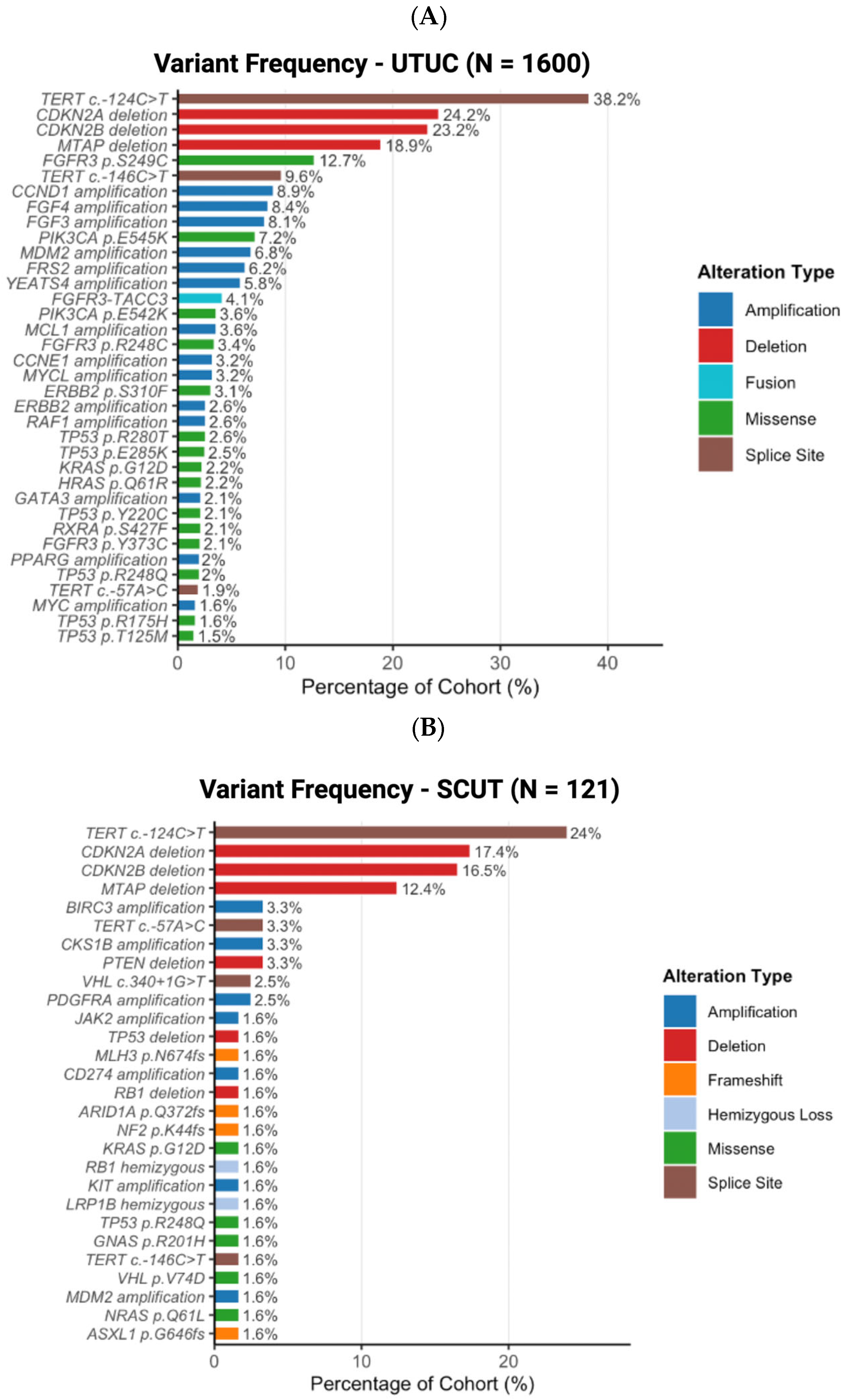
Clinically significant mutational variants identified in the SCUT (N = 121) and UTUC (N = 1600) cohorts were visualized using custom oncoplot-style bar charts. For each cohort, variants were classified by alteration type, including deletions, amplifications, hemizygous loss, missense, frameshift, fusion, and splice site mutations. Categories for specific alterations were based on the standard nomenclature of variant annotations. Variants were then filtered to those occurring at a frequency of ≥1.5% within their respective cohort and ranked in descending order of prevalence. The percentage of affected cases within each cohort was displayed on the x-axis to facilitate proportional comparison across cohorts. This was developed using R Statistical Software (version 4.5.3).

3. Results

3.1. Patient Demographics and Clinicopathologic Features

A total of 1721 patients meeting the inclusion criteria were identified in Tempus Lens, of which 1600 (93%) had UTUC and 121 (7%) had SCUT. Overall, the majority of patients were male (57% UTUC and 66% SCUT) and White (50% UTUC and 46% SCUT). Patients with SCUT were more likely to be younger at diagnosis (median age 61 vs. 71 years, p < 0.001), and have clinical T2 (12% vs. 2%) and T3 (36% vs. 26%) disease at diagnosis compared to patients with UTUC (all p < 0.001). Patients with SCUT were more likely to have T4 (13% vs. 11%), N1 (13% vs. 9%), and M1 (19% vs. 13%) disease at diagnosis compared to patients with UTUC (both p < 0.05). Microsatellite Instability-High (MSI-H) status was more prevalent in patients with UTUC than in those with SCUT (3% vs. 1%), although the difference was not statistically significant (Table 1). Distinct patterns of metastatic spread were observed between groups. In line with a more aggressive clinical course, patients with SCUT were more likely to have visceral disease and metastasis to the lung (44% vs. 21%), pleura (6% vs. 1%), brain (7% vs. 1%), bone (31% vs. 17%), and pancreas (3% vs. <1%) were more common compared to patients with UTUC. Conversely, patients with UTUC predominantly exhibit lymphatic metastatic spread (37% vs. 12%), involving both regional and non-regional lymph nodes, and liver (15% vs. 11%) compared to patients with SCUT (p < 0.001).

3.2. Genomic Landscape

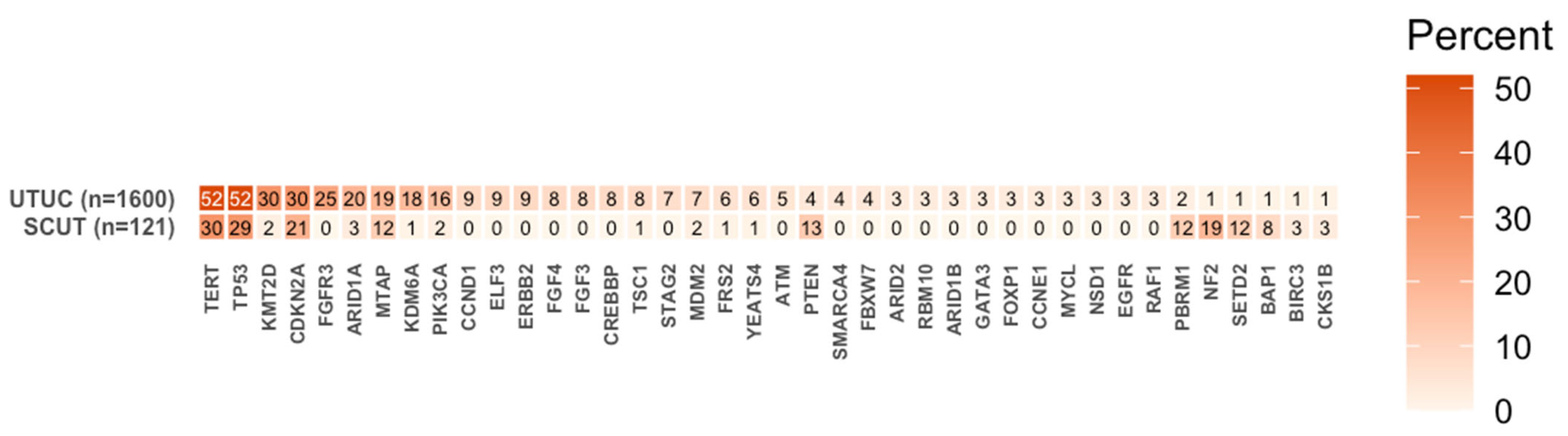
Among SCUT patients, the most prevalent genomic alterations were TERT (30%), TP53 (29%), NF2 (19%), PTEN (13%), SETD2 (12%), PBRM1 (12%), BAP1 (8%), and ARID1A (3%). Among UTUC patients, the most common genomic alterations were TERT (52%), TP53 (52%), KMT2D (30%), FGFR3 (25%), ARID1A (20%), and KDM6A (18%). Comparative analysis demonstrated that SCUT samples were significantly enriched for alterations in NF2, SETD2, PBRM1, PTEN, and BAP1 (all p < 0.05). Compared to UTUC, SCUT samples showed significantly lower mutational rates in FGFR3 and FGF4 (both p < 0.05), with non-significant depletions also observed in ERBB2, ATM, GATA3, FOXP1, and EGFR (Table 2). Among SCUT samples, potentially targetable alterations included NF2, SETD2, and PTEN (Figure 1).
The frequency of clinically significant mutational variants across both cohorts are shown in Figure 2. In both the SCUT and UTUC cohorts, TERT c.-124C>T was the most frequently identified variant, occurring in 24.0% and 38.2% of cases, respectively. Deletions of CDKN2A, CDKN2B, and MTAP were the next most prevalent alterations in both groups, with higher frequencies observed in the UTUC group. Beyond these variants, the two cohorts demonstrated distinct mutational variant landscapes. For example, the UTUC cohort showed a high prevalence of FGFR3 missense variants, including p.S249C (12.7%), p.R248C (3.4%), and p.Y373C (2.1%). In contrast, the SCUT cohort had a higher prevalence of splice-site variants, including VHL c.340+1G>T (2.5%).

4. Discussion

The present study compares the genomic landscape between patients with UTUC and SCUT. Our results show that patients with SCUT are significantly younger at diagnosis and present with a higher disease stage than patients with UTUC. Similarly, patients with SCUT show visceral metastatic tropism, with lung, pleura, bones, brain, and pancreas predominantly affected. Conversely, patients with UTUC predominantly exhibit lymphatic metastatic spread, involving both regional and non-regional lymph nodes. The distinct genomic mutational profiles of these disease types highlight their different biological characteristics and divergent differentiation.
Patients with SCUT show evidence of adverse clinicopathologic features, including a younger age, higher tumor stage, and nodal involvement at diagnosis. Sarcomatoid differentiation results from epithelial-to-mesenchymal transition (EMT), characterized by increased expression of tumor growth factor-β (TGF-β), epithelial growth factor (EGF), and insulin-like growth factor (IGF), along with reduced levels of epithelial cytokeratin markers. This transformation has been associated with advanced disease stages, aggressive clinical behavior, and poor oncologic outcomes in solid malignancies such as lung, breast, and colon cancers cancer [21,22,23]. In the context of bladder cancer, Liu and colleagues evaluated the prognostic significance of sarcomatoid differentiation and found that it was associated with worse 5-year cancer-specific survival and recurrence-free survival compared to patients with non-sarcomatoid carcinoma. Crucially, they observed a prognostic gradient based on the degree of histologic involvement, with patients with a low sarcomatoid component (1–50%) having a better prognosis than those with a high proportion (>50%) [24]. Similarly, Sirgiovanni et al. reported that within a cohort of 73 patients with high-grade urothelial carcinoma, the sarcomatoid subset (8%) universally exhibited advanced tumor stage, namely pT3 (67%) and pT4 (33%), and a high rate of regional lymph node involvement (67%) [15]. After a median follow-up of 33 months, all patients with sarcomatoid differentiation had local recurrence, irrespective of surgical pathology staging [15]. Similarly, Krabbe and colleagues compared outcomes and disease behavior between patients with UTUC and primary pelvicalyceal tumors against primary ureteral tumors. Interestingly, the number of altered markers and alteration status of markers were not significantly different between patients with primary pelvicalyceal versus ureteral tumors when stratified by tumor stage and nodal status, as well as no significant differences in survival outcomes between the two groups when stratified by the number of altered markers (p21, p27, p53, cyclin E, and Ki-67) [25]. Our findings mirror these results, as we identified higher disease stages at diagnosis and a metastatic profile with visceral tropism among patients with SCUT. This may indicate that sarcomatoid histology drives an inherently aggressive disease biology in urothelial carcinoma, regardless of the anatomic origin, and suggests that patients with SCUT could benefit from a more intense treatment pathway, due to the higher prevalence of advanced disease stage at diagnosis and visceral organ involvement.
Identifying specific clusters of signatures that correlate with tumor behavior clinically is of utmost importance for bridging genomic alterations with clinically actionable efforts. To this end, Fuji and colleagues performed a study of 199 UTUC samples to delineate the genetic landscape and classify tumors into five subtypes based on the mutational status of TP53, MDM2, RAS, and FGFR3 [26]. For example, tumors harboring mutations in TP53 and CCND1 were more commonly associated with invasive UTUC, whereas alterations in FGFR3, HRAS, and TERT were more frequent in non-invasive tumors. This allowed them to describe specific clinical patterns associated with genomic alterations, yielding five unique clusters: hypermutated, TP53/MDM2-mutated, RAS-mutated, FGFR3-mutated, and triple-negative. Tumors pertaining to the RAS-mutated cohort frequently showed high-grade histology, squamous differentiation, and an overall aggressive phenotype. While extrapolating these clusters is unfeasible in our present cohort, these results underscore the importance of clustering genomic alterations based on pathways that could mechanistically explain clinical patterns. For example, patients with SCUT histology in our cohort harbored predominantly alterations in TERT and TP53 and were largely devoid of FGFR3 alterations, distinguishing them from the mutational pathways observed in UTUC.
Our results indicate a low incidence of MSI tumors in patients with UTUC and SCUT. MSI status is an important prognostic tumor market, as its presence has been associated with better outcomes and improved response to adjuvant chemotherapy and immunotherapy compared with tumors with stable microsatellite regions [27,28]. One notable example is the phase III KEYNOTE-045 trial of pembrolizumab versus paclitaxel, docetaxel, or vinflunine in recurrent advanced urothelial cancer. A total of 75 patients with upper tract disease were included, with results showing a significant clinical benefit of pembrolizumab over chemotherapy for both patients with upper tract and primary bladder carcinoma. While no histological or MSI substratification was performed for patients with upper tract carcinoma, it does provide an early signal to the potential role of immunotherapy in this patient population [29].
Patients with SCUT and UTUC reveal distinct metastatic patterns, with SCUT patients showing tropism for visceral involvement (lung, brain, bone, and pancreas), and UTUC patients showing lymphatic-predominant spread. This was recently analyzed by Drouaud and colleagues, who evaluated the impact of variant histology on metastatic sites at presentation in patients with bladder cancer. Their results found that distinct variant histology subgroups exhibited independent patterns of organotropism, with sarcomatoid histology showing a predominance of lung involvement as the primary metastatic site, followed by bone, nonregional lymph nodes, liver, and brain. Interestingly, this was in direct contrast to patients with other variant histologies, as patients with squamous cell and small cell variants showed tropism for the brain and liver, respectively, as their primary sites [30]. In a similar study of 398 patients with carcinoma of the upper urinary tract with variant histology, Rink et al. found that histological variants were associated with tumor multifocality, sessile tumor architecture, tumor necrosis, and lymphovascular invasion. Indeed, these adverse tumor-related characteristics are biologic elements that can predict a more aggressive disease course. Importantly, these are not mutually exclusive to any given variant, as tumors can harbor multiple histologic components [31]. To this end, a similar study by Holmäng et al., comparing patients with pure UTUC and squamous histology-predominant carcinoma of the upper urinary tract, found that patients with squamous differentiation had larger tumors at diagnosis and higher tumor stage, despite comparable rates of lymphovascular invasion [32]. Indeed, evidence supports the notion of an overlapping spectrum of adverse macro and microscopic pathologic features in variant histologies of the upper urinary tract. While the distinct mutational profile of SCUT may contribute to its metastatic patterns, further research is necessary to identify specific drivers of organotropism.
The distinct, non-overlapping mutational profiles between UTUC and SCUT highlight their divergent clonal origins and suggest a unique tumor biology that warrants distinct therapeutic considerations. In particular, we found that SCUT samples were depleted in potential targetable alterations, including FGFR3, ERBB2, ATM, FOXP1, and EGFR. The notable absence of FGFR3 alterations in SCUT samples is particularly problematic, as this represents one of the primary therapeutic targets for drug design in upper tract disease. Due to the historical lack of clinical trials dedicated to SCUT, management has largely relied on extrapolating recommendations from conventional urothelial carcinoma. However, our analysis reveals that the non-overlapping profile might render this approach biologically unfounded. Future clinical trials must therefore prioritize designs that target specific genomic alterations enriched in SCUT samples, such as NF2, SETD2, and PTEN. While the therapeutic armamentarium targeting these alterations is currently limited and may be at different stages of the investigational pathway, we summarize a non-exhaustive list of clinical trials evaluating drugs that could potentially target these SCUT-enriched alterations in Table 3. While the present study does not establish causality between specific genomic alterations and clinical behavior, the distinct mutational profiles observed between SCUT and UTUC offer insight into the underlying biology of each entity. For example, TERT promoter mutations were the most frequently found variant across both cohorts. This was further described by Wang et al., who showed that TERT promoter mutations are more characteristic of UTUC and are associated with more aggressive disease [33]. Interestingly, the co-occurrence in the SCUT group further denotes a potential clonal origin between both entities. Conversely, FGFR3-related variants were found almost exclusively in the UTUC group, consistent with the published literature reporting that up to 75% of UTUC tumors exhibit this alteration. Clinically, Katims et al. also describe that FGFR3-mutated patients with UTUC exhibit a T-cell phenotype characterized by more exhausted Th17-like CD4 T cells, fewer regulatory T cells, and more CD8/cytotoxic cells in the naïve state, with a lower response to IL-1 and TNF, which may have clinical implications when exposed to immune system-stimulating drugs [34].
Our study is limited by its retrospective design. Since genomic profiling is not performed routinely across all centers, there may be selection bias in including patients treated at tertiary academic centers or those with more aggressive disease that warrant genomic analysis. Importantly, the dataset includes both tissue-based and blood-based sequencing assays, which can show sensitivity variations. The distribution of tissue-only and blood-only profiling across both cohorts was not available, which can introduce variability in detection rates and should be considered when interpreting inter-cohort genomic comparisons. It is also possible that among patients with SCUT, additional co-occurring histologies were present. Importantly, not all patients had clinical staging or metastatic data available, and comprehensive clinical variables like race, diagnostic and treatment details, and clinical course, were unavailable for patients, reflecting the inherent constraints of real-world genomic databases. Any given patient could have multiple sites of metastatic involvement, impacting the overall rates of metastases. Similarly, patients with predominantly sarcomatoid differentiation were included in the SCUT group, and no stratification based on percentage of sarcomatoid differentiation was performed. Finally, the absence of survival and treatment-related information precludes us from correlating genomic drivers with oncologic outcomes or longitudinal mutational profiles under treatment pressure. As such, all genomic findings should be interpreted as hypothesis-generating and require additional prospective validation before informing clinical decision-making. Additionally, effect sizes and confidence intervals could not be estimated, as alteration frequencies are reported at the cohort level.

5. Conclusions

To our knowledge, this is one of the largest genomic comparisons between SCUT and UTUC. Our findings reveal that SCUT is a distinct biological entity from UTUC, with more aggressive clinicopathologic behavior characterized by a younger age at diagnosis, higher tumor stage, and lymph node involvement. In addition, our results underscore the unique visceral metastatic organotropism of SCUT, which predominantly affects the lung, pleura, brain, bone, and pancreas. The depletion of FGFR3, ERBB2, ATM, FOXP1, and EGFR alterations in SCUT samples underscores the need to design trials tailored to individual mutational profiles and to consider alternative strategies, such as antigen mapping via immunohistochemistry, to leverage novel therapeutic options like immunotherapy and antibody-drug conjugates.

Author Contributions

S.J.-C. and N.J.S. contributed to conceptualization, data curation, formal analysis, writing of the original draft, and writing—review and editing. M.Z., V.G., A.M., K.S., R.W., R.B.-C., J.F., G.Z., B.M., D.V.C., N.D. and A.C.-R. contributed to methodology, data curation, and writing—review and editing. J.H. contributed to methodology, supervision, and project administration. T.D., W.Y. and S.K.P. contributed to supervision, validation, and writing—review and editing. A.T. contributed to conceptualization, supervision, validation, and writing—review and editing. All authors have read and agreed to the published version of the manuscript.

Funding

This research received no external funding.

Institutional Review Board Statement

This is a retrospective study utilizing de-identified data obtained from the Tempus platform data repository, as detailed in the Section 2. Given the retrospective nature of the study and the use of fully de-identified data, IRB approval is not required.

Informed Consent Statement

Given the retrospective nature of the study and the use of fully de-identified data, patient consent forms is not required.

Data Availability Statement

The data that support the findings of this study are available upon reasonable request from the corresponding author.

Conflicts of Interest

A.T. has received consulting fees from Deka Biosciences, Aadi Biosciences, Seattle Genetics/Astellas, Exelixis, Bayer and Gilead Sciences and research funding from Corvus Pharmaceuticals, EMD Serono and Aravive, Inc. S.K.P. reports honoraria and a consulting/advisory role with Novartis, Astellas Pharma, Pfizer, Aveo, Myriad Pharmaceuticals, Roche/Genentech, Exelixis, Bristol-Myers Squibb, Ipsen, and Eisai and honoraria and research funding from Medivation.

References

  1. Rolim, I.; Henriques, V.; Rolim, N.; Blanca, A.; Marques, R.C.; Volavšek, M.; Carvalho, I.; Montironi, R.; Cimadamore, A.; Raspollini, M.R.; et al. Clinicopathologic Analysis of Upper Urinary Tract Carcinoma with Variant Histology. Virchows Arch. 2020, 477, 111–120. [Google Scholar] [CrossRef]
  2. Lonati, C.; Simeone, C.; Suardi, N.; Spiess, P.E.; Necchi, A.; Moschini, M. Genitourinary Manifestations of Lynch Syndrome in the Urological Practice. Asian J. Urol. 2022, 9, 443–450. [Google Scholar] [CrossRef]
  3. Yip, W.; Jaime-Casas, S.; Kothari, A.; Sullivan, M.; Ballas, L.K.; Escobar, D.; Schuckman, A.K.; Rosenberg, J.E.; Coleman, J.A. Urothelial Carcinoma: Perioperative Considerations from Top to Bottom. CA A Cancer J. Clin. 2025, 75, 528–551. [Google Scholar] [CrossRef] [PubMed]
  4. Lonati, C.; Necchi, A.; Gómez Rivas, J.; Afferi, L.; Laukhtina, E.; Martini, A.; Ventimiglia, E.; Colombo, R.; Gandaglia, G.; Salonia, A.; et al. Upper Tract Urothelial Carcinoma in the Lynch Syndrome Tumour Spectrum: A Comprehensive Overview from the European Association of Urology—Young Academic Urologists and the Global Society of Rare Genitourinary Tumors. Eur. Urol. Oncol. 2022, 5, 30–41. [Google Scholar] [CrossRef] [PubMed]
  5. Ohara, K.; Rendeiro, A.F.; Bhinder, B.; Eng, K.W.; Ravichandran, H.; Nguyen, D.; Pisapia, D.; Vosoughi, A.; Fernandez, E.; Shohdy, K.S.; et al. The Evolution of Metastatic Upper Tract Urothelial Carcinoma through Genomic-Transcriptomic and Single-Cell Protein Markers Analysis. Nat. Commun. 2024, 15, 2009. [Google Scholar] [CrossRef] [PubMed]
  6. Mitra, A.P.; Fairey, A.S.; Skinner, E.C.; Boorjian, S.A.; Frank, I.; Schoenberg, M.P.; Bivalacqua, T.J.; Hyndman, M.E.; Reese, A.C.; Steinberg, G.D.; et al. Implications of Micropapillary Urothelial Carcinoma Variant on Prognosis Following Radical Cystectomy: A Multi-Institutional Investigation. Urol. Oncol. Semin. Orig. Investig. 2019, 37, 48–56. [Google Scholar] [CrossRef] [PubMed]
  7. Abufaraj, M.; Foerster, B.; Schernhammer, E.; Moschini, M.; Kimura, S.; Hassler, M.R.; Preston, M.A.; Karakiewicz, P.I.; Remzi, M.; Shariat, S.F. Micropapillary Urothelial Carcinoma of the Bladder: A Systematic Review and Meta-Analysis of Disease Characteristics and Treatment Outcomes. Eur. Urol. 2019, 75, 649–658. [Google Scholar] [CrossRef] [PubMed]
  8. Vetterlein, M.W.; Wankowicz, S.A.M.; Seisen, T.; Lander, R.; Löppenberg, B.; Chun, F.K.-H.; Menon, M.; Sun, M.; Barletta, J.A.; Choueiri, T.K.; et al. Neoadjuvant Chemotherapy Prior to Radical Cystectomy for Muscle-invasive Bladder Cancer with Variant Histology. Cancer 2017, 123, 4346–4355. [Google Scholar] [CrossRef] [PubMed]
  9. Hayashi, H.; Mann, S.; Kao, C.; Grignon, D.; Idrees, M.T. Variant Morphology in Upper Urinary Tract Urothelial Carcinoma: A 14-Year Case Series of Biopsy and Resection Specimens. Hum. Pathol. 2017, 65, 209–216. [Google Scholar] [CrossRef] [PubMed]
  10. Nogueira, L.M.; Yip, W.; Assel, M.J.; Tracey, A.T.; Wong, N.C.; Alvim, R.G.; Al-Ahmadie, H.; Bajorin, D.F.; Coleman, J.A. Survival Impact of Variant Histology Diagnosis in Upper Tract Urothelial Carcinoma. J. Urol. 2022, 208, 813–820. [Google Scholar] [CrossRef] [PubMed]
  11. Genitsch, V.; Kollár, A.; Vandekerkhove, G.; Blarer, J.; Furrer, M.; Annala, M.; Herberts, C.; Pycha, A.; De Jong, J.J.; Liu, Y.; et al. Morphologic and Genomic Characterization of Urothelial to Sarcomatoid Transition in Muscle-Invasive Bladder Cancer. Urol. Oncol. Semin. Orig. Investig. 2019, 37, 826–836. [Google Scholar] [CrossRef] [PubMed]
  12. Amin, M.B. Histological Variants of Urothelial Carcinoma: Diagnostic, Therapeutic and Prognostic Implications. Mod. Pathol. 2009, 22, S96–S118. [Google Scholar] [CrossRef] [PubMed]
  13. Thomsen, M.B.H.; Nordentoft, I.; Lamy, P.; Vang, S.; Reinert, L.; Mapendano, C.K.; Høyer, S.; Ørntoft, T.F.; Jensen, J.B.; Dyrskjøt, L. Comprehensive Multiregional Analysis of Molecular Heterogeneity in Bladder Cancer. Sci. Rep. 2017, 7, 11702. [Google Scholar] [CrossRef] [PubMed]
  14. Wang, X.; MacLennan, G.T.; Zhang, S.; Montironi, R.; Lopez-Beltran, A.; Tan, P.-H.; Foster, S.; Baldridge, L.A.; Cheng, L. Sarcomatoid Carcinoma of the Upper Urinary Tract: Clinical Outcome and Molecular Characterization. Hum. Pathol. 2009, 40, 211–217. [Google Scholar] [CrossRef] [PubMed]
  15. Sirgiovanni, G.; Francesconi, S.; Guida, A.; Caserta, C.; Mosillo, C.; Calandrella, M.L.; Macrini, S.; Cortesi, E.; Ascani, S.; Bracarda, S. Urothelial Carcinoma with Sarcomatoid Variant: A Preliminary Study of Clinical and Pathological Features. J. Clin. Oncol. 2022, 40, 571. [Google Scholar] [CrossRef]
  16. Finkle, J.D.; Boulos, H.; Driessen, T.M.; Lo, C.; Blidner, R.A.; Hafez, A.; Khan, A.A.; Lozac’hmeur, A.; McKinnon, K.E.; Perera, J.; et al. Validation of a Liquid Biopsy Assay with Molecular and Clinical Profiling of Circulating Tumor DNA. npj Precis. Onc. 2021, 5, 63. [Google Scholar] [CrossRef] [PubMed]
  17. Kapilivsky, J.; Islam, F.; Roth, E.K.; Dow, J.; Moran, S.; Scherrer, E.; Hyun, S.W.; Sangli, C. Validation of a Composite Mortality End Point in a Large Clinicogenomic Real-World Database of Patients With Advanced Cancer. JCO Clin. Cancer Inf. 2026, 10, e2500291. [Google Scholar] [CrossRef] [PubMed]
  18. Jani, C.T.; Tran, E.; Jaeger, E.; Dong, J.; Husni, N.; Stoppler, M.C.; Hockenberry, A.J.; Mercer, J.; Pal, S.; Agarwal, N.; et al. Concurrent Tissue and Circulating Tumor DNA Analysis in Renal Cell Carcinoma: Insights from a Multimodal Database. Oncologist 2026, 31, oyag123. [Google Scholar] [CrossRef] [PubMed]
  19. Beaubier, N.; Tell, R.; Lau, D.; Parsons, J.R.; Bush, S.; Perera, J.; Sorrells, S.; Baker, T.; Chang, A.; Michuda, J.; et al. Clinical Validation of the Tempus xT Next-Generation Targeted Oncology Sequencing Assay. Oncotarget 2019, 10, 2384–2396. [Google Scholar] [CrossRef] [PubMed]
  20. Beaubier, N.; Tell, R.; Huether, R.; Bontrager, M.; Bush, S.; Parsons, J.; Shah, K.; Baker, T.; Selkov, G.; Taxter, T.; et al. Clinical Validation of the Tempus xO Assay. Oncotarget 2018, 9, 25826–25832. [Google Scholar] [CrossRef] [PubMed]
  21. Arshi, J.; Yin, F. Sarcomatoid Transformation in Carcinomas: Is Precision Medicine the Answer? J. Cancer Treat. Diagn. 2020, 4, 6–11. [Google Scholar] [CrossRef]
  22. Day, E.; Gavira, J.; Tapia, J.C.; Anguera, G.; Maroto, P. What About Variant Histologies in Bladder Cancer? Eur. Urol. Focus 2024, 10, 227–230. [Google Scholar] [CrossRef] [PubMed]
  23. Gandhi, J.; Chen, J.-F.; Al-Ahmadie, H. Urothelial Carcinoma. Surg. Pathol. Clin. 2022, 15, 641–659. [Google Scholar] [CrossRef] [PubMed]
  24. Liu, S.; Yao, Y.; Wang, Z.-K.; Sun, L.-J.; Zhang, G.-M. Prognostic Value of the Sarcomatoid Component in Bladder Cancer: A Propensity Score Matching Study. Oncol. Lett. 2023, 25, 103. [Google Scholar] [CrossRef] [PubMed]
  25. Krabbe, L.-M.; Bagrodia, A.; Westerman, M.E.; Gayed, B.A.; Haddad, A.Q.; Sagalowsky, A.I.; Shariat, S.F.; Kapur, P.; Lotan, Y.; Margulis, V. Molecular Profile of Urothelial Carcinoma of the Upper Urinary Tract: Are Pelvicalyceal and Ureteral Tumors Different? World J. Urol. 2016, 34, 105–112. [Google Scholar] [CrossRef] [PubMed]
  26. Fujii, Y.; Sato, Y.; Suzuki, H.; Kakiuchi, N.; Yoshizato, T.; Lenis, A.T.; Maekawa, S.; Yokoyama, A.; Takeuchi, Y.; Inoue, Y.; et al. Molecular Classification and Diagnostics of Upper Urinary Tract Urothelial Carcinoma. Cancer Cell 2021, 39, 793–809.e8. [Google Scholar] [CrossRef] [PubMed]
  27. Rouprêt, M.; Fromont, G.; Azzouzi, A.-R.; Catto, J.W.; Vallancien, G.; Hamdy, F.C.; Cussenot, O. Microsatellite Instability as Predictor of Survival in Patients with Invasive Upper Urinary Tract Transitional Cell Carcinoma. Urology 2005, 65, 1233–1237. [Google Scholar] [CrossRef] [PubMed]
  28. Ribic, C.M.; Sargent, D.J.; Moore, M.J.; Thibodeau, S.N.; French, A.J.; Goldberg, R.M.; Hamilton, S.R.; Laurent-Puig, P.; Gryfe, R.; Shepherd, L.E.; et al. Tumor Microsatellite-Instability Status as a Predictor of Benefit from Fluorouracil-Based Adjuvant Chemotherapy for Colon Cancer. N. Engl. J. Med. 2003, 349, 247–257. [Google Scholar] [CrossRef] [PubMed]
  29. Fradet, Y.; Bellmunt, J.; Vaughn, D.J.; Lee, J.L.; Fong, L.; Vogelzang, N.J.; Climent, M.A.; Petrylak, D.P.; Choueiri, T.K.; Necchi, A.; et al. Randomized Phase III KEYNOTE-045 Trial of Pembrolizumab versus Paclitaxel, Docetaxel, or Vinflunine in Recurrent Advanced Urothelial Cancer: Results of >2 years of follow-up. Ann. Oncol. 2019, 30, 970–976. [Google Scholar] [CrossRef] [PubMed]
  30. Drouaud, A.; Xu, V.; Velasquez, A.; Antar, R.; Boyarsky, B.; Weiss, J.; Gonzalez, D.; Silverman, R.; Whalen, M.J. Metastatic Tropism in Urothelial Carcinoma With Variant Histology: A Comprehensive NCDB Analysis. Clin. Genitourin. Cancer 2024, 22, 102179. [Google Scholar] [CrossRef] [PubMed]
  31. Rink, M.; Robinson, B.D.; Green, D.A.; Cha, E.K.; Hansen, J.; Comploj, E.; Margulis, V.; Raman, J.D.; Ng, C.K.; Remzi, M.; et al. Impact of Histological Variants on Clinical Outcomes of Patients with Upper Urinary Tract Urothelial Carcinoma. J. Urol. 2012, 188, 398–404. [Google Scholar] [CrossRef] [PubMed]
  32. Holmäng, S.; Lele, S.M.; Johansson, S.L. Squamous Cell Carcinoma of the Renal Pelvis and Ureter: Incidence, Symptoms, Treatment and Outcome. J. Urol. 2007, 178, 51–56. [Google Scholar] [CrossRef] [PubMed]
  33. Wang, K.; Liu, T.; Liu, L.; Liu, J.; Liu, C.; Wang, C.; Ge, N.; Ren, H.; Yan, K.; Hu, S.; et al. TERT Promoter Mutations in Renal Cell Carcinomas and Upper Tract Urothelial Carcinomas. Oncotarget 2014, 5, 1829–1836. [Google Scholar] [CrossRef] [PubMed]
  34. Katims, A.B.; Kuo, F.; Reisz, P.; Tracey, A.; Thomas, J.; Yip, W.; Merghoub, T.; Bochner, B.H.; Pietzak, E.J.; Solit, D.B.; et al. Characterizing the Immune Phenotype of FGFR3 Mutated Upper Tract Urothelial Carcinoma (UTUC) Using Single-Cell (Sc)RNA-Sequencing (Seq). J. Clin. Oncol. 2023, 41, 558. [Google Scholar] [CrossRef]
Figure 1. Prevalence of genomic alterations for patients with UTUC and SCUT.
Cancers 18 02352 g001
Figure 2. Clinically significant mutational variants frequency for UTUC (A) and SCUT (B) cohorts.
Cancers 18 02352 g002
Table 1. Baseline characteristics for patients with UTUC and SCUT.
Variable UTUC (n = 1600)SCUT (n = 121)p Value
Age (median, IQR) <0.001
71 (64, 77)61 (54, 70)
Sex (No. %) 0.06
Female686 (43%)41 (34%)
Male914 (57%)80 (66%)
Missing0 (0%)0 (0%)
Race (No. %) 0.19
White800 (50%)56 (46%)
Other Race62 (4%)4 (3%)
Black or African American57 (4%)9 (7%)
Asian52 (3%)3 (3%)
Missing629 (39%)49 (41%)
Ethnicity (No. %) 0.07
Not Hispanic or Latino650 (41%)48 (40%)
Hispanic or Latino52 (3%)8 (6%)
Missing898 (56%)65 (54%)
ECOG Score (No. %) 0.91
1627 (39%)46 (38%)
0543 (34%)35 (29%)
2256 (16%)16 (13%)
385 (5%)4 (3%)
413 (1%)0 (0%)
Missing 76 (5%)20 (17%)
Smoking Status (No. %) 0.35
Never smoker245 (16%)26 (21%)
Ex-smoker70 (4%)9 (8%)
Current smoker62 (4%)3 (2%)
Missing 1223 (76%)83 (69%)
MSI (No. %) 0.36
Stable1272 (80%)88 (73%)
High50 (3%)1 (1%)
Missing 278 (17%)32 (26%)
T Stage (No. %) <0.001
Ta/Tis104 (7%)0 (0%)
T1143 (9%)1 (1%)
T2186 (12%)2 (2%)
T3590 (36%)32 (26%)
T4175 (11%)16 (13%)
Missing 402 (25%)70 (58%)
N Stage (No. %) <0.001
N0423 (26%)16 (13%)
N1144 (9%)16 (13%)
N297 (6%)0 (0%)
N36 (<1%)0 (0%)
NX289 (18%)29 (24%)
Missing 641 (40%)60 (50%)
M Stage (No. %) <0.001
M0365 (23%)11 (9%)
M1203 (13%)23 (19%)
Mx109 (7%)2 (2%)
Missing923 (57%)85 (70%)
Metastatic site (No.%) <0.001
Lymph nodes595 (37%)14 (12%)
Lung334 (21%)53 (44%)
Bone264 (17%)38 (31%)
Liver247 (15%)13 (11%)
Soft tissue58 (4%)14 (12%)
Other soft tissue246 (15%)37 (30%)
ECOG, Eastern Cooperative Oncology Group; IQR, interquartile range; MSI, microsatellite instability; SCUT, sarcomatoid carcinoma of the upper tract; UTUC, upper tract urothelial carcinoma.
Table 2. Prevalence of the most common genomic alterations in patients with UTUC and SCUT.
GeneUTUC (n = 1600)SCUT (n = 121)p Value
TERT827 (52%)36 (30%)<0.001
NF218 (1%)23 (19%)<0.001
KMT2D484 (30%)2 (2%)<0.001
FGFR3396 (25%)0 (0%)<0.001
SETD215 (1%)14 (12%)<0.001
TP53832 (52%)35 (29%)<0.001
KDM6A287 (18%)1 (1%)<0.001
PBRM140 (2%)15 (12%)<0.001
ARID1A323 (20%)4 (3%)<0.001
PTEN69 (4%)16 (13%)<0.001
BAP123 (1%)10 (8%)<0.001
PIK3CA261 (16%)3 (2%)<0.001
CCND1143 (9%)0 (0%)0.01
ELF3140 (9%)0 (0%)0.01
ERBB2137 (9%)0 (0%)0.01
FGF4134 (8%)0 (0%)0.01
FGF3129 (8%)0 (0%)0.01
CREBBP121 (8%)0 (0%)0.02
STAG2113 (7%)0 (0%)0.03
TSC1136 (8%)1 (1%)0.03
SMARCA466 (4%)0 (0%)0.07
ATM82 (5%)0 (0%)0.11
FBXW764 (4%)0 (0%)0.11
BIRC312 (1%)4 (3%)0.11
FRS2100 (6%)1 (1%)0.12
ARID254 (3%)0 (0%)0.14
YEATS493 (6%)1 (1%)0.16
CKS1B15 (1%)4 (3%)0.16
MDM2109 (7%)2 (2%)0.16
RBM1049 (3%)0 (0%)0.16
ARID1B48 (3%)0 (0%)0.16
GATA348 (3%)0 (0%)0.16
FOXP150 (3%)0 (0%)0.16
CCNE151 (3%)0 (0%)0.16
MYCL51 (3%)0 (0%)0.16
CDKN2A481 (30%)26 (21%)0.20
NSD142 (3%)0 (0%)0.23
MTAP312 (19%)15 (12%)0.23
EGFR47 (3%)0 (0%)0.23
RAF147 (3%)0 (0%)0.23
Table 3. Non-exhaustive list of clinical trials exploring therapeutic targets for actionable mutations in SCUT.
Mutational PathwayDrugClinical Trial/NCTPopulationDesign
NF2-related pathwaysCrizotinibLCI-GU-URO-CRI-001/NCT02612194Metastatic urothelial carcinoma of the bladder, upper or lower urinary tractsPhase II
EverolimusRAD001/NCT00805129Advanced urothelial carcinomaPhase II
NCT01184326Previously treated advanced urothelial carcinomaPhase I
HCRN GU10-147/NCT01215136Cisplatin ineligible advanced urothelial carcinomaPhase II
SETD2-related pathwaysTalozaparibTALASUR/NCT04678362Platinum-sensitive metastatic or locally advanced urothelial carcinomaPhase II
DecitabineNCT00030615Advanced solid tumorsPhase I
PTEN-related pathwaysX31005NCT03745911Metastatic urothelial carcinomaPhase II
Disclaimer/Publisher’s Note: The statements, opinions and data contained in all publications are solely those of the individual author(s) and contributor(s) and not of MDPI and/or the editor(s). MDPI and/or the editor(s) disclaim responsibility for any injury to people or property resulting from any ideas, methods, instructions or products referred to in the content.