Laparoscopy Prior to Laparotomy in Primary Debulking Surgery for Advanced Epithelial Ovarian Cancer in the U.S.: A National Cancer Database Analysis †


Simple Summary
Abstract
1. Introduction
2. Methods
3. Results
4. Discussion
5. Conclusions
Author Contributions
Funding
Institutional Review Board Statement
Informed Consent Statement
Data Availability Statement
Conflicts of Interest
References
- Cancer of the Ovary—Cancer Stat Facts, SEER. Available online: https://seer.cancer.gov/statfacts/html/ovary.html (accessed on 6 September 2025).
- O’Malley, D.M.; Krivak, T.C.; Kabil, N.; Munley, J.; Moore, K.N. PARP Inhibitors in Ovarian Cancer: A Review. Target Oncol. 2023, 18, 471–503. [Google Scholar] [CrossRef] [Scilit] [PubMed] [PubMed Central]
- Monk, B.J.; Lorusso, D.; Fujiwara, K.; Sehouli, J. Optimal bevacizumab treatment strategy in advanced ovarian cancer: A review. Cancer Treat. Rev. 2025, 137, 102945. [Google Scholar] [CrossRef] [Scilit] [PubMed]
- Bryant, A.; Hiu, S.; Kunonga, P.T.; Gajjar, K.; Craig, D.; Vale, L.; Winter-Roach, B.A.; Elattar, A.; Naik, R. Impact of residual disease as a prognostic factor for survival in women with advanced epithelial ovarian cancer after primary surgery. Cochrane Database Syst. Rev. 2022, 9, CD015048. [Google Scholar] [CrossRef] [Scilit] [PubMed] [PubMed Central]
- Griffiths, C.T. Surgical resection of tumor bulk in the primary treatment of ovarian carcinoma. Natl. Cancer Inst. Monogr. 1975, 42, 101–104. [Google Scholar] [PubMed]
- Lyons, Y.A.; Reyes, H.D.; McDonald, M.E.; Newtson, A.; Devor, E.; Bender, D.P.; Goodheart, M.J.; Gonzalez Bosquet, J. Interval debulking surgery is not worth the wait: A National Cancer Database study comparing primary cytoreductive surgery versus neoadjuvant chemotherapy. Int. J. Gynecol. Cancer 2020, 30, 845–852. [Google Scholar] [CrossRef] [Scilit] [PubMed] [PubMed Central]
- Luo, Y.; Lee, M.; Kim, H.S.; Chung, H.H.; Song, Y.S. Effect of neoadjuvant chemotherapy on platinum resistance in stage IIIC and IV epithelial ovarian cancer. Medicine 2016, 95, e4797. [Google Scholar] [CrossRef] [Scilit] [PubMed]
- Kehoe, S.; Hook, J.; Nankivell, M.; Jayson, G.C.; Kitchener, H.; Lopes, T.; Luesley, D.; Perren, T.; Bannoo, S.; Mascarenhas, M.; et al. Primary chemotherapy versus primary surgery for newly diagnosed advanced ovarian cancer (CHORUS): An open-label, randomised, controlled, non-inferiority trial. Lancet 2015, 386, 249–257. [Google Scholar] [CrossRef] [Scilit] [PubMed]
- Fagotti, A.; Ferrandina, M.G.; Vizzielli, G.; Pasciuto, T.; Fanfani, F.; Gallotta, V.; Margariti, P.A.; Chiantera, V.; Costantini, B.; Gueli Alletti, S.; et al. Randomized trial of primary debulking surgery versus neoadjuvant chemotherapy for advanced epithelial ovarian cancer (SCORPION-NCT01461850). Int. J. Gynecol. Cancer 2020, 30, 1657–1664. [Google Scholar] [CrossRef] [Scilit] [PubMed]
- Onda, T.; Satoh, T.; Ogawa, G.; Saito, T.; Kasamatsu, T.; Nakanishi, T.; Mizutani, T.; Takehara, K.; Okamoto, A.; Ushijima, K.; et al. Comparison of survival between primary debulking surgery and neoadjuvant chemotherapy for stage III/IV ovarian, tubal and peritoneal cancers in phase III randomised trial. Eur. J. Cancer 2020, 130, 114–125. [Google Scholar] [CrossRef] [Scilit] [PubMed]
- Reuss, A.; du Bois, A.; Harter, P.; Fotopoulou, C.; Sehouli, J.; Aletti, G.; Guyon, F.; Greggi, S.; Mosgaard, B.J.; Reinthaller, A.; et al. TRUST: Trial of Radical Upfront Surgical Therapy in advanced ovarian cancer (ENGOT ov33/AGO-OVAR OP7). Int. J. Gynecol. Cancer 2019, 29, 1327–1331. [Google Scholar] [CrossRef] [Scilit] [PubMed]
- Rutten, M.J.; Gaarenstroom, K.N.; Van Gorp, T.; van Meurs, H.S.; Arts, H.J.; Bossuyt, P.M.; Ter Brugge, H.G.; Hermans, R.H.; Opmeer, B.C.; Pijnenborg, J.M.; et al. Laparoscopy to predict the result of primary cytoreductive surgery in advanced ovarian cancer patients (LapOvCa-trial): A multicentre randomized controlled study. BMC Cancer 2012, 12, 31. [Google Scholar] [CrossRef] [Scilit] [PubMed] [PubMed Central]
- Bristow, R.E.; Duska, L.R.; Lambrou, N.C.; Fishman, E.K.; O’Neill, M.J.; Trimble, E.L.; Montz, F.J. A model for predicting surgical outcome in patients with advanced ovarian carcinoma using computed tomography. Cancer 2000, 89, 1532–1540. [Google Scholar] [CrossRef] [Scilit] [PubMed]
- Axtell, A.E.; Lee, M.H.; Bristow, R.E.; Dowdy, S.C.; Cliby, W.A.; Raman, S.; Weaver, J.P.; Gabbay, M.; Ngo, M.; Lentz, S.; et al. Multi-institutional reciprocal validation study of computed tomography predictors of suboptimal primary cytoreduction in patients with advanced ovarian cancer. J. Clin. Oncol. 2007, 25, 384–389. [Google Scholar] [CrossRef] [Scilit] [PubMed]
- Engbersen, M.P.; Van’T Sant, I.; Lok, C.; Lambregts, D.M.J.; Sonke, G.S.; Beets-Tan, R.G.H.; van Driel, W.J.; Lahaye, M.J. MRI with diffusion-weighted imaging to predict feasibility of complete cytoreduction with the peritoneal cancer index (PCI) in advanced stage ovarian cancer patients. Eur. J. Radiol. 2019, 114, 146–151. [Google Scholar] [CrossRef] [Scilit] [PubMed]
- Sala, E.; Rockall, A.; Rangarajan, D.; Kubik-Huch, R.A. The role of dynamic contrast-enhanced and diffusion weighted magnetic resonance imaging in the female pelvis intro. Eur. J. Radiol. 2010, 76, 367–385. [Google Scholar] [CrossRef] [Scilit] [PubMed]
- Garcia Prado, J.; González Hernando, C.; Varillas Delgado, D.; Saiz Martínez, R.; Bhosale, P.; Blazquez Sanchez, J.; Chiva, L. Diffusion-weighted magnetic resonance imaging in peritoneal carcinomatosis from suspected ovarian cancer: Diagnostic performance in correlation with surgical findings. Eur. J. Radiol. 2019, 121, 108696. [Google Scholar] [CrossRef] [Scilit] [PubMed]
- Dai, G.; Liang, K.; Xiao, Z.; Yang, Q.; Yang, S. A meta-analysis on the diagnostic value of diffusion-weighted imaging on ovarian cancer. J. BUON 2019, 24, 2333–2340. [Google Scholar] [PubMed]
- Straubhar, A.M.; Filippova, O.T.; Cowan, R.A.; Lakhman, Y.; Sarasohn, D.M.; Nikolovski, I.; Torrisi, J.M.; Ma, W.; Abu-Rustum, N.R.; Gardner, G.J.; et al. A multimodality triage algorithm to improve cytoreductive outcomes in patients undergoing primary debulking surgery for advanced ovarian cancer: A Memorial Sloan Kettering Cancer Center team ovary initiative. Gynecol. Oncol. 2020, 158, 608–613. [Google Scholar] [CrossRef] [Scilit] [PubMed] [PubMed Central]
- Kahn, R.M.; Murphy, K.; Oladoja, K.; Yeoshoua, E.; Finch, L.; Harlev, C.; Filippova, O.T.; Straubhar, A.M.; Cowan, R.A.; Iasonos, A.; et al. A prospective comparison of resectability scores to enhance preoperative decision-making in the primary management of advanced ovarian cancer: A Memorial Sloan Kettering Cancer Center Team Ovary study. Int. J. Gynecol. Cancer 2025, 35, 102662. [Google Scholar] [CrossRef] [Scilit] [PubMed]
- Narasimhulu, D.M.; Kumar, A.; Weaver, A.L.; McGree, M.E.; Langstraat, C.L.; Cliby, W.A. Using an evidence-based triage algorithm to reduce 90-day mortality after primary debulking surgery for advanced epithelial ovarian cancer. Gynecol. Oncol. 2019, 155, 58–62. [Google Scholar] [CrossRef] [Scilit] [PubMed]
- Lee, C.L.; Kay, N.; Chen, H.L.; Yen, C.F.; Huang, K.G. The roles of laparoscopy in treating ovarian cancer. Taiwan J. Obstet. Gynecol. 2009, 48, 9–14. [Google Scholar] [CrossRef] [Scilit] [PubMed]
- Rutten, M.J.; Leeflang, M.M.; Kenter, G.G.; Mol, B.W.; Buist, M. Laparoscopy for diagnosing resectability of disease in patients with advanced ovarian cancer. Cochrane Database Syst. Rev. 2014, 2014, CD009786, Update in Cochrane Database Syst. Rev. 2019, 3, CD009786. https://doi.org/10.1002/14651858.CD009786.pub3. [Google Scholar] [CrossRef] [Scilit] [PubMed] [PubMed Central]
- Fagotti, A.; Ferrandina, G.; Fanfani, F.; Ercoli, A.; Lorusso, D.; Rossi, M.; Scambia, G. A laparoscopy-based score to predict surgical outcome in patients with advanced ovarian carcinoma: A pilot study. Ann. Surg. Oncol. 2006, 13, 1156–1161. [Google Scholar] [CrossRef] [Scilit] [PubMed]
- Fagotti, A.; Ferrandina, G.; Fanfani, F.; Garganese, G.; Vizzielli, G.; Carone, V.; Salerno, M.G.; Scambia, G. Prospective validation of a laparoscopic predictive model for optimal cytoreduction in advanced ovarian carcinoma. Am. J. Obstet. Gynecol. 2008, 199, 642.e1–642.e6. [Google Scholar] [CrossRef] [Scilit] [PubMed]
- Lee, Y.J.; Chung, Y.S.; Lee, J.Y.; Nam, E.J.; Kim, S.W.; Kim, Y.T.; Kim, S. Role of diagnostic laparoscopy in deciding primary treatment in advanced-stage ovarian cancer. J. Gynecol. Oncol. 2023, 34, e17. [Google Scholar] [CrossRef] [Scilit] [PubMed] [PubMed Central]
- Goncalves, N.; Silver, M.; Hayek, J.; Lamiman, K.; Hanusek, R.; Alagkiozidis, A. Utilization of laparoscopy prior to laparotomy in primary debulking surgery for advanced epithelial ovarian cancer: A National Cancer Database analysis. Gynecol. Oncol. 2025, 200, 238–239. [Google Scholar] [CrossRef] [Scilit]
- Ataseven, B.; Grimm, C.; Harter, P.; Heikaus, S.; Heitz, F.; Traut, A.; Prader, S.; Kahl, A.; Schneider, S.; Kurzeder, C.; et al. Prognostic Impact of Port-Site Metastasis After Diagnostic Laparoscopy for Epithelial Ovarian Cancer. Ann. Surg. Oncol. 2016, 23, 834–840. [Google Scholar] [CrossRef] [Scilit] [PubMed]
- Żyła, D.; Dubaj, M.; Słomczyńska, K.; Kułak, K.; Tarkowski, R.; Furtak-Niczyporuk, M. Pathogenesis and Possible Consequences of Port-Site Metastasis in Gynecologic Oncology After Laparoscopic Procedures. Cancer Manag. Res. 2025, 17, 3259–3272. [Google Scholar] [CrossRef] [Scilit] [PubMed] [PubMed Central]
- Mubarak, F.; Gabriel, E.M.; Bowers, J.C. Use of the National Cancer Database in Identifying Disparities in Cancer Treatment. Am. Surg. 2025, 91, 719–722. [Google Scholar] [CrossRef] [Scilit] [PubMed]



| Laparotomy Only (n = 12,003) | Laparoscopy Before Laparotomy (n = 529) | p-Value | ||
|---|---|---|---|---|
| Age at Diagnosis | 61 (53–69) 1 | 62 (53–69) 1 | 0.911 | |
| Race | White | 10,514 (87.6%) | 454 (85.8%) | 0.117 |
| Black | 834 (6.9%) | 38 (7.2%) | ||
| Asian/Pacific Islander | 444 (3.7%) | 30 (5.7%) | ||
| Other | 142 (1.2%) | 3 (0.6%) | ||
| Unknown | 69 (0.6%) | 4 (0.8%) | ||
| Ethnicity | Not Hispanic | 11,032 (91.9%) | 488 (92.2%) | 0.26 |
| Hispanic/Presumed Hispanic | 690 (5.7%) | 34 (6.4%) | ||
| Unknown | 281 (2.3%) | 7 (1.3%) | ||
| Insurance | No Insurance | 440 (3.7%) | 9 (1.7%) | 0.075 |
| Private Insurance | 6072 (50.6%) | 279 (52.7%) | ||
| Medicaid/Medicare/Other Public | 5344 (44.5%) | 232 (43.9%) | ||
| Unknown | 147 (1.2%) | 9 (1.7%) | ||
| Stage | IIIC | 8984 (74.8%) | 416 (78.6%) | 0.049 |
| IV | 3019 (25.2%) | 113 (21.4%) | ||
| Charlson–Deyo Score ≥ 1 | 2633 (21.9%) | 119 (22.5%) | 0.761 | |
| No Gross Residual | 4228 (35.2%) | 193 (36.5%) | 0.553 | |
| Extensive Surgery | 6642 (55.3%) | 279 (52.7%) | 0.24 | |
| Length of Stay | 6 (5–9) | 6 (4–8) | <0.001 | |
| Readmission within 30 days | 932 (7.8%) | 46 (8.7%) | 0.435 | |
| 90 Day Mortality | 200 (1.7%) | 4 (0.8%) | 0.106 | |
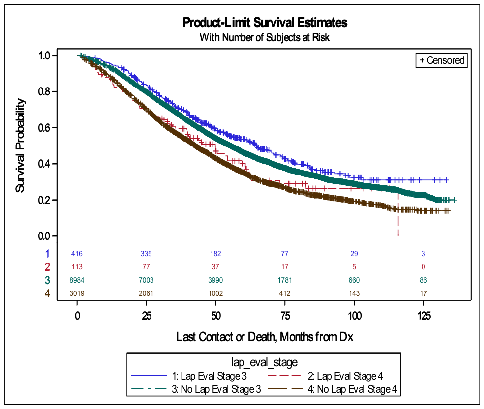
| Overall Survival in Months | 51.6 (50.5–52.7) 2 | 60.8 (54.3–67.2) 2 | 0.012 | |
Disclaimer/Publisher’s Note: The statements, opinions and data contained in all publications are solely those of the individual author(s) and contributor(s) and not of MDPI and/or the editor(s). MDPI and/or the editor(s) disclaim responsibility for any injury to people or property resulting from any ideas, methods, instructions or products referred to in the content. |
© 2026 by the authors. Licensee MDPI, Basel, Switzerland. This article is an open access article distributed under the terms and conditions of the Creative Commons Attribution (CC BY) license.
Share and Cite
Goncalves, N.; Lamiman, K.; Silver, M.; Alagkiozidis, I. Laparoscopy Prior to Laparotomy in Primary Debulking Surgery for Advanced Epithelial Ovarian Cancer in the U.S.: A National Cancer Database Analysis. Cancers 2026, 18, 2195. https://doi.org/10.3390/cancers18142195
Goncalves N, Lamiman K, Silver M, Alagkiozidis I. Laparoscopy Prior to Laparotomy in Primary Debulking Surgery for Advanced Epithelial Ovarian Cancer in the U.S.: A National Cancer Database Analysis. Cancers. 2026; 18(14):2195. https://doi.org/10.3390/cancers18142195
Chicago/Turabian StyleGoncalves, Nicole, Kelly Lamiman, Michael Silver, and Ioannis Alagkiozidis. 2026. "Laparoscopy Prior to Laparotomy in Primary Debulking Surgery for Advanced Epithelial Ovarian Cancer in the U.S.: A National Cancer Database Analysis" Cancers 18, no. 14: 2195. https://doi.org/10.3390/cancers18142195
APA StyleGoncalves, N., Lamiman, K., Silver, M., & Alagkiozidis, I. (2026). Laparoscopy Prior to Laparotomy in Primary Debulking Surgery for Advanced Epithelial Ovarian Cancer in the U.S.: A National Cancer Database Analysis. Cancers, 18(14), 2195. https://doi.org/10.3390/cancers18142195