The Evolving Role of Somatostatin Receptor PET/CT in Medullary Thyroid Carcinoma: An Updated Systematic Review and Meta-Analysis




Simple Summary
Abstract
1. Introduction
2. Materials and Methods
2.1. Protocol
2.2. Literature Search and Study Selection
2.3. Data Extraction and Collection
2.4. Methodological Quality Assessment
2.5. Statistical Analysis
3. Results
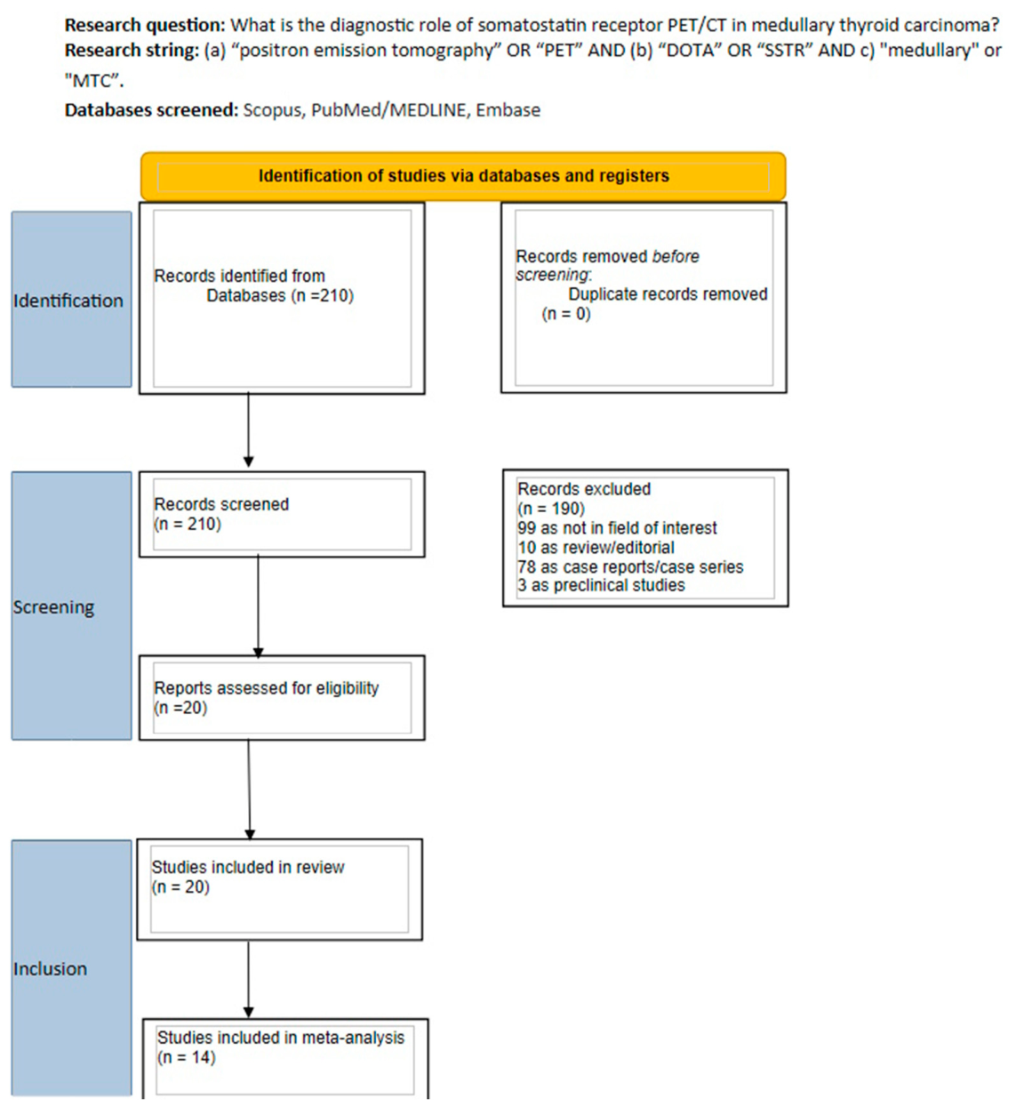
3.1. Study Selection and Quality Report
3.2. Characteristics of Included Studies and Patients
3.3. Characteristics of SSTR PET/CT Imaging
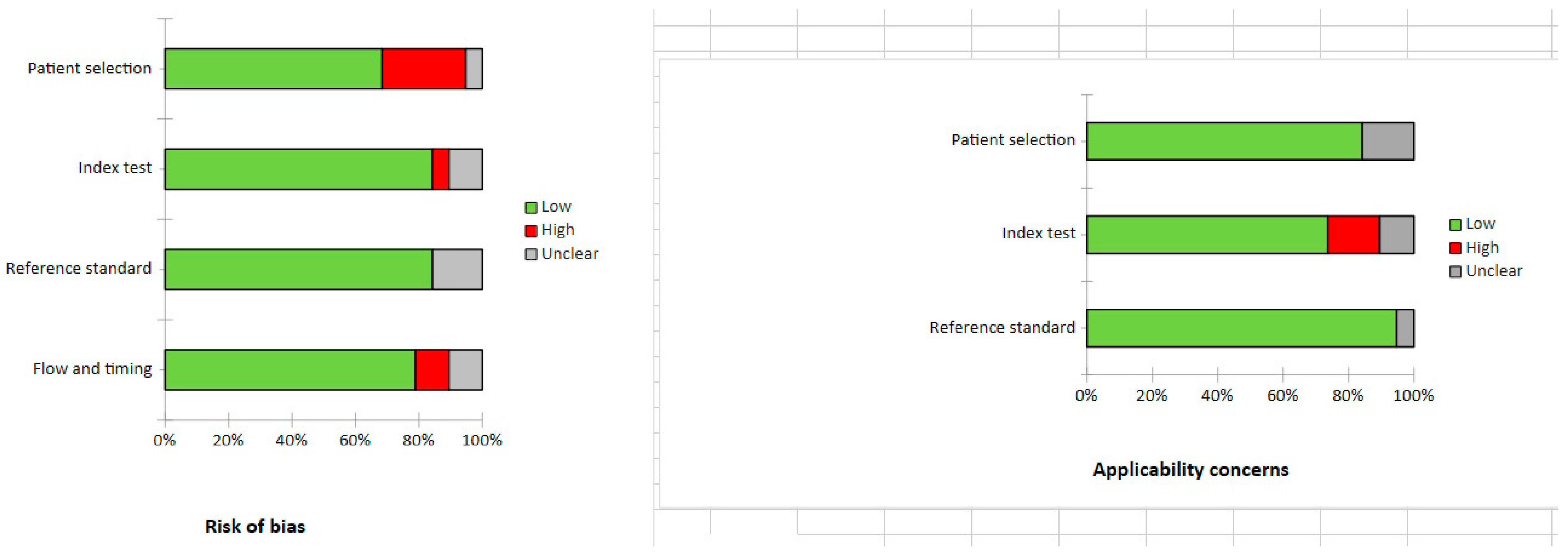
3.4. Risk of Bias and Applicability
3.5. Main Outcomes of SSTR PET/CT Imaging in MTC
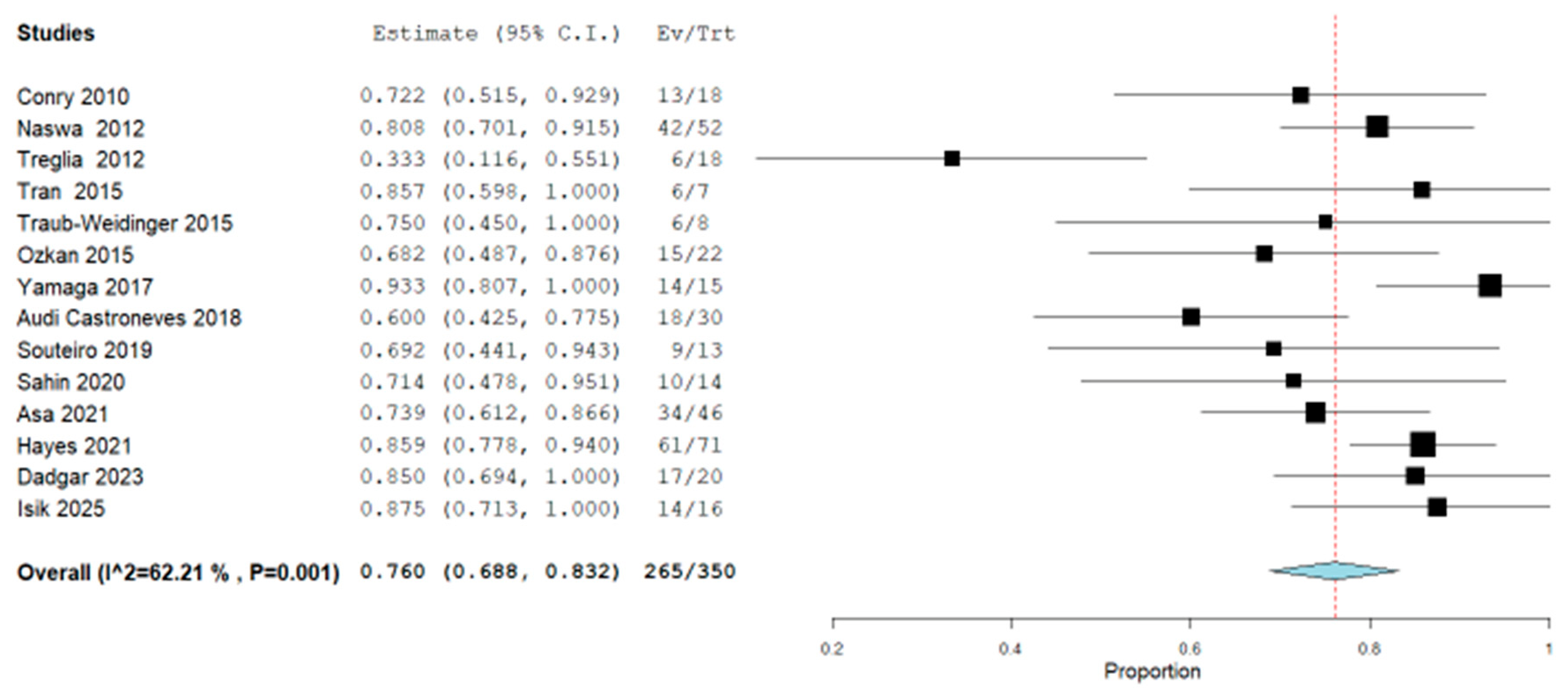
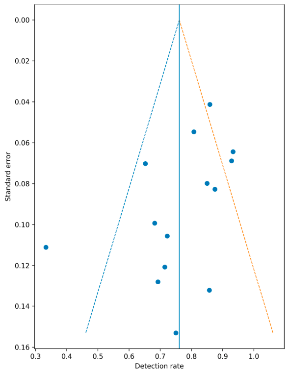
3.6. Quantitative Evaluation (Meta-Analysis)
4. Discussion
5. Conclusions
Supplementary Materials
Author Contributions
Funding
Institutional Review Board Statement
Informed Consent Statement
Data Availability Statement
Conflicts of Interest
Abbreviations
| MTC | medullary thyroid carcinoma |
| SSTR | somatostatin receptors |
| DR | detection rate |
| PET/CT | positron emission tomography/computed tomography |
| CEA | carcinoembryonic antigen |
| PRRT | Peptide Receptor Radionuclide Therapy |
| CI | confidence intervals |
| SPECT | single-photon emission computed tomography |
References
- Caillé, S.; Debreuve-Theresette, A.; Vitellius, G.; Deguelte, S.; La Manna, L.; Zalzali, M. Medullary Thyroid Cancer: Epidemiology and Characteristics According to Data From the Marne-Ardennes Register 1975–2018. J. Endocr. Soc. 2024, 8, bvae084. [Google Scholar] [CrossRef] [PubMed]
- Jara, M.A.; Castroneves, L.A. Overview of management and therapeutic advances in medullary thyroid cancer. Endocr. Oncol. 2025, 5, e240077. [Google Scholar] [CrossRef] [PubMed]
- Pusztaszeri, M.P.; Maleki, Z. The diagnostic challenges of medullary thyroid carcinoma: A practical guide for cytopathologists. Cancer Cytopathol. 2025, 133, e70023. [Google Scholar] [CrossRef] [PubMed]
- Ganeshan, D.; Paulson, E.; Duran, C.; Cabanillas, M.E.; Busaidy, N.L.; Charnsangavej, C. Current Update on Medullary Thyroid Carcinoma. Am. J. Roentgenol. 2013, 20, W867–W876. [Google Scholar] [CrossRef] [PubMed]
- Forma, A.; Kłodnicka, K.; Pająk, W.; Flieger, J.; Teresińska, B.; Januszewski, J.; Baj, J. Thyroid Cancer: Epidemiology, Classification, Risk Factors, Diagnostic and Prognostic Markers, and Current Treatment Strategies. Int. J. Mol. Sci. 2025, 26, 5173. [Google Scholar] [CrossRef] [PubMed]
- Park, H.; Park, J.; Choi, M.S.; Kim, J.; Kim, H.; Shin, J.H.; Kim, J.H.; Kim, J.S.; Kim, S.W.; Chung, J.H.; et al. Preoperative Serum Calcitonin and Its Correlation with Extent of Lymph Node Metastasis in Medullary Thyroid Carcinoma. Cancers 2020, 12, 2894. [Google Scholar] [CrossRef] [PubMed]
- Kushchayev, S.V.; Kushchayeva, Y.S.; Tella, S.H.; Glushko, T.; Pacak, K.; Teytelboym, O.M. Medullary Thyroid Carcinoma: An Update on Imaging. J. Thyroid Res. 2019, 2019, 1893047. [Google Scholar] [CrossRef] [PubMed]
- Treglia, G.; Rufini, V.; Piccardo, A.; Imperiale, A. Update on Management of Medullary Thyroid Carcinoma: Focus on Nuclear Medicine. Semin. Nucl. Med. 2023, 53, 481–489. [Google Scholar] [CrossRef] [PubMed]
- Giovanella, L.; Treglia, G.; Iakovou, I.; Mihailovic, J.; Verburg, F.A.; Luster, M. EANM practice guideline for PET/CT imaging in medullary thyroid carcinoma. Eur. J. Nucl. Med. Mol. Imaging 2019, 47, 61–77. [Google Scholar] [CrossRef] [PubMed]
- de Vries, L.H.; Lodewijk, L.; Willems, S.M.; Dreijerink, K.M.A.; de Keizer, B.; van Diest, P.J.; Schepers, A.; Bonenkamp, H.J.; van Engen-van Grunsven, I.A.C.H.; Kruijff, S.; et al. SSTR2A expression in medullary thyroid carcinoma is correlated with longer survival. Endocrine 2018, 62, 639–647. [Google Scholar] [CrossRef] [PubMed]
- Imperiale, A.; Jha, A.; Meuter, L.; Nicolas, G.P.; Taïeb, D.; Pacak, K. The Emergence of Somatostatin Antagonist–Based Theranostics: Paving the Road Toward Another Success? J. Nucl. Med. 2023, 64, 682–684. [Google Scholar] [CrossRef] [PubMed]
- Sadeghi, R.; Treglia, G. Systematic reviews and meta-analyses of diagnostic studies: A practical guideline. Clin. Transl. Imaging 2017, 5, 83–87. [Google Scholar]
- Page, M.J.; McKenzie, J.E.; Bossuyt, P.M.; Boutron, I.; Hoffmann, T.C.; Mulrow, C.D.; Shamseer, L.; Tetzlaff, J.M.; Akl, E.A.; Brennan, S.E.; et al. The PRISMA 2020 statement: An updated guideline for reporting systematic reviews. BMJ 2021, 372, n71. [Google Scholar] [CrossRef] [PubMed]
- Whiting, P.F.; Rutjes, A.W.; Westwood, M.E.; Mallett, S.; Deeks, J.J.; Reitsma, J.B.; Leeflang, M.M.G.; Sterne, J.A.C.; Bossuyt, P.M.M.; QUADAS-2 Group. QUADAS-2: A revised tool for the quality assessment of diagnostic accuracy studies. Ann. Intern. Med. 2011, 155, 529–536. [Google Scholar] [CrossRef] [PubMed]
- Pałyga, I.; Kowalska, A.; Gąsior-Perczak, D.; Tarnawska-Pierścińska, M.; Słuszniak, J.; Sygut, J.; Góźdź, S. The role of PET-CT scan with somatostatin analogue labelled with gallium-68 (68Ga-DOTA-TATE PET-CT) in diagnosing patients with disseminated medullary thyroid carcinoma (MTC). Endokrynol. Pol. 2010, 61, 507–511. [Google Scholar] [PubMed]
- Conry, B.G.; Papathanasiou, N.D.; Prakash, V.; Kayani, I.; Caplin, M.; Mahmood, S.; Bomanji, J.B. Comparison of (68)Ga-DOTATATE and (18)F-fluorodeoxyglucose PET/CT in the detection of recurrent medullary thyroid carcinoma. Eur. J. Nucl. Med. Mol. Imaging 2010, 37, 49–57. [Google Scholar] [CrossRef] [PubMed]
- Naswa, N.; Sharma, P.; Suman Kc, S.; Lata, S.; Kumar, R.; Malhotra, A.; Bal, C. Prospective evaluation of 68Ga-DOTA-NOC PET-CT in patients with recurrent medullary thyroid carcinoma: Comparison with 18F-FDG PET-CT. Nucl. Med. Commun. 2012, 33, 766–774. [Google Scholar] [CrossRef] [PubMed]
- Tran, K.; Khan, S.; Taghizadehasl, M.; Palazzo, F.; Frilling, A.; Todd, J.F.; Al-Nahhas, A. Gallium-68 Dotatate PET/CT is superior to other imaging modalities in the detection of medullary carcinoma of the thyroid in the presence of high serum calcitonin. Hell. J. Nucl. Med. 2015, 18, 19–24. [Google Scholar] [CrossRef] [PubMed]
- Traub-Weidinger, T.; Putzer, D.; von Guggenberg, E.; Dobrozemsky, G.; Nilica, B.; Kendler, D.; Bale, R.; Virgolini, I.J. Multiparametric PET imaging in thyroid malignancy characterizing tumour heterogeneity: Somatostatin receptors and glucose metabolism. Eur. J. Nucl. Med. Mol. Imaging 2015, 42, 1995–2001. [Google Scholar] [CrossRef] [PubMed]
- Ozkan, Z.G.; Kuyumcu, S.; Uzum, A.K.; Gecer, M.F.; Ozel, S.; Aral, F.; Adalet, I. Comparison of 68Ga-DOTATATE PET-CT, 18F-FDG PET-CT and 99mTc-(V)DMSA scintigraphy in the detection of recurrent or metastatic medullary thyroid carcinoma. Nucl. Med. Commun. 2015, 36, 242–250. [Google Scholar] [CrossRef] [PubMed]
- Yamaga, L.Y.I.; Cunha, M.L.; Campos Neto, G.C.; Garcia, M.R.T.; Yang, J.H.; Camacho, C.P.; Wagner, J.; Funari, M.B.G. 68Ga-DOTATATE PET/CT in recurrent medullary thyroid carcinoma: A lesion-by-lesion comparison with 111In-octreotide SPECT/CT and conventional imaging. Eur. J. Nucl. Med. Mol. Imaging 2017, 44, 1695–1701. [Google Scholar] [CrossRef] [PubMed]
- Castroneves, L.A.; Coura Filho, G.; de Freitas, R.M.C.; Salles, R.; Moyses, R.A.; Lopez, R.V.M.; Pereira, M.A.A.; Tavares, M.R.; Jorge, A.A.L.; Buchpiguel, C.A.; et al. Comparison of 68Ga PET/CT to Other Imaging Studies in Medullary Thyroid Cancer: Superiority in Detecting Bone Metastases. J. Clin. Endocrinol. Metab. 2018, 103, 3250–3259. [Google Scholar] [CrossRef] [PubMed]
- Souteiro, P.; Gouveia, P.; Ferreira, G.; Belo, S.; Costa, C.; Carvalho, D.; Duarte, H.; Sampaio, I.L. 68Ga-DOTANOC and 18F-FDG PET/CT in metastatic medullary thyroid carcinoma: Novel correlations with tumoral biomarkers. Endocrine 2019, 64, 322–329. [Google Scholar] [CrossRef] [PubMed]
- Şahin, E.; Elboğa, U. The role of tumour biomarkers in choosing the appropriate positron emission tomography imaging in follow-up of medullary thyroid cancer. J. Med. Imaging Radiat. Oncol. 2020, 64, 756–761. [Google Scholar] [CrossRef] [PubMed]
- Arici, S.; Karyagar, S.S.; Erdemir, R.U.; Cekin, R.; Atcı, M.M.; Güven, O.; Karyagar, S.; Cihan, Ş. The correlation between tumor markers and Ga-68 DOTA-TATE PET/CT findings in patients with advanced medullary thyroid cancer. Rev. Esp. Med. Nucl. Imagen Mol. 2021, 40, 161–166. [Google Scholar] [CrossRef] [PubMed]
- Asa, S.; Sonmezoglu, K.; Uslu-Besli, L.; Sahin, O.E.; Karayel, E.; Pehlivanoglu, H.; Sager, S.; Kabasakal, L.; Ocak, M.; Sayman, H.B. Evaluation of F-18 DOPA PET/CT in the detection of recurrent or metastatic medullary thyroid carcinoma: Comparison with GA-68 DOTA-TATE PET/CT. Ann. Nucl. Med. 2021, 35, 900–915. [Google Scholar] [CrossRef] [PubMed]
- Hayes, A.R.; Crawford, A.; Al Riyami, K.; Tang, C.; Bomanji, J.; Baldeweg, S.E.; Wild, D.; Morganstein, D.; Harry, A.; Grozinsky-Glasberg, S.; et al. Metastatic Medullary Thyroid Cancer: The Role of 68Gallium-DOTA-Somatostatin Analogue PET/CT and Peptide Receptor Radionuclide Therapy. J. Clin. Endocrinol. Metab. 2021, 106, e4903–e4916. [Google Scholar] [CrossRef] [PubMed]
- Serfling, S.E.; Zhi, Y.; Megerle, F.; Fassnacht, M.; Buck, A.K.; Lapa, C.; Werner, R.A. Somatostatin receptor-directed molecular imaging for therapeutic decision-making in patients with medullary thyroid carcinoma. Endocrine 2022, 78, 169–176. [Google Scholar] [CrossRef] [PubMed]
- Ballal, S.; Yadav, M.P.; Roesch, F.; Raju, S.; Satapathy, S.; Sheokand, P.; Moon, E.S.; Martin, M.; Awarwal, S.; Tripathi, M.; et al. Head-to-Head Comparison of [68Ga]Ga-DOTA.SA.FAPi and [68Ga]Ga-DOTANOC Positron Emission Tomography/Computed Tomography Imaging for the Follow-Up Surveillance of Patients with Medullary Thyroid Cancer. Thyroid. Off. J. Am. Thyroid. Assoc. 2023, 33, 974–982. [Google Scholar] [CrossRef] [PubMed]
- Dadgar, H.; Jafari, E.; Ahmadzadehfar, H.; Rekabpour, S.J.; Ravanbod, M.R.; Kalantarhormozi, M.; Nabipour, I.; Assadi, M. Feasibility and therapeutic potential of the 68Ga/177Lu-DOTATATE theranostic pair in patients with metastatic medullary thyroid carcinoma. Ann. Endocrinol. 2023, 84, 45–51. [Google Scholar] [CrossRef] [PubMed]
- Gild, M.L.; Kumar, S.; Fuchs, T.L.; Glover, A.; Sidhu, S.; Sywak, M.; Tsang, V.; Gill, A.J.; Robinson, B.G.; Schembri, G.; et al. The Clinical Utility of Gallium-68-DOTATATE Positron Emission Tomography Scanning in Medullary Thyroid Cancer. Endocr. Pract. 2024, 30, 218–224. [Google Scholar] [CrossRef] [PubMed]
- Isik, E.G.; Has Simsek, D.; Gul, N.; Erturk, S.M.; Buyukkaya, F.; Soyluk Selcukbiricik, O.; Iscan, A.Y.; Özkan, Z.G.; Sanli, Y.; Mudun, A.; et al. Head-to-Head Comparison of 68Ga-FAPI-04 and 68Ga-DOTA-TATE PET/CT in Recurrent Medullary Thyroid Cancer. Clin. Nucl. Med. 2025, 50, e80–e86. [Google Scholar] [CrossRef] [PubMed]
- Kunte, S.C.; Wenter, V.; Toms, J.; Lindner, S.; Unterrainer, M.; Eilsberger, F.; Jurkschat, K.; Wängler, C.; Wängler, B.; Schirrmacher, R.; et al. PET/CT imaging of differentiated and medullary thyroid carcinoma using the novel SSTR-targeting peptide [18F]SiTATE—First clinical experiences. Eur. J. Nucl. Med. Mol. Imaging 2025, 52, 900–912. [Google Scholar] [CrossRef] [PubMed]
- Treglia, G.; Tamburello, A.; Giovanella, L. Detection rate of somatostatin receptor PET in patients with recurrent medullary thyroid carcinoma: A systematic review and a meta-analysis. Hormones 2017, 16, 362–372. [Google Scholar] [CrossRef] [PubMed]
- Treglia, G.; Cocciolillo, F.; Di Nardo, F.; Poscia, A.; de Waure, C.; Giordano, A.; Rufini, V. Detection Rate of Recurrent Medullary Thyroid Carcinoma Using Fluorine-18 Dihydroxyphenylalanine Positron Emission Tomography. A Meta-analysis. Acad. Radiol. 2012, 19, 1290–1299. [Google Scholar] [CrossRef] [PubMed]
- Imperiale, A.; Berti, V.; Burgy, M.; Cazzato, R.L.; Piccardo, A.; Treglia, G. Molecular imaging and related therapeutic options for medullary thyroid carcinoma: State of the art and future opportunities. Rev. Endocr. Metab. Disord. 2023, 25, 187–202. [Google Scholar] [CrossRef] [PubMed]
- Trimboli, P.; Mian, C.; Piccardo, A.; Treglia, G. Diagnostic tests for medullary thyroid carcinoma: An umbrella review. Endocrine 2023, 81, 183–193. [Google Scholar] [CrossRef] [PubMed]
- Grossrubatscher, E.; Fanciulli, G.; Pes, L.; Sesti, F.; Dolci, C.; de Cicco, F.; Colao, A.; Faggiano, A. Advances in the Management of Medullary Thyroid Carcinoma: Focus on Peptide Receptor Radionuclide Therapy. J. Clin. Med. 2020, 9, 3507. [Google Scholar] [CrossRef] [PubMed]




| First Author | Year | Country | Funding Source | Study Design | N° Patients | Age Mean (Range) | Gender M (%) | Calcitonin, Median/Mean (Range), pg/mL | Purpose/s |
|---|---|---|---|---|---|---|---|---|---|
| Palyga I [15] | 2010 | Poland | None reported | R | 8 | 55.6 (41–72) | 4 (50%) | 265 (53–720) | Staging in patients with negative conventional imaging (ultrasound, CT, MRI, FDG PET) |
| Conry BG [16] | 2010 | UK | None reported | R | 18 | 54 (34–75) | 13 (72%) | 134.9 (1.5–550) | Comparison with FDG PET/CT in recurrent MTC |
| Naswa N [17] | 2012 | India | None reported | P | 52 | 44.7 (18–73) | 38 (73%) | 4017.3 (50–30,000) | Comparison with FDG PET/CT in recurrent MTC |
| Treglia G [18] | 2012 | Italy | Non reported | R | 18 | 53 (24–86) | 6 (33%) | 527 (66.7–14,186) | Comparison with FDG PET/CT and DOPA PET/CT in recurrent MTC |
| Tran [19] | 2015 | UK | None reported | R | 7 | 45 (31–66) | 3 (42%) | 8320 (672–37,180) | Detection rate in recurrent MTC |
| Traub-Weidinger [20] | 2015 | Austria | None reported | R | 31 * | 63 (24–85) | 20 (64%) | na | Detection rate in recurrent MTC |
| Ozkan ZG [21] | 2015 | Turkey | None reported | R | 22 | 42.9(20–69) | 11 (50%) | 871.5 (99.4–7370) | Comparison with FDG PET/CT and DMSA scintigraphy in recurrent MTC |
| Yamaga [22] | 2017 | Brazil | None reported | P | 15 | 43.6 (20–68) | 6 (40%) | 10.990 (417–100,000) | Comparison with 111ln-Octreotide scintigraphy and conventional imaging in recurrent MTC |
| Audi Castroneves L [23] | 2018 | Brazil | None reported | P | 30 | 48 (19–78) | 13 (43%) | 133 (12–1162) in staging group 8323 (564–101,083) in metastatic group | Diagnostic performances and comparison with CT, MRI, and bone scan in metastases detection |
| Souteiro P [24] | 2019 | Portugal | None reported | R | 13 | 50.9 (39–78) | 5 (38%) | 828 (20.4–231,696) | Comparison with FDG PET/CT in metastatic MTC |
| Sahin E [25] | 2020 | Turkey | None reported | R | 14 | 54.4 (27–81) | 6 (43%) | 1201.2 (24.3–7292) | Comparison with FDG PET/CT in recurrent MTC and correlation with tumor markers (calcitonin and CEA) |
| Arici S [26] | 2021 | Turkey | None reported | R | 28 | 55 (35–82) | 9 (32%) | 6671.6 (2–102,148) | Correlation between tumor burden and tumor markers (calcitonin and CEA) |
| Asa S [27] | 2021 | Turkey | yes | P | 46 | 53 (27–82) | 21 (46%) | 2032 (62.4–14,000) | Comparison with 18F-DOPA PET/CT in recurrent and metastatic MTC |
| Hayes [28] | 2021 | UK | yes | R | 71 | 44 (16–84) | 37 (52%) | 3477 (6.8–174,000) | Detection rate in metastatic and recurrent MTC and eligibility for PRRT |
| Serfling SE [29] | 2022 | Germany | None reported | R | 23 | 51.9 (32–81) | 12 (52%) | 1400 (26–42,700) | Staging and therapeutic decision |
| Ballal S [30] | 2023 | India | None reported | R | 27 | 42.4 (14–66) | 21 (78%) | 666.5 (389–1145) | Comparison with FAPI PET/CT in follow-up |
| Dadgar H [31] | 2023 | Iran | None reported | R | 20 | 48.5 (27–71) | 10 (50%) | 256 (14–2000) | Restaging in suspected relapse |
| Gild ML [32] | 2024 | Australia | None reported | R | 37 | na | na | na | Prognostication |
| Isik EG [33] | 2025 | Turkey | None reported | R | 16 | 50 (18–76) | 8 (50%) | 6234 (245–96,880) | Comparison with FAPI PET/CT in recurrent MTC |
| Kunte SC [34] | 2025 | Germany | yes | P | 21 ** | 62.1 (40–73) | 4 (40%) | na | Feasibility |
| First Author | Device | Radiopharmaceutical | Mean Radiotracer Injected Dose, MBq | Mean Uptake Time (Min) | Image Analysis | Semiquantitative Variables |
|---|---|---|---|---|---|---|
| Palyga I [15] | PET/CT | [68Ga]DOTA-TATE | 120–185 | 60 | Visual | |
| Conry BG [16] | PET/CT | [68Ga]DOTA-TATE | 90–220 | 45–60 | Visual and semiquantitative | SUVmax |
| Naswa N [17] | PET/CT | [68Ga]DOTA-NOC | 148–222 | 45–60 | Visual and semiquantitative | SUVmax |
| Treglia G [18] | PET/CT | [68Ga]DOTA-NOC n = 14 | 1.5–2/kg | 60 | Visual | |
| [68Ga]DOTA-TOC n = 4 | 2.5/kg | |||||
| Tran [19] | PET/CT | [68Ga]DOTA-TATE | 110–148 | 45–60 | Visual | |
| Traub-Weidinger [20] | PET/CT | [68Ga]DOTA-TATE | 185 | 60 | Visual | |
| Ozkan ZG [21] | PET/CT | [68Ga]DOTA-TATE | 185 | nr | Visual and semiquantitative | SUVmax |
| Yamaga [22] | PET/CT | [68Ga]DOTA-TATE | 72 | 45 | Visual | |
| Audi Castroneves L [23] | PET | [68Ga]DOTA-LAN & [68Ga]DOTA-TOC | 95–150 100–150 | 90 | Visual | |
| Souteiro P [24] | PET/CT | [68Ga]DOTA-NOC | 122 | 76 | Visual | |
| Sahin E [25] | PET/CT | [68Ga]DOTA-TATE | 2–3/kg | 45 | Visual | |
| Arici S [26] | PET/CT | [68Ga]DOTA-TATE | 2/kg | 60 | Visual and semiquantitative | MTV, TLV |
| Asa S [27] | PET/CT | [68Ga]DOTA-TATE | 176 | 45–60 | Visual and semiquantitative | sumSUVmax |
| Hayes [28] | PET/CT | [68Ga]DOTA-TATE & [68Ga]DOTA-TOC & [68Ga]DOTA-NOC | nr | nr | Visual and semiquantitative | SUVmax |
| Serfling SE [29] | PET/CT | [68Ga]DOTA-TATE & [68Ga]DOTA-TOC | 120 | 60 | Visual and semiquantitative | SUVpeak, TV |
| Ballal S [30] | PET/CT | [68Ga]DOTA-NOC | 185 | 60 | Visual and semiquantitative | SUVpeak, TBR |
| Dadgar H [31] | PET/CT | [68Ga]DOTA-TATE | 148–185 | 60 | Visual and semiquantitative | SUVmax |
| Gild ML [32] | PET/CT | [68Ga]DOTA-TATE | 120–180 | 50 | Visual and semiquantitative | SUVmax, SUVmean, MTV and TLA |
| Isik EG [33] | PET/CT | [68Ga]DOTA-TATE | 185 | 60 | Visual and semiquantitative | SUVmax |
| Kunte SC [34] | PET/CT | [18F]SiTATE | 215.5 | 90 | Visual and semiquantitative | SUVmax, SUVmean, TTV, wb-SUV |
| First Author | PET Positive/Total PET (%) | Mean SUVmax | Management Impact n (%) | Kind of Impact | Correlation with Calcitonin Levels |
|---|---|---|---|---|---|
| Conry BG [16] | 13/18 (72%) | na | na | na | yes |
| Naswa N [17] | 42/52 (81%) | Local recurrence: 3 Cervical metastasis: 3.4 Mediastinal node metastasis: 6.2 Liver metastasis: 6 Skeletal metastasis: 12.9 | na | na | >500 pg/mL (lack significance analysis) |
| Treglia G [18] | 6/18 (33%) | na | 3 (16.6%) | Guide to biopsy | yes |
| Tran [19] | 6/7 (86%) | na | na | Confirmation or ruling out recurrence | na |
| Traub-Weidinger [20] | 6/8 (75%) | na | na | na | na |
| Ozkan ZG [21] | 15/22 (68%) | na | 18.20% | Guide to surgery and PRRT | yes |
| Yamaga [22] | 14/15 (93%) | na | na | na | na |
| Audi Castroneves L [23] | 5/16 (31%) in a biochemical recurrence group; 13/14 (93%) in a metastatic disease group | 6.4 (median) in a metastatic disease group | 6.25% in a biochemical recurrence group; 35.7% in a metastatic disease group | Initiation of treatments such as bone antiresorptives, radiation therapy, or a change of radiological workup during follow-up. | na |
| Souteiro P [24] | 9/13 (69%) | na | na | Guide to surgery or PRRT | yes |
| Sahin E [25] | 10/14 (71%) | na | na | na | yes (with total tumor burden) |
| Asa S [27] | 30/46 (65%) | 31.1 * | 68.40% | Guide to surgery, PRRT, RFA or combined treatment. | yes |
| Hayes [28] | 61/71 (86%) | na | na | na | na |
| Ballal S [30] | na | na | 94.4% for primary tumours 95% for lymph nodes 100% for brain metastases 68.9% for lung nodules 46.4% for liver metastases 76.5% for bone metastases 0% for pleural metastases | na | na |
| Dadgar H [31] | 17/20 (85%) | 8.77 | 20% | Guide to PRRT | na |
| Isik EG [33] | 14/16 (87.5%) | 9.7 | na | na | na |
Disclaimer/Publisher’s Note: The statements, opinions and data contained in all publications are solely those of the individual author(s) and contributor(s) and not of MDPI and/or the editor(s). MDPI and/or the editor(s) disclaim responsibility for any injury to people or property resulting from any ideas, methods, instructions or products referred to in the content. |
© 2026 by the authors. Licensee MDPI, Basel, Switzerland. This article is an open access article distributed under the terms and conditions of the Creative Commons Attribution (CC BY) license.
Share and Cite
Tasevski, S.; Imperiale, A.; Treglia, G.; Albano, D. The Evolving Role of Somatostatin Receptor PET/CT in Medullary Thyroid Carcinoma: An Updated Systematic Review and Meta-Analysis. Cancers 2026, 18, 2096. https://doi.org/10.3390/cancers18132096
Tasevski S, Imperiale A, Treglia G, Albano D. The Evolving Role of Somatostatin Receptor PET/CT in Medullary Thyroid Carcinoma: An Updated Systematic Review and Meta-Analysis. Cancers. 2026; 18(13):2096. https://doi.org/10.3390/cancers18132096
Chicago/Turabian StyleTasevski, Slavko, Alessio Imperiale, Giorgio Treglia, and Domenico Albano. 2026. "The Evolving Role of Somatostatin Receptor PET/CT in Medullary Thyroid Carcinoma: An Updated Systematic Review and Meta-Analysis" Cancers 18, no. 13: 2096. https://doi.org/10.3390/cancers18132096
APA StyleTasevski, S., Imperiale, A., Treglia, G., & Albano, D. (2026). The Evolving Role of Somatostatin Receptor PET/CT in Medullary Thyroid Carcinoma: An Updated Systematic Review and Meta-Analysis. Cancers, 18(13), 2096. https://doi.org/10.3390/cancers18132096