Article

AXL-mRNA Overexpression in Size-Based Enriched Circulating Tumor Cells as a Potential Biomarker for Anti-AXL Targeted Therapies in Non-Small-Cell Lung Cancer


1
Analysis of Circulating Tumor Cells Laboratory, Laboratory of Analytical Chemistry, Department of Chemistry, National and Kapodistrian University of Athens, 15771 Athens, Greece
2
7th Department of Pulmonary Diseases, “Sotiria” General Hospital of Athens, 11527 Athens, Greece
3
Oncology Unit, 2nd Department of Surgery, Aretaieio Hospital, Medical School, National and Kapodistrian University of Athens, 11528 Athens, Greece
4
Medical Oncology Unit, 2nd Department of Internal Medicine, “Attikon” General Hospital of Athens, 12462 Athens, Greece
5
Department of Medical Oncology, University General Hospital of Alexandroupolis, Medical School, Democritus University of Thrace, 68100 Alexandroupolis, Greece
6
Department of Medical Oncology, University General Hospital of Larissa, 41334 Larissa, Greece
7
Hellenic Oncology Research Group (HORG), 11471 Athens, Greece
*
Authors to whom correspondence should be addressed.
Cancers 2026, 18(11), 1759; https://doi.org/10.3390/cancers18111759
Submission received: 6 May 2026 / Revised: 20 May 2026 / Accepted: 20 May 2026 / Published: 28 May 2026

Simple Summary

AXL, a tyrosine kinase receptor, is involved in epithelial-to-mesenchymal transition (EMT), cell survival, invasion, metastasis, and resistance to tyrosine kinase inhibitors of epidermal growth factor receptor (EGFR-TKIs) and immune checkpoint inhibitors (ICIs). Recent clinical studies have shown promising results in non-small-cell lung cancer (NSCLC) patients treated with AXL inhibitors. In the present study, we evaluated, for the first time, AXL-mRNA overexpression in size-based enriched circulating tumor cell (CTC) fractions in longitudinal liquid biopsy samples from two independent groups of NSCLC patients: (a) under osimertinib treatment and (b) during immunotherapy. Our results indicate that AXL-mRNA overexpression in CTC fractions deserves to be further evaluated through larger clinical studies as a potential biomarker for anti-AXL targeted therapies in NSCLC.

Abstract

Background: AXL, a tyrosine kinase receptor, is involved in epithelial-to-mesenchymal transition (EMT), cell survival, invasion, metastasis, and resistance to EGFR-TKIs and immune checkpoint inhibitors (ICIs). Recent clinical studies have shown promising results in NSCLC patients treated with AXL inhibitors. Methods: We evaluated AXL-mRNA overexpression in CTC fractions of NSCLC patients under osimertinib treatment (Group A, n = 39), (collected in the context of a multicenter Phase II clinical study (ClinicalTrials.gov number: NCT02771314) or immunotherapy (Group B, n = 116) at different time points collected in the context of a prospective, multicenter study (ClinicalTrials.gov number: NCT04490564). Size-based CTC enrichment (Parsortix, CelLBx Health, Guildford, UK) was used, and AXL-mRNA overexpression was evaluated using RT-qPCR. Results: In Group A, AXL-mRNA overexpression in CTC fractions was detected in 5/39 (12.8%) patients’ samples at baseline, in 5/31 (16.1%) after one cycle of treatment, in 7/79 (8.9%) during treatment and in 4/32 (12.5%) at progression of disease (PD). In Group B, AXL-mRNA overexpression in CTC fractions was detected in 8/116 (6.9%) samples before immunotherapy, in 8/71 (11.3%) after three or four cycles, while no AXL transcripts were detected at PD. Conclusions: Our results indicate that AXL-mRNA overexpression in CTC fractions deserve to be further evaluated through larger clinical studies as a potential biomarker for anti-AXL targeted therapies in NSCLC.

1. Introduction

During the last few decades, the treatment landscape of non-small-cell lung cancer (NSCLC) has been successfully altered. The development of immune checkpoint inhibitors (ICIs) has revolutionized the NSCLC treatment approach leading to improved clinical outcomes of cancer patients [1,2]. Specific targeted therapies, including those against somatic mutations found in the tyrosine kinase (TK) domain of epidermal growth factor receptor (EGFR), have also shown improved efficacy to EGFR-mutant NSCLC patients compared to conventional chemotherapeutic agents [3]. However, there is a common challenge that patients need to overcome; treatment resistance remains a major obstacle, limiting long-term response to treatments and prolonged survival rates [4]. Tumor heterogeneity, driver mutations, tumor microenvironment, and activation of alternative signaling pathways are some of the significant factors of resistance. The detection of novel biomarkers and the development of new anti-target therapies are common strategies to overcome resistance [4,5].
AXL, a member of the TAM (TYRO3, AXL, and MERTK) receptor tyrosine kinases (RTKs) family, has gained attention through its versatile roles in cell proliferation and survival, metastasis, tumor progression and treatment resistance [6]. Its implication in epithelial-to-mesenchymal transition (EMT), renders it a significant EMT marker in various types of cancer [7]. AXL is found to be highly expressed in NSCLC and more specifically its overexpression has been linked to the EGFR signaling pathway [8]. One of the alternative signaling mechanisms of acquired resistance to osimertinib is the bypass activation of AXL [9]. Moreover, there is a growing body of evidence that AXL plays a key role in resistance to immunotherapy in NSCLC [10].
Liquid biopsy is now an established minimal invasive approach that has revolutionized the management of NSCLC patients, through integration of circulating tumor DNA (ctDNA) analysis into clinical practice to facilitate personalized treatment [11,12]. In liquid biopsy, CTC analysis has a unique advantage over ctDNA analysis, since it can provide highly important information at the protein and gene expression level, for important therapeutic targets such as PD-L1, AR-V7, and AXL. Through molecular characterization, CTC analysis unveils important information on the molecular heterogeneity of disease and tumor clonal evolution, leading to the identification of resistance mechanisms and the development of novel therapeutic approaches [13]. Previous recent studies have shown that AXL is overexpressed in CTCs in advanced NSCLC [14] and also in metastatic breast cancer, suggesting liquid biopsy as a promising approach during AXL inhibition [15]. AXL tissue expression in combination with CTC enumeration could predict patient outcomes in resected lung adenocarcinomas [16]. Our group was the first to report a possible EMT role of AXL in the development of acquired EGFR-TKI resistance based on AXL-mRNA expression levels in CTC fractions of NSCLC patients [17,18].
In a recent review, it is reported that AXL inhibition could be a promising therapeutic intervention in EGFR-mutant NSCLC or during immunotherapy based on preclinical and clinical studies [19]. At present, several clinical trials are examining the effectiveness of AXL-inhibitors combined with EGFR-TKIs or ICIs [19]. Bemcentinib is the only selective small-molecule AXL inhibitor that obtained US FDA fast-track designation for treating advanced or metastatic NSCLC patients when used alongside a PD-L1 inhibitor in the phase 2 BGBC008 trial (NCT03184571) [20].
Based on these recent findings, in the present study, we evaluated, for the first time, whether AXL could be a novel CTC biomarker and therapeutic target in NSCLC. To this end, we evaluated, for the first time, AXL-mRNA overexpression in size-based enriched CTC fractions in longitudinal liquid biopsy samples from two independent groups of NSCLC patients, (a) under osimertinib treatment and (b) during immunotherapy. We further investigated the role of AXL-mRNA overexpression in CTC fractions as a potential biomarker for anti-AXL targeted therapies in NSCLC.

2. Materials and Methods

2.1. Patients

In the present study, clinical samples from two independent cohorts of NSCLC patients were analyzed: (a) under osimertinib treatment (Group A) and (b) under immunotherapy (Group B). Patients and healthy donors (HD) gave their written informed consent to participate in two different clinical studies, respectively. Both studies were conducted in accordance with the Declaration of Helsinki.
Group A: Thirty-nine patients with histologically or cytologically confirmed EGFR-mutant lung adenocarcinomas received second-line osimertinib (AZD9291; Astra Zeneca, Cambridge, UK) as part of a multicenter Phase II clinical trial (ClinicalTrials.gov number: NCT02771314, registration date: 13 May 2016) and EudraCT number: 2016-001335-12, registration date: 13 April 2016) organized by the Hellenic Oncology Research Group (HORG). The research has received approval from the National Drug Administration of Greece (EOF), the National Ethics Committee (35/00-03/16, 35/03-11/16), and the Institutional Ethical Committees of the centers participating in HORG. At the start of this clinical study, osimertinib was given as a second-line therapy to NSCLC patients with EGFR mutations, following the existing guidelines at that time.
Group B: Peripheral blood (PB) samples from 116 patients with NSCLC were collected at five clinical centers in Greece as part of a prospective, multicenter research study (ClinicalTrials.gov identifier: NCT04490564, registration date: 28 July 2020). Eligibility criteria for patients with metastatic non-small-cell lung cancer (NSCLC): starting PD-1 inhibitors typically required male and female individuals aged 18 and above. The Ethics committees of all involved institutions granted approval for the study: Attikon University Hospital (7th meeting of the Scientific Advisory Board 10 September 2019), “SOTIRIA” General Hospital of Athens (19261, 28 August 2019); Aretaieion University Hospital, (137/28/11/19); General University Hospital of Alexandroupolis, (6th regular meeting of the Scientific Advisory Board, 19 June 2019).

2.2. Collection of PB Samples and Processing

PB was obtained in EDTA tubes after rejecting the initial 5 mL of blood draw to prevent contamination from skin epithelial cells. In Group A, 15 mL of PB were collected from NSCLC patients with EGFR mutations at different intervals as follows: (a) 39 samples at baseline, prior to starting treatment with osimertinib; (b) 31 samples after the first cycle of therapy; (c) 79 samples approximately every 3 months during treatment; and (d) 31 samples upon disease progression (PD). In Group B, PB (10 mL) was obtained from NSCLC patients at 3 distinct time points as follows: (a) 116 samples prior to immunotherapy, (b) 71 samples following 3 or 4 cycles of ICIs, and (c) 10 samples at PD. PB samples collected from a control group consisting of 46 HDs were processed and analyzed using the same experimental procedure. All preanalytical conditions for sample collection, transportation and storage conditions were strictly controlled and standardized, as the ACTC lab is ISO-15189 certified [21].

2.3. CTC Enrichment

PB samples were centrifuged at 530× g (10 min at room temperature); the supernatant plasma was extracted and stored at −70 °C for subsequent analysis. In the remaining pellet of blood cells, an equal volume of phosphate-buffered saline (PBS; pH 7.3) was added to the plasma that was removed. Then, the FDA-cleared Parsortix™ (CelLBxHealth, Guildford, UK) device was used for CTC enrichment, using a 6.5 μm separation cassette. The harvested cells were collected in a final volume of 210 μL PBS.

2.4. Total RNA Extraction—cDNA Synthesis

Following CTC enrichment, TRIzol LS reagent (Thermo Fisher Scientific, Waltham, MA, USA) was used for total RNA extraction from the size-based enriched CTC fractions in RNAase free conditions. RNA was dissolved in Ambion RNA Storage Solution (Thermo Fisher Scientific), quantified using a NanoDrop ND-1000 UV–Vis Spectrophotometer (Thermo Fisher Scientific), and maintained at −70 °C. A High-Capacity RNA-to-cDNA kit (Thermo Fisher Scientific) was used for cDNA synthesis in a total volume of 20 μL, following the manufacturer’s instructions and previously outlined methods [17].

2.5. In Silico Design of Primers and Probes

In silico design for the AXL primers and probe was performed using Primer Premier 5.0 software (Premier Biosoft, Palo Alto, CA, USA). The Basic Local Alignment Search Tool (BLAST), version 2.11.0 (NCBI, NIH) was used for the design of primers and probes to prevent primer–dimer formation, false priming locations, hairpin structure formation, and hybridization with genomic DNA. The sequences of primers and probes are the following: Forward primer: 5′-AGAAGGAGACCCGTTATGGAGA-3′; Reverse primer: 5′-ATGCCCAGGCTGTTCAAGG-3′; Taqman probe: 5′-FAM-CTTCAGTGGTCCGACGACTGTAGGACTT-BHQ-3′.

2.6. RT-qPCR

RT-qPCR was performed to evaluate AXL mRNA expression, as follows: the amplification reaction mixture for AXL contained 4 μL of the PCR synthesis buffer (5×, Promega, Madison, WI, USA), 1.2 μL MgCl2 (25 Mm, Promega), 0.2 μL dNTPs (10 mM, Thermo Fisher Scientific), 0.1 μL Hot-Start DNA polymerase (5 U/μL, Promega), 0.3 μL of forward and reverse primer (10 μΜ, IDT, Coralville, IA, USA), 0.83 μL hydrolysis probe (3 μM, IDT) and DEPC–H2O, to a final volume of 10 μL. PCR cycling conditions for AXL were: 95 °C for 2 min, 45 cycles of 95 °C for 10 s, annealing at 58 °C for 20 s, extension at 72 °C for 20 s, and a final cooling cycle at 40 °C for 30 s.
The expression of Beta-2 microglobulin (B2M) was used as a reference gene for ensuring the presence of amplifiable material in all samples and for normalization of AXL mRNA expression, as previously described [17]. All RT-qPCR assays were performed in the cobas z480 system (Roche Molecular Systems, Inc., Pleasanton, CA, USA).

2.7. Normalization of RT-qPCR Data for AXL Expression

RT-qPCR data for AXL expression were normalized with respect to B2M expression in the same cDNAs, using the 2−ΔΔCq method [22]. CTCs were enriched using the size-based Parsortix technology (CelLBx, Guildford, UK); to correct the background noise due to the co-isolation of peripheral blood mononuclear cells (PBMC), present in all CTC enrichment systems, PB from a group of HDs was analyzed in exactly the same way as the patients’ and the results were used to establish a cut-off value through normalization of AXL expression with respect to B2M expression. The inclusion of a healthy donor group is very important, since CTCs are enriched through Parsortix and not isolated as pure cancer cells; therefore, every sample contains also a number of PBMCs, and this background should be taken into account. The cut-off value for AXL transcripts was calculated as the mean value of signals derived in the HDs group plus two standard deviations (2SD), as previously described [23]. Based on this approach, a clinical sample is defined as AXL overexpressed (AXL-positive) based on the fold change in AXL expression in the CTC fraction with respect to the corresponding PBMC fraction in the group of HDs.

2.8. Statistical Analysis

Statistical analysis was conducted utilizing IBM SPSS Statistics for Windows, version 31.0 (IBM Corp., Armonk, NY, USA). The McNemar test was used to evaluate the concordance of gene expression across various time points. Every p-value is two-tailed. A p-value of less than 0.05 is considered statistically significant.

3. Results

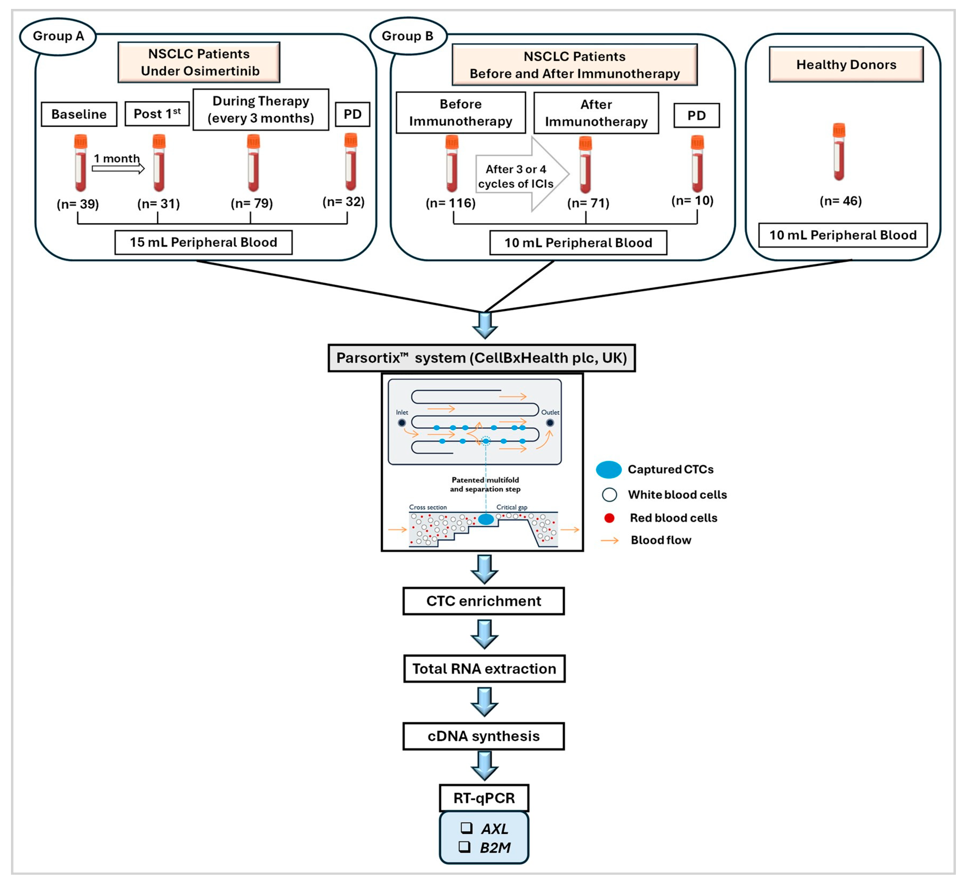
The outline of the study is shown in Figure 1.

3.1. AXL-mRNA Expression Levels in Size-Based Enriched CTC Fractions

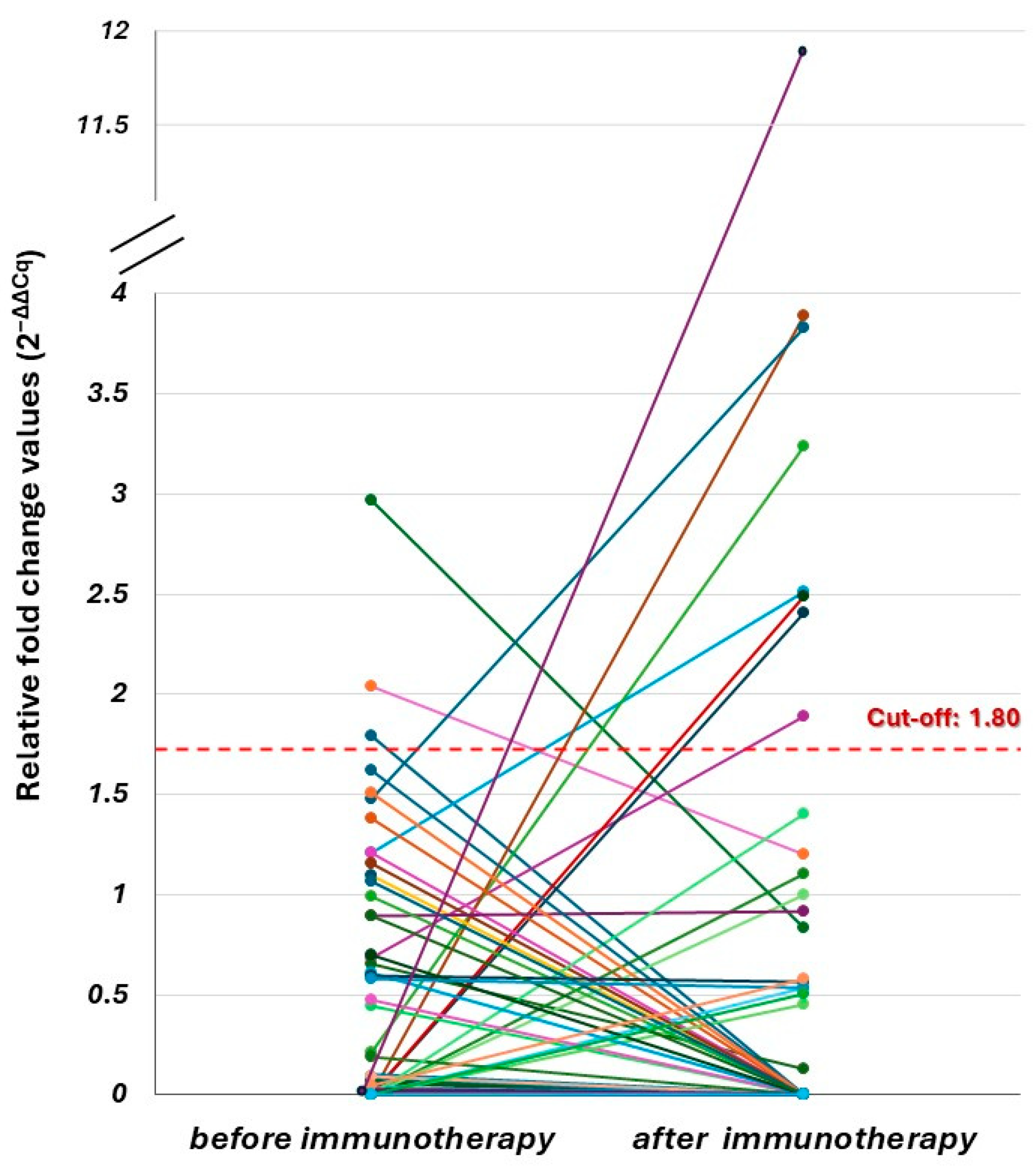
AXL transcripts were detected at very low levels in PB samples from HDs (n = 46) that were analyzed in the same way. Thus, the 2−ΔΔCq method was used for normalizing AXL-mRNA expression levels in patients’ samples with respect to the expression of B2M as reference gene, as previously described [17,22,23,24]. Since the total PB volume used for CTC enrichment was different in these two groups, the samples for the control group of HDs were analyzed in exactly the same way, using 15 mL PB for Group A and 10 mL PB for Group B. For Group A, the cut-off value for AXL transcripts was calculated as the mean value of signals derived in the HDs group (n = 10), plus two standard deviations (2SD), as previously described (15 mL PB, cut-off: 2−ΔΔCq = 1.55) [17]. The same approach was used for Group B analyzing PB samples from 36 HDs (10 mL PB, cut-off: 2−ΔΔCq = 1.80).

3.2. AXL-mRNA Overexpression in Size-Based Enriched CTC Fractions of NSCLC Patients Under Osimertinib

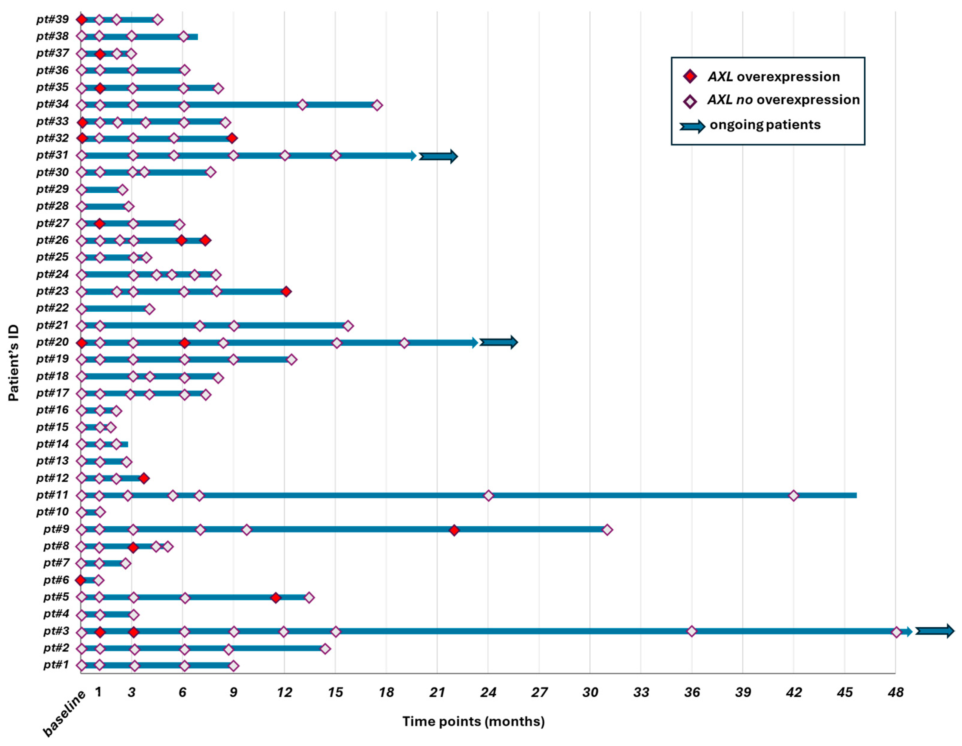
Group A: NSCLC patients were longitudinally monitored one month after osimertinib initiation and then approximately every 3 months and until PD. The expression of AXL was also evaluated at these different time-points (Figure 2). In most patients, AXL-mRNA overexpression was detected only at a single time point; whereas, in some cases (Pt#3, Pt#20, Pt#26, and Pt#32) was detected at two distinct time points during treatment. AXL-mRNA overexpression in CTC fractions was detected in 5/39 (12.8%) patients’ samples at baseline; whereas, after one cycle of treatment, AXL-mRNA overexpression was detected in 5/31 (16.1%) samples. AXL-mRNA overexpression in CTC fractions was detected in 7/79 (8.9%) samples during various stages of treatment and in 4/32 (12.5%) samples at PD. At the time of analysis, three patients were still under osimertinib treatment (ongoing).

3.3. Longitudinal Monitoring of AXL-mRNA Overexpression in CTC Fractions During Osimertinib

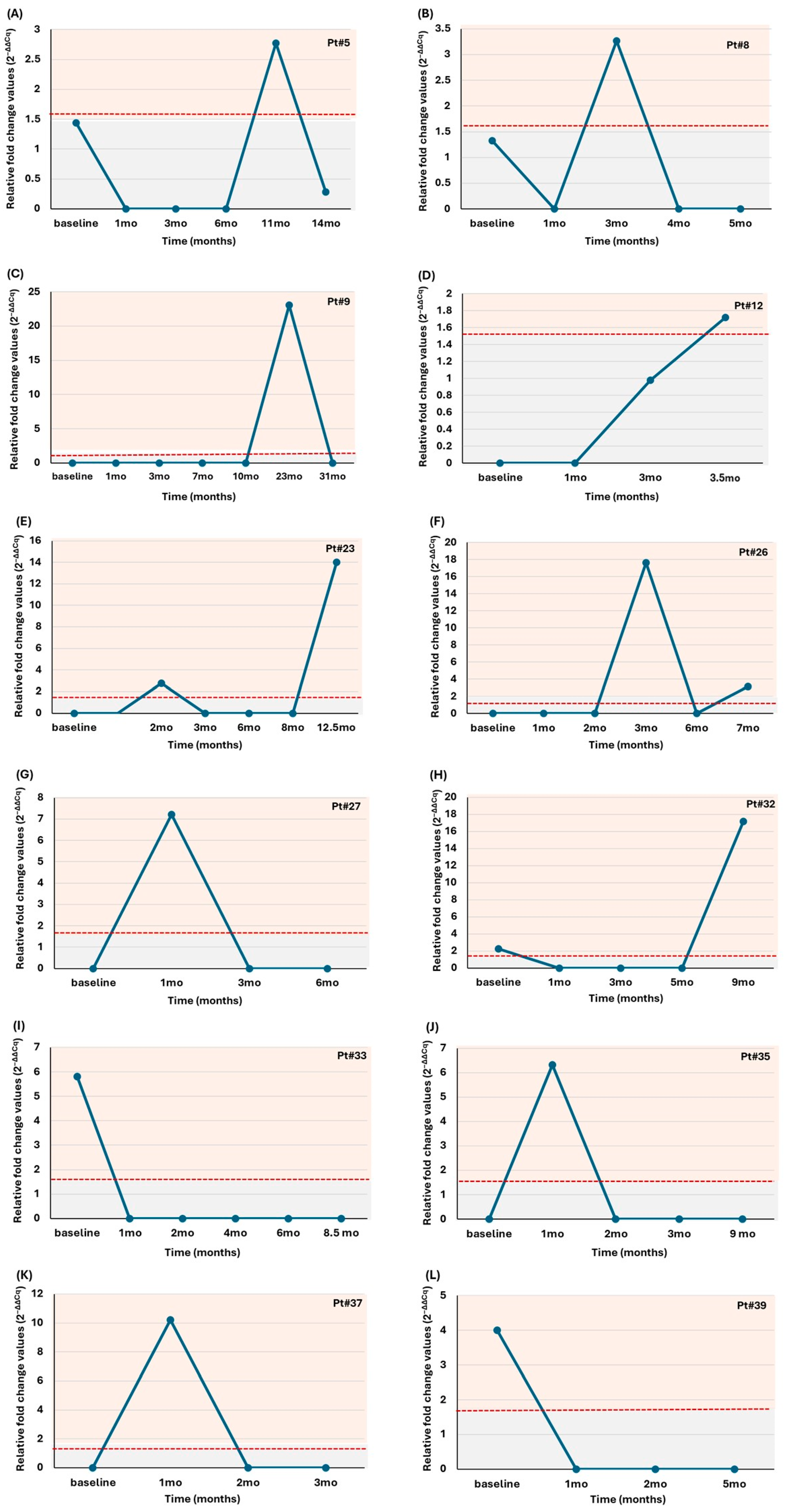
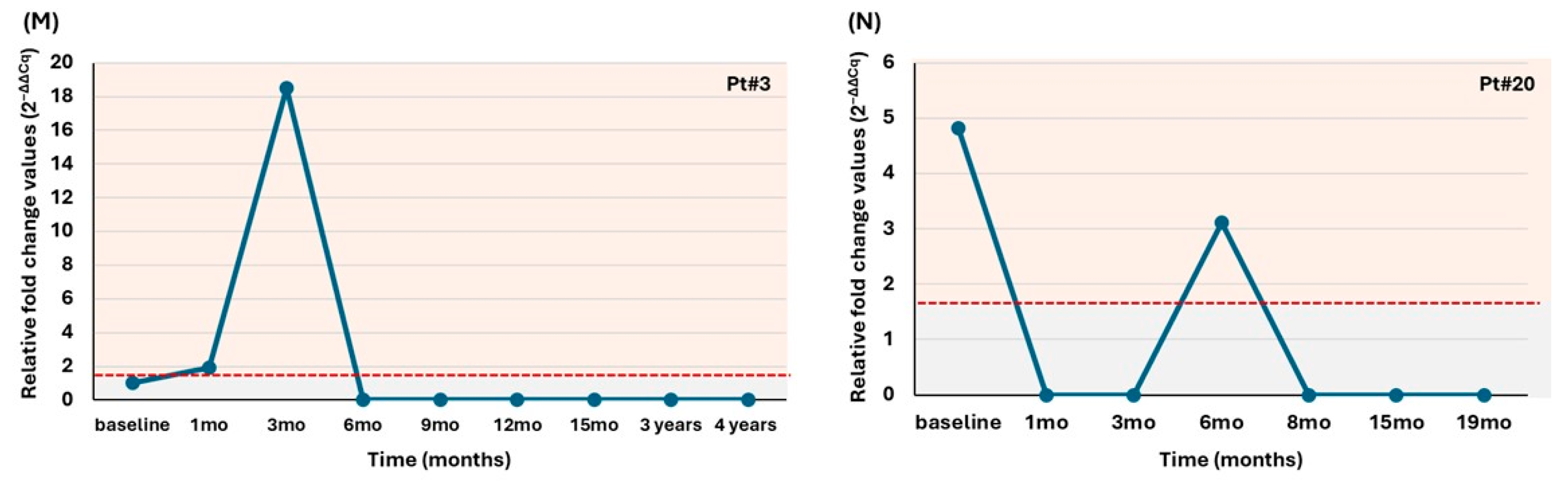
Next, we assessed the levels of AXL-mRNA overexpression and their deviations over time during osimertinib treatment in a subset of 12 NSCLC patients that reached PD (Figure 3A–L) and in two NSCLC patients that were still under osimertinib treatment (ongoing) at the time of analysis (Figure 3M,N).
AXL-mRNA overexpression was detected in CTC fractions of patients Pt#32, Pt#33, Pt#39 at baseline; whereas, in patients Pt#27, Pt#35, and Pt#37 AXL-positive CTC fractions were detected after one cycle of treatment (within the first month after osimertinib initiation). CTC fractions of Pt#12, Pt#23, Pt#26 and Pt#32 were positive for AXL-mRNA overexpression at PD. In Pt#9, AXL-positive CTC fractions were detected after 23 months of osimertinib treatment and the patient progressed almost 8 months later. AXL-mRNA overexpression was detected in CTC fractions of Pt#8 and Pt#26 after 3 months of osimertinib treatment and both these patients progressed after almost 4 months (Figure 3A–L). Longitudinal monitoring of ongoing NSCLC patients did not reveal any specific pattern of AXL-mRNA overexpression. More specifically, Pt#3 had AXL-positive CTC fraction after 3 months of treatment and subsequently the levels of AXL-mRNA overexpression were below the cut-off value. AXL-mRNA overexpression was detected in Pt#20 before treatment (baseline) and then after six months of therapy at lower levels. Between these two time-points, after six months and at the time of analysis, no AXL-positive CTC fraction was detected (Figure 3M,N).

3.4. AXL-mRNA Overexpression in Size-Based Enriched CTC Fractions of NSCLC Patients Before and After Immunotherapy

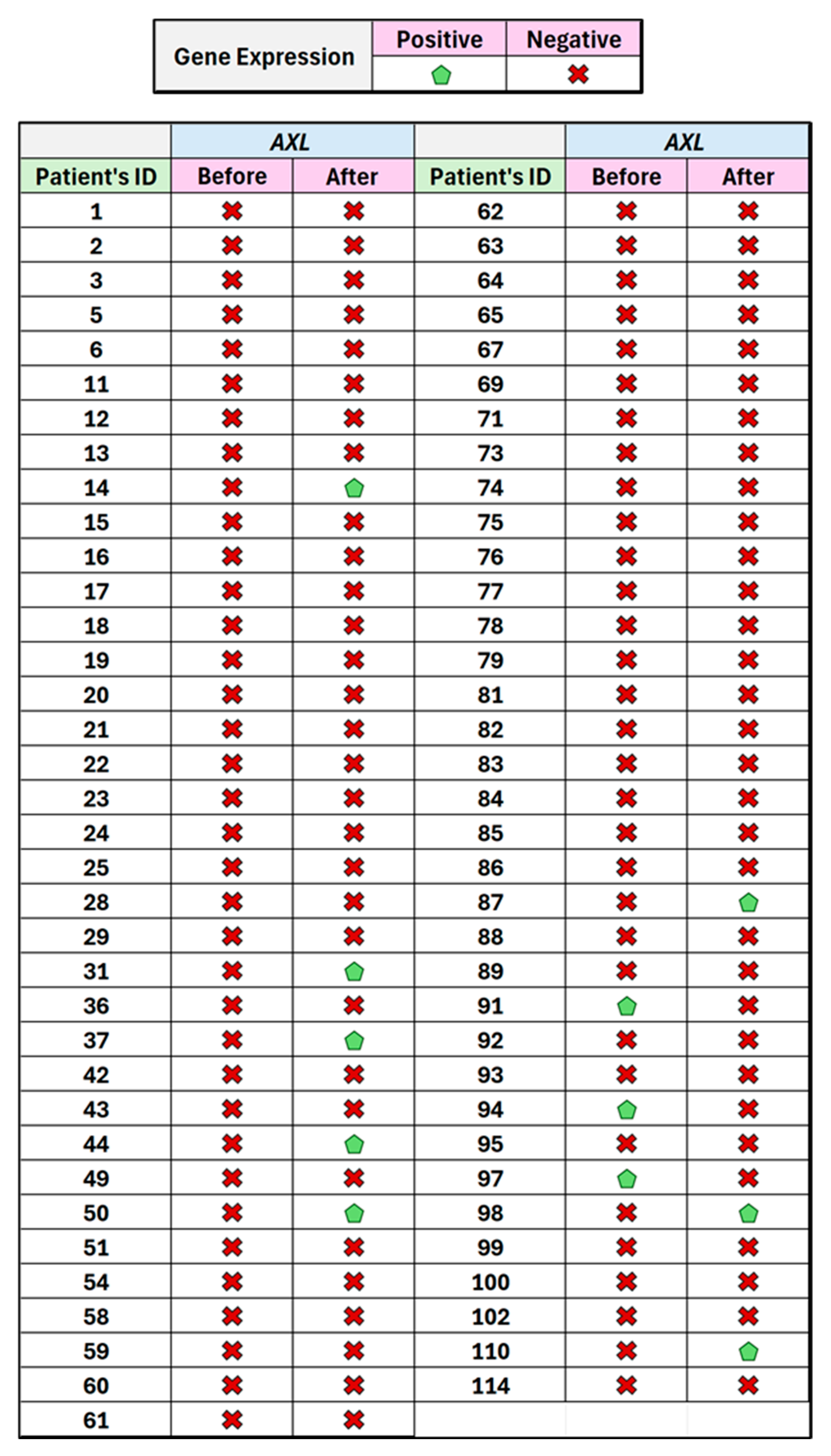
Group B: AXL-mRNA overexpression in CTC fractions was detected in 8/116 (6.9%) patients prior to treatment, in 8/71 (11.3%) after three or four cycles of treatment with ICIs, while no AXL transcripts were detected in any of the 10 patients at the time of PD. AXL-mRNA overexpression was not detected in matched samples before and after immunotherapy for any of the NSCLC patients. No statistically significant differences were observed between the time points (before and after immunotherapy: McNemar test, p = 0.227).
Direct comparison of AXL-mRNA overexpression detected before and after immunotherapy for 71 NSCLC patients revealed that three patients (Pt#91, Pt#94, Pt#97) had relative fold change values for AXL-mRNA overexpression detected in CTC fractions below the cut-off value after immunotherapy compared to the higher levels detected before immunotherapy. Conversely, AXL-mRNA overexpression in CTC fractions was detected in higher levels after immunotherapy for eight patients (Pt#14, Pt#31, Pt#37, Pt#44, Pt#50, Pt#87, Pt#98, and Pt#110). Pt#110 had the highest difference in levels of AXL-mRNA overexpression between the two time-points (Figure 4 and Figure 5).

4. Discussion

In this study, we evaluated AXL-mRNA overexpression in size-based enriched CTC fractions from NSCLC patients under osimertinib treatment and for the first time in NSCLC patients before and after immunotherapy.
Since the first report indicating AXL kinase as a novel mechanism of acquired resistance in EGFR-mutant NSCLC [25], many other preclinical and clinical studies have confirmed these primary data and further highlighted the role of AXL in EMT and drug resistance in NSCLC [26,27]. Numerous studies have reported that AXL confers resistance to EGFR-TKIs either as a bypass signaling pathway or by disrupting feedback signals [19,28]. Recently, the interest shifted to treating EGFR-mutant NSCLC patients with third-generation EGFR-TKIs, such as osimertinib [29]. In vitro and in vivo experiments have identified AXL as an important resistance mechanism to osimertinib. These results were also confirmed by data showing the effective outcomes of AXL inhibition in drug resistant cells [8,9,30,31]. AXL transcript levels significantly correlated with the tumor mutations burden and the emergence of the T790M, as this was also shown by other groups [32]. Unlike other driver genes, AXL expression is mediated through alternative mechanisms rather than gene mutations or amplifications [33]. Many different signaling pathways have been proposed to be involved in AXL-mediated resistance to EGFR-TKIs [30,32,34,35,36,37,38,39].
Our study was the first to detect AXL-mRNA overexpression in CTC fractions of NSCLC patients under osimertinib treatment [17]. In the present study, in Group A, an increased number of patients was detected with AXL-positive enriched CTCs after first cycle of treatment (16.1%) compared to baseline or later cycles of treatment and PD. Our results confirm our previous observation, although a smaller number of NSCLC patients was analyzed then [17]. Taniguchi et al. have shown that AXL overexpression was detected either in the initial or in the tolerant phase of cells treated with osimertinib [9]. Longitudinal monitoring of ongoing NSCLC patients during osimertinib, after one month of treatment and before PD, did not reveal any specific pattern of AXL-mRNA overexpression in CTC fractions. Compared to baseline, post 1st cycle and PD data, AXL-mRNA overexpression was detected in relatively low percentages (8.9%) during the follow-up of patients which can be explained by the effect of osimertinib on CTCs during treatment, as was shown in our previous study [18]. In addition, following the NSCLC patients during osimertinib treatment through AXL-mRNA overexpression in CTC fractions, there was a heterogeneous pattern of gene expression among them, further highlighting the significance of personalized treatment.
CTCs act as precursors of metastasis and their analysis unravels oncogenic alterations related to metastasis or to treatment resistance [13,40]. Moreover, CTC analysis offers a great potential for unveiling the heterogeneous tumor profile and novel resistance mechanisms in NSCLC [41,42]. In EGFR-mutant NSCLC, distinct CTC subpopulations exhibit a more aggressive or stem-like character [17,43,44]. Thus, the development and use of “size-based” CTC enrichment technologies like Parsortix, was proved to be optimal for detecting mesenchymal CTCs in NSCLC [14,17,24]. It has been demonstrated that AXL overexpression is also involved in EMT-mediated resistance to EGFR-TKIs [7,27]. Accordingly, it has been found that AXL inhibition can reverse EMT by resensitizing cells [26,27]. The most frequently observed CTC marker related to EMT is vimentin (VIM) demonstrating the key role of EMT in EGFR-TKI resistance [17,43,44,45]. Upregulation of AXL is often observed concurrently with the expression of VIM whereas VIM knockdown decreased AXL expression and restored erlotinib sensitivity [25]. Conversely, AXL inhibition did not affect EMT or VIM levels, suggesting that AXL upregulation in EGFR-resistant NSCLC cells may occur downstream of EMT [46]. EMT also modulates cancer cells by conferring metastatic capacity and transforming them into more aggressive cells with the aim of migrating to a potential metastatic site [47]. AXL overexpression was detected concurrently with VIM-positive CTCs, further enhancing the role of AXL in EMT and its implication in acquired EGFR-TKI resistance [14,17,25]. In our previous study, we have examined in parallel AXL and VIM overexpression in a small number of size-based enriched CTC fractions. According to our findings, in 11/81 (13.6%) samples, VIM was co-expressed with AXL—1/81 (1.2%) was only AXL-positive, and 41/81 (50.6%) were only VIM-positive [17]. This could possibly be explained by the fact that CTCs are highly heterogeneous, and our results were based on bulk analysis of CTCs, while previously published data reported a correlation of AXL expression with VIM expression in primary tumors.
Tumor progression and therapeutic resistance is not only a matter of cancer cells but also an interplay with the tumor microenvironment (TME) [48]. There is an increased body of evidence that major cellular and non-cellular components of the TME such as regulatory T-cells, dendritic cells (DCs), tumor-associated macrophages (TAMs), natural killer (NK) cells, fibroblasts, and other immune-stromal cells are modified by AXL overexpression1 [10,49,50]. A very recent study that is mainly based on animal models has shown that AXL is linked to negative loops of these immune cells and that AXL inhibition could enhance immune responses [51]. Resistance to combined immunotherapy and radiation therapy was related to AXL overexpression in tumors, while sensitivity to immunotherapy was increased through AXL inhibition [52]. In preclinical models, it was found that all tumors expressing AXL were also expressing PD-L1 and thus cooperative targeting boosts antitumor immunity [53]. mRNA profiles from NSCLC datasets revealed correlation also with CXCR4 and CXCR6 expressions [54]. High AXL expression in the tumor tissue of NSCLC patients revealed predictive significance of survival in patients treated with ICI [55] and after chemotherapy progression [56].
Based on the above evidence that AXL-mRNA overexpression plays a key role in the modulation of the immunosuppressive microenvironment, in this study we evaluated for, the first time, AXL-mRNA overexpression in enriched CTC fractions of NSCLC patients before and during immunotherapy in NSCLC. According to our findings, AXL was overexpressed in enriched CTC fractions in an increased number of NSCLC patients of Group B after three or four cycles of anti-PD-1 therapy compared to baseline; whereas, no AXL-mRNA overexpression was detected in enriched CTC fractions at PD analyzed only for 10 patients. However, this is a retrospective analysis and, thus, the small group of NSCLC patients that reached PD may not be representative of the presence of AXL-mRNA overexpression at this time point. It is recommended that a larger cohort study could further clarify the implication of AXL-mRNA overexpression at disease progression and resistance to immunotherapy.
It is important to highlight that in Group A, AXL-mRNA overexpression was detected in 12.8% of patients at baseline and increased to 16.1% after the first cycle, indicating an early adaptive response to osimertinib, while in Group B, AXL-mRNA overexpression rose from 6.9% at baseline to 11.3% during immunotherapy. The absence of AXL-mRNA overexpression at PD in Group B contrasts sharply with Group A, where 12.5% of PD samples remained AXL-positive. This could be due to the very small sample size (n = 10) at PD for Group B, or it may indicate a different resistance trajectory under immunotherapy, potentially expected within the broader context of NSCLC heterogeneity [57].
One of the limitations of our study is the significant discrepancy in the size of the two treatment cohorts, (39 patients in the osimertinib group versus 116 patients in the immunotherapy group). This imbalance limits the ability to compare the clinical utility of AXL as a biomarker across different treatment modalities. Moreover, the small number of patient samples that we analyzed did not allow us to evaluate the clinical significance based on Kaplan–Meier curves. However, based on our results, on previous evidence and also on the growing interest of the development and efficacy of AXL-inhibitors, we suggest that evaluation of AXL-mRNA expression in CTCs is a very promising biomarker that should be further tested in future clinical studies including larger cohorts of patients, since it could serve as a novel therapeutic target during longitudinal monitoring of NSCLC patients for guiding treatment decisions.
Lately, many clinical trials are testing the efficacy of various types of AXL-inhibitors in NSCLC patients as monotherapy or in combination with other agents [8]. The FDA has granted a fast track designation to bemcentinib (BGB324), an oral small molecule that is an inhibitor of AXL kinase, in combination with an anti-PD-L1 agent for patients with STK11 altered advanced or metastatic NSCLC (NCT03184571) [20]. Combinations of bemcentinib with erlotinib (NCT02424617), docetaxel (NCT02922777), and pembrolizumab (NCT03184571) exhibited tolerability in advanced NSCLC. Mecbotamab vedotin (BA3011), a conditionally active biologic anti-AXL antibody drug conjugate (CAB-AXL-ADC) is currently being investigated in combination with PD-1 or PD-L1 inhibitor in NSCLC (phase II, NCT04681131). In the SAFFRON-103 study (NCT03666143), sitravatinib combined with the anti-PD-1 antibody tislelizumab exhibited tolerance and objective responses in patients with advanced NSCLC [58]. Currently, sitravatinib and nivolumab are under evaluation in a phase III trial (NCT04921358) for advanced NSCLC. A novel AXL inhibitor, ONO-7475 combined with osimertinib proved effective to suppress resistance in EGFR-mutant NSCLC cells with AXL overexpression [59]. Very recently, the results from phase I/II study of Enapotamab Vedotin, an AXL-specific ADC in solid tumors including NSCLC, showed manageable safety profiles. However, antitumor activity did not show superior clinically meaningful responses as monotherapy compared to other therapies [60].
It is now well established that CTC molecular characterization at the mRNA level has a strong potential not only to provide information on tumor heterogeneity and alternative treatment resistance alterations but also to guide therapeutic interventions in NSCLC [61,62,63]. Based on our findings, we suggest that AXL-mRNA overexpression in CTCs should be further evaluated as a potential biomarker for anti-AXL targeted therapies in NSCLC. Considering the recent growing interest in the development and evaluation of new AXL-inhibitors, our observations could support the implementation of larger clinical studies to clarify the role of AXL-mRNA overexpression in CTC fractions as a potential novel therapeutic target during longitudinal monitoring of patients with NSCLC.

5. Conclusions

Our results indicate that AXL-mRNA overexpression in CTC fractions deserves to be further evaluated through larger clinical studies as a potential biomarker for anti-AXL targeted therapies in NSCLC.

Author Contributions

Conceptualization, E.L. and A.N.; methodology, A.N., E.T. (Elena Themistokli), A.S. (Areti Strati) and M.Z.; validation, A.N. and E.T. (Elena Themistokli); formal analysis, E.L. and A.N.; investigation, E.L. and A.N.; resources, E.T. (Emilia Tsaroucha), A.S. (Aggeliki Sfika), A.P., I.B., A.K. and V.G.; data curation, A.N., E.T. (Elena Themistokli) and A.S. (Areti Strati); writing—original draft preparation, A.N. and E.T. (Elena Themistokli); writing—review and editing, A.N. and E.L.; visualization, E.L.; supervision, E.L.; project administration, E.L.; funding acquisition, E.L. All authors have read and agreed to the published version of the manuscript.

Funding

This work was partially supported by the Stavros Niarchos Foundation within the framework of a grant to the National and Kapodistrian University of Athens [no. 16785]. Osimertinib (AZD9291; Astra Zeneca, UK) was administered to the patients in the context of a multicenter Phase II clinical study [ClinicalTrials.gov number, NCT02771314] supported by Astra Zeneca and sponsored by the Hellenic Oncology Research Group (HORG). Part of the study that included patients during immunotherapy was financially supported by the Hellenic Foundation for Research and Innovation and the General Secretariat for Research and Technology under Grant Agreement No. 1964 and from the European Union and Greek national funds through the Operational Program Competitiveness, Entrepreneurship and Innovation, under the call RESEARCH-CREATE-INNOVATE (project code: T1RCI-02935).

Institutional Review Board Statement

The study was conducted in accordance with the Declaration of Helsinki. The study for Group A was approved by the National Ethics Committee (35/00-03/16, 35/03-11/16), and the Institutional Ethical Committees of the centers participating in HORG. The study for Group B was approved by the ethics committees of all involved institutions granted approval for the study: Attikon University Hospital (7th meeting of the Scientific Advisory Board 10 September 2019), “SOTIRIA” General Hospital of Athens (19261, 28 August 2019); Aretaieion University Hospital, (137/28/11/19); General University Hospital of Alexandroupolis, (6th regular meeting of the Scientific Advisory Board, 19 June 2019).

Informed Consent Statement

Informed consent was obtained from all subjects involved in the study.

Data Availability Statement

The data presented in this study are available on request from the corresponding authors. The data are not publicly available due to ethical restrictions.

Acknowledgments

We acknowledge the contributions of all the patients who participated in this clinical study and the healthy volunteers for providing blood samples.

Conflicts of Interest

The authors declare no conflicts of interest.

Abbreviations

The following abbreviations are used in this manuscript:
B2MBeta-2 microglobulin
CTCCirculating tumor cells
ctDNACirculating tumor DNA
EGFR-TKIsEpidermal Growth Factor Receptor-Tyrosine Kinase Inhibitors
EMTEpithelial-to-mesenchymal transition
HDHealthy donors
ICIImmune checkpoint inhibitors
NSCLCNon-small-cell lung cancer
PBPeripheral blood
PBMCPeripheral blood mononuclear cells
PDProgression of disease
TMETumor microenvironment
VIMVimentin

References

  1. Otano, I.; Ucero, A.C.; Zugazagoitia, J.; Paz-Ares, L. At the crossroads of immunotherapy for oncogene-addicted subsets of NSCLC. Nat. Rev. Clin. Oncol. 2023, 3, 143–159. [Google Scholar] [CrossRef] [Scilit] [PubMed]
  2. Su, P.L.; Furuya, N.; Asrar, A.; Rolfo, C.; Li, Z.; Carbone, D.P.; He, K. Recent advances in therapeutic strategies for non-small cell lung cancer. J. Hematol. Oncol. 2025, 18, 35. [Google Scholar] [CrossRef] [Scilit] [PubMed]
  3. Ntzifa, A.; Marras, T.; Georgoulias, V.; Lianidou, E. Liquid biopsy for the management of NSCLC patients under osimertinib treatment treatment. Crit. Rev. Clin. Lab. Sci. 2024, 61, 347–369. [Google Scholar] [CrossRef] [Scilit]
  4. Wang, M.; Herbst, R.S.; Boshoff, C. Toward personalized treatment approaches for non-small-cell lung cancer. Nat. Med. 2021, 27, 1345–1356. [Google Scholar] [CrossRef] [Scilit]
  5. Li, M.S.C.; Mok, K.K.S.; Mok, T.S.K. Developments in targeted therapy & immunotherapy-how non-small cell lung cancer management will change in the next decade: A narrative review. Ann. Transl. Med. 2023, 11, 358. [Google Scholar] [CrossRef] [Scilit]
  6. Linger, R.M.A.; Keating, A.K.; Earp, H.S.; Graham, D.K. TAM Receptor Tyrosine Kinases: Biologic Functions, Signaling, and Potential Therapeutic Targeting in Human Cancer. Adv. Cancer Res. 2008, 100, 35–83. [Google Scholar] [CrossRef] [Scilit]
  7. Antony, J.; Huang, R.Y.J. AXL-driven EMT state as a targetable conduit in cancer. Cancer Res. 2017, 77, 3725–3732. [Google Scholar] [CrossRef] [Scilit]
  8. Namba, K.; Shien, K.; Takahashi, Y.; Torigoe, H.; Sato, H.; Yoshioka, T.; Takeda, T.; Kurihara, E.; Ogoshi, Y.; Yamamoto, H.; et al. Activation of AXL as a preclinical acquired resistance mechanism against osimertinib treatment in EGFR-mutant non-small cell lung cancer cells. Mol. Cancer Res. 2019, 17, 499–507. [Google Scholar] [CrossRef] [Scilit]
  9. Taniguchi, H.; Yamada, T.; Wang, R.; Tanimura, K.; Adachi, Y.; Nishiyama, A.; Tanimoto, A.; Takeuchi, S.; Araujo, L.H.; Boroni, M.; et al. AXL confers intrinsic resistance to osimertinib and advances the emergence of tolerant cells. Nat. Commun. 2019, 10, 259. [Google Scholar] [CrossRef] [Scilit]
  10. Engelsen, A.S.T.; Lotsberg, M.L.; Abou Khouzam, R.; Thiery, J.P.; Lorens, J.B.; Chouaib, S.; Terry, S. Dissecting the Role of AXL in Cancer Immune Escape and Resistance to Immune Checkpoint Inhibition. Front. Immunol. 2022, 13, 869676. [Google Scholar] [CrossRef] [Scilit] [PubMed]
  11. Malapelle, U.; Pisapia, P.; Pepe, F.; Russo, G.; Buono, M.; Russo, A.; Gomez, J.; Khorshid, O.; Mack, P.C.; Rolfo, C.; et al. The evolving role of liquid biopsy in lung cancer. Lung Cancer 2022, 172, 53–64. [Google Scholar] [CrossRef] [Scilit]
  12. Malapelle, U.; Pisapia, P.; Addeo, A.; Arrieta, O.; Bellosillo, B.; Cardona, A.F.; Cristofanilli, M.; De Miguel-Perez, D.; Denninghoff, V.; Durán, I.; et al. Liquid biopsy from research to clinical practice: Focus on non-small cell lung cancer. Expert Rev. Mol. Diagn. 2021, 21, 1165–1178. [Google Scholar] [CrossRef] [Scilit]
  13. Strati, A.; Markou, A.; Kyriakopoulou, E.; Lianidou, E. Detection and Molecular Characterization of Circulating Tumour Cells: Challenges for the Clinical Setting. Cancers 2023, 15, 2185. [Google Scholar] [CrossRef] [Scilit]
  14. Ikeda, M.; Koh, Y.; Teraoka, S.; Sato, K.; Kanai, K.; Hayata, A.; Tokudome, N.; Akamatsu, H.; Ozawa, Y.; Akamatsu, K.; et al. Detection of AXL expression in circulating tumor cells of lung cancer patients using an automated microcavity array system. Cancer Med. 2020, 9, 2122–2133. [Google Scholar] [CrossRef] [Scilit] [PubMed]
  15. Bardol, T.; Eslami-S, Z.; Masmoudi, D.; Alexandre, M.; Duboys de Labarre, M.; Bobrie, A.; D’Hondt, V.; Guiu, S.; Kurma, K.; Cayrefourcq, L.; et al. First evidence of AXL expression on circulating tumor cells in metastatic breast cancer patients: A proof-of-concept study. Cancer Med. 2024, 13, e6843. [Google Scholar] [CrossRef] [Scilit] [PubMed]
  16. De Miguel-Pérez, D.; Bayarri-Lara, C.I.; Ortega, F.G.; Russo, A.; Rodriguez, M.J.M.; Alvarez-Cubero, M.J.; Serrano, E.M.; Lorente, J.A.; Rolfo, C.; Serrano, M.J. Post-surgery circulating tumor cells and AXL overexpression as new poor prognostic biomarkers in resected lung adenocarcinoma. Cancers 2019, 11, 1750. [Google Scholar] [CrossRef] [Scilit]
  17. Ntzifa, A.; Strati, A.; Kallergi, G.; Kotsakis, A.; Georgoulias, V.; Lianidou, E. Gene expression in circulating tumor cells reveals a dynamic role of EMT and PD-L1 during osimertinib treatment in NSCLC patients. Sci. Rep. 2021, 11, 2313. [Google Scholar] [CrossRef] [Scilit] [PubMed]
  18. Kallergi, G.; Kontopodis, E.; Ntzifa, A.; Jordana-Ariza, N.; Karachaliou, N.; Pantazaka, E.; Charalambous, H.A.; Psyrri, A.; Tsaroucha, E.; Boukovinas, I.; et al. Effect of Osimertinib on CTCs and ctDNA in EGFR Mutant Non-Small Cell Lung Cancer Patients: The Prognostic Relevance of Liquid Biopsy. Cancers 2022, 14, 1574. [Google Scholar] [CrossRef] [Scilit]
  19. Yadav, M.; Sharma, A.; Patne, K.; Tabasum, S.; Suryavanshi, J.; Rawat, L.; Machaalani, M.; Eid, M.; Singh, R.P.; Choueiri, T.K.; et al. AXL signaling in cancer: From molecular insights to targeted therapies. Signal Transduct. Target. Ther. 2025, 10, 37. [Google Scholar] [CrossRef] [Scilit]
  20. Krebs, M.G.; Helland, A.; Costa, E.C.; Aperribay, E.A.; Gomez, M.D.; Perez, J.T.; Thompson, J.; Strauss, J.; Granados, A.L.O.; Felip, E.; et al. OA01.07 A Phase II Study of the Oral Selective AXL Inhibitor Bemcentinib with Pembrolizumab in Patients with Advanced NSCLC. J. Thorac. Oncol. 2021, 16, S103. [Google Scholar] [CrossRef] [Scilit]
  21. Standard No. 15189:2022; Medical Laboratories—Requirements for Quality and Competence. 4th ed. International Organization for Standardization: Geneva, Switzerland, 2022; Volume 4. Available online: https://www.iso.org/standard/76677.html (accessed on 19 May 2026).
  22. Livak, K.J.; Schmittgen, T.D. Analysis of relative gene expression data using real-time quantitative PCR and the 2−ΔΔCT method. Methods 2001, 25, 402–408. [Google Scholar] [CrossRef] [Scilit]
  23. Zavridou, M.; Mastoraki, S.; Strati, A.; Koutsodontis, G.; Klinakis, A.; Psyrri, A.; Lianidou, E. Direct comparison of size-dependent versus EpCAM-dependent CTC enrichment at the gene expression and DNA methylation level in head and neck squamous cell carcinoma. Sci. Rep. 2020, 16, 6551. [Google Scholar] [CrossRef] [Scilit]
  24. Strati, A.; Zavridou, M.; Economopoulou, P.; Gkolfinopoulos, S.; Psyrri, A.; Lianidou, E. Development and Analytical Validation of a Reverse Transcription Droplet Digital PCR (RT-ddPCR) Assay for PD-L1 Transcripts in Circulating Tumor Cells. Clin. Chem. 2021, 67, 642–652. [Google Scholar] [CrossRef] [Scilit]
  25. Zhang, Z.; Lee, J.C.; Lin, L.; Olivas, V.; Au, V.; Laframboise, T.; Abdel-Rahman, M.; Wang, X.; Levine, A.D.; Rho, J.K.; et al. Activation of the AXL kinase causes resistance to EGFR-targeted therapy in lung cancer. Nat. Genet. 2013, 44, 852–860. [Google Scholar] [CrossRef] [Scilit]
  26. Byers, L.A.; Diao, L.; Wang, J.; Saintigny, P.; Girard, L.; Peyton, M.; Shen, L.; Fan, Y.; Giri, U.; Tumula, P.K.; et al. An epithelial-mesenchymal transition (EMT) gene signature predicts resistance to EGFR and PI3K inhibitors and identifies Axl as a therapeutic target for overcoming EGFR inhibitor resistance. Clin. Cancer Res. 2013, 19, 279–290. [Google Scholar] [CrossRef] [Scilit] [PubMed]
  27. Wu, F.; Li, J.; Jang, C.; Wang, J.; Xiong, J. The role of Axl in drug resistance and epithelial-to-mesenchymal transition of non-small cell lung carcinoma. Int. J. Clin. Exp. Pathol. 2014, 7, 6653–6661. [Google Scholar]
  28. Jiang, Z.; Gu, Z.; Yu, X.; Cheng, T.; Liu, B. Research progress on the role of bypass activation mechanisms in resistance to tyrosine kinase inhibitors in non-small cell lung cancer. Front. Oncol. 2024, 14, 1447678. [Google Scholar] [CrossRef] [Scilit] [PubMed]
  29. Le, X.; Puri, S.; Negrao, M.V.; Nilsson, M.B.; Boyle, T.; Hicks, J.K.; Lovinger, K.L.; Roarty, E.; Tang, M.; Sun, H.; et al. Landscape of EGFR-dependent and -independent resistance mechanisms to osimertinib and continuation therapy post-progression in EGFR-mutant NSCLC. Clin. Cancer Res. 2019, 24, 6195–6203. [Google Scholar] [CrossRef] [Scilit]
  30. Karachaliou, N.; Chaib, I.; Cardona, A.F.; Berenguer, J.; Bracht, J.W.P.; Yang, J.; Cai, X.; Wang, Z.; Hu, C.; Drozdowskyj, A.; et al. Common Co-activation of AXL and CDCP1 in EGFR-mutation-positive Non-Small Cell Lung Cancer Associated with Poor Prognosis. EBioMedicine 2018, 29, 112–127. [Google Scholar] [CrossRef] [Scilit] [PubMed]
  31. Kim, D.; Bach, D.H.; Fan, Y.H.; Luu, T.T.T.; Hong, J.Y.; Park, H.J.; Lee, S.K. AXL degradation in combination with EGFR-TKI can delay and overcome acquired resistance in human non-small cell lung cancer cells. Cell Death Dis. 2019, 10, 361. [Google Scholar] [CrossRef] [Scilit]
  32. Noronha, A.; Nataraj, N.B.; Lee, J.S.; Zhitomirsky, B.; Oren, Y.; Oster, S.; Lindzen, M.; Mukherjee, S.; Will, R.; Ghosh, S.; et al. AXL and Error-Prone DNA Replication Confer Drug Resistance and Offer Strategies to Treat EGFR-Mutant Lung Cancer. Cancer Discov. 2022, 12, 2666–2683. [Google Scholar] [CrossRef] [Scilit]
  33. Tang, Y.; Zang, H.; Wen, Q.; Fan, S. AXL in cancer: A modulator of drug resistance and therapeutic target. J. Exp. Clin. Cancer Res. 2023, 42, 148. [Google Scholar] [CrossRef] [Scilit]
  34. Tian, Y.; Zhang, Z.; Miao, L.; Yang, Z.; Yang, J.; Wang, Y.; Qian, D.; Cai, H.; Wang, Y. Anexelekto (AXL) Increases Resistance to EGFR-TKI and Activation of AKT and ERK1/2 in Non-Small Cell Lung Cancer Cells. Oncol. Res. 2016, 24, 295–303. [Google Scholar] [CrossRef] [Scilit]
  35. Ghiso, E.; Migliore, C.; Ciciriello, V.; Morando, E. YAP-Dependent AXL Overexpression Mediates Resistance to EGFR Inhibitors in NSCLC. Neoplasia 2017, 19, 1012–1021. [Google Scholar] [CrossRef] [Scilit]
  36. Saab, S.; Chang, O.S.; Nagaoka, K.; Hung, M.; Yamaguchi, H. The potential role of YAP in Axl-mediated resistance to EGFR tyrosine kinase inhibitors. Am. J. Cancer Res. 2019, 9, 2719–2729. [Google Scholar]
  37. Liu, Y.N.; Tsai, M.F.; Wu, S.G.; Chang, T.H.; Tsai, T.H.; Gow, C.H.; Chang, Y.L.; Shih, J.Y. Acquired resistance to EGFR tyrosine kinase inhibitors is mediated by the reactivation of STC2/JUN/AXL signaling in lung cancer. Int. J. Cancer 2019, 145, 1609–1624. [Google Scholar] [CrossRef] [Scilit] [PubMed]
  38. Sang, Y.B.; Kim, J.H.; Kim, C.G.; Hong, M.H.; Kim, H.R.; Cho, B.C.; Lim, S.M. The Development of AXL Inhibitors in Lung Cancer: Recent Progress and Challenges. Front. Oncol. 2022, 12, 811247. [Google Scholar] [CrossRef] [Scilit]
  39. Vouri, M.; Kennedy, S.P.; An, Q.; Pilkington, G.J.; Ha, S. Axl-EGFR receptor tyrosine kinase hetero-interaction provides EGFR with access to pro-invasive signalling in cancer cells. Oncogenesis 2016, 5, e266. [Google Scholar] [CrossRef] [Scilit] [PubMed]
  40. Micalizzi, D.S.; Haber, D.A.; Maheswaran, S. Cancer metastasis through the prism of epithelial-to-mesenchymal transition in circulating tumor cells. Mol. Oncol. 2017, 11, 770–780. [Google Scholar] [CrossRef] [Scilit] [PubMed]
  41. Pawlikowska, P.; Faugeroux, V.; Oulhen, M.; Aberlenc, A.; Tayoun, T.; Pailler, E.; Farace, F. Circulating tumor cells (CTCs) for the noninvasive monitoring and personalization of non-small cell lung cancer (NSCLC) therapies. J. Thorac. Dis. 2019, 11, S45–S56. [Google Scholar] [CrossRef] [Scilit]
  42. Gallo, M.; De Luca, A.; Frezzetti, D.; Passaro, V.; Maiello, M.R.; Normanno, N. The potential of monitoring treatment response in non-small cell lung cancer using circulating tumour cells. Expert Rev. Mol. Diagn. 2019, 19, 683–694. [Google Scholar] [CrossRef] [Scilit]
  43. Lindsay, C.R.; Faugeroux, V.; Michiels, S.; Pailler, E.; Facchinetti, F.; Ou, D.; Bluthgen, M.V.; Pannet, C.; Ngo-Camus, M.; Bescher, G.; et al. A Prospective Examination of Circulating Tumor Cell Profiles in Non-Small Cell Lung Cancer Molecular Subgroups. Ann. Oncol. 2017, 28, 1523–1531. [Google Scholar] [CrossRef] [Scilit]
  44. Hanssen, A.; Wagner, J.; Gorges, T.M.; Taenzer, A.; Uzunoglu, F.G.; Driemel, C.; Stoecklein, N.H.; Knoefel, W.T.; Angenendt, S.; Hauch, S.; et al. Characterization of different CTC subpopulations in non-small cell lung cancer. Sci. Rep. 2016, 6, 28010. [Google Scholar] [CrossRef] [Scilit]
  45. Morrow, C.J.; Trapani, F.; Metcalf, R.L.; Bertolini, G.; Hodgkinson, C.L.; Khandelwal, G.; Kelly, P.; Galvin, M.; Carter, L.; Simpson, K.L.; et al. Tumourigenic non-small-cell lung cancer mesenchymal circulating tumour cells: A clinical case study. Ann. Oncol. 2016, 27, 1155–1160. [Google Scholar] [CrossRef] [Scilit] [PubMed]
  46. Dela Carmela, M.C.P.; Medina, P.M.B. Epithelial–mesenchymal transition (EMT) and its role in acquired epidermal growth factor receptor-tyrosine kinase inhibitor (EGFR-TKI) chemoresistance in non-small cell lung cancer (NSCLC). Cancer Pathog. Ther. 2025, 3, 215–225. [Google Scholar] [CrossRef] [Scilit]
  47. Dongre, A.; Weinberg, R.A. New insights into the mechanisms of epithelial–mesenchymal transition and implications for cancer. Nat. Rev. Mol. Cell Biol. 2019, 20, 69–84. [Google Scholar] [CrossRef] [Scilit]
  48. Li, Z.; Li, J.; Bai, X.; Huang, X.; Wang, Q. Tumor microenvironment as a complex milieu driving cancer progression: A mini review. Clin. Transl. Oncol. 2025, 27, 1943–1952. [Google Scholar] [CrossRef] [Scilit]
  49. DeRyckere, D.; Huelse, J.M.; Earp, H.S.; Graham, D.K. TAM family kinases as therapeutic targets at the interface of cancer and immunity. Nat. Rev. Clin. Oncol. 2023, 20, 755–779. [Google Scholar] [CrossRef] [Scilit]
  50. Liu, Y.; Xu, L.; Dou, Y.; He, Y. AXL: Shapers of tumor progression and immunosuppressive microenvironments. Mol. Cancer 2025, 24, 11. [Google Scholar] [CrossRef] [Scilit] [PubMed]
  51. Yang, J.; Chen, G.; Wang, R.; Song, C.; Yi, H. Navigating TAM receptor dynamics in tumour immunotherapy. Cancer Immunol. Immunother. 2025, 74, 146. [Google Scholar] [CrossRef] [Scilit] [PubMed]
  52. Aguilera, T.A.; Rafat, M.; Castellini, L.; Shehade, H.; Kariolis, M.S.; Hui, A.B.; Stehr, H.; Von Eyben, R.; Jiang, D.; Ellies, L.G.; et al. Reprogramming the immunological microenvironment through radiation. Nat. Commun. 2016, 7, 13898. [Google Scholar] [CrossRef] [Scilit]
  53. Boshuizen, J.; Pencheva, N.; Krijgsman, O.; Altimari, D.D.; Castro, P.G.; de Bruijn, B.; Ligtenberg, M.A.; Gresnigt-Van den Heuvel, E.; Vredevoogd, D.W.; Song, J.Y.; et al. Cooperative Targeting of Immunotherapy-Resistant Melanoma and Lung Cancer by an AXL-Targeting Antibody-Drug Conjugate and Immune Checkpoint Blockade. Cancer Res. 2021, 81, 1775–1787. [Google Scholar] [CrossRef] [Scilit]
  54. Tsukita, Y.; Fujino, N.; Miyauchi, E.; Saito, R.; Fujishima, F.; Itakura, K.; Kyogoku, Y.; Okutomo, K.; Yamada, M.; Okazaki, T.; et al. Axl kinase drives immune checkpoint and chemokine signalling pathways in lung adenocarcinomas. Clin. Cancer Res. 2019, 22, 4585–4593. [Google Scholar] [CrossRef] [Scilit]
  55. Morimoto, K.; Yamada, T.; Sawada, R.; Azuma, K.; Goto, Y.; Harada, T.; Shiotsu, S.; Tamiya, N.; Chihara, Y.; Takeda, T.; et al. Predictive value of p53 and AXL immunostaining for the efficacy of immune checkpoint inhibitor-based therapy after osimertinib treatment in patients with epidermal growth factor-mutant non-small cell lung cancer. Cancer Immunol. Immunother. 2023, 72, 1699–1707. [Google Scholar] [CrossRef] [Scilit]
  56. Rayford, A.; Gärtner, F.; Ramnefjell, M.P.; Lorens, J.B.; Micklem, D.R.; Aanerud, M.; Engelsen, A.S.T. AXL expression reflects tumor-immune cell dynamics impacting outcome in non-small cell lung cancer patients treated with immune checkpoint inhibitor monotherapy. Front. Immunol. 2024, 15, 1444007. [Google Scholar] [CrossRef] [Scilit]
  57. Xiang, Y.; Liu, X.; Wang, Y.; Zheng, D. Mechanisms of resistance to targeted therapy and immunotherapy in non-small cell lung cancer: Promising strategies to overcoming challenges. Front. Immunol. 2024, 15, 1366260. [Google Scholar] [CrossRef] [Scilit]
  58. Zhao, J.; Yu, X.; Huang, D.; Ma, Z.; Gao, B.; Cui, J. SAFFRON-103: A phase 1b study of the safety and efficacy of sitravatinib combined with tislelizumab in patients with locally advanced or metastatic non-small cell lung cancer. J. Immunother. Cancer 2023, 11, e006055. [Google Scholar] [CrossRef] [Scilit]
  59. Okura, N.; Nishioka, N.; Yamada, T.; Taniguchi, H.; Tanimura, K.; Katayama, Y.; Yoshimura, A.; Watanabe, S.; Kikuchi, T.; Shiotsu, S.; et al. ONO-7475, a Novel AXL Inhibitor, Suppresses the Adaptive Resistance to Initial EGFR-TKI Treatment in EGFR-Mutated Non-Small Cell Lung Cancer. Clin. Cancer Res. 2020, 26, 224–2256. [Google Scholar] [CrossRef] [Scilit]
  60. Rohrberg, K.S.; Lopez, J.S.; Milhem, M.M.; Blank, C.U.; Reijers, I.; Thistlethwaite, F.; Plummer, R.; Piha-Paul, S.A.; Jänne, P.A.; Shum, E.; et al. Phase I/II Study of AXL-Specific Antibody-Drug Conjugate Enapotamab Vedotin in Patients with Advanced Solid Tumors. Cancer Res. Commun. 2025, 5, 2066–2078. [Google Scholar] [CrossRef] [Scilit]
  61. Lianidou, E.; Pantel, K. Liquid biopsies. Genes Chromosom. Cancer 2019, 58, 219–232. [Google Scholar] [CrossRef] [Scilit]
  62. Kilgour, E.; Rothwell, D.G.; Brady, G.; Dive, C. Liquid Biopsy-Based Biomarkers of Treatment Response and Resistance. Cancer Cell 2020, 37, 485–495. [Google Scholar] [CrossRef] [Scilit]
  63. Ntzifa, A.; Marras, T.; Kallergi, G. Comprehensive liquid biopsy analysis for monitoring NSCLC patients under second-line osimertinib treatment. Front. Oncol. 2024, 14, 1435537. [Google Scholar] [CrossRef] [Scilit]
Figure 1. Experimental flowchart of the study.
Cancers 18 01759 g001
Figure 2. Group A: AXL-mRNA overexpression in size-based enriched CTC fractions with respect to B2M expression of NSCLC patients at different time points during osimertinib treatment.
Cancers 18 01759 g002
Figure 3. Relative fold change values of AXL-mRNA overexpression in CTC fractions of NSCLC patients who presented PD (AL) over time and of two ongoing NSCLC patients (M,N) over time (red line depicts cut-off value: 2−ΔΔCq = 1.55, reddish area: above cut-off value, grayish area: below cut-off value).
Cancers 18 01759 g003aCancers 18 01759 g003b
Figure 4. Group B: Direct comparison of AXL-mRNA overexpression detected in CTC fractions before and after immunotherapy for 71 metastatic NSCLC patients.
Cancers 18 01759 g004
Figure 5. Relative fold change values of AXL-mRNA overexpression in CTC fractions from metastatic NSCLC patients in paired samples (n = 71) before and after immunotherapy (each line represents an individual patient, red dashed line depicts cut-off value: 2−ΔΔCq = 1.80).
Cancers 18 01759 g005
Disclaimer/Publisher’s Note: The statements, opinions and data contained in all publications are solely those of the individual author(s) and contributor(s) and not of MDPI and/or the editor(s). MDPI and/or the editor(s) disclaim responsibility for any injury to people or property resulting from any ideas, methods, instructions or products referred to in the content.