Circulating Tumor DNA as Emerging Predictive and Prognostic Biomarker in Prostate Cancer


Simple Summary
Abstract
1. Introduction
2. Detection Methods and Technologies for Liquid Biopsies
- i.
- Significantly shorter mean fragment lengths compared with localized disease (p < 0.05);
- ii.
- An inverse correlation between fragment size and circulating tumor fraction (p < 0.0001);
- iii.
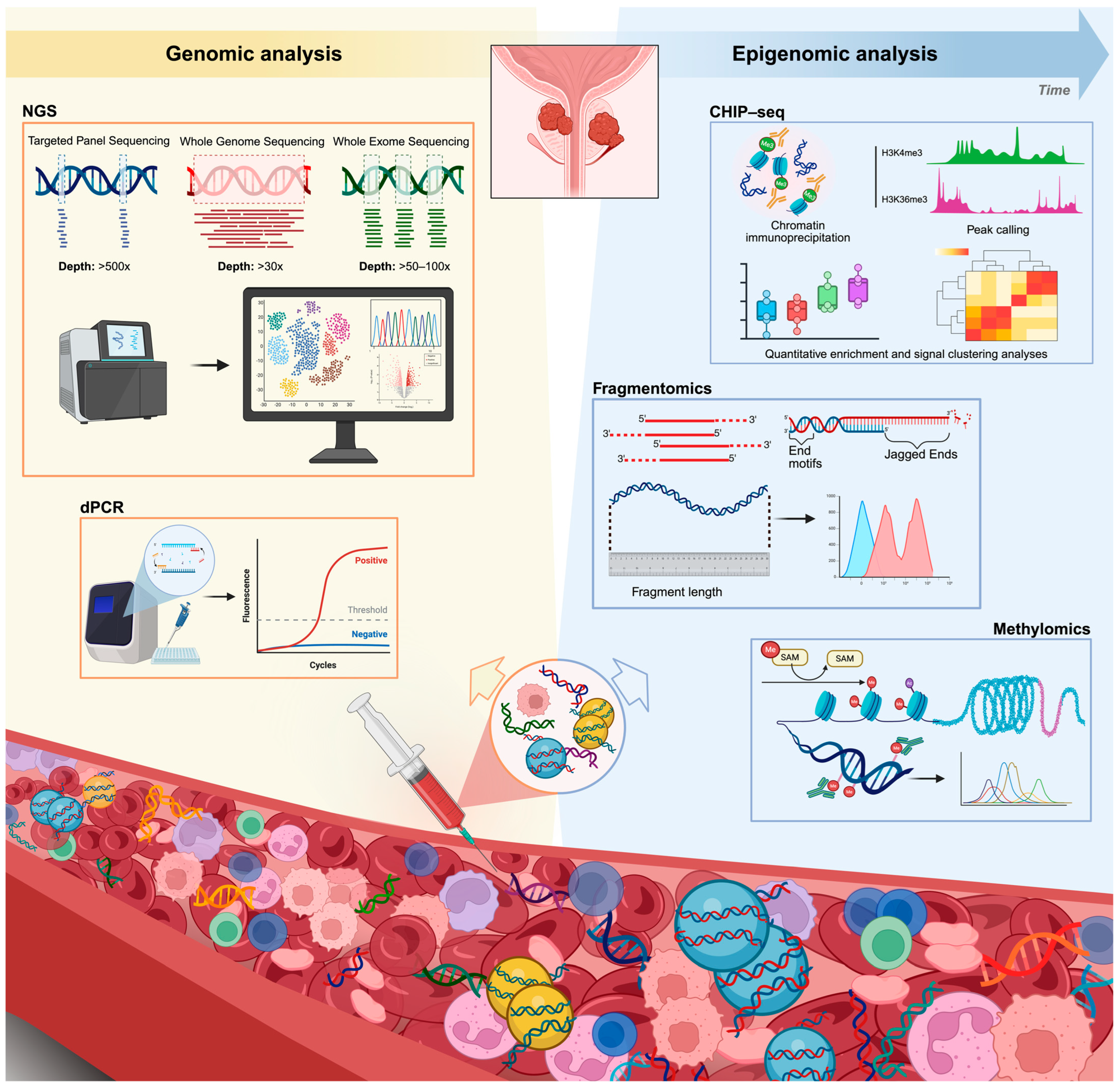
- A shorter PFS and OS in patients with higher proportions of longer fragments (p = 0.008 and p = 0.027, respectively) [57]. Notably, integrating fragment size profiles with cfDNA end-motif patterns enables the reconstruction of nucleosome footprints (i.e., “nucleosomics”) that reflect chromatin accessibility and transcription factor binding, thus informing gene expression and tissue of origin [28]. De Sarkar et al. applied patient-derived xenograft (PDX) nucleosome-footprint signatures to classify human mCRPC phenotypes. In 101 cfDNA samples from the Dana-Farber Cancer Institute database, the model distinguished adenocarcinoma from neuroendocrine subtypes with an overall accuracy of 0.96 (AUC 0.97 for high tumor fraction, TFx ≥ 0.10; 0.76 for low tumor fraction, TFx < 0.10) [62].
3. Role of ctDNA in Localized Prostate Cancer
4. Role of ctDNA in Advanced Prostate Cancer and Its Clinical Application
5. ctDNA as Prognostic Biomarker
6. ctDNA in Metastatic Hormone-Sensitive Prostate Cancer
7. ctDNA in Metastatic Castration-Resistant Prostate Cancer
8. ctDNA as Predictive Biomarker
9. Using Liquid Biopsies to Predict Resistance to Systemic Treatments for Advanced Prostate Cancer
10. NEPC Marker Detection with Liquid Biopsies
11. ctDNA and Its Emerging Integration with Epigenomic Profiling
12. Challenges and Limitations with ctDNA
13. Future Directions and Conclusions
Author Contributions
Funding
Institutional Review Board Statement
Informed Consent Statement
Data Availability Statement
Conflicts of Interest
Abbreviations
| ADT | Androgen deprivation therapy |
| AR | Androgen receptor |
| ARPIs | Androgen receptor pathway inhibitors |
| AR-V7 | Androgen receptor splice variant 7 |
| BCTs | Blood collection tubes |
| BEAMing | Beads, Emulsion, Amplification, and Magnetics |
| CAPP-Seq | Cancer Personalized Profiling by deep Sequencing |
| cfChIP-seq | cell-free chromatin immunoprecipitation sequencing |
| cfDNA | cell-free DNA |
| CH | Clonal hematopoiesis |
| CHIP | Clonal hematopoiesis of indeterminate potential |
| CIN | Chromosomal instability |
| CNAs | Copy number alterations |
| CNV | Copy number variation |
| CRPC-NE | Castration-resistant prostate cancer with neuroendocrine phenotype |
| CT | Computed tomography |
| CTC | Circulating tumor cell |
| ctDNA | circulating tumor DNA |
| ddPCR | droplet digital PCR |
| DDR | DNA damage response |
| dMMR | mismatch repair deficiency |
| EDI | Evolutionary dynamic index |
| EZH2 | Enhancer of zeste homolog 2 |
| GWA | Genome-wide aneuploidy score |
| HRR | homologous recombination repair |
| LBD | Ligand-binding domain |
| LDH | Lactate dehydrogenase |
| mCRPC | metastatic castration-resistant prostate cancer |
| mFAST-SeqS | modified Fast Aneuploidy Screening Test-Sequencing |
| mHSPC | metastatic hormone-sensitive prostate cancer |
| MRD | minimal residual disease |
| MSI-H | microsatellite instability-high |
| NEMO | Neuroendocrine detection and monitoring |
| NEPC | Neuroendocrine prostate cancer |
| NGS | Next-generation sequencing |
| OS | overall survival |
| PARP | Poly(ADP-ribose) polymerase |
| PARPi | PARP inhibitors |
| PCR | polymerase chain reaction |
| PDX | Patient-derived xenograft |
| PET | Positron emission tomography |
| PFS | progression-free survival |
| PSA | Prostate-specific antigen |
| PSMA | Prostate-specific membrane antigen |
| qPCR | quantitative PCR |
| rPFS | radiographic progression-free survival |
| TEC-Seq | Targeted Error Correction Sequencing |
| TFx | tumor fraction |
| TMB | tumor mutational burden |
| UMIs | unique molecular identifiers |
| VAF | variant allele frequency |
| WES | whole-exome sequencing |
| WGS | whole-genome sequencing |
References
- Chu, F.; Chen, L.; Guan, Q.; Chen, Z.; Ji, Q.; Ma, Y.; Ji, J.; Sun, M.; Huang, T.; Song, H.; et al. Global Burden of Prostate Cancer: Age-Period-Cohort Analysis from 1990 to 2021 and Projections until 2040. World J. Surg. Oncol. 2025, 23, 98. [Google Scholar] [CrossRef] [PubMed]
- Bray, F.; Laversanne, M.; Sung, H.; Ferlay, J.; Siegel, R.L.; Soerjomataram, I.; Jemal, A. Global Cancer Statistics 2022: GLOBOCAN Estimates of Incidence and Mortality Worldwide for 36 Cancers in 185 Countries. CA Cancer J. Clin. 2024, 74, 229–263. [Google Scholar] [CrossRef]
- Lowentritt, B.H.; Du, S.; Rossi, C.; Muser, E.; Khilfeh, I.; Kinkead, F.; Korsiak, J.; Lefebvre, P.; Pilon, D.; Agarwal, N. Real-World Clinical Outcomes among Patients with Metastatic Castration-Sensitive Prostate Cancer Initiating Apalutamide. Future Oncol. Lond. Engl. 2024, 20, 2005–2013. [Google Scholar] [CrossRef]
- Fizazi, K.; Tran, N.; Fein, L.; Matsubara, N.; Rodriguez-Antolin, A.; Alekseev, B.Y.; Özgüroğlu, M.; Ye, D.; Feyerabend, S.; Protheroe, A.; et al. Abiraterone Acetate plus Prednisone in Patients with Newly Diagnosed High-Risk Metastatic Castration-Sensitive Prostate Cancer (LATITUDE): Final Overall Survival Analysis of a Randomised, Double-Blind, Phase 3 Trial. Lancet Oncol. 2019, 20, 686–700. [Google Scholar] [CrossRef]
- Zhang, A.Y.; Davis, I.D.; Thomas, H.; McLaughlin, R.A.; Tan, T.H.; Pook, D.W.; Marx, G.M.; Zielinski, R.R.; Sandhu, S.; Thomson, A.; et al. 8-Year Outcomes of Enzalutamide (ENZA) versus a Non-Steroidal Anti-Androgen (NSAA) for Metastatic, Hormone-Sensitive Prostate Cancer (ENZAMET; ANZUP 1304). J. Clin. Oncol. 2025, 43, 5090. [Google Scholar] [CrossRef]
- Agarwal, N.; Bjartell, A.; Juárez, Á.; Kerßenboom, R.; Perualila, N.; Wigfield, P.; Chi, K.N. Estimating Median Overall Survival of Apalutamide Compared to Placebo in Metastatic Hormone-Sensitive Prostate Cancer (mHSPC) Populations: Statistical Extrapolations of the TITAN Study. J. Clin. Oncol. 2024, 42, 223. [Google Scholar] [CrossRef]
- Spratt, D.E.; Srinivas, S.; Adra, N.; Ahmed, B.; An, Y.; Bitting, R.; Chapin, B.; Cheng, H.H.; Cho, S.Y.; D’Amico, A.V.; et al. Prostate Cancer, Version 3.2026, NCCN Clinical Practice Guidelines in Oncology. J. Natl. Compr. Canc. Netw. 2025, 23, 469–493. [Google Scholar] [CrossRef]
- Freedland, S.J.; Davis, M.; Epstein, A.J.; Arondekar, B.; Ivanova, J.I. Real-World Treatment Patterns and Overall Survival among Men with Metastatic Castration-Resistant Prostate Cancer (mCRPC) in the US Medicare Population. Prostate Cancer Prostatic Dis. 2024, 27, 327–333. [Google Scholar] [CrossRef]
- Zedan, A.H.; Hansen, T.; Timm, S. Survival Outcomes for Metastatic Castration Sensitive Prostate Cancer: Real World Data from a Danish Cohort 2016–2022. J. Clin. Oncol. 2024, 42, e17088. [Google Scholar] [CrossRef]
- VanderWeele, D.J.; Finney, R.; Katayama, K.; Gillard, M.; Paner, G.; Imoto, S.; Yamaguchi, R.; Wheeler, D.; Lack, J.; Cam, M.; et al. Genomic Heterogeneity Within Individual Prostate Cancer Foci Impacts Predictive Biomarkers of Targeted Therapy. Eur. Urol. Focus 2019, 5, 416–424. [Google Scholar] [CrossRef]
- Haffner, M.C.; Zwart, W.; Roudier, M.P.; True, L.D.; Nelson, W.G.; Epstein, J.I.; De Marzo, A.M.; Nelson, P.S.; Yegnasubramanian, S. Genomic and Phenotypic Heterogeneity in Prostate Cancer. Nat. Rev. Urol. 2021, 18, 79–92. [Google Scholar] [CrossRef]
- Bernard-Tessier, A.; Beltran, H. Exploring the Biology of Metastatic Hormone-Sensitive Prostate Cancer: On the Road to Precision Medicine. J. Clin. Investig. 2026, 136, e200920. [Google Scholar] [CrossRef] [PubMed]
- Karzai, F.H.; Madan, R.A.; Figg, W.D. Beyond PSA: Managing Modern Therapeutic Options in Metastatic Castration-Resistant Prostate Cancer. South. Med. J. 2015, 108, 224–228. [Google Scholar] [CrossRef]
- Lapini, A.; Caffo, O.; Pappagallo, G.; Iacovelli, R.; D’Angelillo, R.M.; Vavassori, V.; Ceccarelli, R.; Bracarda, S.; Jereczek-Fossa, B.A.; Da Pozzo, L.; et al. Monitoring Patients with Metastatic Hormone-Sensitive and Metastatic Castration-Resistant Prostate Cancer: A Multidisciplinary Consensus Document. Cancers 2019, 11, 1908. [Google Scholar] [CrossRef]
- Bryce, A.H.; Alumkal, J.J.; Armstrong, A.; Higano, C.S.; Iversen, P.; Sternberg, C.N.; Rathkopf, D.; Loriot, Y.; de Bono, J.; Tombal, B.; et al. Radiographic Progression with Nonrising PSA in Metastatic Castration-Resistant Prostate Cancer: Post Hoc Analysis of PREVAIL. Prostate Cancer Prostatic Dis. 2017, 20, 221–227. [Google Scholar] [CrossRef] [PubMed]
- Dao, J.; Conway, P.J.; Subramani, B.; Meyyappan, D.; Russell, S.; Mahadevan, D. Using cfDNA and ctDNA as Oncologic Markers: A Path to Clinical Validation. Int. J. Mol. Sci. 2023, 24, 13219. [Google Scholar] [CrossRef]
- Pessoa, L.S.; Heringer, M.; Ferrer, V.P. ctDNA as a Cancer Biomarker: A Broad Overview. Crit. Rev. Oncol. Hematol. 2020, 155, 103109. [Google Scholar] [CrossRef]
- Ye, D.; Zhao, Y.; Yang, L.; Sun, X.; Cao, T.; Yuan, M.; Qiao, T.; Xing, L.; Sun, X. Prognostic Significance of Circulating Tumor DNA in Metastatic Prostate Cancer: A Systematic Review and Meta-Analysis. Clin. Genitourin. Cancer 2026, 24, 102545. [Google Scholar] [CrossRef]
- González-Billalabeitia, E.; Conteduca, V.; Wetterskog, D.; Jayaram, A.; Attard, G. Circulating Tumor DNA in Advanced Prostate Cancer: Transitioning from Discovery to a Clinically Implemented Test. Prostate Cancer Prostatic Dis. 2019, 22, 195–205. [Google Scholar] [CrossRef]
- Sánchez-Herrero, E.; Serna-Blasco, R.; Robado de Lope, L.; González-Rumayor, V.; Romero, A.; Provencio, M. Circulating Tumor DNA as a Cancer Biomarker: An Overview of Biological Features and Factors That May Impact on ctDNA Analysis. Front. Oncol. 2022, 12, 943253. [Google Scholar] [CrossRef] [PubMed]
- Fonseca, N.M.; Maurice-Dror, C.; Herberts, C.; Tu, W.; Fan, W.; Murtha, A.J.; Kollmannsberger, C.; Kwan, E.M.; Parekh, K.; Schönlau, E.; et al. Prediction of Plasma ctDNA Fraction and Prognostic Implications of Liquid Biopsy in Advanced Prostate Cancer. Nat. Commun. 2024, 15, 1828. [Google Scholar] [CrossRef] [PubMed]
- Kopytov, S.A.; Sagitova, G.R.; Guschin, D.Y.; Egorova, V.S.; Zvyagin, A.V.; Rzhevskiy, A.S. Circulating Tumor DNA in Prostate Cancer: A Dual Perspective on Early Detection and Advanced Disease Management. Cancers 2025, 17, 2589. [Google Scholar] [CrossRef]
- Trujillo, B.; Wu, A.; Wetterskog, D.; Attard, G. Blood-Based Liquid Biopsies for Prostate Cancer: Clinical Opportunities and Challenges. Br. J. Cancer 2022, 127, 1394–1402. [Google Scholar] [CrossRef] [PubMed]
- Bartolomucci, A.; Nobrega, M.; Ferrier, T.; Dickinson, K.; Kaorey, N.; Nadeau, A.; Castillo, A.; Burnier, J.V. Circulating Tumor DNA to Monitor Treatment Response in Solid Tumors and Advance Precision Oncology. npj Precis. Oncol. 2025, 9, 84. [Google Scholar] [CrossRef]
- Pedrani, M.; Barizzi, J.; Salfi, G.; Nepote, A.; Testi, I.; Merler, S.; Castelo-Branco, L.; Mestre, R.P.; Turco, F.; Tortola, L.; et al. The Emerging Predictive and Prognostic Role of Aggressive-Variant-Associated Tumor Suppressor Genes Across Prostate Cancer Stages. Int. J. Mol. Sci. 2025, 26, 318. [Google Scholar] [CrossRef]
- Chanhih, N.; Laraqui, A.; Hassine, S.; Ameur, A.; Hamedoun, L.; El Annaz, H.; Abi, R.; Tagajdid, M.R.; Amine, I.L.; Ennibi, K.; et al. Circulating Tumor DNA as a Biomarker for Precision Medicine in Prostate Cancer: A Systematic Review. Int. J. Mol. Sci. 2025, 26, 11049. [Google Scholar] [CrossRef]
- Im, Y.R.; Tsui, D.W.Y.; Diaz, L.A.; Wan, J.C.M. Next-Generation Liquid Biopsies: Embracing Data Science in Oncology. Trends Cancer 2021, 7, 283–292. [Google Scholar] [CrossRef]
- Hasenleithner, S.O.; Speicher, M.R. A Clinician’s Handbook for Using ctDNA throughout the Patient Journey. Mol. Cancer 2022, 21, 81. [Google Scholar] [CrossRef] [PubMed]
- Gilson, P. Enrichment and Analysis of ctDNA. In Tumor Liquid Biopsies; Recent Results in Cancer Research; Springer: Cham, Switzerland, 2020; Volume 215, pp. 181–211. [Google Scholar] [CrossRef]
- Gezer, U.; Bronkhorst, A.J.; Holdenrieder, S. The Clinical Utility of Droplet Digital PCR for Profiling Circulating Tumor DNA in Breast Cancer Patients. Diagnostics 2022, 12, 3042. [Google Scholar] [CrossRef]
- Perkins, G.; Lu, H.; Garlan, F.; Taly, V. Droplet-Based Digital PCR: Application in Cancer Research. Adv. Clin. Chem. 2017, 79, 43–91. [Google Scholar] [CrossRef]
- Diehl, F.; Li, M.; He, Y.; Kinzler, K.W.; Vogelstein, B.; Dressman, D. BEAMing: Single-Molecule PCR on Microparticles in Water-in-Oil Emulsions. Nat. Methods 2006, 3, 551–559. [Google Scholar] [CrossRef] [PubMed]
- Lu, C.; Brown, L.C.; Antonarakis, E.S.; Armstrong, A.J.; Luo, J. Androgen Receptor Variant-Driven Prostate Cancer II: Advances in Laboratory Investigations. Prostate Cancer Prostatic Dis. 2020, 23, 381–397. [Google Scholar] [CrossRef] [PubMed]
- Antonarakis, E.S.; Lu, C.; Wang, H.; Luber, B.; Nakazawa, M.; Roeser, J.C.; Chen, Y.; Mohammad, T.A.; Chen, Y.; Fedor, H.L.; et al. AR-V7 and Resistance to Enzalutamide and Abiraterone in Prostate Cancer. N. Engl. J. Med. 2014, 371, 1028–1038. [Google Scholar] [CrossRef] [PubMed]
- Stitz, R.; Stoiber, F.; Silye, R.; Vlachos, G.; Andaloro, S.; Rebhan, E.; Dunzinger, M.; Pühringer, F.; Gallo, C.; El-Heliebi, A.; et al. Clinical Implementation of a Noninvasive, Multi-Analyte Droplet Digital PCR Test to Screen for Androgen Receptor Alterations. J. Mol. Diagn. 2024, 26, 467–478. [Google Scholar] [CrossRef]
- Bronkhorst, A.J.; Holdenrieder, S. The Changing Face of Circulating Tumor DNA (ctDNA) Profiling: Factors That Shape the Landscape of Methodologies, Technologies, and Commercialization. Med. Genet. 2023, 35, 201–235. [Google Scholar] [CrossRef]
- Kinde, I.; Wu, J.; Papadopoulos, N.; Kinzler, K.W.; Vogelstein, B. Detection and Quantification of Rare Mutations with Massively Parallel Sequencing. Proc. Natl. Acad. Sci. USA 2011, 108, 9530–9535. [Google Scholar] [CrossRef]
- Newman, A.M.; Bratman, S.V.; To, J.; Wynne, J.F.; Eclov, N.C.W.; Modlin, L.A.; Liu, C.L.; Neal, J.W.; Wakelee, H.A.; Merritt, R.E.; et al. An Ultrasensitive Method for Quantitating Circulating Tumor DNA with Broad Patient Coverage. Nat. Med. 2014, 20, 548–554. [Google Scholar] [CrossRef]
- Newman, A.M.; Lovejoy, A.F.; Klass, D.M.; Kurtz, D.M.; Chabon, J.J.; Scherer, F.; Stehr, H.; Liu, C.L.; Bratman, S.V.; Say, C.; et al. Integrated Digital Error Suppression for Improved Detection of Circulating Tumor DNA. Nat. Biotechnol. 2016, 34, 547–555. [Google Scholar] [CrossRef]
- Phallen, J.; Sausen, M.; Adleff, V.; Leal, A.; Hruban, C.; White, J.; Anagnostou, V.; Fiksel, J.; Cristiano, S.; Papp, E.; et al. Direct Detection of Early-Stage Cancers Using Circulating Tumor DNA. Sci. Transl. Med. 2017, 9, eaan2415. [Google Scholar] [CrossRef]
- Dang, H.X.; Chauhan, P.S.; Ellis, H.; Feng, W.; Harris, P.K.; Smith, G.; Qiao, M.; Dienstbach, K.; Beck, R.; Atkocius, A.; et al. Cell-Free DNA Alterations in the AR Enhancer and Locus Predict Resistance to AR-Directed Therapy in Patients with Metastatic Prostate Cancer. JCO Precis. Oncol. 2020, 4, 680–713. [Google Scholar] [CrossRef]
- Walmsley, C.S.; Jonsson, P.; Cheng, M.L.; McBride, S.; Kaeser, C.; Vargas, H.A.; Laudone, V.; Taylor, B.S.; Kappagantula, R.; Baez, P.; et al. Convergent Evolution of BRCA2 Reversion Mutations under Therapeutic Pressure by PARP Inhibition and Platinum Chemotherapy. npj Precis. Oncol. 2024, 8, 34. [Google Scholar] [CrossRef] [PubMed]
- Quigley, D.; Alumkal, J.J.; Wyatt, A.W.; Kothari, V.; Foye, A.; Lloyd, P.; Aggarwal, R.; Kim, W.; Lu, E.; Schwartzman, J.; et al. Analysis of Circulating Cell-Free DNA Identifies Multiclonal Heterogeneity of BRCA2 Reversion Mutations Associated with Resistance to PARP Inhibitors. Cancer Discov. 2017, 7, 999–1005. [Google Scholar] [CrossRef]
- Loehr, A.; Hussain, A.; Patnaik, A.; Bryce, A.H.; Castellano, D.; Font, A.; Shapiro, J.; Zhang, J.; Sautois, B.; Vogelzang, N.J.; et al. Emergence of BRCA Reversion Mutations in Patients with Metastatic Castration-Resistant Prostate Cancer After Treatment with Rucaparib. Eur. Urol. 2023, 83, 200–209. [Google Scholar] [CrossRef] [PubMed]
- Goodall, J.; Mateo, J.; Yuan, W.; Mossop, H.; Porta, N.; Miranda, S.; Perez-Lopez, R.; Dolling, D.; Robinson, D.R.; Sandhu, S.; et al. Circulating Cell-Free DNA to Guide Prostate Cancer Treatment with PARP Inhibition. Cancer Discov. 2017, 7, 1006–1017. [Google Scholar] [CrossRef]
- Belic, J.; Koch, M.; Ulz, P.; Auer, M.; Gerhalter, T.; Mohan, S.; Fischereder, K.; Petru, E.; Bauernhofer, T.; Geigl, J.B.; et al. Rapid Identification of Plasma DNA Samples with Increased ctDNA Levels by a Modified FAST-SeqS Approach. Clin. Chem. 2015, 61, 838–849. [Google Scholar] [CrossRef]
- Ulz, P.; Belic, J.; Graf, R.; Auer, M.; Lafer, I.; Fischereder, K.; Webersinke, G.; Pummer, K.; Augustin, H.; Pichler, M.; et al. Whole-Genome Plasma Sequencing Reveals Focal Amplifications as a Driving Force in Metastatic Prostate Cancer. Nat. Commun. 2016, 7, 12008. [Google Scholar] [CrossRef]
- Francini, E.; Ou, F.-S.; Rhoades, J.; Wolfe, E.G.; O’Connor, E.P.; Ha, G.; Gydush, G.; Kelleher, K.M.; Bhatt, R.S.; Balk, S.P.; et al. Circulating Cell-Free DNA as Biomarker of Taxane Resistance in Metastatic Castration-Resistant Prostate Cancer. Cancers 2021, 13, 4055. [Google Scholar] [CrossRef] [PubMed]
- Isebia, K.T.; Mostert, B.; Deger, T.; Kraan, J.; de Weerd, V.; Oomen-de Hoop, E.; Hamberg, P.; Haberkorn, B.C.M.; Helgason, H.H.; de Wit, R.; et al. mFast-SeqS-Based Aneuploidy Score in Circulating Cell-Free DNA Is a Prognostic Biomarker in Prostate Cancer. Mol. Oncol. 2023, 17, 1898–1907. [Google Scholar] [CrossRef]
- Dor, Y.; Cedar, H. Principles of DNA Methylation and Their Implications for Biology and Medicine. Lancet Lond. Engl. 2018, 392, 777–786. [Google Scholar] [CrossRef]
- Wang, B.; Wang, M.; Lin, Y.; Zhao, J.; Gu, H.; Li, X. Circulating Tumor DNA Methylation: A Promising Clinical Tool for Cancer Diagnosis and Management. Clin. Chem. Lab. Med. 2024, 62, 2111–2127. [Google Scholar] [CrossRef]
- Shin, H.J.; Hua, J.T.; Li, H. Recent Advances in Understanding DNA Methylation of Prostate Cancer. Front. Oncol. 2023, 13, 1182727. [Google Scholar] [CrossRef]
- Mahon, K.L.; Qu, W.; Lin, H.-M.; Spielman, C.; Cain, D.; Jacobs, C.; Stockler, M.R.; Higano, C.S.; de Bono, J.S.; Chi, K.N.; et al. Serum Free Methylated Glutathione S-Transferase 1 DNA Levels, Survival, and Response to Docetaxel in Metastatic, Castration-Resistant Prostate Cancer: Post Hoc Analyses of Data from a Phase 3 Trial. Eur. Urol. 2019, 76, 306–312. [Google Scholar] [CrossRef] [PubMed]
- Wu, A.; Cremaschi, P.; Wetterskog, D.; Conteduca, V.; Franceschini, G.M.; Kleftogiannis, D.; Jayaram, A.; Sandhu, S.; Wong, S.Q.; Benelli, M.; et al. Genome-Wide Plasma DNA Methylation Features of Metastatic Prostate Cancer. J. Clin. Investig. 2020, 130, 1991–2000. [Google Scholar] [CrossRef] [PubMed]
- Wong, J.; Tian, Y.; Patel, M.S.; Avasthi, K.; Hanson, C.; Larsen, M.; Ampaw, E.; Fadlullah, M.Z.H.; Finklestein, J.; Tan, A.C.; et al. Plasma Cell-Free DNA Methylation-Based Prognosis in Metastatic Castrate-Resistant Prostate Cancer. Res. Sq. 2025. [Google Scholar] [CrossRef]
- Franceschini, G.M.; Quaini, O.; Mizuno, K.; Orlando, F.; Ciani, Y.; Ku, S.-Y.; Sigouros, M.; Rothmann, E.; Alonso, A.; Benelli, M.; et al. Noninvasive Detection of Neuroendocrine Prostate Cancer through Targeted Cell-Free DNA Methylation. Cancer Discov. 2024, 14, 424–445. [Google Scholar] [CrossRef]
- Chen, S.; Petricca, J.; Ye, W.; Guan, J.; Zeng, Y.; Cheng, N.; Gong, L.; Shen, S.Y.; Hua, J.T.; Crumbaker, M.; et al. The Cell-Free DNA Methylome Captures Distinctions between Localized and Metastatic Prostate Tumors. Nat. Commun. 2022, 13, 6467. [Google Scholar] [CrossRef]
- Katsman, E.; Orlanski, S.; Martignano, F.; Fox-Fisher, I.; Shemer, R.; Dor, Y.; Zick, A.; Eden, A.; Petrini, I.; Conticello, S.G.; et al. Detecting Cell-of-Origin and Cancer-Specific Methylation Features of Cell-Free DNA from Nanopore Sequencing. Genome Biol. 2022, 23, 158. [Google Scholar] [CrossRef] [PubMed]
- Lo, Y.M.D.; Han, D.S.C.; Jiang, P.; Chiu, R.W.K. Epigenetics, Fragmentomics, and Topology of Cell-Free DNA in Liquid Biopsies. Science 2021, 372, eaaw3616. [Google Scholar] [CrossRef]
- Thierry, A.R. Circulating DNA Fragmentomics and Cancer Screening. Cell Genom. 2023, 3, 100242. [Google Scholar] [CrossRef]
- Mouliere, F.; Robert, B.; Arnau Peyrotte, E.; Del Rio, M.; Ychou, M.; Molina, F.; Gongora, C.; Thierry, A.R. High Fragmentation Characterizes Tumour-Derived Circulating DNA. PLoS ONE 2011, 6, e23418. [Google Scholar] [CrossRef]
- De Sarkar, N.; Patton, R.D.; Doebley, A.-L.; Hanratty, B.; Adil, M.; Kreitzman, A.J.; Sarthy, J.F.; Ko, M.; Brahma, S.; Meers, M.P.; et al. Nucleosome Patterns in Circulating Tumor DNA Reveal Transcriptional Regulation of Advanced Prostate Cancer Phenotypes. Cancer Discov. 2023, 13, 632–653. [Google Scholar] [CrossRef]
- Vandekerkhove, G.; Struss, W.J.; Annala, M.; Kallio, H.M.L.; Khalaf, D.; Warner, E.W.; Herberts, C.; Ritch, E.; Beja, K.; Loktionova, Y.; et al. Circulating Tumor DNA Abundance and Potential Utility in De Novo Metastatic Prostate Cancer. Eur. Urol. 2019, 75, 667–675. [Google Scholar] [CrossRef]
- Conteduca, V.; Wetterskog, D.; Scarpi, E.; Romanel, A.; Gurioli, G.; Jayaram, A.; Lolli, C.; Tandefelt, D.G.; Schepisi, G.; Casadei, C.; et al. Plasma Tumour DNA as an Early Indicator of Treatment Response in Metastatic Castration-Resistant Prostate Cancer. Br. J. Cancer 2020, 123, 982–987. [Google Scholar] [CrossRef] [PubMed]
- Pope, B.; Park, G.; Lau, E.; Belic, J.; Lach, R.; George, A.; McCoy, P.; Nguyen, A.; Grima, C.; Campbell, B.; et al. Ultrasensitive Detection of Circulating Tumour DNA Enriches for Patients with a Greater Risk of Recurrence of Clinically Localised Prostate Cancer. Eur. Urol. 2024, 85, 407–410. [Google Scholar] [CrossRef] [PubMed]
- Garofoli, M.; Maiorano, B.A.; Bruno, G.; Giordano, G.; Falagario, U.G.; Necchi, A.; Carrieri, G.; Landriscina, M.; Conteduca, V. Circulating Tumor DNA: A New Research Frontier in Urological Oncology from Localized to Metastatic Disease. Eur. Urol. Oncol. 2025, 8, 805–817. [Google Scholar] [CrossRef]
- Hennigan, S.T.; Trostel, S.Y.; Terrigino, N.T.; Voznesensky, O.S.; Schaefer, R.J.; Whitlock, N.C.; Wilkinson, S.; Carrabba, N.V.; Atway, R.; Shema, S.; et al. Low Abundance of Circulating Tumor DNA in Localized Prostate Cancer. JCO Precis. Oncol. 2019, 3, 1–13. [Google Scholar] [CrossRef]
- Lama, D.; Huang, Z.; Jaime-Casas, S.; Contente-Cuomo, T.; Stampar, M.; Marshall, A.; Dinwiddie, D.; Berens, M.E.; Pond, S.; Trent, J.M.; et al. Feasibility of Enriched Amplicon Circulating Tumor DNA Sequencing to Detect Minimal Residual Disease (MRD) after Prostatectomy in Localized Prostate Cancer. J. Clin. Oncol. 2025, 43, 402. [Google Scholar] [CrossRef]
- Lau, E.; McCoy, P.; Reeves, F.; Chow, K.; Clarkson, M.; Kwan, E.M.; Packwood, K.; Northen, H.; He, M.; Kingsbury, Z.; et al. Detection of ctDNA in Plasma of Patients with Clinically Localised Prostate Cancer Is Associated with Rapid Disease Progression. Genome Med. 2020, 12, 72. [Google Scholar] [CrossRef] [PubMed]
- Barbieri, C.E.; Baca, S.C.; Lawrence, M.S.; Demichelis, F.; Blattner, M.; Theurillat, J.-P.; White, T.A.; Stojanov, P.; Van Allen, E.; Stransky, N.; et al. Exome Sequencing Identifies Recurrent SPOP, FOXA1 and MED12 Mutations in Prostate Cancer. Nat. Genet. 2012, 44, 685–689. [Google Scholar] [CrossRef]
- Cancer Genome Atlas Research Network. The Molecular Taxonomy of Primary Prostate Cancer. Cell 2015, 163, 1011–1025. [Google Scholar] [CrossRef]
- Stopsack, K.H.; Nandakumar, S.; Wibmer, A.G.; Haywood, S.; Weg, E.S.; Barnett, E.S.; Kim, C.J.; Carbone, E.A.; Vasselman, S.E.; Nguyen, B.; et al. Oncogenic Genomic Alterations, Clinical Phenotypes, and Outcomes in Metastatic Castration-Sensitive Prostate Cancer. Clin. Cancer Res. Off. J. Am. Assoc. Cancer Res. 2020, 26, 3230–3238. [Google Scholar] [CrossRef]
- Robinson, D.; Van Allen, E.M.; Wu, Y.-M.; Schultz, N.; Lonigro, R.J.; Mosquera, J.-M.; Montgomery, B.; Taplin, M.-E.; Pritchard, C.C.; Attard, G.; et al. Integrative Clinical Genomics of Advanced Prostate Cancer. Cell 2015, 161, 1215–1228. [Google Scholar] [CrossRef]
- Fizazi, K.; Clarke, N.W.; De Santis, M.; Uemura, H.; Fay, A.P.; Karadurmus, N.; Kwiatkowski, M.; Alvarez-Fernandez, C.; Jiang, S.; Sotelo, M.; et al. Capivasertib plus Abiraterone in PTEN-Deficient Metastatic Hormone-Sensitive Prostate Cancer: CAPItello-281 Phase III Study. Ann. Oncol. Off. J. Eur. Soc. Med. Oncol. 2025, 37, 53–68. [Google Scholar] [CrossRef]
- de Bono, J.S.; He, M.; Shi, Z.; Nowicka, M.; Bracarda, S.; Sternberg, C.N.; Chi, K.N.; Olmos, D.; Sandhu, S.; Massard, C.; et al. Final Overall Survival and Molecular Data Associated with Clinical Outcomes in Patients Receiving Ipatasertib and Abiraterone in the Phase 3 IPATential150 Trial. Eur. Urol. 2025, 87, 672–682. [Google Scholar] [CrossRef] [PubMed]
- Sweeney, C.; Bracarda, S.; Sternberg, C.N.; Chi, K.N.; Olmos, D.; Sandhu, S.; Massard, C.; Matsubara, N.; Alekseev, B.; Parnis, F.; et al. Ipatasertib plus Abiraterone and Prednisolone in Metastatic Castration-Resistant Prostate Cancer (IPATential150): A Multicentre, Randomised, Double-Blind, Phase 3 Trial. Lancet 2021, 398, 131–142. [Google Scholar] [CrossRef]
- Francini, E.; Agarwal, N.; Castro, E.; Cheng, H.H.; Chi, K.N.; Clarke, N.; Mateo, J.; Rathkopf, D.; Saad, F.; Tombal, B. Intensification Approaches and Treatment Sequencing in Metastatic Castration-Resistant Prostate Cancer: A Systematic Review. Eur. Urol. 2025, 87, 29–46. [Google Scholar] [CrossRef] [PubMed]
- Pedrani, M.; Salfi, G.; Merler, S.; Testi, I.; Cani, M.; Turco, F.; Trevisi, E.; Tortola, L.; Treglia, G.; Di Tanna, G.L.; et al. Prognostic and Predictive Role of SPOP Mutations in Prostate Cancer: A Systematic Review and Meta-Analysis. Eur. Urol. Oncol. 2024, 7, 1199–1215. [Google Scholar] [CrossRef] [PubMed]
- Olmos, D.; Lorente, D.; Jambrina, A.; Tello-Velasco, D.; Ovejero-Sánchez, M.; Gonzalez-Ginel, I.; Romero-Laorden, N.; Nunes-Carneiro, D.; Balongo, M.; Gutierrez-Pecharromán, A.M.; et al. BRCA1/2 and Homologous Recombination Repair Alterations in High- and Low-Volume Metastatic Hormone-Sensitive Prostate Cancer: Prevalence and Impact on Outcomes. Ann. Oncol. Off. J. Eur. Soc. Med. Oncol. 2025, 36, 1190–1202. [Google Scholar] [CrossRef]
- Chi, K.N.; Barnicle, A.; Sibilla, C.; Lai, Z.; Corcoran, C.; Barrett, J.C.; Adelman, C.A.; Qiu, P.; Easter, A.; Dearden, S.; et al. Detection of BRCA1, BRCA2, and ATM Alterations in Matched Tumor Tissue and Circulating Tumor DNA in Patients with Prostate Cancer Screened in PROfound. Clin. Cancer Res. Off. J. Am. Assoc. Cancer Res. 2023, 29, 81–91. [Google Scholar] [CrossRef]
- Vandekerkhove, G.; Giri, V.N.; Halabi, S.; McNair, C.; Hamade, K.; Bitting, R.L.; Wyatt, A.W. Toward Informed Selection and Interpretation of Clinical Genomic Tests in Prostate Cancer. JCO Precis. Oncol. 2024, 8, e2300654. [Google Scholar] [CrossRef]
- Bang, S.; Won, D.; Shin, S.; Cho, K.S.; Park, J.W.; Lee, J.; Choi, Y.D.; Kang, S.; Lee, S.-T.; Choi, J.R.; et al. Circulating Tumor DNA Analysis on Metastatic Prostate Cancer with Disease Progression. Cancers 2023, 15, 3998. [Google Scholar] [CrossRef]
- Shiota, M.; Matsubara, N.; Kato, T.; Eto, M.; Osawa, T.; Abe, T.; Shinohara, N.; Nishimoto, K.; Yasumizu, Y.; Tanaka, N.; et al. Genomic Profiling and Clinical Utility of Circulating Tumor DNA in Metastatic Prostate Cancer: SCRUM-Japan MONSTAR SCREEN Project. BJC Rep. 2024, 2, 28. [Google Scholar] [CrossRef]
- Kohli, M.; Tan, W.; Zheng, T.; Wang, A.; Montesinos, C.; Wong, C.; Du, P.; Jia, S.; Yadav, S.; Horvath, L.G.; et al. Clinical and Genomic Insights into Circulating Tumor DNA-Based Alterations across the Spectrum of Metastatic Hormone-Sensitive and Castrate-Resistant Prostate Cancer. eBioMedicine 2020, 54, 102728. [Google Scholar] [CrossRef]
- Yu, E.Y.; Rumble, R.B.; Agarwal, N.; Cheng, H.H.; Eggener, S.E.; Bitting, R.L.; Beltran, H.; Giri, V.N.; Spratt, D.; Mahal, B.; et al. Germline and Somatic Genomic Testing for Metastatic Prostate Cancer: ASCO Guideline. J. Clin. Oncol. Off. J. Am. Soc. Clin. Oncol. 2025, 43, 748–758. [Google Scholar] [CrossRef]
- Hussain, M.; Corcoran, C.; Sibilla, C.; Fizazi, K.; Saad, F.; Shore, N.; Sandhu, S.; Mateo, J.; Olmos, D.; Mehra, N.; et al. Tumor Genomic Testing for >4,000 Men with Metastatic Castration-Resistant Prostate Cancer in the Phase III Trial PROfound (Olaparib). Clin. Cancer Res. Off. J. Am. Assoc. Cancer Res. 2022, 28, 1518–1530. [Google Scholar] [CrossRef] [PubMed]
- Kwan, E.M.; Ng, S.W.S.; Tolmeijer, S.H.; Emmett, L.; Sandhu, S.; Buteau, J.P.; Iravani, A.; Joshua, A.M.; Francis, R.J.; Subhash, V.; et al. Lutetium-177-PSMA-617 or Cabazitaxel in Metastatic Prostate Cancer: Circulating Tumor DNA Analysis of the Randomized Phase 2 TheraP Trial. Nat. Med. 2025, 31, 2722–2736. [Google Scholar] [CrossRef]
- Azad, A.; Fizazi, K.; Laird, D.; Matsubara, N.; De Giorgi, U.; ButtIgliero, C.; Karsh, L.I.; Joung, J.Y.; Yip, S.; Zschaebitz, S.; et al. Utility of ctDNA Burden as a Prognostic Biomarker for Efficacy in TALAPRO-2: A Phase 3 Study of Talazoparib (TALA) + Enzalutamide (ENZA) vs Placebo (PBO) + ENZA as First-Line (1L) Treatment in Patients (Pts) with Metastatic Castration-Resistant Prostate Cancer (mCRPC). J. Clin. Oncol. 2024, 42, 5020. [Google Scholar] [CrossRef]
- De Bono, J.S.; Morris, M.J.; Sartor, A.O.; Wei, X.X.; Fizazi, K.; Herrmann, K.; Piulats, J.M.; Mahammedi, H.; Logothetis, C.; George, D.J.; et al. Association of Baseline and On-Treatment ctDNA Fraction with Clinical Outcomes in Patients with mCRPC in the PSMAfore Study of 177Lu-PSMA-617. J. Clin. Oncol. 2025, 43, 16. [Google Scholar] [CrossRef]
- Liu, H. The Prognostic Significance of Circulating Tumor DNA in Prostate Cancer: A Systematic Review and Meta-Analysis. J. Clin. Oncol. 2025, 43, 214. [Google Scholar] [CrossRef]
- Yang, B.; Zhao, T.; Dong, B.; Chen, W.; Yang, G.; Xie, J.; Guo, C.; Wang, R.; Wang, H.; Huang, L.; et al. Circulating Tumor DNA and Tissue Complementarily Detect Genomic Alterations in Metastatic Hormone-Sensitive Prostate Cancer. iScience 2024, 27, 108931. [Google Scholar] [CrossRef]
- Grist, E.; Dutey-Magni, P.; Parry, M.A.; Mendes, L.; Sachdeva, A.; Proudfoot, J.A.; Hamid, A.A.; Ismail, M.; Howlett, S.; Friedrich, S.; et al. Tumor Transcriptome-Wide Expression Classifiers Predict Treatment Sensitivity in Advanced Prostate Cancers. Cell 2025, 188, 5717–5734.e10. [Google Scholar] [CrossRef]
- Du, X.; Fei, X.; Wang, J.; Dong, Y.; Fan, L.; Yang, B.; Chen, W.; Gong, Y.; Xia, B.; Zhu, H.; et al. Early Serial Circulating Tumor DNA Sequencing Predicts the Efficacy of Chemohormonal Therapy in Patients with Metastatic Hormone-Sensitive Prostate Cancer. Transl. Oncol. 2023, 34, 101701. [Google Scholar] [CrossRef] [PubMed]
- Lee, C.U.; Cho, E.; Lee, J.; Lim, J.E.; Chung, J.H.; Song, W.; Kang, M.; Sung, H.H.; Jeong, B.C.; Seo, S.I.; et al. Chromosomal Instability in Cell-Free DNA as a Prognostic Biomarker of Metastatic Hormone-Sensitive Prostate Cancer Treated with Androgen Deprivation Therapy. Eur. Urol. Focus 2023, 9, 89–95. [Google Scholar] [CrossRef] [PubMed]
- Crippa, A.; Laere, B.D.; Discacciati, A.; Larsson, B.; Persson, M.; Johansson, S.; D’hondt, S.; Hjälm-Eriksson, M.; Pettersson, L.; Enblad, G.; et al. Prognostic Value of the Circulating Tumor DNA Fraction in Metastatic Castration-Resistant Prostate Cancer: Results from the ProBio Platform Trial. Eur. Urol. Oncol. 2025, 8, 1486–1495. [Google Scholar] [CrossRef] [PubMed]
- Sweeney, C.J.; Petry, R.; Xu, C.; Childress, M.; He, J.; Fabrizio, D.; Gjoerup, O.; Morley, S.; Catlett, T.; Assaf, Z.J.; et al. Circulating Tumor DNA Assessment for Treatment Monitoring Adds Value to PSA in Metastatic Castration-Resistant Prostate Cancer. Clin. Cancer Res. 2024, 30, 4115–4122. [Google Scholar] [CrossRef]
- Annala, M.; Fu, S.; Bacon, J.V.W.; Sipola, J.; Iqbal, N.; Ferrario, C.; Ong, M.; Wadhwa, D.; Hotte, S.J.; Lo, G.; et al. Cabazitaxel versus Abiraterone or Enzalutamide in Poor Prognosis Metastatic Castration-Resistant Prostate Cancer: A Multicentre, Randomised, Open-Label, Phase II Trial. Ann. Oncol. 2021, 32, 896–905. [Google Scholar] [CrossRef]
- Annala, M.; Vandekerkhove, G.; Khalaf, D.; Taavitsainen, S.; Beja, K.; Warner, E.W.; Sunderland, K.; Kollmannsberger, C.; Eigl, B.J.; Finch, D.; et al. Circulating Tumor DNA Genomics Correlate with Resistance to Abiraterone and Enzalutamide in Prostate Cancer. Cancer Discov. 2018, 8, 444–457. [Google Scholar] [CrossRef]
- Fettke, H.; Kostos, L.; Docanto, M.; Bukczynska, P.; Ng, N.; Medhurst, E.; Awad, M.; Buteau, J.P.; Eliceiry, S.C.; Manoharan, S.; et al. Baseline and Early On-Treatment Circulating Tumour DNA Fraction Are a Key Prognostic Biomarker in Metastatic Castration-Resistant Prostate Cancer Treated with [177Lu]Lu-PSMA-617. Eur. Urol. 2025. [Google Scholar] [CrossRef]
- Zhao, Y.; Ramesh, N.; Xu, P.; Sei, E.; Hu, M.; Bai, S.; Troncoso, P.; Aparicio, A.M.; Logothetis, C.J.; Corn, P.G.; et al. Longitudinal Profiling of Circulating Tumor DNA Reveals the Evolutionary Dynamics of Metastatic Prostate Cancer during Serial Therapy. Cancer Res. 2025, 85, 1680–1695. [Google Scholar] [CrossRef]
- Shaya, J.; Nonato, T.; Cabal, A.; Randall, J.M.; Millard, F.; Stewart, T.; McKay, R.R. Analysis of the Prognostic Significance of Circulating Tumor DNA in Metastatic Castrate Resistant Prostate Cancer. Clin. Genitourin. Cancer 2021, 19, 564.e1–564.e10. [Google Scholar] [CrossRef]
- Knutson, T.P.; Luo, B.; Kobilka, A.; Lyman, J.; Guo, S.; Munro, S.A.; Li, Y.; Heer, R.; Gaughan, L.; Morris, M.J.; et al. AR Alterations Inform Circulating Tumor DNA Detection in Metastatic Castration Resistant Prostate Cancer Patients. Nat. Commun. 2024, 15, 10648. [Google Scholar] [CrossRef]
- Antonarakis, E.S.; Zhang, N.; Saha, J.; Nevalaita, L.; Ikonen, T.; Tsai, L.J.; Garratt, C.; Fizazi, K. Prevalence and Spectrum of AR Ligand-Binding Domain Mutations Detected in Circulating-Tumor DNA Across Disease States in Men with Metastatic Castration-Resistant Prostate Cancer. JCO Precis. Oncol. 2024, 8, e2300330. [Google Scholar] [CrossRef]
- Tripathi, N.; Thomas, V.M.; Sayegh, N.; Gebrael, G.; Chigarira, B.; Jo, Y.; Li, H.; Sahu, K.K.; Nussenzveig, R.; Nordblad, B.; et al. Impact of Androgen Receptor Alterations on Cell-Free DNA Genomic Profiling on Survival Outcomes in Metastatic Castration-Resistant Prostate Cancer. Prostate 2023, 83, 1602–1609. [Google Scholar] [CrossRef] [PubMed]
- Kwan, E.M.; Dai, C.; Fettke, H.; Hauser, C.; Docanto, M.M.; Bukczynska, P.; Ng, N.; Foroughi, S.; Graham, L.-J.K.; Mahon, K.; et al. Plasma Cell–Free DNA Profiling of PTEN-PI3K-AKT Pathway Aberrations in Metastatic Castration-Resistant Prostate Cancer. JCO Precis. Oncol. 2021, 5, 622–637. [Google Scholar] [CrossRef] [PubMed]
- Singh, V.M.; Salunga, R.C.; Huang, V.J.; Tran, Y.; Erlander, M.; Plumlee, P.; Peterson, M.R. Analysis of the Effect of Various Decalcification Agents on the Quantity and Quality of Nucleic Acid (DNA and RNA) Recovered from Bone Biopsies. Ann. Diagn. Pathol. 2013, 17, 322–326. [Google Scholar] [CrossRef] [PubMed]
- Herberts, C.; Annala, M.; Sipola, J.; Ng, S.W.S.; Chen, X.E.; Nurminen, A.; Korhonen, O.V.; Munzur, A.D.; Beja, K.; Schönlau, E.; et al. Deep Whole-Genome ctDNA Chronology of Treatment-Resistant Prostate Cancer. Nature 2022, 608, 199–208. [Google Scholar] [CrossRef]
- Choudhury, A.D.; Werner, L.; Francini, E.; Wei, X.X.; Ha, G.; Freeman, S.S.; Rhoades, J.; Reed, S.C.; Gydush, G.; Rotem, D.; et al. Tumor Fraction in Cell-Free DNA as a Biomarker in Prostate Cancer. JCI Insight 2018, 3, e122109. [Google Scholar] [CrossRef]
- Mehra, N.; Dolling, D.; Sumanasuriya, S.; Christova, R.; Pope, L.; Carreira, S.; Seed, G.; Yuan, W.; Goodall, J.; Hall, E.; et al. Plasma Cell-Free DNA Concentration and Outcomes from Taxane Therapy in Metastatic Castration-Resistant Prostate Cancer from Two Phase III Trials (FIRSTANA and PROSELICA). Eur. Urol. 2018, 74, 283–291. [Google Scholar] [CrossRef]
- Tolmeijer, S.H.; Boerrigter, E.; Sumiyoshi, T.; Kwan, E.M.; Ng, S.W.S.; Annala, M.; Donnellan, G.; Herberts, C.; Benoist, G.E.; Hamberg, P.; et al. Early On-Treatment Changes in Circulating Tumor DNA Fraction and Response to Enzalutamide or Abiraterone in Metastatic Castration-Resistant Prostate Cancer. Clin. Cancer Res. 2023, 29, 2835–2844. [Google Scholar] [CrossRef]
- Jayaram, A.; Wingate, A.; Wetterskog, D.; Wheeler, G.; Sternberg, C.N.; Jones, R.; Berruti, A.; Lefresne, F.; Lahaye, M.; Thomas, S.; et al. Plasma Tumor Gene Conversions after One Cycle Abiraterone Acetate for Metastatic Castration-Resistant Prostate Cancer: A Biomarker Analysis of a Multicenter International Trial. Ann. Oncol. 2021, 32, 726–735. [Google Scholar] [CrossRef]
- De Giorgi, U.; Sansovini, M.; Severi, S.; Nicolini, S.; Monti, M.; Gurioli, G.; Foca, F.; Casadei, C.; Conteduca, V.; Celli, M.; et al. Circulating Androgen Receptor Gene Amplification and Resistance to 177Lu-PSMA-617 in Metastatic Castration-Resistant Prostate Cancer: Results of a Phase 2 Trial. Br. J. Cancer 2021, 125, 1226–1232. [Google Scholar] [CrossRef]
- Berchuck, J.E.; Ravi, P.; D’Ippolito, A.; Gorthi, A.; Savignano, H.; Stoltenberg, H.; Tran, B.N.; Tamakloe, T.; Painter, C.; Cibulskis, K.; et al. Plasma Epigenomic Profiling to Reveal Molecular Correlates of Response and Resistance to 177Lu-PSMA-617 in Metastatic Castration-Resistant Prostate Cancer (mCRPC). J. Clin. Oncol. 2025, 43, 5074. [Google Scholar] [CrossRef]
- Halabi, S.; Guo, S.; Luo, B.; Yu, C.; Knutson, T.P.; Kobilka, A.; Lyman, J.; Beltran, H.; Antonarakis, E.S.; Galsky, M.D.; et al. Pathogenic Genomic Alterations in Circulating Tumor DNA Predict Overall Survival in Men with Metastatic Castrate-Resistant Prostate Cancer. Eur. Urol. 2025, 89, 332–342. [Google Scholar] [CrossRef] [PubMed]
- Herberts, C.; Murtha, A.J.; Fu, S.; Wang, G.; Schönlau, E.; Xue, H.; Lin, D.; Gleave, A.; Yip, S.; Angeles, A.; et al. Activating AKT1 and PIK3CA Mutations in Metastatic Castration-Resistant Prostate Cancer. Eur. Urol. 2020, 78, 834–844. [Google Scholar] [CrossRef]
- Torquato, S.; Pallavajjala, A.; Goldstein, A.; Valda Toro, P.; Silberstein, J.L.; Lee, J.; Nakazawa, M.; Waters, I.; Chu, D.; Shinn, D.; et al. Genetic Alterations Detected in Cell-Free DNA Are Associated with Enzalutamide and Abiraterone Resistance in Castration-Resistant Prostate Cancer. JCO Precis. Oncol. 2019, 3, 1–14. [Google Scholar] [CrossRef]
- De Bono, J.S.; Morris, M.J.; Sartor, O.; Wei, X.X.; Fizazi, K.; Herrmann, K.; Piulats, J.M.; Mahammedi, H.; Logothetis, C.; George, D.J.; et al. Baseline ctDNA Analyses and Associations with Outcomes in Taxane-Naive Patients with mCRPC Treated with 177Lu-PSMA-617 versus Change of ARPI in PSMAfore. J. Clin. Oncol. 2024, 42, 5008. [Google Scholar] [CrossRef]
- Ravindranathan, D.; Russler, G.A.; Yantorni, L.; Drusbosky, L.M.; Bilen, M.A. Detection of Microsatellite Instability via Circulating Tumor DNA and Response to Immunotherapy in Metastatic Castration-Resistant Prostate Cancer: A Case Series. Case Rep. Oncol. 2021, 14, 190–196. [Google Scholar] [CrossRef]
- Barata, P.; Agarwal, N.; Nussenzveig, R.; Gerendash, B.; Jaeger, E.; Hatton, W.; Ledet, E.; Lewis, B.; Layton, J.; Babiker, H.; et al. Clinical Activity of Pembrolizumab in Metastatic Prostate Cancer with Microsatellite Instability High (MSI-H) Detected by Circulating Tumor DNA. J. Immunother. Cancer 2020, 8, e001065. [Google Scholar] [CrossRef]
- Azad, A.A.; Volik, S.V.; Wyatt, A.W.; Haegert, A.; Le Bihan, S.; Bell, R.H.; Anderson, S.A.; McConeghy, B.; Shukin, R.; Bazov, J.; et al. Androgen Receptor Gene Aberrations in Circulating Cell-Free DNA: Biomarkers of Therapeutic Resistance in Castration-Resistant Prostate Cancer. Clin. Cancer Res. Off. J. Am. Assoc. Cancer Res. 2015, 21, 2315–2324. [Google Scholar] [CrossRef]
- Sumiyoshi, T.; Mizuno, K.; Yamasaki, T.; Miyazaki, Y.; Makino, Y.; Okasho, K.; Li, X.; Utsunomiya, N.; Goto, T.; Kobayashi, T.; et al. Clinical Utility of Androgen Receptor Gene Aberrations in Circulating Cell-Free DNA as a Biomarker for Treatment of Castration-Resistant Prostate Cancer. Sci. Rep. 2019, 9, 4030. [Google Scholar] [CrossRef] [PubMed]
- Wyatt, A.W.; Azad, A.A.; Volik, S.V.; Annala, M.; Beja, K.; McConeghy, B.; Haegert, A.; Warner, E.W.; Mo, F.; Brahmbhatt, S.; et al. Genomic Alterations in Cell-Free DNA and Enzalutamide Resistance in Castration-Resistant Prostate Cancer. JAMA Oncol. 2016, 2, 1598–1606. [Google Scholar] [CrossRef]
- Jayaram, A.; Wingate, A.; Wetterskog, D.; Conteduca, V.; Khalaf, D.; Sharabiani, M.T.A.; Calabrò, F.; Barwell, L.; Feyerabend, S.; Grande, E.; et al. Plasma Androgen Receptor Copy Number Status at Emergence of Metastatic Castration-Resistant Prostate Cancer: A Pooled Multicohort Analysis. JCO Precis. Oncol. 2019, 3, 1–13. [Google Scholar] [CrossRef] [PubMed]
- Rathkopf, D.E.; Smith, M.R.; Ryan, C.J.; Berry, W.R.; Shore, N.D.; Liu, G.; Higano, C.S.; Alumkal, J.J.; Hauke, R.; Tutrone, R.F.; et al. Androgen Receptor Mutations in Patients with Castration-Resistant Prostate Cancer Treated with Apalutamide. Ann. Oncol. Off. J. Eur. Soc. Med. Oncol. 2017, 28, 2264–2271. [Google Scholar] [CrossRef]
- Valentín López, J.C.; Yu, C.; Knutson, T.P.; Kobilka, A.; Lyman, J.; Guo, S.; Luo, B.; Munro, S.A.; Li, Y.; Heer, R.; et al. Evolution of AR and Non-AR Alterations in Circulating Tumor DNA of Metastatic Prostate Cancer Patients Treated in the Phase 3 Alliance A031201 Trial. Clin. Cancer Res. Off. J. Am. Assoc. Cancer Res. 2026. online ahead of print. [Google Scholar] [CrossRef]
- Lozano, R.; Castro, E.; Aragón, I.M.; Cendón, Y.; Cattrini, C.; López-Casas, P.P.; Olmos, D. Genetic Aberrations in DNA Repair Pathways: A Cornerstone of Precision Oncology in Prostate Cancer. Br. J. Cancer 2021, 124, 552–563. [Google Scholar] [CrossRef]
- Catalano, M.; Generali, D.; Gatti, M.; Riboli, B.; Paganini, L.; Nesi, G.; Roviello, G. DNA Repair Deficiency as Circulating Biomarker in Prostate Cancer. Front. Oncol. 2023, 13, 1115241. [Google Scholar] [CrossRef]
- Muniz, M.; Tsai, L.J.; Orme, J.J.; Bucheit, L.A.; Basourakos, S.P.; Wei, N.; Koch, R.M.; Scharf, Z.; Gupta, S.; Kase, A.M.; et al. Evolution of Potentially Actionable Genomic Alterations in Advanced Prostate Cancer: A Real-World Analysis of Serial Circulating Tumor DNA Testing. Cancers 2025, 17, 3048. [Google Scholar] [CrossRef] [PubMed]
- Tukachinsky, H.; Madison, R.W.; Chung, J.H.; Gjoerup, O.V.; Severson, E.A.; Dennis, L.; Fendler, B.J.; Morley, S.; Zhong, L.; Graf, R.P.; et al. Genomic Analysis of Circulating Tumor DNA in Patients with Advanced Prostate Cancer Identifies Targetable BRCA Alterations and AR Resistance Mechanisms. Clin. Cancer Res. Off. J. Am. Assoc. Cancer Res. 2021, 27, 3094–3105. [Google Scholar] [CrossRef]
- Thapa, B.; Sarkar, N.D.; Giri, S.; Sharma, K.; Kim, M.; Kilari, D. Integrating PARP Inhibitors in mCRPC Therapy: Current Strategies and Emerging Trends. Cancer Manag. Res. 2024, 16, 1267–1283. [Google Scholar] [CrossRef]
- Fizazi, K.; Piulats, J.M.; Reaume, M.N.; Ostler, P.; McDermott, R.; Gingerich, J.R.; Pintus, E.; Sridhar, S.S.; Bambury, R.M.; Emmenegger, U.; et al. Rucaparib or Physician’s Choice in Metastatic Prostate Cancer. N. Engl. J. Med. 2023, 388, 719–732. [Google Scholar] [CrossRef] [PubMed]
- Smith, M.R.; Scher, H.I.; Sandhu, S.; Efstathiou, E.; Lara, P.N.; Yu, E.Y.; George, D.J.; Chi, K.N.; Saad, F.; Ståhl, O.; et al. Niraparib in Patients with Metastatic Castration-Resistant Prostate Cancer and DNA Repair Gene Defects (GALAHAD): A Multicentre, Open-Label, Phase 2 Trial. Lancet Oncol. 2022, 23, 362–373. [Google Scholar] [CrossRef] [PubMed]
- Bono, J.d.; Mateo, J.; Fizazi, K.; Saad, F.; Shore, N.; Sandhu, S.; Chi, K.N.; Sartor, O.; Agarwal, N.; Olmos, D.; et al. Olaparib for Metastatic Castration-Resistant Prostate Cancer. N. Engl. J. Med. 2020, 382, 2091–2102. [Google Scholar] [CrossRef] [PubMed]
- Agarwal, N.; Azad, A.A.; Carles, J.; Fay, A.P.; Matsubara, N.; Heinrich, D.; Szczylik, C.; De Giorgi, U.; Young Joung, J.; Fong, P.C.C.; et al. Talazoparib plus Enzalutamide in Men with First-Line Metastatic Castration-Resistant Prostate Cancer (TALAPRO-2): A Randomised, Placebo-Controlled, Phase 3 Trial. Lancet Lond. Engl. 2023, 402, 291–303. [Google Scholar] [CrossRef]
- Chi, K.N.; Rathkopf, D.; Smith, M.R.; Efstathiou, E.; Attard, G.; Olmos, D.; Lee, J.Y.; Small, E.J.; Pereira de Santana Gomes, A.J.; Roubaud, G.; et al. Niraparib and Abiraterone Acetate for Metastatic Castration-Resistant Prostate Cancer. J. Clin. Oncol. 2023, 41, 3339–3351. [Google Scholar] [CrossRef]
- Clarke, N.W.; Armstrong, A.J.; Thiery-Vuillemin, A.; Oya, M.; Shore, N.; Loredo, E.; Procopio, G.; de Menezes, J.; Girotto, G.; Arslan, C.; et al. Abiraterone and Olaparib for Metastatic Castration-Resistant Prostate Cancer. NEJM Evid. 2022, 1, EVIDoa2200043. [Google Scholar] [CrossRef] [PubMed]
- Attard, G.; Agarwal, N.; Graff, J.N.; Sandhu, S.; Efstathiou, E.; Özgüroğlu, M.; Pereira de Santana Gomes, A.J.; Vianna, K.; Luo, H.; Gotto, G.T.; et al. Niraparib and Abiraterone Acetate plus Prednisone for HRR-Deficient Metastatic Castration-Sensitive Prostate Cancer: A Randomized Phase 3 Trial. Nat. Med. 2025, 31, 4109–4118. [Google Scholar] [CrossRef]
- Willis, J.; Lefterova, M.I.; Artyomenko, A.; Kasi, P.M.; Nakamura, Y.; Mody, K.; Catenacci, D.V.T.; Fakih, M.; Barbacioru, C.; Zhao, J.; et al. Validation of Microsatellite Instability Detection Using a Comprehensive Plasma-Based Genotyping Panel. Clin. Cancer Res. Off. J. Am. Assoc. Cancer Res. 2019, 25, 7035–7045. [Google Scholar] [CrossRef]
- Abida, W.; Cheng, M.L.; Armenia, J.; Middha, S.; Autio, K.A.; Vargas, H.A.; Rathkopf, D.; Morris, M.J.; Danila, D.C.; Slovin, S.F.; et al. Analysis of the Prevalence of Microsatellite Instability in Prostate Cancer and Response to Immune Checkpoint Blockade. JAMA Oncol. 2019, 5, 471–478. [Google Scholar] [CrossRef]
- Lemery, S.; Keegan, P.; Pazdur, R. First FDA Approval Agnostic of Cancer Site—When a Biomarker Defines the Indication. N. Engl. J. Med. 2017, 377, 1409–1412. [Google Scholar] [CrossRef]
- Marabelle, A.; Fakih, M.; Lopez, J.; Shah, M.; Shapira-Frommer, R.; Nakagawa, K.; Chung, H.C.; Kindler, H.L.; Lopez-Martin, J.A.; Miller, W.H.; et al. Association of Tumour Mutational Burden with Outcomes in Patients with Advanced Solid Tumours Treated with Pembrolizumab: Prospective Biomarker Analysis of the Multicohort, Open-Label, Phase 2 KEYNOTE-158 Study. Lancet Oncol. 2020, 21, 1353–1365. [Google Scholar] [CrossRef]
- Marabelle, A.; Le, D.T.; Ascierto, P.A.; Di Giacomo, A.M.; De Jesus-Acosta, A.; Delord, J.-P.; Geva, R.; Gottfried, M.; Penel, N.; Hansen, A.R.; et al. Efficacy of Pembrolizumab in Patients with Noncolorectal High Microsatellite Instability/Mismatch Repair–Deficient Cancer: Results from the Phase II KEYNOTE-158 Study. J. Clin. Oncol. 2020, 38, 1–10. [Google Scholar] [CrossRef] [PubMed]
- Mosalem, O.; Tan, W.; Bryce, A.H.; Dronca, R.S.; Childs, D.S.; Pagliaro, L.C.; Orme, J.J.; Kase, A.M. A Real-World Experience of Pembrolizumab Monotherapy in Microsatellite Instability-High and/or Tumor Mutation Burden-High Metastatic Castration-Resistant Prostate Cancer: Outcome Analysis. Prostate Cancer Prostatic Dis. 2025, 28, 138–144. [Google Scholar] [CrossRef]
- Park, J.J.; Chu, A.; Li, J.; Ali, A.; McKay, R.R.; Hwang, C.; Labriola, M.K.; Jang, A.; Kilari, D.; Mo, G.; et al. Repeat Next-Generation Sequencing Testing on Progression in Men with Metastatic Prostate Cancer Can Identify New Actionable Alterations. JCO Precis. Oncol. 2024, 8, e2300567. [Google Scholar] [CrossRef]
- Pascual, J.; Attard, G.; Bidard, F.-C.; Curigliano, G.; De Mattos-Arruda, L.; Diehn, M.; Italiano, A.; Lindberg, J.; Merker, J.D.; Montagut, C.; et al. ESMO Recommendations on the Use of Circulating Tumour DNA Assays for Patients with Cancer: A Report from the ESMO Precision Medicine Working Group. Ann. Oncol. Off. J. Eur. Soc. Med. Oncol. 2022, 33, 750–768. [Google Scholar] [CrossRef]
- Quistini, A.; Chierigo, F.; Fallara, G.; Depalma, M.; Tozzi, M.; Maggi, M.; Jannello, L.M.I.; Pellegrino, F.; Mantica, G.; Terracciano, D.; et al. Androgen Receptor Signalling in Prostate Cancer: Mechanisms of Resistance to Endocrine Therapies. Res. Rep. Urol. 2025, 17, 211–223. [Google Scholar] [CrossRef]
- Kwan, E.M.; Wyatt, A.W. Androgen Receptor Genomic Alterations and Treatment Resistance in Metastatic Prostate Cancer. Prostate 2022, 82, S25–S36. [Google Scholar] [CrossRef] [PubMed]
- Taplin, M.E.; Bubley, G.J.; Ko, Y.J.; Small, E.J.; Upton, M.; Rajeshkumar, B.; Balk, S.P. Selection for Androgen Receptor Mutations in Prostate Cancers Treated with Androgen Antagonist. Cancer Res. 1999, 59, 2511–2515. [Google Scholar]
- Zengin, Z.B.; Henderson, N.C.; Park, J.J.; Ali, A.; Nguyen, C.; Hwang, C.; Barata, P.C.; Bilen, M.A.; Graham, L.; Mo, G.; et al. Clinical Implications of AR Alterations in Advanced Prostate Cancer: A Multi-Institutional Collaboration. Prostate Cancer Prostatic Dis. 2025, 28, 378–384. [Google Scholar] [CrossRef]
- Du, P.; Liao, J.; Watkins, A.; Cristescu, R.; Barkley, N.; Markensohn, J. Real-World Prevalence and Outcomes of Androgen Receptor Ligand-Binding Domain Mutations in Prostate Cancer. J. Clin. Oncol. 2025, 43, 93. [Google Scholar] [CrossRef]
- Leone, G.; Orlando, F.; Dutey-Magni, P.; Vainauskas, O.; Grist, E.; Ciani, Y.; Lall, S.; Thakali, S.; Wingate, A.; Wetterskog, D.; et al. Plasma AR Alterations and Timing of Intensified Hormone Treatment for Prostate Cancer: The STAMPEDE Phase 3 Randomized Clinical Trial. JAMA Oncol. 2025, 11, 426–429. [Google Scholar] [CrossRef]
- Fenton, M.A.; Shuster, T.D.; Fertig, A.M.; Taplin, M.E.; Kolvenbag, G.; Bubley, G.J.; Balk, S.P. Functional Characterization of Mutant Androgen Receptors from Androgen-Independent Prostate Cancer. Clin. Cancer Res. Off. J. Am. Assoc. Cancer Res. 1997, 3, 1383–1388. [Google Scholar]
- Veldscholte, J.; Ris-Stalpers, C.; Kuiper, G.G.; Jenster, G.; Berrevoets, C.; Claassen, E.; van Rooij, H.C.; Trapman, J.; Brinkmann, A.O.; Mulder, E. A Mutation in the Ligand Binding Domain of the Androgen Receptor of Human LNCaP Cells Affects Steroid Binding Characteristics and Response to Anti-Androgens. Biochem. Biophys. Res. Commun. 1990, 173, 534–540. [Google Scholar] [CrossRef]
- Romanel, A.; Gasi Tandefelt, D.; Conteduca, V.; Jayaram, A.; Casiraghi, N.; Wetterskog, D.; Salvi, S.; Amadori, D.; Zafeiriou, Z.; Rescigno, P.; et al. Plasma AR and Abiraterone-Resistant Prostate Cancer. Sci. Transl. Med. 2015, 7, 312re10. [Google Scholar] [CrossRef]
- Ledet, E.M.; Lilly, M.B.; Sonpavde, G.; Lin, E.; Nussenzveig, R.H.; Barata, P.C.; Yandell, M.; Nagy, R.J.; Kiedrowski, L.; Agarwal, N.; et al. Comprehensive Analysis of AR Alterations in Circulating Tumor DNA from Patients with Advanced Prostate Cancer. Oncologist 2020, 25, 327–333. [Google Scholar] [CrossRef] [PubMed]
- Hara, T.; Miyazaki, J.; Araki, H.; Yamaoka, M.; Kanzaki, N.; Kusaka, M.; Miyamoto, M. Novel Mutations of Androgen Receptor: A Possible Mechanism of Bicalutamide Withdrawal Syndrome. Cancer Res. 2003, 63, 149–153. [Google Scholar]
- Prekovic, S.; van Royen, M.E.; Voet, A.R.D.; Geverts, B.; Houtman, R.; Melchers, D.; Zhang, K.Y.J.; Van den Broeck, T.; Smeets, E.; Spans, L.; et al. The Effect of F877L and T878A Mutations on Androgen Receptor Response to Enzalutamide. Mol. Cancer Ther. 2016, 15, 1702–1712. [Google Scholar] [CrossRef]
- Joseph, J.D.; Lu, N.; Qian, J.; Sensintaffar, J.; Shao, G.; Brigham, D.; Moon, M.; Maneval, E.C.; Chen, I.; Darimont, B.; et al. A Clinically Relevant Androgen Receptor Mutation Confers Resistance to Second-Generation Antiandrogens Enzalutamide and ARN-509. Cancer Discov. 2013, 3, 1020–1029. [Google Scholar] [CrossRef]
- Balbas, M.D.; Evans, M.J.; Hosfield, D.J.; Wongvipat, J.; Arora, V.K.; Watson, P.A.; Chen, Y.; Greene, G.L.; Shen, Y.; Sawyers, C.L. Overcoming Mutation-Based Resistance to Antiandrogens with Rational Drug Design. eLife 2013, 2, e00499. [Google Scholar] [CrossRef]
- Ong, M.; Chi, K.N.; Hotte, S.J.; Winquist, E.; Saad, F.; Ferrario, C.; Macfarlane, R.J.; Kolinsky, M.P.; Hansen, A.R.; Salim, M.; et al. Substudy C of the Canadian Cancer Trials Group (CCTG) IND.234: PC_BETS (Prostate Cancer Biomarker Enrichment and Treatment Selection)—A Phase II Study of Darolutamide (DARO) Selected by Androgen-Receptor (AR) Circulating Tumor DNA (ctDNA) in Patients (PTS) with Metastatic Castration-Resistant Prostate Cancer (mCRPC) after Prior AR Pathway Inhibitors (ARPIs). J. Clin. Oncol. 2025, 43, 5056. [Google Scholar] [CrossRef]
- Paschalis, A.; Sharp, A.; Welti, J.C.; Neeb, A.; Raj, G.V.; Luo, J.; Plymate, S.R.; de Bono, J.S. Alternative Splicing in Prostate Cancer. Nat. Rev. Clin. Oncol. 2018, 15, 663–675. [Google Scholar] [CrossRef] [PubMed]
- Armstrong, A.J.; Luo, J.; Nanus, D.M.; Giannakakou, P.; Szmulewitz, R.Z.; Danila, D.C.; Healy, P.; Anand, M.; Berry, W.R.; Zhang, T.; et al. Prospective Multicenter Study of Circulating Tumor Cell AR-V7 and Taxane Versus Hormonal Treatment Outcomes in Metastatic Castration-Resistant Prostate Cancer. JCO Precis. Oncol. 2020, 4, 1285–1301. [Google Scholar] [CrossRef]
- Sharp, A.; Coleman, I.; Yuan, W.; Sprenger, C.; Dolling, D.; Rodrigues, D.N.; Russo, J.W.; Figueiredo, I.; Bertan, C.; Seed, G.; et al. Androgen Receptor Splice Variant-7 Expression Emerges with Castration Resistance in Prostate Cancer. J. Clin. Investig. 2019, 129, 192–208. [Google Scholar] [CrossRef]
- Hille, C.; Gorges, T.M.; Riethdorf, S.; Mazel, M.; Steuber, T.; Von Amsberg, G.; König, F.; Peine, S.; Alix-Panabières, C.; Pantel, K. Detection of Androgen Receptor Variant 7 (ARV7) mRNA Levels in EpCAM-Enriched CTC Fractions for Monitoring Response to Androgen Targeting Therapies in Prostate Cancer. Cells 2019, 8, 1067. [Google Scholar] [CrossRef]
- Cheng, H.H.; Salipante, S.J.; Nelson, P.S.; Montgomery, B.; Pritchard, C.C. Polyclonal BRCA2 Reversion Mutations Detected in Circulating Tumor DNA After Platinum Chemotherapy in a Patient with Metastatic Prostate Cancer. JCO Precis. Oncol. 2018, 2, 1–5. [Google Scholar] [CrossRef]
- Abida, W.; Patnaik, A.; Campbell, D.; Shapiro, J.; Bryce, A.H.; McDermott, R.; Sautois, B.; Vogelzang, N.J.; Bambury, R.M.; Voog, E.; et al. Rucaparib in Men with Metastatic Castration-Resistant Prostate Cancer Harboring a BRCA1 or BRCA2 Gene Alteration. J. Clin. Oncol. Off. J. Am. Soc. Clin. Oncol. 2020, 38, 3763–3772. [Google Scholar] [CrossRef]
- Aggarwal, R.; Huang, J.; Alumkal, J.J.; Zhang, L.; Feng, F.Y.; Thomas, G.V.; Weinstein, A.S.; Friedl, V.; Zhang, C.; Witte, O.N.; et al. Clinical and Genomic Characterization of Treatment-Emergent Small-Cell Neuroendocrine Prostate Cancer: A Multi-Institutional Prospective Study. J. Clin. Oncol. Off. J. Am. Soc. Clin. Oncol. 2018, 36, 2492–2503. [Google Scholar] [CrossRef] [PubMed]
- Abida, W.; Cyrta, J.; Heller, G.; Prandi, D.; Armenia, J.; Coleman, I.; Cieslik, M.; Benelli, M.; Robinson, D.; Van Allen, E.M.; et al. Genomic Correlates of Clinical Outcome in Advanced Prostate Cancer. Proc. Natl. Acad. Sci. USA 2019, 116, 11428–11436. [Google Scholar] [CrossRef]
- Davies, A.H.; Beltran, H.; Zoubeidi, A. Cellular Plasticity and the Neuroendocrine Phenotype in Prostate Cancer. Nat. Rev. Urol. 2018, 15, 271–286. [Google Scholar] [CrossRef]
- Quintanal-Villalonga, Á.; Chan, J.M.; Yu, H.A.; Pe’er, D.; Sawyers, C.L.; Sen, T.; Rudin, C.M. Lineage Plasticity in Cancer: A Shared Pathway of Therapeutic Resistance. Nat. Rev. Clin. Oncol. 2020, 17, 360–371. [Google Scholar] [CrossRef] [PubMed]
- Berchuck, J.E.; Baca, S.C.; McClure, H.M.; Korthauer, K.; Tsai, H.K.; Nuzzo, P.V.; Kelleher, K.M.; He, M.; Steinharter, J.A.; Zacharia, S.; et al. Detecting Neuroendocrine Prostate Cancer through Tissue-Informed Cell-Free DNA Methylation Analysis. Clin. Cancer Res. Off. J. Am. Assoc. Cancer Res. 2022, 28, 928–938. [Google Scholar] [CrossRef] [PubMed]
- Zhao, S.G.; Sperger, J.M.; Schehr, J.L.; McKay, R.R.; Emamekhoo, H.; Singh, A.; Schultz, Z.D.; Bade, R.M.; Stahlfeld, C.N.; Gilsdorf, C.S.; et al. A Clinical-Grade Liquid Biomarker Detects Neuroendocrine Differentiation in Prostate Cancer. J. Clin. Investig. 2022, 132, e161858. [Google Scholar] [CrossRef]
- Daniel, M.; Knutson, T.P.; Sperger, J.M.; Li, Y.; Singh, A.; Stahlfeld, C.N.; Passow, C.; Auch, B.; Lang, J.M.; Dehm, S.M. AR Gene Rearrangement Analysis in Liquid Biopsies Reveals Heterogeneity in Lethal Prostate Cancer. Endocr. Relat. Cancer 2021, 28, 645–655. [Google Scholar] [CrossRef] [PubMed]
- Sperger, J.M.; Emamekhoo, H.; McKay, R.R.; Stahlfeld, C.N.; Singh, A.; Chen, X.E.; Kwak, L.; Gilsdorf, C.S.; Wolfe, S.K.; Wei, X.X.; et al. Prospective Evaluation of Clinical Outcomes Using a Multiplex Liquid Biopsy Targeting Diverse Resistance Mechanisms in Metastatic Prostate Cancer. J. Clin. Oncol. Off. J. Am. Soc. Clin. Oncol. 2021, 39, 2926–2937. [Google Scholar] [CrossRef] [PubMed]
- Rahbar, K.; Kidd, M.S.; Drozdov, I.A.; Malczewska, A.; Bodei, L.; Rajwa, P.; Boegemann, M.; Bernemann, C.; Kitz, A.; Modlin, I.M. A Genomic Blood Test (NETest) Identifies Neuroendocrine Transformation of Prostate Cancer. J. Clin. Oncol. 2020, 38, e17511. [Google Scholar] [CrossRef]
- Baca, S.C.; Seo, J.-H.; Davidsohn, M.P.; Fortunato, B.; Semaan, K.; Sotudian, S.; Lakshminarayanan, G.; Diossy, M.; Qiu, X.; El Zarif, T.; et al. Liquid Biopsy Epigenomic Profiling for Cancer Subtyping. Nat. Med. 2023, 29, 2737–2741. [Google Scholar] [CrossRef]
- Semaan, K.; Nawfal, R.; Canniff, J.; Savignano, H.; Seo, J.-H.; Siegmund, S.E.; Wu, S.J.; Rotow, J.K.; Lee, G.-S.M.; El Hajj Chehade, R.; et al. Plasma Epigenomic Profiling Reveals Treatment-Emergent Squamous Transformation in Prostate Cancer. npj Precis. Oncol. 2025, 9, 233. [Google Scholar] [CrossRef]
- Silva, R.; Moran, B.; Russell, N.M.; Fahey, C.; Vlajnic, T.; Manecksha, R.P.; Finn, S.P.; Brennan, D.J.; Gallagher, W.M.; Perry, A.S. Evaluating Liquid Biopsies for Methylomic Profiling of Prostate Cancer. Epigenetics 2020, 15, 715–727. [Google Scholar] [CrossRef]
- Venkadakrishnan, V.B.; Presser, A.G.; Singh, R.; Booker, M.A.; Traphagen, N.A.; Weng, K.; Voss, N.C.E.; Mahadevan, N.R.; Mizuno, K.; Puca, L.; et al. Lineage-Specific Canonical and Non-Canonical Activity of EZH2 in Advanced Prostate Cancer Subtypes. Nat. Commun. 2024, 15, 6779. [Google Scholar] [CrossRef]
- Margueron, R.; Reinberg, D. The Polycomb Complex PRC2 and Its Mark in Life. Nature 2011, 469, 343–349. [Google Scholar] [CrossRef] [PubMed]
- Sipola, J.; Munzur, A.D.; Kwan, E.M.; Seo, C.C.Y.; Hauk, B.J.; Parekh, K.; Liao, Y.J.R.; Bernales, C.Q.; Donnellan, G.; Bloise, I.; et al. Plasma Cell–Free DNA Chromatin Immunoprecipitation Profiling Depicts Phenotypic and Clinical Heterogeneity in Advanced Prostate Cancer. Cancer Res. 2025, 85, 791–807. [Google Scholar] [CrossRef] [PubMed]
- Xiao, L.; Tien, J.C.; Vo, J.; Tan, M.; Parolia, A.; Zhang, Y.; Wang, L.; Qiao, Y.; Shukla, S.; Wang, X.; et al. Epigenetic Reprogramming with Antisense Oligonucleotides Enhances the Effectiveness of Androgen Receptor Inhibition in Castration-Resistant Prostate Cancer. Cancer Res. 2018, 78, 5731–5740. [Google Scholar] [CrossRef]
- Schade, A.E.; Kuzmickas, R.; Rodriguez, C.L.; Mattioli, K.; Enos, M.; Gardner, A.; Cichowski, K. Combating Castration-Resistant Prostate Cancer by Co-Targeting the Epigenetic Regulators EZH2 and HDAC. PLoS Biol. 2023, 21, e3002038. [Google Scholar] [CrossRef]
- Illana, F.J.; Fernández-Galán, E.; Muñoz-Bravo, J.L.; Valiña Amado, L.; García Martín, C.; González-Fernández, C.; Miró-Cañís, S.; Trapé, J.; Martínez-Peinado, A.; Filella, X.; et al. Circulating Tumor DNA in Patients with Cancer: Insights from Clinical Laboratory. Adv. Lab. Med. 2025, 6, 259–276. [Google Scholar] [CrossRef] [PubMed]
- Haselmann, V.; Ahmad-Nejad, P.; Geilenkeuser, W.J.; Duda, A.; Gabor, M.; Eichner, R.; Patton, S.; Neumaier, M. Results of the First External Quality Assessment Scheme (EQA) for Isolation and Analysis of Circulating Tumour DNA (ctDNA). Clin. Chem. Lab. Med. 2018, 56, 220–228. [Google Scholar] [CrossRef] [PubMed]
- Wong, D.; Moturi, S.; Angkachatchai, V.; Mueller, R.; DeSantis, G.; van den Boom, D.; Ehrich, M. Optimizing Blood Collection, Transport and Storage Conditions for Cell Free DNA Increases Access to Prenatal Testing. Clin. Biochem. 2013, 46, 1099–1104. [Google Scholar] [CrossRef]
- Schmidt, B.; Reinicke, D.; Reindl, I.; Bork, I.; Wollschläger, B.; Lambrecht, N.; Fleischhacker, M. Liquid Biopsy-Performance of the PAXgene® Blood ccfDNA Tubes for the Isolation and Characterization of Cell-Free Plasma DNA from Tumor Patients. Clin. Chim. Acta Int. J. Clin. Chem. 2017, 469, 94–98. [Google Scholar] [CrossRef]
- Diaz, I.M.; Nocon, A.; Held, S.A.E.; Kobilay, M.; Skowasch, D.; Bronkhorst, A.J.; Ungerer, V.; Fredebohm, J.; Diehl, F.; Holdenrieder, S.; et al. Pre-Analytical Evaluation of Streck Cell-Free DNA Blood Collection Tubes for Liquid Profiling in Oncology. Diagnostics 2023, 13, 1288. [Google Scholar] [CrossRef]
- Risberg, B.; Tsui, D.W.Y.; Biggs, H.; Ruiz-Valdepenas Martin de Almagro, A.; Dawson, S.-J.; Hodgkin, C.; Jones, L.; Parkinson, C.; Piskorz, A.; Marass, F.; et al. Effects of Collection and Processing Procedures on Plasma Circulating Cell-Free DNA from Cancer Patients. J. Mol. Diagn. 2018, 20, 883–892. [Google Scholar] [CrossRef]
- Bronkhorst, A.J.; Aucamp, J.; Pretorius, P.J. Cell-Free DNA: Preanalytical Variables. Clin. Chim. Acta Int. J. Clin. Chem. 2015, 450, 243–253. [Google Scholar] [CrossRef] [PubMed]
- Malentacchi, F.; Pizzamiglio, S.; Verderio, P.; Pazzagli, M.; Orlando, C.; Ciniselli, C.M.; Günther, K.; Gelmini, S. Influence of Storage Conditions and Extraction Methods on the Quantity and Quality of Circulating Cell-Free DNA (ccfDNA): The SPIDIA-DNAplas External Quality Assessment Experience. Clin. Chem. Lab. Med. 2015, 53, 1935–1942. [Google Scholar] [CrossRef]
- Antonarakis, E.S.; Tierno, M.; Fisher, V.; Tukachinsky, H.; Alexander, S.; Hamdani, O.; Hiemenz, M.C.; Huang, R.S.P.; Oxnard, G.R.; Graf, R.P. Clinical and Pathological Features Associated with Circulating Tumor DNA Content in Real-World Patients with Metastatic Prostate Cancer. Prostate 2022, 82, 867–875. [Google Scholar] [CrossRef] [PubMed]
- Fridlich, O.; Peretz, A.; Fox-Fisher, I.; Pyanzin, S.; Dadon, Z.; Shcolnik, E.; Sadeh, R.; Fialkoff, G.; Sharkia, I.; Moss, J.; et al. Elevated cfDNA after Exercise Is Derived Primarily from Mature Polymorphonuclear Neutrophils, with a Minor Contribution of Cardiomyocytes. Cell Rep. Med. 2023, 4, 101074. [Google Scholar] [CrossRef]
- Dadam, M.N.; Hien, L.T.; Makram, E.M.; Sieu, L.V.; Morad, A.; Khalil, N.; Tran, L.; Makram, A.M.; Huy, N.T. Role of Cell-Free DNA Levels in the Diagnosis and Prognosis of Sepsis and Bacteremia: A Systematic Review and Meta-Analysis. PLoS ONE 2024, 19, e0305895. [Google Scholar] [CrossRef] [PubMed]
- Liu, F.; Su, Y.; Liu, X.; Zhao, L.; Wu, Z.; Liu, Y.; Zhang, L. Cell-Free DNA: A Metabolic Byproduct with Diagnostic and Prognostic Potential in Rheumatic Disorders. Front. Pharmacol. 2025, 16, 1537934. [Google Scholar] [CrossRef]
- Weeks, L.D.; Ebert, B.L. Causes and Consequences of Clonal Hematopoiesis. Blood 2023, 142, 2235–2246. [Google Scholar] [CrossRef] [PubMed]
- Hu, Y.; Ulrich, B.C.; Supplee, J.; Kuang, Y.; Lizotte, P.H.; Feeney, N.B.; Guibert, N.M.; Awad, M.M.; Wong, K.-K.; Jänne, P.A.; et al. False-Positive Plasma Genotyping Due to Clonal Hematopoiesis. Clin. Cancer Res. Off. J. Am. Assoc. Cancer Res. 2018, 24, 4437–4443. [Google Scholar] [CrossRef]
- Magee, D.; Domenyuk, V.; Abraham, J.; Perdigones Borderias, N.; Swensen, J.; Solipuram, P.; Ayanambakkam, A.; Mehdi, R.; Challagalla, J.; Heath, E.; et al. Characterization of Plasma Cell-Free DNA Variants as of Tumor or Clonal Hematopoiesis Origin in 16,812 Advanced Cancer Patients. Clin. Cancer Res. Off. J. Am. Assoc. Cancer Res. 2025, 31, 2710–2718. [Google Scholar] [CrossRef]
- Chan, H.T.; Nagayama, S.; Chin, Y.M.; Otaki, M.; Hayashi, R.; Kiyotani, K.; Fukunaga, Y.; Ueno, M.; Nakamura, Y.; Low, S.-K. Clinical Significance of Clonal Hematopoiesis in the Interpretation of Blood Liquid Biopsy. Mol. Oncol. 2020, 14, 1719–1730. [Google Scholar] [CrossRef]
- Jensen, K.; Konnick, E.Q.; Schweizer, M.T.; Sokolova, A.O.; Grivas, P.; Cheng, H.H.; Klemfuss, N.M.; Beightol, M.; Yu, E.Y.; Nelson, P.S.; et al. Association of Clonal Hematopoiesis in DNA Repair Genes with Prostate Cancer Plasma Cell-Free DNA Testing Interference. JAMA Oncol. 2021, 7, 107–110. [Google Scholar] [CrossRef]
- Abbosh, C.; Swanton, C.; Birkbak, N.J. Clonal Haematopoiesis: A Source of Biological Noise in Cell-Free DNA Analyses. Ann. Oncol. 2019, 30, 358–359. [Google Scholar] [CrossRef]
- Marass, F.; Stephens, D.; Ptashkin, R.; Zehir, A.; Berger, M.F.; Solit, D.B.; Diaz, L.A.; Tsui, D.W.Y. Fragment Size Analysis May Distinguish Clonal Hematopoiesis from Tumor-Derived Mutations in Cell-Free DNA. Clin. Chem. 2020, 66, 616–618. [Google Scholar] [CrossRef]
- Arango-Argoty, G.; Haghighi, M.; Sun, G.J.; Choe, E.Y.; Markovets, A.; Barrett, J.C.; Lai, Z.; Jacob, E. An Artificial Intelligence-Based Model for Prediction of Clonal Hematopoiesis Variants in Cell-Free DNA Samples. npj Precis. Oncol. 2025, 9, 147. [Google Scholar] [CrossRef]
- Lockwood, C.M.; Borsu, L.; Cankovic, M.; Earle, J.S.L.; Gocke, C.D.; Hameed, M.; Jordan, D.; Lopategui, J.R.; Pullambhatla, M.; Reuther, J.; et al. Recommendations for Cell-Free DNA Assay Validations: A Joint Consensus Recommendation of the Association for Molecular Pathology and College of American Pathologists. J. Mol. Diagn. 2023, 25, 876–897. [Google Scholar] [CrossRef] [PubMed]
- Batool, S.M.; Yekula, A.; Khanna, P.; Hsia, T.; Gamblin, A.S.; Ekanayake, E.; Escobedo, A.K.; You, D.G.; Castro, C.M.; Im, H.; et al. The Liquid Biopsy Consortium: Challenges and Opportunities for Early Cancer Detection and Monitoring. Cell Rep. Med. 2023, 4, 101198. [Google Scholar] [CrossRef] [PubMed]
- Ezeife, D.A.; Spackman, E.; Juergens, R.A.; Laskin, J.J.; Agulnik, J.S.; Hao, D.; Laurie, S.A.; Law, J.H.; Le, L.W.; Kiedrowski, L.A.; et al. The Economic Value of Liquid Biopsy for Genomic Profiling in Advanced Non-Small Cell Lung Cancer. Ther. Adv. Med. Oncol. 2022, 14, 17588359221112696. [Google Scholar] [CrossRef] [PubMed]
- Fizazi, K.; Gillessen, S.; ESMO Guidelines Committee. Electronic address: Clinicalguidelines@esmo.org Updated Treatment Recommendations for Prostate Cancer from the ESMO Clinical Practice Guideline Considering Treatment Intensification and Use of Novel Systemic Agents. Ann. Oncol. Off. J. Eur. Soc. Med. Oncol. 2023, 34, 557–563. [Google Scholar] [CrossRef]
- Wyatt, A.W.; Litiere, S.; Bidard, F.-C.; Cabel, L.; Dyrskjøt, L.; Karlovich, C.A.; Pantel, K.; Petrie, J.; Philip, R.; Andrews, H.S.; et al. Plasma ctDNA as a Treatment Response Biomarker in Metastatic Cancers: Evaluation by the RECIST Working Group. Clin. Cancer Res. Off. J. Am. Assoc. Cancer Res. 2024, 30, 5034–5041. [Google Scholar] [CrossRef]
- Kwan, E.M.; Rushton, M.K.; Tu, W.; Hotte, S.J.; Mukherjee, S.D.; Ong, M.; Kolinsky, M.P.; Hamilou, Z.; Winquist, E.; Ferrario, C.; et al. Prospective ctDNA Genotyping for Treatment Selection in Metastatic Castration-Resistant Prostate Cancer (mCRPC): The Canadian Cancer Trials Group Phase II PC-BETS Umbrella Study. J. Clin. Oncol. 2023, 41, 218. [Google Scholar] [CrossRef]
- Mahal, B.A.; Alshalalfa, M.; Kensler, K.H.; Chowdhury-Paulino, I.; Kantoff, P.; Mucci, L.A.; Schaeffer, E.M.; Spratt, D.; Yamoah, K.; Nguyen, P.L.; et al. Racial Differences in Genomic Profiling of Prostate Cancer. N. Engl. J. Med. 2020, 383, 1083–1085. [Google Scholar] [CrossRef]
- Zimmerman, R.; Bilen, M.A.; Heath, E.I.; Nandagopal, L.; Swami, U.; Kessel, A.; Jaeger, E.; Wesolowski, S.; Hernanadez, E.J.; Chipman, J.; et al. Comprehensive Genomic Profiling of Cell-Free DNA in Men with Advanced Prostate Cancer: Differences in Genomic Landscape Based on Race. Oncologist 2022, 27, e815–e818. [Google Scholar] [CrossRef]
- Rai, R.; Yadav, S.S.; Pan, H.; Khan, I.; O’Connor, J.; Alshalalfa, M.; Davicioni, E.; Taioli, E.; Elemento, O.; Tewari, A.K.; et al. Epigenetic Analysis Identifies Factors Driving Racial Disparity in Prostate Cancer. Cancer Rep. 2018, 2, e1153. [Google Scholar] [CrossRef] [PubMed]
- Rayford, W.; Beksac, A.T.; Alger, J.; Alshalalfa, M.; Ahmed, M.; Khan, I.; Falagario, U.G.; Liu, Y.; Davicioni, E.; Spratt, D.E.; et al. Comparative Analysis of 1152 African-American and European-American Men with Prostate Cancer Identifies Distinct Genomic and Immunological Differences. Commun. Biol. 2021, 4, 670. [Google Scholar] [CrossRef] [PubMed]
- Abraham, J.; Domenyuk, V.; Perdigones, N.; Klimov, S.; Antani, S.; Yoshino, T.; Heath, E.I.; Lou, E.; Liu, S.V.; Marshall, J.L.; et al. Validation of an AI-Enabled Exome/Transcriptome Liquid Biopsy Platform for Early Detection, MRD, Disease Monitoring, and Therapy Selection for Solid Tumors. Sci. Rep. 2025, 15, 21173. [Google Scholar] [CrossRef] [PubMed]
- Widman, A.J.; Shah, M.; Frydendahl, A.; Halmos, D.; Khamnei, C.C.; Øgaard, N.; Rajagopalan, S.; Arora, A.; Deshpande, A.; Hooper, W.F.; et al. Ultrasensitive Plasma-Based Monitoring of Tumor Burden Using Machine Learning-Guided Signal Enrichment. Nat. Med. 2024, 30, 1655–1666. [Google Scholar] [CrossRef]


| Studies | Design | N | ctDNA Assay | Systemic Tx | Timing of Sampling | Prognostic Impact of Genomic Alterations | Prognostic Impact of ctDNA Fraction Dynamics |
|---|---|---|---|---|---|---|---|
| Kohli M et al. [84] | Longitudinal prospective cohort study | 250 (NGS analyzable). mHSPC (untreated n = 73, on ADT n = 33); mCRPC (biochemical n = 75, clinical n = 69). Includes high- and low-volume mHSPC | 120-gene PredicineLDT hybrid-capture panel, with matched germline DNA | ADT | Baseline, 3 mo post ADT, and at progression | Untreated mHSPC: DDR gene alterations (ATM, BRCA1/2, CHEK2) shorten OS and ADT failure time mCRPC: AR gain and TP53 mutations correlate with poorer OS | High-volume disease with high ctDNA TFx associated with the worst outcomes, while low-volume disease with low ctDNA TFx shows the best results. High-volume disease with low ctDNA TFx has intermediate outcomes |
| Yang B et al. [91] | Retrospective multicenter Chinese cohort study | 182, HSPC (localized [n = 24], lymph node disease [n = 44], low-volume mHSPC [n = 42], high-volume mHSPC [n = 72]) | The custom, hybrid-capture next-generation sequencing panels run at Glorious MeClinical Laboratory | ADT +/− Abiraterone/chemotherapy | 71% treatment-naïve at ctDNA sampling; 29.1% had prior systemic therapy | BRCA1/2, TP53, PTEN, RB1, or CDK12 associated with shorter time to CRPC in high-volume mHSPC | NR |
| Shiota M et al. [83] | Prospective observational study | 163, mHSPC (n = 68) and mCRPC (n = 95) | FoundationOne Liquid CDx | ADT, ARPI, Taxanes | Pre-treatment and post-progression | HRR alterations in mHSPC are associated with shorter time to CRPC in ADT group. AR amplification/mutation associated with shorter time to treatment failure in mCRPC | ctDNA TFx 10% in mHSPC is associated with shorter time to CRPC |
| Lee CU et al. [94] | Prospective study | 75, mHSPC | Low-depth WGS of plasma cfDNA | ADT | Before ADT initiation | cfDNA-based chromosomal instability (CIN). Shorted time to progression on ADT and higher risk of progression to mCRPC strongly linked to High CIN Mutations not reported in this study | NA |
| Vandekerkhove G et al. [63] | Prospective study | 53, De novo mHSPC | Targeted sequencing (73 prostate cancer driver genes) | ADT, ADT + Docetaxel or ADT + ARPI | Before and after ADT initiation | TP53 mutations, DDR gene alterations associated with earlier progression to CRPC | NR This study examined the feasibility and complementarity of ctDNA compared to tissue samples |
| Fonseca NM et al. [21] | Meta-analysis of prospective trials and biobanking | 491, mCRPC, 738 plasma cfDNA samples | A custom, research-grade hybrid-capture targeted NGS assay on plasma cfDNA with matched WBC sequencing | Abiraterone, Enzalutamide and Taxanes (Docetaxel, Cabazitaxel) | Prior to 1st-, 2nd-, or 3rd-line therapy | NR | ctDNA% strongly prognostic for OS, clinical/r PFS across treatment lines. High baseline ctDNA% (>30%) linked to 5.6× greater risk of death vs. <2% |
| Crippa A et al. [95] | Prospective randomized platform trial (substudy) | 220, mCRPC (139 with detectable ctDNA, 81 with undetectable ctDNA) | Hybridization-based capture (prostate-specific design) | ARPI or Taxanes | Baseline before treatment | NR | Undetectable ctDNA levels at baseline predict a better prognosis, regardless of therapy. Each 10% increase in ctDNA tumor fraction corresponds to a 10% reduction in survival time |
| Sweeney CJ et al. [96] | Retrospective analysis of a phase III trial (IMbassador250) | 494, mCRPC (post-Abiraterone) | FoundationOne Monitor (tissue-naïve assay) | Enzalutamide +/− immunotherapy | Baseline and cycle 3 day 1 (C3D1) | NA | Detection of ctDNA TFx at C3D1 associated with worse rPFS and OS. Discordant cases (undetected TFx but no PSA reduction) had favorable outcomes |
| Annala M et al. [97] | Prospective randomized phase II trial | 95, mCRPC with poor prognosis features | Research-grade deep targeted sequencing (73-gene assay) | Cabazitaxel or Abiraterone, Enzalutamide | Baseline, cycle 4, and progression | RB1, TP53, and AR alterations enriched in poor prognosis patients. AR amplification associated with shorter OS and TTP | Baseline ctDNA TFx > 30% had markedly shorter OS than undetectable ctDNA. On treatment ctDNA increase linked to shorter TTP |
| Annala M et al. [98] | Prospective randomized phase II study | 202, mCRPC (treatment-naïve) | Whole-exome and targeted 72-gene sequencing | Abiraterone, Enzalutamide | Baseline prior to systemic therapy | BRCA2, ATM, and TP53 are strongly associated with poor outcomes and primary resistance. AR structural rearrangements associated with primary resistance | ctDNA TFx > 30% associated with poor therapy response, TTP independent of clinical factors |
| Fettke H et al. [99] | Prospective registry study | 150, mCRPC | Customized 78-gene panel | Lu-PSMA (177Lu-PSMA-617) | Baseline, week 6 (cycle 2), week 18 (cycle 4), and progression | AR and TP53 were frequent alterations. FOLH1 alterations associated with resistance. WNT pathway alterations associated with shorter PSA-PFS | Baseline ctDNA TFx is an independent prognostic factor for OS. Undetectable ctDNA at week 6 linked to superior benefit |
| Zhao Y et al. [100] | Longitudinal prospective cohort study | 60, mCRPC (277 serial samples) | Sparse whole-genome sequencing and deep whole-exome sequencing | ARPI (Abiraterone, Enzalutamide), Taxanes (Docetaxel, Cabazitaxel), and Radium-223 | Before, during, and upon progression | ARPI resistance associated with ZFHX3, FANCA, RB1, PIK3CB, PTEN, BRCA2, and AR aberrations. Taxane resistance linked to CTCF (Chr16q22.1) alterations and PTEN loss, while TP53 mutations are often acquired | Higher ctDNA TFx (>13.7%) at baseline associated with worse OS. Evolutionary Dynamic Index low levels associated with poor survival |
| Goodall J et al. [45] | Prospective phase II trial (TOPARP-A) | 50, mCRPC (46 for cfDNA analysis) | Targeted sequencing (custom panel) and whole-exome sequencing | Olaparib | Baseline, weeks 1, 4, 8, and 16, and progression | Emergence of second-hit reversion mutations in BRCA2 and PALB2 due to Olaparib at progression | A 50% decrease in cfDNA concentration after 8 weeks independently associated with longer OS (HR 0.19) |
| Shaya J et al. [101] | Retrospective study | 63, mCRPC | Commercial assays (Guardant 360 or Tempus xF) | ARSI, Taxanes | Heterogeneous sample collection (mostly during progression) | More than one alteration is linked to worse OS compared to 0–1 alterations. Actionable alterations with FDA-approved options for HRR or MMR genes were found in 16% of patients | Maximum allelic fraction (mAF) > 6.4% associated with inferior OS (8 vs. 25 mo, HR 3.9), though mAF lost significance in multivariable models |
| Knutson TP [102] | Prospective (Alliance A031201 phase 3 trial) | 776, mCRPC | AR-ctDETECT (custom targeted DNA-seq) and ichorCNA | Enzalutamide +/− Abiraterone | Baseline, single pre-treatment timepoint | ctDNA-positive status associated with worse OS (27 vs. 47 mo, HR 2.0) and rPFS (18 vs. 33 mo). TP53, PTEN, and RB1 mutations or copy number loss were common in ctDNA-positive patients, especially those with high aneuploidy, and associated with poorer prognosis | Higher ctDNA TFx (ctDNA aneuploidy fraction) was a strong prognostic marker for worse outcomes |
| Antonarakis ES et al. [103] | Retrospective (real-world database analysis) | 8420, mCRPC | Commercial assay (Guardant360) | Abiraterone, Enzalutamide, Apalutamide, Darolutamide | ctDNA was not sampled at predetermined protocol-defined times | AR-LBD mutation prevalence increases with treatment lines (15% to 24%). AR-LBD+ patients had shorter OS (50 vs. 61 mo, p = 0.013). High enrichment of HRR, PI3K, and RB1 mutations in AR-LBD+ cohort | NR AR-LBD+ cohort had higher mean ctDNA burden (VAF 18.94% vs. 10.04%) |
| Tripathi N et al. [104] | Retrospective | 137, mCRPC | Commercial assay (Guardant360 or Tempus xT) | Abiraterone/Enzalutamide | cfDNA sampling either before or after initiation of the first-line ARPI | AR alterations associated with significantly inferior OS post-progression on first ARPI (15 vs. 30 mo, p < 0.001). Taxane efficacy independent of AR status | NR |
| Kwan EM et al. [105] | Prospective | 231, mCRPC | Predicine targeted NGS assay | ARPI or Taxane | Baseline | PTEN loss (37%) and AR gain (42%) independently confer poor OS. Combined AR gain + PTEN loss + PIK3CA gain associated with highest risk (HR 3.2). Cumulative CNV burden (PTEN/PI3K/AR) significantly associated with worse OS | Higher ctDNA TFx associated with worse clinical outcomes |
| Studies | Design | N | Platform/Assay for ctDNA Analysis | Systemic Tx | Timing of Sampling | Predictive Impact of Genomic Alterations | Predictive Impact of ctDNA Fraction Dynamics | Comments |
|---|---|---|---|---|---|---|---|---|
| Du X et al. [93] | Retrospective study | 66; (mHSPC low volume 80%, high volume 20%) | In-house targeted gene NGS panel | ADT plus Docetaxel | Baseline (pre-treatment) and a second sample after 1 cycle of treatment | De novo HRR pathway alterations (ATM, BRCA2) after treatment associated with shorter time to castration resistance (8 vs. 13 mo) and worse clinical outcome | Elevated ctDNA fraction after 1 cycle of chemotherapy predicts shorter time to castration resistance (8 vs. 18 mo, p < 0.001) | Early serial ctDNA monitoring provides predictive value for chemohormonal therapy efficacy in mHSPC |
| Herberts C et al. [115] | Retrospective study | 599; mCRPC | Targeted cfDNA sequencing (custom panel) | Abiraterone, Enzalutamide, Docetaxel | Baseline and serial. Sampling not at a single uniform timepoint | AKT1/PIK3CA mutations (6% prevalence) delineate subtype with low AR copy gain. OS and PSA-PFS similar to wild-type. AKT1 mutations are mutually exclusive with PTEN alterations | NR | Clonal mutations were consistent across serial ctDNA collections. AKT1/PIK3CA mutation fraction correlates with ipatasertib response in case report |
| Torquato S et al. [116] | Prospective study | 62; mCRPC | Deep NGS (46 genes) | Enzalutamide, Abiraterone | Baseline and at progression | AR LBD mutations are associated with shorter PFS (HR 2.51, p = 0.020). TP53 alterations associated with worse OS (HR 2.70, p = 0.009). PI3K pathway alterations associated with worse OS | High baseline ctDNA TFx and rising TFx over time associated with resistance, poor benefit with ARPI and worse clinical outcomes (PFS, OS) | TP53 and RB1 alterations together were associated with significantly worse OS (HR 4.56). Lower or stable ctDNA TFx associated with better responses |
| De Bono et al. [117] | Prospective phase 3 study (PSMA-fore) | 468 (156 with ctDNA >1%) | Targeted NGS (585 genes) | 177Lu-PSMA-617 vs. ARPI change (Taxane-naïve mCRPC) | Baseline and early in treatment | Presence of baseline 8q amplifications, AR amplifications, and TP53 deleterious alterations were associated with shorter rPFS and decreased tumor response in the 177Lu-PSMA-617 arm | Early ctDNA clearance was strongly associated with longer rPFS and improved tumor response | Baseline ctDNA fraction >1% adversely associated with rPFS, RECIST, and PSA50 response across both arms. 177Lu-PSMA-617 prolonged rPFS regardless of baseline ctDNA |
| Azad et al. [88] | Prospective phase 3 study (TALAPRO-2) | 678 | FoundationOne Liquid CDx | Talazoparib + Enzalutamide vs. placebo + Enzalutamide (1st-line mCRPC) | Baseline and week 9 | Not reported | Conversion from high-to-low at W9 prognostic of improved rPFS vs. remaining high (Talazoparib: 16.6 vs. 5.5 mo; placebo: 10.9 vs. 2.6 mo). Remaining low showed greater rPFS benefit vs. high-to-low conversion (Talazoparib HR = 0.45, p = 0.0003; placebo HR = 0.34, p < 0.0001) | High baseline ctDNA burden was adversely prognostic for rPFS across both arms. Study was limited by the prototype tumor fraction algorithm’s limit of quantification |
| De Giorgi U et al. [112] | Prospective phase 2 study | 40; mCRPC | Digital droplet PCR | Lu-PSMA (177Lu-PSMA-617) | Within 28 days of treatment initiation | AR gene gain/amplification associated with early progression and shorter OS and PFS | NA | Patients with no AR alterations showed durable benefit to Lu-PSMA |
| Halabi S et al. [114] | Prospective (Alliance A031201 phase 3 trial) | 776; mCRPC | AR-ctDETECT assay (targeted ctDNA sequencing) | Enzalutamide +/− Abiraterone | Baseline, single pre-treatment timepoint | PGAs (gains in AR, AR enhancer, MYC; losses in PTEN, TP53, RB1, NKX3–1) associated with poorer OS (27 vs. 47 mo). Clinical genetic model improved OS prediction accuracy | NA | ctDNA-derived PGAs enhance OS predictions by 30% and correlate with poorer clinical variables |
| Ravindranathan D et al. [118] | Retrospective case series | 2; mCRPC | Commercial assay (Guardant360) | Pembrolizumab | Baseline and serial | ctDNA detected MSI-H status. Both cases showed excellent response | NR | Repeat ctDNA showed complete clearance of somatic alterations and MSI-H status in Case 1, and 15% to 0.5% reduction in Case 2 |
| Barata P et al. [119] | Retrospective case series | 9; mCRPC; | Commercial assay (Guardant360) | Pembrolizumab | Baseline ctDNA for clinical decision-making | MSI-H predicted robust response: 44% PSA50 response rate Co-occurring genomic alterations include TP53, AR, BRCA1/2, PIK3CA, ATM. Responses associated with combined MSI-H and DDR gene alterations | NA ctDNA was used to detect MSI-H and co-occurring alterations rather than to track ctDNA dynamics over time | ctDNA is feasible to identify MSI-H status where tissue biopsy is difficult |
| Azad AA et al. [120] | Retrospective | 62; mCRPC | Array comparative genomic hybridization and Roche 454 NGS | Abiraterone, Enzalutamide | Baseline, at progression | AR amplification linked to Enzalutamide resistance (53% in progressors) AR aberrations in pre-treatment cfDNA associated with lower PSA response rates and shorter PFS | NR | cfDNA shows high concordance with metastatic tumor biopsies for AR status and indicates therapeutic resistance |
| Sumiyoshi T et al. [121] | Prospective/retrospective | 102; mCRPC (Japanese patients) | dPCR and target sequencing | Abiraterone, Enzalutamide, Taxanes | Baseline and serial | AR aberrations (gain/mutations L702H, T878A, H875Y) associated with poor response to Abiraterone (median PSA-PFS 67 vs. 342 days), but not Enzalutamide | NR | Baseline AR alterations diminished with effective treatment; new AR amplifications/mutations (T878A, L702H, H875Y) emerged at progression |
| Wyatt AW et al. [122] | Temporal observation cohort | 65; mCRPC | aCGH and deep AR exon 2–8 sequencing (Illumina MiSeq) | Enzalutamide | Baseline, 12 weeks, and at progression | AR amplification, heavily mutated AR (≥2 mutations) and RB1 loss associated with worse PFS. TP53 and CTNNB1 mutations identified at progression | NR | Clonal selection observed during treatment; emergence of AR L702H, H875Y and T878A in patients with prior Abiraterone/prednisone |
| Jayaram A et al. [123] | Pooled analysis of prospective cohorts | 501; mCRPC | ddPCR and targeted capture NGS | Abiraterone or Enzalutamide | Single baseline (before first-line ARPI) | AR copy number (CN) ≥1.92 identifies aggressive disease with shorter OS and shorter PFS. AR gain associated with shorter prior response to primary ADT | NA | AR CN gain correlates with increased total cfDNA yield and tumor volume markers but is used unadjusted for TFx in clinical dichotomization |
| Rathkopf DE et al. [124] | Phase I/II study | 93, (51 nmCRPC, 46 mCRPC) | BEAMing (digital PCR) | Apalutamide | Baseline before starting Apalutamide and at progression | AR T878A associated with Abiraterone resistance Acquisition of AR F877L at progression in 3.7% of patients. Decrease/loss of T878A mutation in 2 of 3 post-Abiraterone patients on Apalutamide | NR | AR-LBD mutations like F877L and T878A are not common contributors to Apalutamide resistance |
| Kwan EM et al. [87] | Post hoc biomarker analysis of a randomized phase II trial (TheraP) | 180, mCRPC (biomarker population, n = 178) | Custom targeted panel (76 genes + whole-genome backbone) | Lu-PSMA (Lutetium-177-PSMA-617) vs. Cabazitaxel | Baseline (pre-treatment) and at progression | PTEN alterations with worse outcomes while on Cabazitaxel. ATM and BRCA2 defects are found in exceptional responders, while TP53 is linked to reduced survival, and AR status is non-predictive | NR | PSMA-PET (SUVmean) and ctDNA% were independent predictors; ctDNA TFx < 2% predicts better outcomes on Lu-PSMA and overall survival |
| José C. Valentín López et al. [125] | Prospective (secondary analysis of Alliance A031201 phase 3 trial) | 327; mCRPC | AR-ctDETECT targeted cfDNA sequencing assay (820 kb across 69 genes) | Enzalutamide or Enzalutamide + Abiraterone | Baseline and at radiographic progression | Non-AR alterations (TP53, PTEN, RB1, FANCA) indicate primary resistance in rapid progressors (rPFS < 6 mo), while AR alterations (copy gains, GSRs) are more common in delayed progressors (rPFS > 30 mo) | NR | Identified an AR extrachromosomal DNA signature (AR gain + 2+ GSRs) that promotes AR structural diversification and LBD-truncating variants during treatment resistance |
Disclaimer/Publisher’s Note: The statements, opinions and data contained in all publications are solely those of the individual author(s) and contributor(s) and not of MDPI and/or the editor(s). MDPI and/or the editor(s) disclaim responsibility for any injury to people or property resulting from any ideas, methods, instructions or products referred to in the content. |
© 2026 by the authors. Licensee MDPI, Basel, Switzerland. This article is an open access article distributed under the terms and conditions of the Creative Commons Attribution (CC BY) license.
Share and Cite
Thapa, B.; Venturini, J.; Choudhury, A.D.; Francini, E. Circulating Tumor DNA as Emerging Predictive and Prognostic Biomarker in Prostate Cancer. Cancers 2026, 18, 1702. https://doi.org/10.3390/cancers18111702
Thapa B, Venturini J, Choudhury AD, Francini E. Circulating Tumor DNA as Emerging Predictive and Prognostic Biomarker in Prostate Cancer. Cancers. 2026; 18(11):1702. https://doi.org/10.3390/cancers18111702
Chicago/Turabian StyleThapa, Bicky, Jacopo Venturini, Atish D. Choudhury, and Edoardo Francini. 2026. "Circulating Tumor DNA as Emerging Predictive and Prognostic Biomarker in Prostate Cancer" Cancers 18, no. 11: 1702. https://doi.org/10.3390/cancers18111702
APA StyleThapa, B., Venturini, J., Choudhury, A. D., & Francini, E. (2026). Circulating Tumor DNA as Emerging Predictive and Prognostic Biomarker in Prostate Cancer. Cancers, 18(11), 1702. https://doi.org/10.3390/cancers18111702