Squamous Cancers and Precancers of the Vulva: Emerging Diagnostic, Prognostic and Predictive Biomarkers in Pathology
Simple Summary
Abstract
1. Introduction
2. Etiology and Pathogenesis of Vulvar Squamous Cell Carcinoma
2.1. HPV-Associated Pathway
2.2. HPV-Independent Pathway
3. Invasive Vulvar Squamous Cell Carcinoma
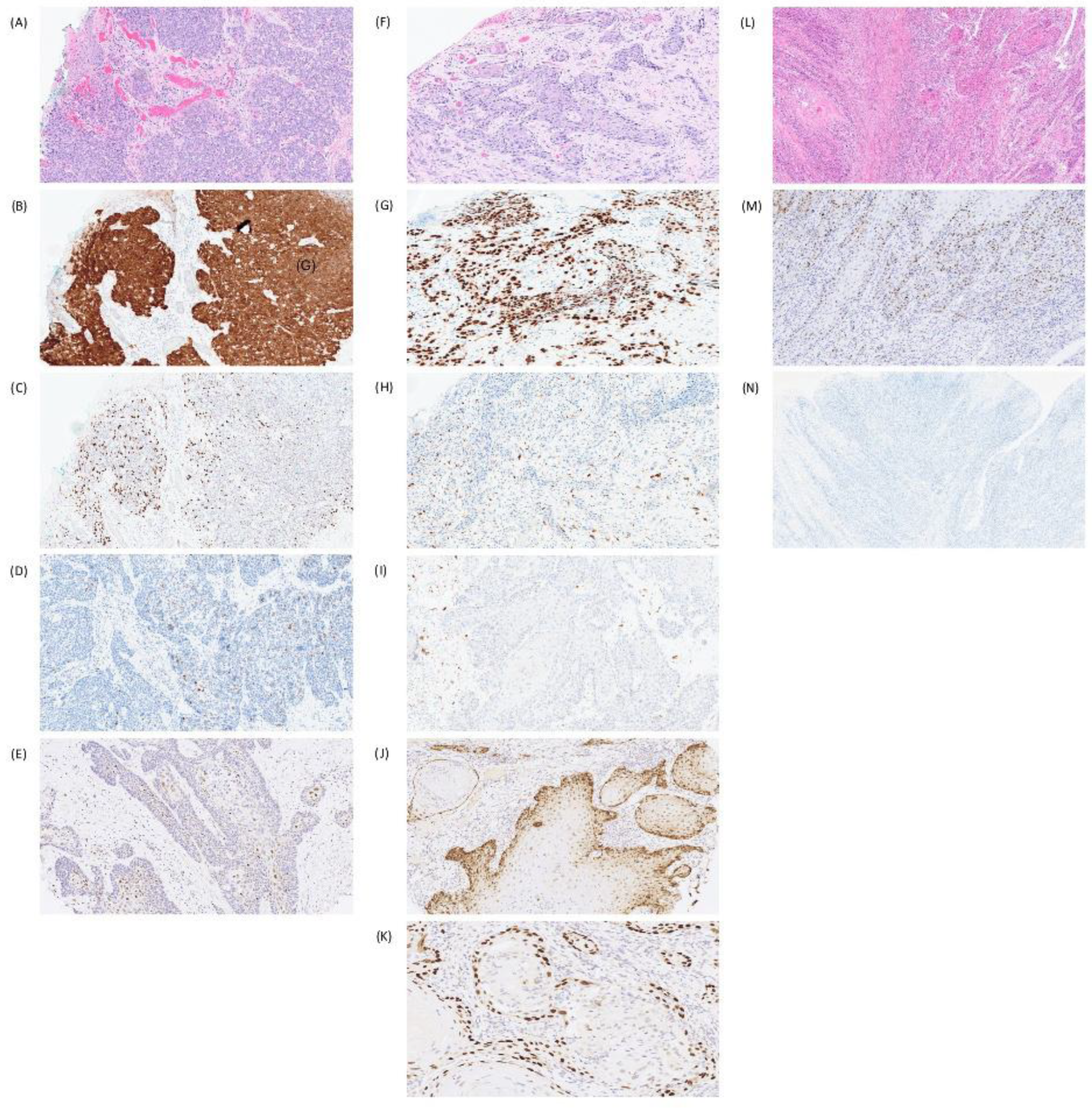
3.1. p16 as a Diagnostic, Prognostic and Predictive Biomarker
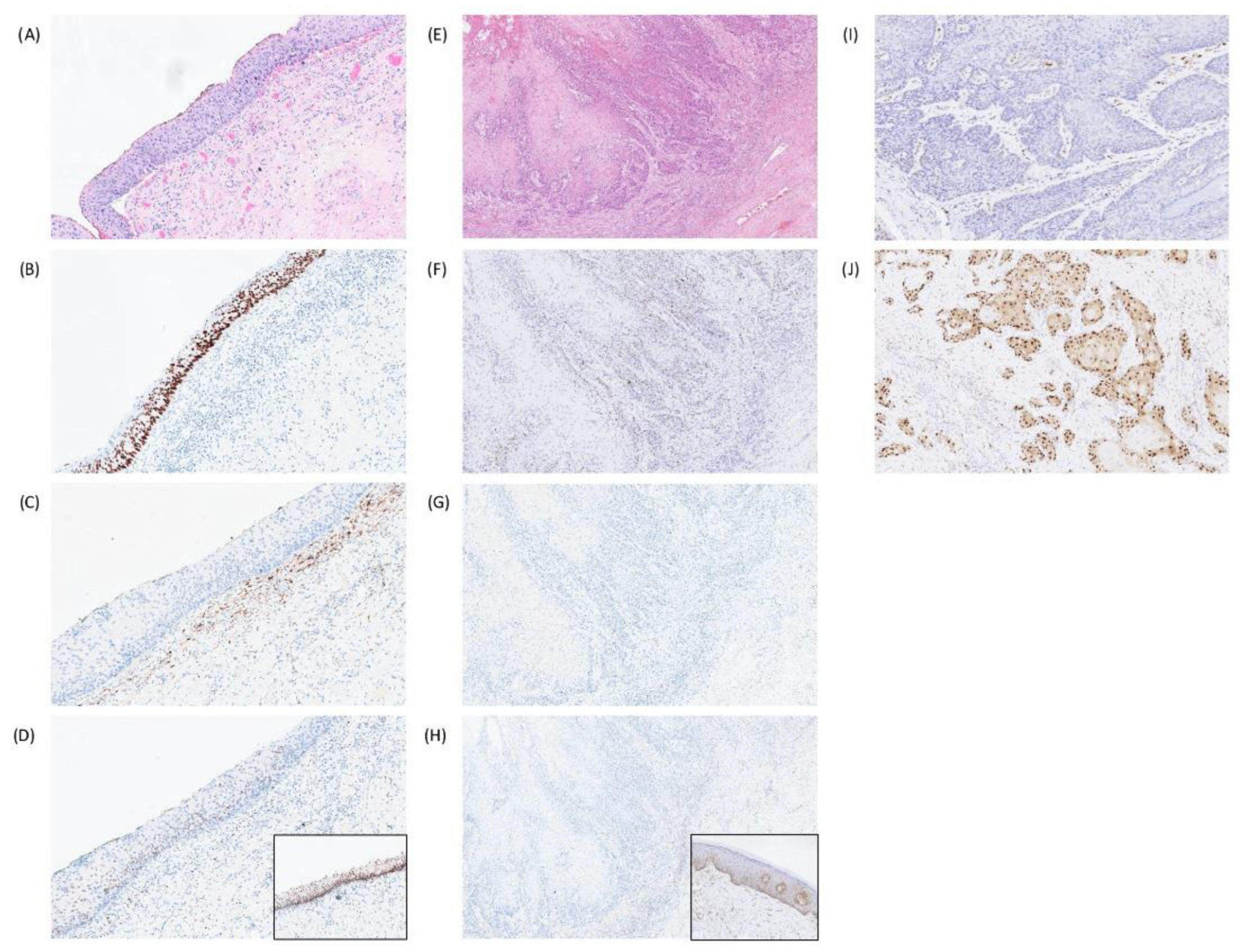
3.2. p53 as a Diagnostic and Prognostic Marker
3.3. Cyclin D1 as a Prognostic Marker
3.4. Other Prognostic Markers for VSCC in Limited Studies
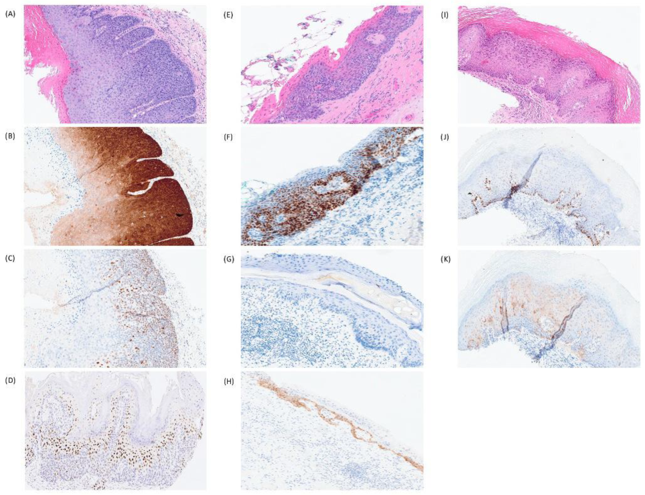
4. Precursors to HPVI Vulvar Squamous Cell Carcinoma
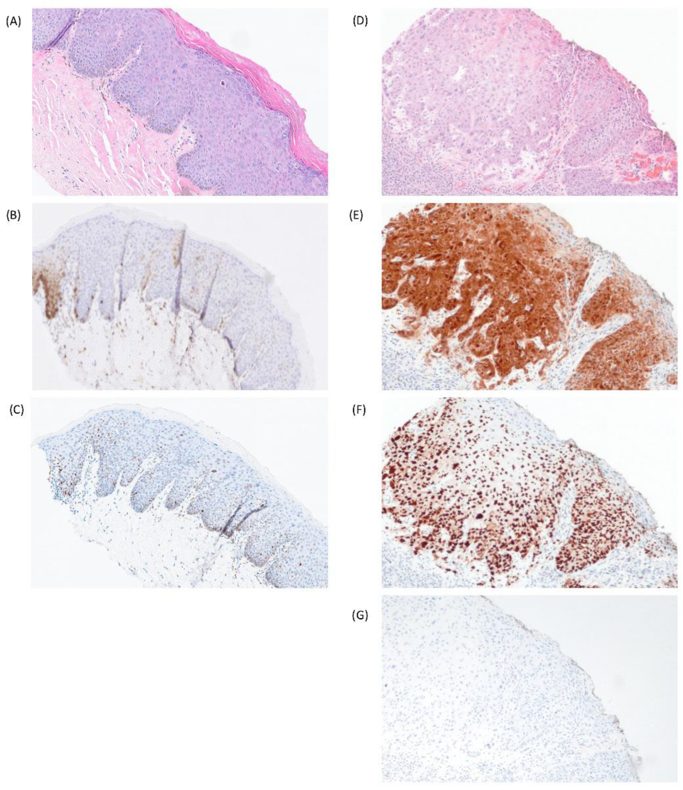
4.1. p53
4.2. CK17
4.3. CK13
4.4. SOX2
4.5. GATA3
4.6. GLUT1
4.7. Other Diagnostic Markers for HPVI VIN in Limited Studies
5. Summary
Author Contributions
Funding
Institutional Review Board Statement
Informed Consent Statement
Data Availability Statement
Conflicts of Interest
References
- Bray, F.; Laversanne, M.; Sung, H.; Ferlay, J.; Siegel, R.L.; Soerjomataram, I.; Jemal, A. Global Cancer Statistics 2022: GLOBOCAN Estimates of Incidence and Mortality Worldwide for 36 Cancers in 185 Countries. CA. Cancer J. Clin. 2024, 74, 229–263. [Google Scholar] [CrossRef] [PubMed]
- Olawaiye, A.B.; Cuello, M.A.; Rogers, L.J. Cancer of the Vulva: 2021 Update. Int. J. Gynecol. Obstet. 2021, 155, 7–18. [Google Scholar] [CrossRef]
- Preti, M.; Anderson, K.; Venturino, E.; Maggino, T.; Carozzi, F.; Robba, E.; Vieira-Baptista, P.; Borella, F.; Barchi, L.; Bevilacqua, F.; et al. Vulvar Inspection During Cervical Cancer Screening Procedures: The Ugly Reality. J. Low. Genit. Tract Dis. 2024, 28, 391–393. [Google Scholar] [CrossRef]
- Preti, M.; Joura, E.; Vieira-Baptista, P.; Van Beurden, M.; Bevilacqua, F.; Bleeker, M.C.G.; Bornstein, J.; Carcopino, X.; Chargari, C.; Cruickshank, M.E.; et al. The European Society of Gynaecological Oncology (ESGO), the International Society for the Study of Vulvovaginal Disease (ISSVD), the European College for the Study of Vulval Disease (ECSVD) and the European Federation for Colposcopy (EFC) Consensus Statements on Pre-Invasive Vulvar Lesions. Int. J. Gynecol. Cancer Off. J. Int. Gynecol. Cancer Soc. 2022, 32, 830–845. [Google Scholar] [CrossRef]
- McCluggage, W.G.; Bosse, T.; Focchi, G.; Gilks, C.B.; Hoang, L.N.; Howitt, B.; McAlpine, J.N.; Ordi, J.; Singh, N.; Wong, R.W.C.; et al. Carcinoma of the Vulva Histopathology Reporting Guide, 2nd ed.; International Collaboration on Cancer Reporting: Sydney, Australia, 2023; ISBN 978-1-922324-37-5. [Google Scholar]
- Abu-Rustum, N.R.; Yashar, C.M.; Arend, R.; Barber, E.; Bradley, K.; Brooks, R.; Campos, S.M.; Chino, J.; Chon, H.S.; Crispens, M.A.; et al. Vulvar Cancer, Version 3.2024, NCCN Clinical Practice Guidelines in Oncology. J. Natl. Compr. Cancer Netw. JNCCN 2024, 22, 117–135. [Google Scholar] [CrossRef]
- Olawaiye, A.B.; Cuello, M.A.; Beriwal, S.; Rogers, L.J. Cancer of the Vulva: 2025 Update: FIGO Cancer Report 2025. Int. J. Gynaecol. Obstet. Off. Organ Int. Fed. Gynaecol. Obstet. 2025, 171, 36–47. [Google Scholar] [CrossRef]
- Cancer Protocol Templates. College of American Pathologists. Available online: https://www.cap.org/protocols-and-guidelines/cancer-reporting-tools/cancer-protocol-templates (accessed on 5 February 2026).
- Ayala-Peacock, D.N.; Chadha, M. Advances in Vulvar Cancer: A Radiation Oncology Perspective. Cancers 2025, 17, 2415. [Google Scholar] [CrossRef] [PubMed]
- Yang, H.; Almadani, N.; Thompson, E.F.; Tessier-Cloutier, B.; Chen, J.; Ho, J.; Senz, J.; McConechy, M.K.; Chow, C.; Ta, M.; et al. Classification of Vulvar Squamous Cell Carcinoma and Precursor Lesions by P16 and P53 Immunohistochemistry: Considerations, Caveats, and an Algorithmic Approach. Mod. Pathol. 2023, 36, 100145. [Google Scholar] [CrossRef]
- Faber, M.T.; Sand, F.L.; Albieri, V.; Norrild, B.; Kjaer, S.K.; Verdoodt, F. Prevalence and Type Distribution of Human Papillomavirus in Squamous Cell Carcinoma and Intraepithelial Neoplasia of the Vulva. Int. J. Cancer 2017, 141, 1161–1169. [Google Scholar] [CrossRef]
- Wong, R.W.-C.; Palicelli, A.; Hoang, L.; Singh, N. Interpretation of P16, P53 and Mismatch Repair Protein Immunohistochemistry in Gynaecological Neoplasia. Diagn. Histopathol. 2020, 26, 257–277. [Google Scholar] [CrossRef]
- Salama, A.; Momeni Boroujeni, A.; Vanderbilt, C.; Soslow, R. Molecular Landscape of HPV Negative Vulvar Squamous Cell Carcinoma Including NOTCH Alterations. Abstracts from USCAP 2020: Gynecologic and Obstetric Pathology (1047–1234). Mod. Pathol. 2020, 33, 1164–1337. [Google Scholar] [CrossRef]
- Corey, L.; Wallbillich, J.J.; Wu, S.; Farrell, A.; Hodges, K.; Xiu, J.; Nabhan, C.; Guastella, A.; Kheil, M.; Gogoi, R.; et al. The Genomic Landscape of Vulvar Squamous Cell Carcinoma. Int. J. Gynecol. Pathol. Off. J. Int. Soc. Gynecol. Pathol. 2023, 42, 515–522. [Google Scholar] [CrossRef]
- Hørding, U.; Junge, J.; Daugaard, S.; Lundvall, F.; Poulsen, H.; Bock, J.E. Vulvar Squamous Cell Carcinoma and Papillomaviruses: Indications for Two Different Etiologies. Gynecol. Oncol. 1994, 52, 241–246. [Google Scholar] [CrossRef] [PubMed]
- Day, T.; Marzol, A.; Pagano, R.; Jaaback, K.; Scurry, J. Clinicopathologic Diagnosis of Differentiated Vulvar Intraepithelial Neoplasia and Vulvar Aberrant Maturation. J. Low. Genit. Tract Dis. 2020, 24, 392–398. [Google Scholar] [CrossRef]
- Nooij, L.S.; Ter Haar, N.T.; Ruano, D.; Rakislova, N.; van Wezel, T.; Smit, V.T.H.B.M.; Trimbos, B.J.B.M.Z.; Ordi, J.; van Poelgeest, M.I.E.; Bosse, T. Genomic Characterization of Vulvar (Pre)Cancers Identifies Distinct Molecular Subtypes with Prognostic Significance. Clin. Cancer Res. Off. J. Am. Assoc. Cancer Res. 2017, 23, 6781–6789. [Google Scholar] [CrossRef]
- Parra-Herran, C.; Nucci, M.R.; Singh, N.; Rakislova, N.; Howitt, B.E.; Hoang, L.; Gilks, C.B.; Bosse, T.; Watkins, J.C. HPV-Independent, P53-Wild-Type Vulvar Intraepithelial Neoplasia: A Review of Nomenclature and the Journey to Characterize Verruciform and Acanthotic Precursor Lesions of the Vulva. Mod. Pathol. 2022, 35, 1317–1326. [Google Scholar] [CrossRef]
- Thuijs, N.B.; van Beurden, M.; Duin, S.; Heideman, D.A.M.; Berkhof, J.; Steenbergen, R.D.M.; Bleeker, M.C.G. High-Grade Vulvar Intraepithelial Neoplasia: Comprehensive Characterization and Long-Term Vulvar Carcinoma Risk. Histopathology 2024, 84, 301–314. [Google Scholar] [CrossRef] [PubMed]
- Bigby, S.M.; Eva, L.J.; Fong, K.L.; Jones, R.W. The Natural History of Vulvar Intraepithelial Neoplasia, Differentiated Type: Evidence for Progression and Diagnostic Challenges. Int. J. Gynecol. Pathol. Off. J. Int. Soc. Gynecol. Pathol. 2016, 35, 574–584. [Google Scholar] [CrossRef]
- Ordi, O.; Saco, A.; Peñuelas, N.; Blanco-Irazuegui, O.; Pino, M.D.; Carreras-Dieguez, N.; Marimon, L.; Rodrigo-Calvo, M.T.; Morató, A.; Sisuashvili, L.; et al. Whole-Exome Sequencing of Vulvar Squamous Cell Carcinomas Reveals an Impaired Prognosis in Patients with TP53 Mutations and Concurrent CCND1 Gains. Mod. Pathol. 2024, 37, 100574. [Google Scholar] [CrossRef]
- Dasgupta, S.; Koljenović, S.; Van Den Bosch, T.; Swagemakers, S.; Van Der Hoeven, N.; Van Marion, R.; Van Der Spek, P.; Van Doorn, H.; Van Kemenade, F.; Ewing-Graham, P. Evaluation of Immunohistochemical Markers, CK17 and SOX2, as Adjuncts to P53 for the Diagnosis of Differentiated Vulvar Intraepithelial Neoplasia (dVIN). Pharmaceuticals 2021, 14, 324. [Google Scholar] [CrossRef]
- Darragh, T.M.; Colgan, T.J.; Cox, J.T.; Heller, D.S.; Henry, M.R.; Luff, R.D.; McCalmont, T.; Nayar, R.; Palefsky, J.M.; Stoler, M.H.; et al. The Lower Anogenital Squamous Terminology Standardization Project for HPV-Associated Lesions: Background and Consensus Recommendations from the College of American Pathologists and the American Society for Colposcopy and Cervical Pathology. J. Low. Genit. Tract Dis. 2012, 16, 205–242. [Google Scholar] [CrossRef] [PubMed]
- Rakislova, N.; Clavero, O.; Alemany, L.; Saco, A.; Quirós, B.; Lloveras, B.; Alejo, M.; Pawlita, M.; Quint, W.; Del Pino, M.; et al. Histological Characteristics of HPV-Associated and -Independent Squamous Cell Carcinomas of the Vulva: A Study of 1594 Cases. Int. J. Cancer 2017, 141, 2517–2527. [Google Scholar] [CrossRef]
- Kaderly Rasmussen, E.L.; Kjær, S.K.; Larsen, L.G.; Mejlgaard, E.; Franzmann, M.B.; Kjær, A.K.; Salinas, N.V.; Schledermann, D.; Frederiksen, K.; Hansen, T.; et al. Six-Pattern P53 Interpretation in 1293 Vulvar Squamous Cell Carcinomas: Inter-Pathologist Variation and Pattern Distribution According to P16 Status. Histopathology 2025, 87, 815–826. [Google Scholar] [CrossRef] [PubMed]
- Thompson, E.F.; Chen, J.; Huvila, J.; Pors, J.; Ren, H.; Ho, J.; Chow, C.; Ta, M.; Proctor, L.; McAlpine, J.N.; et al. P53 Immunohistochemical Patterns in HPV-Related Neoplasms of the Female Lower Genital Tract Can Be Mistaken for TP53 Null or Missense Mutational Patterns. Mod. Pathol. 2020, 33, 1649–1659. [Google Scholar] [CrossRef] [PubMed]
- Sisuashvili, L.; Saco, A.; Carreras-Dieguez, N.; Celada, C.; Marimon, L.; Montironi, C.; Ordi, J.; Rakislova, N. Confusing Histopathological Features and HPV Testing Results in Vulvar Squamous Cell Carcinoma Arising in a Young Woman: A Case Solved Using Next-Generation Sequencing. Int. J. Gynecol. Pathol. Off. J. Int. Soc. Gynecol. Pathol. 2025, 44, 120–124. [Google Scholar] [CrossRef]
- Thompson, E.F.; Hoang, L.; Höhn, A.K.; Palicelli, A.; Talia, K.L.; Tchrakian, N.; Senz, J.; Rusike, R.; Jordan, S.; Jamieson, A.; et al. Molecular Subclassification of Vulvar Squamous Cell Carcinoma: Reproducibility and Prognostic Significance of a Novel Surgical Technique. Int. J. Gynecol. Cancer Off. J. Int. Gynecol. Cancer Soc. 2022, 32, 977–985. [Google Scholar] [CrossRef]
- Höhn, A.K.; Forberger, M.; Alfaraidi, M.; Gilks, C.B.; Brambs, C.E.; Höckel, M.; Hoang, L.; Singh, N.; Horn, L.-C. High Concordance of Molecular Subtyping between Pre-Surgical Biopsy and Surgical Resection Specimen (Matched-Pair Analysis) in Patients with Vulvar Squamous Cell Carcinoma Using P16- and P53-Immunostaining. Gynecol. Oncol. 2024, 185, 17–24. [Google Scholar] [CrossRef]
- Cheng, A.S.; Karnezis, A.N.; Jordan, S.; Singh, N.; McAlpine, J.N.; Gilks, C.B. P16 Immunostaining Allows for Accurate Subclassification of Vulvar Squamous Cell Carcinoma into HPV-Associated and HPV-Independent Cases. Int. J. Gynecol. Pathol. Off. J. Int. Soc. Gynecol. Pathol. 2015, 35, 385–393. [Google Scholar] [CrossRef]
- Allo, G.; Yap, M.L.; Cuartero, J.; Milosevic, M.; Ferguson, S.; Mackay, H.; Kamel-Reid, S.; Weinreb, I.; Ghazarian, D.; Pintilie, M.; et al. HPV-Independent Vulvar Squamous Cell Carcinoma Is Associated with Significantly Worse Prognosis Compared with HPV-Associated Tumors. Int. J. Gynecol. Pathol. Off. J. Int. Soc. Gynecol. Pathol. 2020, 39, 391–399. [Google Scholar] [CrossRef]
- Dong, F.; Kojiro, S.; Borger, D.R.; Growdon, W.B.; Oliva, E. Squamous Cell Carcinoma of the Vulva: A Subclassification of 97 Cases by Clinicopathologic, Immunohistochemical, and Molecular Features (P16, P53, and EGFR). Am. J. Surg. Pathol. 2015, 39, 1045–1053. [Google Scholar] [CrossRef]
- Kortekaas, K.E.; Bastiaannet, E.; van Doorn, H.C.; de Vos van Steenwijk, P.J.; Ewing-Graham, P.C.; Creutzberg, C.L.; Akdeniz, K.; Nooij, L.S.; van der Burg, S.H.; Bosse, T.; et al. Vulvar Cancer Subclassification by HPV and P53 Status Results in Three Clinically Distinct Subtypes. Gynecol. Oncol. 2020, 159, 649–656. [Google Scholar] [CrossRef]
- Kjær, S.K.; Frederiksen, K.; Rasmussen, C.L.; Thomsen, L.T.; Madsen, E.M.; Franzmann, M.B.; Kjær, A.K.; Larsen, L.G.; Salinas, N.V.; Schledermann, D.; et al. Prognostic Impact of P16 and High-risk HPV DNA in ~1300 Patients with Vulvar Cancer. Int. J. Cancer 2025, 157, 1354–1362. [Google Scholar] [CrossRef]
- Proctor, L.; Hoang, L.; Moore, J.; Thompson, E.; Leung, S.; Natesan, D.; Chino, J.; Gilks, B.; McAlpine, J.N. Association of Human Papilloma Virus Status and Response to Radiotherapy in Vulvar Squamous Cell Carcinoma. Int. J. Gynecol. Cancer Off. J. Int. Gynecol. Cancer Soc. 2020, 30, 100–106. [Google Scholar] [CrossRef]
- Lee, L.J.; Howitt, B.; Catalano, P.; Tanaka, C.; Murphy, R.; Cimbak, N.; DeMaria, R.; Bu, P.; Crum, C.; Horowitz, N.; et al. Prognostic Importance of Human Papillomavirus (HPV) and P16 Positivity in Squamous Cell Carcinoma of the Vulva Treated with Radiotherapy. Gynecol. Oncol. 2016, 142, 293–298. [Google Scholar] [CrossRef]
- Dohopolski, M.J.; Horne, Z.D.; Pradhan, D.; Bhargava, R.; Edwards, R.P.; Kelley, J.L.; Comerci, J.T.; Olawaiye, A.B.; Courtney-Brooks, M.; Berger, J.L.; et al. The Prognostic Significance of P16 Status in Patients with Vulvar Cancer Treated with Vulvectomy and Adjuvant Radiation. Int. J. Radiat. Oncol. Biol. Phys. 2019, 103, 152–160. [Google Scholar] [CrossRef] [PubMed]
- Carreras-Dieguez, N.; Saco, A.; Del Pino, M.; Marimon, L.; López Del Campo, R.; Manzotti, C.; Fusté, P.; Pumarola, C.; Torné, A.; Garcia, A.; et al. Human Papillomavirus and P53 Status Define Three Types of Vulvar Squamous Cell Carcinomas with Distinct Clinical, Pathological, and Prognostic Features. Histopathology 2023, 83, 17–30. [Google Scholar] [CrossRef] [PubMed]
- Dongre, H.N.; Elnour, R.; Tornaas, S.; Fromreide, S.; Thomsen, L.C.V.; Kolseth, I.B.M.; Nginamau, E.S.; Johannessen, A.C.; Vintermyr, O.K.; Costea, D.E.; et al. TP53 Mutation and Human Papilloma Virus Status as Independent Prognostic Factors in a Norwegian Cohort of Vulva Squamous Cell Carcinoma. Acta Obstet. Gynecol. Scand. 2024, 103, 165–175. [Google Scholar] [CrossRef] [PubMed]
- Tessier-Cloutier, B.; Kortekaas, K.E.; Thompson, E.; Pors, J.; Chen, J.; Ho, J.; Prentice, L.M.; McConechy, M.K.; Chow, C.; Proctor, L.; et al. Major P53 Immunohistochemical Patterns in In Situ and Invasive Squamous Cell Carcinomas of the Vulva and Correlation with TP53 Mutation Status. Mod. Pathol. 2020, 33, 1595–1605. [Google Scholar] [CrossRef]
- Kortekaas, K.E.; Solleveld-Westerink, N.; Tessier-Cloutier, B.; Rutten, T.A.; Poelgeest, M.I.E.; Gilks, C.B.; Hoang, L.N.; Bosse, T. Performance of the Pattern-Based Interpretation of P53 Immunohistochemistry as a Surrogate for TP53 Mutations in Vulvar Squamous Cell Carcinoma. Histopathology 2020, 77, 92–99. [Google Scholar] [CrossRef]
- Jeffus, S.K.; Wooldridge, J.T.; Hoang, L.; Parra-Herran, C.; Hamza, M.; Lindsey, M.; Verret, M.; Zoumberos, N.; Fogel, B.; Wyeth, A.; et al. Interpretation of P16 and P53 in the Classification of Squamous Cell Carcinoma of the Vulva-An Interobserver Agreement Study. Am. J. Surg. Pathol. 2025, 49, 176–187. [Google Scholar] [CrossRef] [PubMed]
- Köbel, M.; Ronnett, B.M.; Singh, N.; Soslow, R.A.; Gilks, C.B.; McCluggage, W.G. Interpretation of P53 Immunohistochemistry in Endometrial Carcinomas: Toward Increased Reproducibility. Int. J. Gynecol. Pathol. Off. J. Int. Soc. Gynecol. Pathol. 2019, 38, S123–S131. [Google Scholar] [CrossRef] [PubMed]
- Sand, F.L.; Nielsen, D.M.B.; Frederiksen, M.H.; Rasmussen, C.L.; Kjaer, S.K. The Prognostic Value of P16 and P53 Expression for Survival after Vulvar Cancer: A Systematic Review and Meta-Analysis. Gynecol. Oncol. 2019, 152, 208–217. [Google Scholar] [CrossRef]
- Gioacchini, F.M.; Alicandri-Ciufelli, M.; Kaleci, S.; Magliulo, G.; Presutti, L.; Re, M. The Prognostic Value of Cyclin D1 Expression in Head and Neck Squamous Cell Carcinoma. Eur. Arch. Otorhinolaryngol. 2016, 273, 801–809. [Google Scholar] [CrossRef] [PubMed]
- Carreras-Dieguez, N.; Ordi, O.; Peñuelas, N.; Del Pino, M.; Diez-Ahijado, L.; Sisuashvili, L.; Darecka, K.; Marimon, L.; Vega, N.; Torné, A.; et al. Cyclin D1 Overexpression Predicts Poor Disease-Specific Survival in Human Papillomavirus-Independent Vulvar Squamous Cell Carcinoma. Mod. Pathol. 2025, 38, 100833. [Google Scholar] [CrossRef] [PubMed]
- Choschzick, M.; Hess, S.; Tennstedt, P.; Holst, F.; Bohlken, H.; Gieseking, F.; Mahner, S.; Woelber, L.; Simon, R.; Sauter, G. Role of Cyclin D1 Amplification and Expression in Vulvar Carcinomas. Hum. Pathol. 2012, 43, 1386–1393. [Google Scholar] [CrossRef] [PubMed]
- Brambs, C.E.; Horn, L.-C.; Mende, M.; Höckel, M.; Eckey, C.; Hiller, G.G.R.; Höhn, A.K. Epithelial–Mesenchymal Transition (EMT) in Vulvar Cancer with and without Inguinal Lymph Node Involvement. J. Cancer Res. Clin. Oncol. 2022, 148, 1183–1193. [Google Scholar] [CrossRef]
- Knopp, S.; Bjørge, T.; Nesland, J.M.; Tropé, C.; Holm, R. Cyclins D1, D3, E, and A in Vulvar Carcinoma Patients. Gynecol. Oncol. 2005, 97, 733–739. [Google Scholar] [CrossRef]
- Zannoni, G.F.; Prisco, M.G.; Vellone, V.G.; De Stefano, I.; Vizzielli, G.; Tortorella, L.; Fagotti, A.; Scambia, G.; Gallo, D. Cytoplasmic Expression of Oestrogen Receptor Beta (ERβ) as a Prognostic Factor in Vulvar Squamous Cell Carcinoma in Elderly Women. Histopathology 2011, 59, 909–917. [Google Scholar] [CrossRef]
- Buchholz, A.; Vattai, A.; Fürst, S.; Vilsmaier, T.; Kuhn, C.; Schmoeckel, E.; Mayr, D.; Dannecker, C.; Mahner, S.; Jeschke, U.; et al. EP4 as a Negative Prognostic Factor in Patients with Vulvar Cancer. Cancers 2021, 13, 1410. [Google Scholar] [CrossRef]
- Buchholz, A.; Vattai, A.; Fürst, S.; Vilsmaier, T.; Zati Zehni, A.; Steger, A.; Kuhn, C.; Schmoeckel, E.; Dannecker, C.; Mahner, S.; et al. Prostaglandin E2 Receptor EP1 Expression in Vulvar Cancer. J. Cancer Res. Clin. Oncol. 2023, 149, 5369–5376. [Google Scholar] [CrossRef]
- Ansorge, N.; Dannecker, C.; Jeschke, U.; Schmoeckel, E.; Mayr, D.; Heidegger, H.H.; Vattai, A.; Burgmann, M.; Czogalla, B.; Mahner, S.; et al. Combined COX-2/PPARγ Expression as Independent Negative Prognosticator for Vulvar Cancer Patients. Diagnostics 2021, 11, 491. [Google Scholar] [CrossRef] [PubMed]
- Podoll, M.B.; Singh, N.; Gilks, C.B.; Moghadamfalahi, M.; Sanders, M.A. Assessment of CK17 as a Marker for the Diagnosis of Differentiated Vulvar Intraepithelial Neoplasia. Int. J. Gynecol. Pathol. 2017, 36, 273–280. [Google Scholar] [CrossRef] [PubMed]
- Dasgupta, S.; Ewing-Graham, P.C.; Van Kemenade, F.J.; Van Doorn, H.C.; Noordhoek Hegt, V.; Koljenović, S. Differentiated Vulvar Intraepithelial Neoplasia (dVIN): The Most Helpful Histological Features and the Utility of Cytokeratins 13 and 17. Virchows Arch. 2018, 473, 739–747. [Google Scholar] [CrossRef] [PubMed]
- McMullen-Tabry, E.R.; Schechter, S.A.; Wang, G.Y.; Sciallis, A.P.; Hrycaj, S.M.; Chan, M.P.; Skala, S.L. P53/CK17 Dual Stain Improves Accuracy of Distinction Between Differentiated Vulvar Intraepithelial Neoplasia and Its Mimics. Int. J. Gynecol. Pathol. 2022, 41, 298–306. [Google Scholar] [CrossRef]
- Hartsough, E.M.; Watkins, J.; Nazarian, R.M. D2-40 and CK17 Immunohistochemistry as a Diagnostic Adjunct for HPV-Independent Squamous Lesions in the Vulva and Their Role in Defining Atypical Lichen Sclerosus. Am. J. Surg. Pathol. 2024, 48, 1617–1623. [Google Scholar] [CrossRef]
- Cook, E.; Van De Vijver, K.; Parra-Herran, C. Diagnosis of Verruciform Acanthotic Vulvar Intra-epithelial Neoplasia (vaVIN) Using CK17, SOX2 and GATA3 Immunohistochemistry. Histopathology 2024, 84, 1212–1223. [Google Scholar] [CrossRef]
- Thuijs, N.B.; Voss, F.O.; Ewing-Graham, P.C.; Dasgupta, S.; Berkhof, J.; Bulten, J.; Van De Vijver, K.; Bleeker, M.C.G. Expression of CK17 and SOX2 in Vulvar Intraepithelial Neoplasia: A Comprehensive Analysis of 150 Vulvar Lesions. Cancers 2024, 16, 3966. [Google Scholar] [CrossRef]
- Hartsough, E.M.; Nazarian, R.M.; Watkins, J.C. CK17 Immunohistochemistry Is a Useful Adjunct in the Diagnosis of HPV-Independent, TP53-Wild-Type Verruciform/Acanthotic Vulvar Intraepithelial Neoplasia (vaVIN). Am. J. Surg. Pathol. 2025, 49, 658–662. [Google Scholar] [CrossRef]
- Altarawneh, B.K.; Quddus, M.R.; Singh, K.; Sung, C.J.; Marketkar, S. Histomorphology and Utility of CK17, P53 Dual Stain with CK 13 in the Diagnosis of Differentiated Vulvar Intraepithelial Neoplasia. Ann. Diagn. Pathol. 2026, 80, 152561. [Google Scholar] [CrossRef]
- Brustmann, H.; Brunner, A. Immunohistochemical Expression of SOX2 in Vulvar Intraepithelial Neoplasia and Squamous Cell Carcinoma. Int. J. Gynecol. Pathol. Off. J. Int. Soc. Gynecol. Pathol. 2013, 32, 323–328. [Google Scholar] [CrossRef]
- Goyal, A.; Zhang, G.; Yang, B. Differential Expression Patterns of GATA3 in Usual and Differentiated Types of Vulvar Intraepithelial Neoplasia: Potential Diagnostic Implications. Mod. Pathol. 2018, 31, 1131–1140. [Google Scholar] [CrossRef]
- Zare, S.Y.; Fard, E.V.; Fadare, O. GATA3 Immunohistochemistry as a Diagnostic Adjunct for Differentiated Vulvar Intraepithelial Neoplasia: Utility and Limitations. Hum. Pathol. 2023, 139, 55–64. [Google Scholar] [CrossRef]
- Zhang, G.; Yang, B. Distinct Patterns of GLUT1 Expression in Human Papillomavirus Associated and Human Papillomavirus Independent Vulvar Intraepithelial Neoplasia and Squamous Cell Carcinoma. Mod. Pathol. 2025, 38, 100836. [Google Scholar] [CrossRef]
- Zhang, G.; Soslow, R.A.; Yang, B. GLUT1 Expression Patterns in Verruciform Acanthotic Vulvar Squamous Intraepithelial Neoplasia, HPV-Independent P53 Wild-Type Squamous Cell Carcinoma and Benign Vulvar Lesions. Mod. Pathol. 2025, 38, 100900. [Google Scholar] [CrossRef]
- Brustmann, H.; Hinterholzer, S.; Brunner, A. Immunohistochemical Expression of Survivin and γ-H2AX in Vulvar Intraepithelial Neoplasia and Low-Stage Squamous Cell Carcinoma. Int. J. Gynecol. Pathol. 2011, 30, 583–590. [Google Scholar] [CrossRef]
- Wellenhofer, A.; Brustmann, H. Expression of Human Telomerase Reverse Transcriptase in Vulvar Intraepithelial Neoplasia and Squamous Cell Carcinoma: An Immunohistochemical Study with Survivin and P53. Arch. Pathol. Lab. Med. 2012, 136, 1359–1365. [Google Scholar] [CrossRef][Green Version]
- Pinto, A.P.; Degen, M.; Barron, P.; Crum, C.P.; Rheinwald, J.G. Phosphorylated S6 as an Immunohistochemical Biomarker of Vulvar Intraepithelial Neoplasia. Mod. Pathol. 2013, 26, 1498–1507. [Google Scholar] [CrossRef] [PubMed]
- Hofstetter, G.; Mildner, M.; Tschandl, P.; Pammer, J. ORF1p Is a Potential Novel Diagnostic Marker for Differentiated Vulvar Intraepithelial Neoplasia. Int. J. Gynecol. Pathol. 2023, 42, 201–206. [Google Scholar] [CrossRef] [PubMed]
- Zhang, C.; Arentz, G.; Winderbaum, L.; Lokman, N.; Klingler-Hoffmann, M.; Mittal, P.; Carter, C.; Oehler, M.; Hoffmann, P. MALDI Mass Spectrometry Imaging Reveals Decreased CK5 Levels in Vulvar Squamous Cell Carcinomas Compared to the Precursor Lesion Differentiated Vulvar Intraepithelial Neoplasia. Int. J. Mol. Sci. 2016, 17, 1088. [Google Scholar] [CrossRef] [PubMed]
- Chen, H.; Gonzalez, J.L.; Brennick, J.B.; Liu, M.; Yan, S. Immunohistochemical Patterns of ProEx C in Vulvar Squamous Lesions: Detection of Overexpression of MCM2 and TOP2A. Am. J. Surg. Pathol. 2010, 34, 1250–1257. [Google Scholar] [CrossRef]
- Liu, Y.A.; Ji, J.X.; Almadani, N.; Crawford, R.I.; Gilks, C.B.; Kinloch, M.; Hoang, L. Comparison of P53 Immunohistochemical Staining in Differentiated Vulvar Intraepithelial Neoplasia (dVIN) with That in Inflammatory Dermatoses and Benign Squamous Lesions in the Vulva. Histopathology 2021, 78, 424–433. [Google Scholar] [CrossRef]
- Wei, K.; Almadani, N.; Torlakovic, E.; Haupt, R.; Anderson, L.; Crawford, R.; Focchi, G.; Gilks, B.; Horn, L.-C.; Kellow, M.; et al. Proficiency Testing of P53 Immunohistochemistry Pattern Read-out in Vulvar Biopsies Demonstrates Frequent Basal Overexpression Interpretation in TP53 Wild-Type Cases. Int. J. Gynecol. Pathol. Off. J. Int. Soc. Gynecol. Pathol. 2025, 44, 485–495. [Google Scholar] [CrossRef]
- Mayer, A.; Schmidt, M.; Seeger, A.; Serras, A.F.; Vaupel, P.; Schmidberger, H. GLUT-1 Expression Is Largely Unrelated to Both Hypoxia and the Warburg Phenotype in Squamous Cell Carcinomas of the Vulva. BMC Cancer 2014, 14, 760. [Google Scholar] [CrossRef]
- Krasinskas, A.M.; Bartlett, D.L.; Cieply, K.; Dacic, S. CDKN2A and MTAP Deletions in Peritoneal Mesotheliomas Are Correlated with Loss of P16 Protein Expression and Poor Survival. Mod. Pathol. 2010, 23, 531–538. [Google Scholar] [CrossRef]
- Ji, J.X.; El Maghrabi, A.; Ren, H.C.; Lee, L.H.; Young, S.; Yip, S.T.; Moodley, J.; Lai, C.; Shirsat, H.; Hahn, E.; et al. Utility of P53, P16, and MTAP Immunohistochemistry in Oral Epithelial Dysplasia with Concurrent Candidiasis: A Novel Pattern-Based Approach. Mod. Pathol. 2026, 39, 100959. [Google Scholar] [CrossRef]
- Thuijs, N.B.; Berkhof, J.; Özer, M.; Duin, S.; Van Splunter, A.P.; Snoek, B.C.; Heideman, D.A.M.; Van Beurden, M.; Steenbergen, R.D.M.; Bleeker, M.C.G. DNA Methylation Markers for Cancer Risk Prediction of Vulvar Intraepithelial Neoplasia. Int. J. Cancer 2021, 148, 2481–2488. [Google Scholar] [CrossRef]
- Voss, F.O.; Thuijs, N.B.; Duin, S.; Özer, M.; Van Beurden, M.; Berkhof, J.; Steenbergen, R.D.M.; Bleeker, M.C.G. Clinical Validation of Methylation Biomarkers for Optimal Detection of High-grade Vulvar Intraepithelial Neoplasia. Int. J. Cancer 2023, 153, 783–791. [Google Scholar] [CrossRef]
- Thuijs, N.B.; Voss, F.O. DNA Methylation Testing for Vulvar Cancer Risk Stratification in Patients with High-Grade Vulvar Intraepithelial Neoplasia: A Population-Based Cohort Study. Br. J. Dermatol. 2025, 193, 1138–1145. [Google Scholar] [CrossRef]
- Voss, F.O.; Berkhof, J.; Duin, S.; Fons, G.; Van Beurden, M.; Steenbergen, R.D.M.; Bleeker, M.C.G. DNA Methylation and P53 Immunohistochemistry as Prognostic Biomarkers for Vulvar Lichen Sclerosus. Mod. Pathol. 2024, 37, 100553. [Google Scholar] [CrossRef]



| Biomarkers | Number of Studies | Interpretation Criteria | Total Number of Cases (and % Positive) by Interpretation Criteria | Sensitivity † | Specificity † |
|---|---|---|---|---|---|
| CK17 Summary | 9 | Positive expression is Diffuse, moderate to strong cytoplasmic staining, suprabasal or full-thickness expression. | 300 VIN 217 non-dysplastic lesions including LS | Relatively high sensitivity (70–100%) | Wide range of specificity (27–100%) |
| Podoll 2016 [54] | intermediate to strong diffuse immunoreactivity, confined to the upper half of the epithelium | 29 (93%) dVIN 9 (0%) HSIL 7 (29%) LSC 8 (63%) LS | 93% | 53% | |
| Dasgupta 2018 [55] | diffuse, strong staining, suprabasal to full thickness | 54 (88.9%) dVIN 14 (0%) LS 30 (0%) NDEL | 89% | 100% | |
| Dasgupta 2021 [22] | diffuse, moderate-strong, cytoplasmic staining, suprabasal to full thickness | 56 (80%) dVIN 8 (88%) deVIL 27 (63%) HSIL 46 (20%) NDEL | 80% for diagnosis of DVIN (by ROC) | 80% for diagnosis of DVIN (by ROC) | |
| McMullen-Tabry 2022 [56] | moderate to strong, partial thickness or full thickness staining | 30 (100%) dVIN 30 (73%) NDEL (LS, LSC, PEH) 20 (75%) LS progressed to dVIN 20 (50%) LS did not progress to dVIN 5 (0%) normal | 100% | 27% | |
| Hartsough 2024 [57] | superficial to suprabasal expression | 10 (70%) vaVIN (vLSC, DE-VIL, VAAD) | 70% | n/a | |
| Cook 2024 [58] | Full thickness | 16 (81%) HPVI p53wt VIN (vaVINs) 34 (32%) NDEL (verruciform xanthoma, LSC *, LS *, psorasis, PEH *) | 81% | 68% | |
| Thuijs 2024 [59] | diffuse (>50%) and moderate-to-strong intensity, partial to full thickness | 30 (73%) HPVI p53abn VIN 16 (100%) HPVI p53wt VIN 58 (14%) HSIL 4 (0%) LSIL 37 (24%) NDEL 5 nonconclusive | 83% | 75% | |
| Hartsough 2025 [60] | Scored from 0 to 3 based on intensity and thickness (superficial, suprabasal, diffuse) | 11 VSCC HPVI 19 dVIN 8 atypical LS 11 LS (increasing scores with progression) | n/a | n/a | |
| Altarawneh 2026 [61] | Full thickness | 32 (90.6%) dVIN [85.7% if p53abn dVIN] | 91% | n/a | |
| CK13 Summary | 2 | Reduced expression | 86 dVIN 44 non-dysplastic | Low sensitivity ~15% | Uncertain |
| Altarawneh 2026 [61] | either lack of staining or diffuse full-thickness | Lack of staining: 32 (12.5%) dVIN [9.5% of p53abn dVIN] Diffuse full thickness: 32 (46.9%) dVIN [38% of p53abn dVIN] | n/a | n/a | |
| Dasgupta 2018 [55] | complete lack of staining | 54 (15%) dVIN 14 (0%) LS 30 (0%) NDEL | 15% | 100% | |
| SOX2 Summary | 4 | Diffuse moderate to strong nuclear staining | 140 HPVI VIN 122 non-dysplastic lesions | Wide range 33–86% | Wide range 20 to 100% |
| Brustmann 2013 [62] | modified score combining proportion and intensity divided into negative, moderately positive (score 3–4) and strongly positive (score 5–6) | 33 (100%) SCC 18 (100%) dVIN 16 (100%) HSIL 9 (56%) LS 25 (88%) normal (using score of 3–4) | 44–100% (depending on cut-off of score 3–4 or 5–6) | 20–100% (depending on cut-off of score 3–4 or 5–6) | |
| Dasgupta 2021 [22] | diffuse, moderate to strong nuclear expression, in basal/suprabasal layers to full thickness | 56 (86%) dVIN 8 (88%) deVIL 27 (88%) HSIL 46 (19%) NDEL | Sensitivity 86% for diagnosis of DVIN (by ROC) | specificity 81% for diagnosis of DVIN (by ROC) | |
| Cook 2024 [58] | strong staining ≥ 10% cells | 12 (75%) vaVINs 30 (17%) NDEL (verruciform xanthoma, LSC, LS, psorasis, PEH) | 75% | 83% | |
| Thuijs 2024 [59] | diffuse >50% and moderate-to-strong intensity, partial to full thickness | 30 (43%) HPVI p53abn VIN 16 (13%) HPVI p53wt VIN 58 (2%) HSIL 4 (0%) LSIL 37 (3%) NDEL (LS, inflammation, reactive, fibroepithelial polyps, normal) | 33% | 97% | |
| GATA3 Summary | 3 | Positive is loss of staining in >25% of basal layer | 77 HPVI VIN 113 non-dysplastic lesions | ~90% for dVIN 58–90% for vaVIN | ~95% for dVIN 78–90% for vaVIN |
| Goyal 2018 [63] | loss in >25% of basal cells | 23 (87%) VSCC 34 (88%) dVIN 30 (0%) HSIL 20 (0%) LS 12 (0%) LSC 45 (0%) normal | 88% | 100% | |
| Zare 2023 [64] | loss in >25% of basal cells | 21 (90%) dVIN 10 (90%) VAM 44 (16%) HSIL 49 (4%) NDEL (* LS, LSC, SD, LP, inflammation) 75 (0%) normal | 90% | 96% | |
| Cook 2024 [58] | loss in >25% of basal cells | 12 (58%) vaVINs 32 (22%) NDEL (verruciform xanthoma *, LSC, LS, psorasis, PEH) | 58% | 78% | |
| GLUT1 | 2 | Moderate to strong diffuse staining in basal to mid-epithelial layers | 89 HPVI VIN 88 non-dysplastic lesions | Very high sensitivity reported so far | Very high specificity reported so far |
| Zhang 2025a [65] | Moderate to strong staining compared to nonneoplastic vulva | 90 VSCC (88.9–100%) 65 (96.9%) HPVI VIN [98% of p53abn and 92% of p53wt] 45 (82.2%) HSIL 40 (0%) NDEL (LS, inflammation, FEP, other) | 97% | 100% | |
| Zhang 2025b [66] | diffuse confluent staining with moderate to strong intensity, in basal to intermediate cell layers | 24 (100%) vaVIN (all p53wt) 48 (0%) NDEL (resection margins, inflammation, LSC, seborrheic keratosis, PEH, condyloma) | 100% | 100% | |
| Survivin | 2 | Moderate to strong nuclear over-expression in >5% of cells | 52 VSCC 16–34 HPVI VIN ‡ 20 non-dysplastic lesions, including LS ‡ | High sensitivity depending on cut-off used to define positivity | Uncertain (not many non-dysplastic epithelial lesions studied) |
| Brustmann 2011 [67] | Moderate to strong nuclear over-expression in >5% of cells | 20 (100%) VSCC 16 (87%) dVIN 16 (100%) HSIL 10 (40%) LS 25 (20%) normal | 87% | n/a | |
| Wellenhofer 2012 [68] | >5% nuclear immunoreactive cells (score of 2+ or 3+) | 32 (100%) VSCC 18 (100%) dVIN 16 (100%) HSIL 10 (40%) LS 25 (20%) normal | 100% | n/a | |
| γ-H2AX Brustmann 2011 [67] | 1 | Moderate to strong nuclear over-expression in >5% of cells | 20 (40%) VSCC 16 (69%) dVIN 16 (69%) HSIL 10 (30%) LS 25 (0%) normal | 69% | n/a |
| hTERT Wellenhofer 2012 [68] | 1 | >5% nuclear immunoreactive cells (score of 2+ or 3+) | 32 (100%) VSCC 18 (100%) dVIN 16 (100%) HSIL 10 (40%) LS 25 (0%) normal | 100% | n/a |
| P-S6 Pinto 2013 [69] | 1 | Positive basal-layer staining | 7 (100%) dVIN 9 (77%) HSIL | n/a | n/a |
| ORF1p Hofstetter 2023 [70] | 1 | Moderate-to-strong basal or full-thickness staining | 29 (93%) dVIN 26 (77%) HSIL 20 (30%) inflammatory 22 LS (18%) 20 (0%) normal | 93% | 76% |
| CK5 Zhang 2016 [71] | 1 | Reduced/decreased staining in VSCC compared to healthy epithelium and DVIN (mass spectrometry and later IHC) | 6–8 VSCC with dVIN and normal skin | n/a | n/a |
| ProEx C (MCM2/TOP2A) Chen 2010 [72] | 1 | Nuclear overexpression extends beyond basal/parabasal layer | 18 VSCC 3 verrucous carcinoma 6 dVIN 23 HSIL 14 condyloma 13 LS 22 normal | 0% | n/a |
Disclaimer/Publisher’s Note: The statements, opinions and data contained in all publications are solely those of the individual author(s) and contributor(s) and not of MDPI and/or the editor(s). MDPI and/or the editor(s) disclaim responsibility for any injury to people or property resulting from any ideas, methods, instructions or products referred to in the content. |
© 2026 by the authors. Licensee MDPI, Basel, Switzerland. This article is an open access article distributed under the terms and conditions of the Creative Commons Attribution (CC BY) license.
Share and Cite
Alsolami, S.; Ji, J.; Hoang, L. Squamous Cancers and Precancers of the Vulva: Emerging Diagnostic, Prognostic and Predictive Biomarkers in Pathology. Cancers 2026, 18, 1518. https://doi.org/10.3390/cancers18101518
Alsolami S, Ji J, Hoang L. Squamous Cancers and Precancers of the Vulva: Emerging Diagnostic, Prognostic and Predictive Biomarkers in Pathology. Cancers. 2026; 18(10):1518. https://doi.org/10.3390/cancers18101518
Chicago/Turabian StyleAlsolami, Somayah, Jennifer Ji, and Lynn Hoang. 2026. "Squamous Cancers and Precancers of the Vulva: Emerging Diagnostic, Prognostic and Predictive Biomarkers in Pathology" Cancers 18, no. 10: 1518. https://doi.org/10.3390/cancers18101518
APA StyleAlsolami, S., Ji, J., & Hoang, L. (2026). Squamous Cancers and Precancers of the Vulva: Emerging Diagnostic, Prognostic and Predictive Biomarkers in Pathology. Cancers, 18(10), 1518. https://doi.org/10.3390/cancers18101518